53 minute read
The production methods of selenium nanoparticles
ACTA UNIV. SAPIENTIAE, ALIMENTARIA, 14 (2021) 14–43
DOI: 10.2478/ausal-2021-0002
Advertisement
B. Khandsuren1,2 J. Prokisch1,2
e-mail: b_khandsuren@muls.edu.mn e-mail: jprokisch@agr.unideb.hu 1 Institute of Animal Science, Biotechnology and Nature Conversation, Faculty of Agricultural and Food Sciences and Environmental Management, University of Debrecen, 138 Böszörményi Street, 4032 Debrecen, Hungary 2 Doctoral School of Animal Science, University of Debrecen, 138 Böszörményi Street, 4032 Debrecen, Hungary
Abstract. In recent years, the application of selenium nanoparticles has been increasing in medicine, agriculture, engineering, and food science. Therefore, researchers are converting inorganic selenium sources into nano form by various methods. Particularly both probiotics and pathogenic bacterial strains have the ability to synthesize selenium nanoparticles under aerobic and anaerobic conditions. Amazingly, dose-dependent selenium nanoparticles have antibacterial activity against their own pathogenic producer, even when added externally. Also, plant extracts and conventional chemical reducing agents continue to make a signifi cant contribution to the production of selenium nanoparticles in an economic, eco-friendly, simple, and rapid way. Biological and chemical methods are suitable for the biological applications of selenium nanoparticles such as functional food or nutritional supplements and nanomedicine.
Keywords and phrases: selenium nanoparticles, bacteria, plants, fungi, reducing agents
1. Introduction
Selenium is an essential nutrient element required for the production of amino acids and enzymes, reducing cell and tissue damage caused by free radicals in the human and animal body. However, not all living organisms can produce it, so it is necessary to be obtained from the diet; and there is a narrow gap between its essential and toxic effects. Naturally, selenium is found as inorganic selenium (selenate, selenite, selenide, elemental selenium) and organic selenium
(selenocystine, Se-methylselenocysteine), with selenate and selenite showing the highest toxic effects for their high solubility and bioavailability (FernándezLlamosas et al., 2016). Actually, the toxicity effect of selenium has been reduced by synthesising its nanoparticles with various conversion methods. Recently, selenium nanoparticles have attracted even more attention in food supplements (Garousi, 2017; Tóth & Csapó, 2018) and in nanomedicine based on their higher bioavailability (Zhang et al., 2008) and lower toxicity (Wang et al., 2009). Compared to other general forms, nanoparticles and their applicability are dominated by several signifi cant characteristics such as size or shape, while heat treatment has a measurable effect on the size, structure, and surface charge. Selenium nanoparticles and fortifi ed food supplements have a positive impact on growth and antioxidant status (Shi et al., 2011), rumen fermentation (Galbraith et al., 2016), and fertility (Fernandes et al., 2012; Giadinis et al., 2016). They also exhibit anti-tumour activities both in vitro (Ramamurthy et al., 2013) and in vivo (Yazdi et al., 2012) by inducing mitochondria-mediated apoptosis (Chen et al., 2008) and stimulating immune reaction against cancer cells (Yazdi et al., 2012). In addition, their application is correlated to the protective effect against the toxicity of many toxic metals such as chromium (Hao et al., 2017), mercury (Cogun et al., 2012; Wang et al., 2017), and arsenic (Prasad & Selvaraj, 2014). Actually, one of the challenges to use selenium-nanoparticle-enriched food supplements is related to fi nd a suitable matrix, which is a fl oating microbubble. Therefore, the present review focused on integrating the current knowledge regarding the capability of microorganisms, plants, and chemical agents with selenite for the synthesis of selenium nanoparticles by the simple, rapid, economic, and effi cient methods and on presenting it to future researchers.
2. Selenite reduction with bacteria
The biological synthesis of selenium nanoparticles was obtained with the help of secondary metabolites, which were synthesised by the plants and microbes. Metabolites contain phenols, and alkaloids help in the reduction and stability role in nanoparticle synthesis. The biosynthesis of nanoparticles using bacteria is more effective than the chemical way, which has a high purity of selenium, is a cheaper and faster process, and offers a better possibility to control the parameters (Eszenyi et al., 2011). Selenium-tolerant bacterial strains can change selenite and selenate when grown in a selenium-enriched medium; this resistance action is achieved through two different processes: reduction to red elemental selenium form (Eszenyi et al., 2011) or metabolic conversion to organic selenium such as selenocysteine and selenomethionine (Andreoni et al., 2000).
The mechanisms of bacterial synthesis of selenium nanoparticles is explained by several stages: (1) transport of Se oxyanions into the cell; (2) the redox reactions of selenium oxyanions; (3) export of elementary Se0 nuclei out of the cell; (4) assembly of elementary Se0 into nanoparticles at the nuclei (Tugarova & Kamnev, 2017). The fi rst step in selenium metabolism, the transport of selenate and selenite into bacterial cells (and, in particular, the intracellular reduction of these oxyanions), has been little documented. In the fi nal stage, some authors suggested that larger-sized nanoparticles could form by the aggregation of small ones (Kessi & Hanselmann, 2004).
Many strains of Gram-positive (Lactobacillus sp., Bifi dobacterium, Streptococcus sp., Enterococcus sp., Staphylococcus sp., Actinobacteria sp., Bacillus sp.) and Gram-negative (Escherichia coli, Ralstonia eutropha, Enterobacter cloacae, Pseudomonas aeruginosa, Pantoea agglomerans, Zooglea ramigera, Klebsiella pneumoniae) bacteria are able to reduce selenite (Se+IV) to less toxic elemental selenium (Se0) with the formation of selenium nanoparticles. Probiotic bacterial strains are mostly used for the biosynthesis of selenium nanoparticles in the fermentation process. Eszenyi et al. (2011) and Prokisch & Zommara (2011) investigated how probiotic bacterial strains of Lactobacillus casei, Lactobacillus acidophilus LA-5, Lactobacillus helveticus LH-B02, Streptococcus thermophilus, and Bifi dobacterium BB-12 transform the inorganic selenium compounds into an organic compound. In brief, 20 mL of 10,000 mg/L sodium hydrogen selenite stock solution was added into 980 mL of MRS broth. Then, fresh bacterial culture was added into 10 mL of mixture broth, and the culture was incubated at 37 °C for 24–36 hrs. Then nanoparticles of selenium were recovered by acidic hydrolysis with 37% (m/m) hydrochloric acid for 5 days at room temperature. In the next step, it was centrifuged at 6,000 rpm and washed with distilled water, while the fi nal step was removing the cell fragments by fi ltering.
It has been found that certain bacteria defended themselves against the toxicity of selenite ion; elemental selenium was produced within the cell and stored in small, nano-sized spheres (SeNPs). During the fermentation, the transparent nutrient solution becomes red in the nano-selenium produced by the proliferating bacteria. The nanoparticles formed in the bacterium can be recovered and used after purifi cation of the cell wall (Figure 1).
By the use of microorganisms belonging to the genus Bifi dobacterium, grey selenium comprising 400–500 nm-sized nanospheres were produced. By the use of microorganisms belonging to the genus Lactobacillus, red selenium comprising 100–300 nm-sized nanospheres were produced. Also, red selenium is produced comprising 50–100 nm-sized nanospheres by the use of microorganisms belonging to the species S. thermophilus. After purifi cation, the suspension of selenium nanoparticles contained 800 mg/kg of selenium in the form of 250 nm-sized
red elemental selenium. Studies on animals involving sheep, chicken, and fi sh and on humans, it was proved that this selenium form has a signifi cantly better antioxidant effect than other selenium compounds; it cannot be overdosed and is the least toxic form of selenium. Nano-selenium contained LactoMicroSel© brand product was prepared by lactic acid bacteria in the yogurt-making process, and another product, nano-selenium was used for further nanotechnological experiments in a different type of medicinal supplement (Eszenyi et al., 2011; Prokisch & Zommara, 2011).
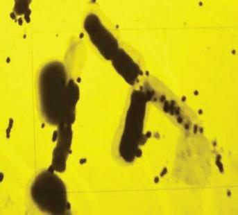
Figure 1. SEM and TEM pictures of the partially digested bacteria with selenium nanoparticles (Eszenyi et al., 2011)
Lactobacillus casei ATCC 393 (L. casei 393) was used for the bioconversion of selenium (Xu et al., 2018). The fresh culture medium of L. casei 393 was cultivated with 200 mg/mL of sodium selenite at 37 °C for 24 hrs under anaerobic conditions, without shaking. At the end of the fermentation, the culture medium was centrifuged at 12,500 rpm for 10 min, and then pellets were washed twice with phosphate buffer solution. Finally, red selenium nanoparticles with a size of 50–80 nm accumulated intracellularly. As a result, L. casei 393 enriched with selenium nanoparticles promoted the growth and proliferation of porcine intestinal epithelial cells (IPEC-J2), human colonic epithelial cells (NCM460), and human acute monocytic leukaemia-cell (THP-1) derived macrophagocyte, and it inhibited the growth of human liver tumour cell line (HepG2).
Similarly, Visha et al. (2015) synthesised selenium nanoparticles with 15–50 nm by a strain of Lactobacillus acidophilus. The fermentation method was the same as in the previously mentioned studies. The culture medium was autoclaved at 121 °C for 20 min to disrupt the bacterial cell wall and release the selenium nanoparticles. Then the mixture centrifuged at 14,000 rpm for 15 min and washed thrice with
distilled water and fi nally ultrasonicated for 15 min in order to disintegrate the cohesive selenium spheres.
Enterococcus faecalis (ATCC NO.: 29212) was used as reducing bacteria for the synthesis of selenium nanoparticles (Shoeibi & Mashreghi, 2017). The concentrations of sodium selenite (0.19 mM–2.97 mM) were added into the culture medium (LB broth) at 37 °C on a rotary shaker (150 rpm) for 24 hrs and 48 hrs. At the end of the fermentation process, the converted nanoparticles were isolated from the culture by centrifugation at 10,000 rpm for 10 min and washed with distilled water several times. Finally, the bio-fabricated nanoparticles were spherical in shape and ranged from 29 to 195 nm in size. This study showed that low concentrations of sodium selenite (such as 0.95 mM) could produce a larger amount of selenium nanoparticles compared with higher concentrations. Also, the authors wish to point out that the extracellular synthesis of selenium nanospheres easily separated from the bacterial mass, without any physical or chemical treatment. The produced selenium nanoparticles inhibited the growth of S. aureus.
Some studies reported that Staphylococcus sp. can convert inorganic selenium into its elemental form. For example, S. carnosus was used for the synthesis of selenium nanoparticles with sodium selenite. The culture medium with different concentrations (1–5 mM) of sodium selenite was incubated at 37 °C for 72 hrs under constant shaking at 180 rpm. Then the reaction mixture was centrifuged for 10 min at 2,000 rpm, and the pellet was resuspended in phosphate-buffered saline. Finally, cells were lysed by sonication, employing 5 cycles of 2 min each. The solution was centrifuged for 30 min at 10,000 rpm, and the pellet was washed with ethanol and distilled water. This produced particles washed with ethanol, having an average diameter of 439 nm, and particles washed with water, having an average diameter of 525 nm. This study showed the concentration-dependent activity of the different selenium particles against E. coli, S. cerevisiae, and Steinernema feltiae. In addition, a high concentration of selenium nanoparticles (1,000 μM) generated biologically are also toxic against its own Gram-positive producer, even when added externally (Estevam et al., 2017).
Actinobacteria (Streptomyces minutiscleroticus M10A62) also synthesised selenium nanoparticles (10–250 nm). Preparation method: 5 g of previously prepared wet bacterial biomass was dissolved in 100 mL of an aqueous solution of 1 mM selenite and incubated in a rotary shaker for 72 hrs. After the incubation period, the reaction mixture was centrifuged at 20,000 rpm for 1 h and fi ltered. The synthesised selenium nanoparticles showed good antiviral activity against Dengue virus (Ramya et al., 2015).
Selenium-tolerant aerobic microorganisms may provide an opportunity to overcome these limitations in the biosynthetic processes. Some aquatic and soil bacterial
strains have been shown to resist selenium oxyanions and reduce them to elemental selenium or methylated selenium forms, which become this way less bioavailable and toxic. For example, the generation of selenium nanoparticles by soil bacteria Bacillus sp. and Pseudomonas aeruginosa under aerobic conditions has recently been reported; however, these studies include only the partial characterization of selenium nanospheres (Dhanjal & Cameotra, 2010; Tejo Prakash et al., 2009).
Bacillus subtilis synthesised semiconductor monoclinic selenium nanoparticles with diameters ranging from 50 to 400 nm for the detection of H2O2 biosensors. In this case, 100 mL medium with 4 mM sodium selenite and 1 mL activated B. subtilis were incubated at 35 °C for 48 hrs on a rotary shaker (170 rpm). At the end of the growing time, it was centrifuged at 10,000 rpm for 6 min and then washed with doubledistilled water and absolute ethanol several times. Also, they converted spherical monoclinic selenium nanoparticles into a highly anisotropic, one-dimensional (1D) trigonal structure after one day at room temperature (Wang et al., 2010).
Bacillus mycoides SeITE01, which was isolated from the rhizosphere of the selenium hyper accumulator legume Astragalus bisulcatus, synthesised selenium nanoparticles with sizes ranging from 50 to 400 nm. In this procedure, 100 mL of nutrient broth with concentrations of 0.5 and 2 mM of sodium selenite was incubated at 28 °C for 48 hrs on a rotary shaker (200 rpm). After growth, the culture medium was centrifuged at 10,020 g for 10 min. Then the cell-free medium was centrifuged at 41,410 g for 30 min and washed with water, and then the two centrifugation steps were repeated. Extra- and intracellular elemental selenium production was detected in this reaction. This study showed that the size of selenium nanoparticles was dependent on the incubation times, showing a direct relationship between incubation time and the nanoparticle size. For example, the average diameter of the selenium nanoparticles was 50–100 nm and 50–400 nm after 6 hrs and 48 hrs of the incubation period respectively (Lampis et al., 2014).
Cremonini et al. (2016) prepared selenium nanoparticles using Bacillus mycoides having a size of 160.6 ± 52.24 nm, and by using Stenotrophomonas maltophilia the nanoparticles’ size was 170.6 ± 35.12 nm. In the method adopted for the preparation of nanoparticles, 105 CFU/mL B. mycoides and 107 CFU/mL S. maltophilia and 2 mM sodium selenite with the nutrient broth were incubated aerobically at 27 °C in a rotary shaker at 150 rpm for 6 hrs. Then the mixture medium was centrifuged at 10,000 g for 10 min, washed twice with 0.9% NaCl, suspended in Tris/HCl buffer (pH 8.2), and the cells were disrupted by ultrasonication for 5 min. Then the suspension was centrifuged at 10,000 g for 30 min. Finally, the nanoparticles were centrifuged at 40,000 g for 30 min, washed twice, and suspended in deionized sterile water. The selenium nanoparticles synthesised by both B. mycoides and S. maltophilia had high antibacterial activities with low MIC values against a clinical
strain of Pseudomonas aeruginosa but no biocidal effect against Candida species of C. albicans and C. parapsilosi (Cremonini et al., 2016).
Bacillus cereus synthesised successfully red elemental selenium from a precursor selenium source that was reported by several studies. The strain AJK3 of Bacillus cereus isolated from a polluted lake was able to produce amorphous selenium nanoparticles of 93 nm. The medium nutrient broth complemented with various sodium selenite concentrations (0.25–1.0 mM) was inoculated at 37 °C for 24–72 hrs. Then the culture medium was separated from the bacterial cells and the nanoparticles by centrifugation at 16,750 rpm for 10 min. The particle size varied from 50 to 150 nm (Kora, 2018). Similarly, amorphous selenium nanospheres between 150 and 200 nm in diameter were synthesised by a strain CM100B of Bacillus cereus under aerobic conditions. In preparation, 100 mL of tryptic soya broth (TSB) with 2 mM of 1 M sodium selenite stock solution was inoculated at 37 °C at 200 rpm. Samples were collected at 2 h intervals and a simple centrifugation step (1,844 rpm) separated supernatants from the bacterial biomass. The authors mentioned the ability of the strain to tolerate high levels of toxic selenite ions, which was studied by challenging the microbe with different concentrations of sodium selenite (0.5–10 mM) (Dhanjal & Cameotra, 2010).
Bacillus megaterium (BSB6 and BSB12) isolated from the Bhitarkanika mangrove soil transformed spherical selenium nanoparticles of sizes around 200 nm. In the conversion process, 100 μl cell suspension with 2.0 mM selenite was inoculated into 100 mL nutrient broth at 37 °C for 48 hrs. Then, the mixture was medium fi ltered through polycarbonate micro-pore fi lters (0.22 μm), washed with Tris–HCl buffer three times, and fi xed with 3% glutaraldehyde in 0.1 M phosphate buffer for 60 min. The suspended solution was centrifuged, washed with deionized water, and subjected to acid digestion for 5 min with 10 mL of HNO3 and 0.5 mL of H2SO4 followed by the reduction of selenite with 6 M HCl at 100 °C for 30 min (Mishra et al., 2011).
The selenium nanoparticles bio-transformed by Pseudomonas aeruginosa ATCC 27853 were spherical, polydisperse, with a size ranging from 47 to 165 nm, and the average particle size was about 95.9 nm under aerobic conditions. In brief, the nutrient broth medium supplemented with different concentrations of sodium selenite (0.25–1.0 mM) was inoculated with 100 mL of bacterial suspension containing 107 CFU/mL and incubated at 37 °C for 24–72 hrs under static conditions. Then the bacterial cells and the nanoparticles were separated from the culture medium by centrifugation at 10,000 rpm for 10 min (Kora & Rastogi, 2016).
Spherical selenium nanoparticles with diameters ranging from 50 to 500 nm were prepared by Pseudomonas alcaliphila. Trigonal nanorods occurred after incubating in an aqueous reaction solution for 24 hrs, as shown in Figure 2. In the production of nanoparticles, 1 mL freshly activated bacterial culture with 2.63 g
selenite (0.1 M) was added into the medium. After 48 hrs of the reaction, the culture was centrifuged at 10,000 rpm for 10 min and washed with double-distilled water. For the purifi cation of nanoparticles, the solution was centrifuged at 10,000 rpm for 10 min in the complete salts solution: 0.25 M NaOH, 0.1 M NaOH, 10 mM Na2HPO4, and carbon-free, distilled, deionized water(Zhang et al., 2011).
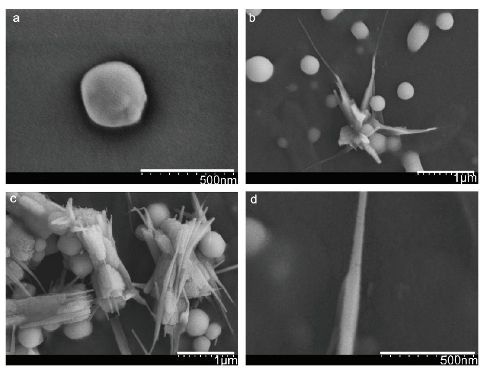
Figure 2.FESEM images of the transformation process from monoclinic selenium nanospheres to trigonal selenium nanorods. (a) An individual selenium nanosphere, (b) the beginning of the transformation, (c) the aggregation of trigonal selenium nanorods, (d) The image of an individual selenium nanorod (Zhang et al., 2011)
Under aerobic conditions at room temperature, Pantoea agglomerans strain UC-32 synthesised selenium nanoparticles smaller than 100 nm. In this study, UC-32cells were grown in TSB supplemented with 1 mM sodium selenite and alkaline lysis was used for the isolation and purifi cation of the nanoparticles from the bacterial cell mass. Then the cell suspensions were sonicated at 100 W for 2 min and centrifuged at 100,009 g for 10 min sequentially in SDS 0.1 %/1 M NaOH. Finally, pellets were resuspended in distilled water. It was also reported that stabilized selenium nanoparticles with L-cysteine (4 mM) had a high antioxidant activity compared to selenite and non-stabilized selenium nanoparticles (Torres et al.,2012).
Besides, Gram-negative soil bacteria Ralstonia eutropha successfully synthesised extracellular, stable, uniform, and spherical selenium nanoparticles of sizes ranging from 40 to 120 nm. In this study, bacterial biomass was used for conversion. After 24 hrs of incubation, the bacterial biomass was harvested by centrifugation at a speed of 5,000 rpm at room temperature for 10 min and washed several times using sterilized Millipore water. Then 2 g of wet R. eutropha biomass with 100 mL aqueous 1.5x10⁻³ M sodium selenite solution was incubated at 30 oC for 48 hrs at 120 rpm. After the incubation time, the mixture reaction was centrifuged at 12,000 rpm for 10 min and washed several times with Millipore water and acetone. These synthesised selenium nanoparticles signifi cantly inhibited the growth of Pseudomonas aeruginosa, Staphylococcus aureus, Escherichia coli, Streptococcus pyogenes, and Aspergillus clavatus (Srivastava & Mukhopadhyay, 2015b). However, these pathogenic bacteria have been used for the biofabrication of nano-selenium. For example, Medina Cruz et al. (2018) used Escherichia coli K-12 HB101, Pseudomonas aeruginosa ATCCVR 27853, Methicillin-resistant Staphylococcus aureus MRSA ATCC 43300, and S. aureus ATCC12600 as reducing agents. Fresh bacterial culture in Luria-Bertani (LB) broth was mixed with an aqueous solution of 2 mM sodium selenite and kept at 37 °C in a shaking incubator at 200 rpm for 72 hrs. At the end of the incubation time, it was centrifuged at 7,500 rpm for 15 min. The supernatant was removed, and the pellet was washed twice with a 0.9% NaCl solution and resuspended in 15 mL of Tris/HCl buffer. Finally, ultrasonication and hyperthermia-based approaches were used for the purifi cation of selenium nanoparticles. The results showed that the average diameter of the synthesised selenium nanoparticles was 90–150 nm; 25 to 250 mg/mL concentrations of selenium nanoparticles showed antibacterial effect against S. aureus and E. coli and no signifi cant cytotoxicity effect against human dermal fi broblasts for 24 hrs (Medina Cruz et al., 2018).
Similarly, Escherichia coli ATCC 35218 biosynthesised red amorphous selenium nanoparticles of sizes ranging from 100 to 183 nm, and the average particle size was about 155 nm. The result showed that the bacterial strain was evident from 89.2% of selenium removal within 72 hrs at a concentration of 1 mM (Kora & Rastogi, 2017).
Zooglea ramigera synthesised extracellularly monoclinic crystalline selenium nanoparticles (30–150 nm). In this procedure, 1 mL of Z. ramigera culture and 3 mM of sodium selenite were added into 100 mL of sterilized N-broth medium and incubated at 30 °C for 48 hrs at 150 rpm. Then, the converted particles were isolated from the mixture by centrifugation at 12,000 rpm for 10 min and washed several times with distilled water and acetone (Srivastava & Mukhopadhyay, 2013).
3. Selenite reduction with fungi
The antifungal activity of selenium nanoparticles is well known; most fungi are sensitive to selenium compounds. However, some studies have shown that few fungi have demonstrated to have the capacity of selenium nanoparticle biosynthesis. Basically, biochemical and molecular mechanisms underlying selenium oxyanions’ reduction into nanoparticles are still unclear, especially when involved in fungal transformation under aerobic conditions (Vetchinkina et al., 2013).
Lentinula edodes converted sodium selenite and the organoselenium compound 1,5-diphenyl-3-selenopentanedione-1,5 (DAPS-25) into elemental form with a diameter of 180.51 ± 16.82 nm. Aqueous solutions of 10-6 mol sodium selenate, 10-2 mol sodium selenite, and 10-7 mol selenium-containing formulation DAPS-25 with 10-3 mol 50% ethanol were added to both beer wort and liquid synthetic medium of fungus separately. Results showed that red selenium nanoparticles accumulated intracellularly in the fungal L. edodes (Vetchinkina et al., 2013).
Aspergillus terreus synthesised spherical selenium nanoparticles with an average size of 47 nm after 60 min of incubation. To complete the formation of the nanoparticles, 20 mL fi ltered supernatants of Aspergillus terreus were added to 80 mL of sodium selenite solution (100 mg/mL), and the reaction mixture was incubated at room temperature for 60 min. Then, the mixture was centrifuged at 20,000 g for 10 min and washed with distilled water three times (Zare et al., 2013).
Gliocladium roseum prepared spherical selenium nanoparticles in the range of 20–80 nm. Also, a crystalline structure was observed in this study. In the preparation method, 100 mL of cell-free fi ltrate and a relevant amount of sodium selenite were mixed to make the overall solution of 1.5 mM sodium selenite salt, and the mixture was incubated at 30 °C for 24 hrs at 120 rpm. After 24 hrs, the mixture was centrifuged at 12,000 rpm for 15 min and washed with distilled water and acetone for the separation and purifi cation from cell mass. All selenium nanoparticles were synthesised extracellularly (Srivastava & Mukhopadhyay, 2015a).
4. Selenite reduction with plant extracts
The selenium nanoparticles have been synthesised by plant extracts because metabolites produced from the plants help in the reduction of precursor molecule. This procedure has several advantages over other biological methods with bacteria and fungi because it is inexpensive, does not need any special condition, and the synthesis method is free of any toxic-reducing agents and organic solvents. In addition, the biomolecules present in the extract are assumed to provide
stabilization and to reduce the nanoparticle, also enhancing its potency as an antimicrobial and antioxidant agent.
Lemon leaf extract successfully synthesised spherical, polydispersed, and crystalline selenium nanoparticles of sizes ranging from 60 to 80 nm. In this method, 2 mL of leaf extract from the homogenisation of 0.5 g of the lemon leaf was added dropwise into 20 mL of 10 mM sodium selenite solution under magnetic stirring. Then the mixture was kept at 30 °C with a rotary shaker operating at 200 rpm for 24 hrs in dark conditions (Prasad et al., 2013).
Clausena dentata is a fl owering plant from the citrus family, which synthesised selenium nanoparticles ranging from 46.32 nm to 78.88 nm. In the conversion steps, 12 mL of plant extract was added to 88 mL of 1 mM aqueous selenium powder and kept until the reaction turned brown. Then the obtained extract was fi ltered through a paper fi lter (No1 Whatman). These produced selenium nanoparticles with low concentration (LC50) signifi cantly controlled mosquito vectors at early stages, including 240.714 mg/L for A. stephensi, 104.13 mg/L for A. aegypti, and 99.602 mg/L for C. quinquefasciatus (Sowndarya et al., 2017).
Citrus reticulata also fabricated spherical selenium nanoparticles with a size of 70 nm. In the preparation, 50 mL of orange peel extract was mixed with 5 mL of 0.1 M sodium selenite in drops until the reaction turned red. The reaction was controlled at temperatures of 30 °C, 40 °C, 50 °C, 60 °C, 70 °C , and 80 °C and at pH 2, 4, 6, 8, 10, and 12. The results showed that selenium nanoparticles were found to be effi cient at 40 °C and at pH 4. The selenium nanoparticles inhibited the bloom of algae (Sasidharan et al., 2015).
Garlic extract, Allium sativum, produced uniform, mono-dispersive, and highly stable selenium particles with the size range of 48–87 nm. Briefl y, 5 mL of the garlic extract was mixed with 50 mL of 20 mM sodium selenite solution under magnetic stirrer with 150 rpm and heated at 60 °C for 24 hrs. Then, the mixture was centrifuged at 10,000 rpm for 30 min, washed with double-distilled water and ethanol three times, and fi nally dried at room temperature. The garlic acid-mediated selenium nanoparticles showed good feed additive material for aquaculture. Therefore, this study suggested that selenium nanoparticles can be used for feeding the larvae of any fi nfi sh and shellfi sh when supplementing the trace elements (Satgurunathan et al., 2017). Allium sativum as a capping and reducing agent is used for the production of selenium nanoparticles with the size range of 40–100 nm. Briefl y, 2 mL of homogenised garlic cloves extract was added dropwise into 20 mL of 10 mM sodium selenite under magnetic stirring conditions and was kept at 36 °C on the rotary shaker at 120 rpm for 5–7 days in dark condition. In this study, the cytotoxicity of this selenium nanoparticle was compared to the chemically synthesised selenium nanoparticle against Vero cells. The results showed that Vero cells treated with chemically synthesised
nanoparticles led to a CC50 of 18.8 ± 0.8 lg/mL, while Vero cells treated with green-synthesised nanoparticles produced a CC50 of 31.8 ± 0.6 lg/mL (Anu et al., 2017).
Allium sativum produced 21–40 nm- and 41–50 nm-sized selenium nanoparticles at 4 hrs and 72 hrs respectively (Sribenjarat et al., 2020). In the preparation process, 0.06 g of garlic extract was dissolved in 20 mL of 10 mM sodium selenite, and 80 mM of ascorbic acid solution was added dropwise until a slightly yellow colour was achieved. After colour changing, the mixture was incubated at 130 rpm for 72 hrs in the dark. The samples were collected by centrifugation at 20,000 g at 4, 24, 48, and 72 hrs of the incubation period and washed twice with distilled water. Biosynthesised selenium nanoparticles showed less cytotoxicity to normal human MRC-5 cell after 72 hrs compared to 24 hrs and inhibited the growth of S. aureus.
Similarly, hollow and spherical selenium nanoparticles of sizes ranging between 7 and 45 nm and between 8 and 52 nm were synthesised by Allium sativum. In brief, 10 drops extract of Allium sativum was added into 25 mL of 5 mM sodium selenite solution until the colour changed on the magnetic stirrer. The biofabricated selenium nanoparticles were stable for more than 2 months and presented high antioxidant activity. 100 μl and 25 μl concentration of selenium nanoparticles synthesised by Allium sativum showed the highest antimicrobial activity against Bacillus subtilis and the least against Staphylococcus aureus (Vyas & Rana, 2017, 2018b). In addition, these authors reported that 9–58 nmsized selenium nanoparticles were produced by Aloe vera leaf extract. In the short method, 10 drops of Aloe vera leaf extract was added into 25 mL of 5 mM sodium selenite solution until the colour changed on the magnetic stirrer (Vyas & Rana, 2018a). Aloe vera extract synthesised spherical selenium nanoparticles of 50 nm. In this case, 4.92 mL extract was added into 13.03 mL 10 mM sodium selenite and autoclaved at 121 °C and 1.5 bar for 15 min. The synthesised selenium nanoparticles indicated high antibacterial (E. coli and S. aureus) and antifungal (C. coccodes and P. digitatum) activities (Fardsadegh & Jafarizadeh-Malmiri, 2019).
Zingiber offi cinale was used for the synthesis of selenium nanoparticles, which was confi rmed by the colour change from pale yellow to red. In brief, 1% of ginger extract was added to 10 mM of sodium selenite solution in the ratio of 9:1 and kept at room temperature for 75 hrs under stirring conditions of 130 rpm. Biosynthesised selenium nanoparticles in 250 μg/mL showed antibacterial activity against Proteus sp. (Menon et al., 2019).
Psidium guajava (guava leaves) extract transformed sodium selenite into selenium nanoparticles with diameters in the range of 8–20 nm. In the formation of nanoparticles, 100 mL of freshly prepared guava leaf extract was mixed with 900 mL of aqueous sodium selenite (25 mM) at 60 °C for 3 hours. Subsequently,
the mixture was centrifuged at 13,280 rpm for 20 min, washed thrice with distilled water, and then air-dried. The synthesised nanoparticles showed the antibacterial effect on both Gram-positive (S. aureus MTCC-3160) and Gramnegative (E. coli MTCC-405) bacteria. Also, they were non-toxic to human cell lines (CHO pro-cells) and had toxic effects against hepatic cell lines (HepG2 cells) (Alam et al., 2019).
Amorphous selenium nanoparticles were synthesised using Emblica offi cinalis fruit extract of sizes ranging from 15 to 40 nm. In short, 2 mL of aqueous fruit extract of E. offi cinalis was added dropwise into 10 mL of 10 mM sodium selenite under magnetic stirring conditions, and the mixture was kept at 27 ± 2 °C for 24 hrs at 120 rpm in dark condition. The fruit-extract-mediated selenium nanoparticles showed antimicrobial and antifungal activities against several strains (Gunti et al., 2019).
Selenium nanoparticles synthesised using Hawthorn (Crataegus hupehensis Sarg.) fruit extract are mono-dispersed and stable, with an average size of 113 nm. In the conversion method, 2 mg/mL of lyophilised extract was mixed with 0.01 M of sodium under magnetic stirring for 12 hrs, and then the mixture was dialysed (MWCO 8000–14000) in ultrapure water for 48 hrs. The prepared selenium nanoparticles indicated anti-tumour activity against HepG2 cells (Cui et al., 2018).
Pelargonium zonale leaf extract synthesised 50 nm of selenium nanoparticles under microwave irradiation. 15 mL 10 mM sodium selenite solution with 1.48 mL plant extract was exposed to microwave radiation for 4 min and at constant power of 800 W. Biosynthesised selenium nanoparticles showed antibacterial and antifungal activities against Escherichia coli, Staphylococcus aureus, Colletotrichum coccodes, and Penicillium digitatum (Fardsadegh et al., 2019).
Spherical selenium nanoparticles with the size of approx. 400 nm were produced with the use of parsley (Petroselinum crispum) leaves extract. In the steps of preparation, lyophilised leaves with distilled water were homogenised in the ratio 1:10 (w/v) and fi ltered. Then 10,000 ppm selenite solution was added (ratio 1:10 and 1:1 v/v) at room temperature. The mixture was centrifuged at 5,000 rpm for 10 min after the reaction had turned red. Then the supernatant was washed with distilled water, followed by repeated centrifugations (three times), fi ltrations, and drying (Fritea et al., 2017).
Azadirachta indica leaves extract synthesised spherical and smooth-surfaced selenium nanoparticles with sizes of ~153 and ~287 nm after 5 and 10 min of reaction period, as shown in Figure 3. In the process, 10 mM sodium selenite solution with 100 mL of aqueous leaves extract was incubated at 37 °C on a rotary shaker at 100 rpm. After 5 and 10 min, selenium nanoparticles were centrifuged at 10,000 rpm for 10 min and washed three times with distilled water. The synthesised
selenium nanoparticles showed dose-dependent antibacterial activity against Pseudomonas aeruginosa (Mulla et al., 2020).
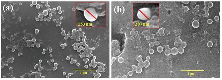
Figure 3. SEM images (FESEM images in insight) of purifi ed spherical selenium nanoparticles at (a) 5 and (b) 10 min of the reduction reaction (Mulla et al., 2020)
Selenium nanoparticles synthesised using Ocimum tenuifl orum leaf extract are spherical selenium nanoparticles in the size range of 15–20 nm. Briefl y, 1% of leaf extract and 10 of mM selenite solution were mixed with a ratio 9:1 at room temperature under stirring conditions of 130 rpm for 72 hrs. The synthesised selenium nanoparticles inhibited the aggregation and growth of urinary stone (CaC2O4 monohydrate (COM) crystals) (Liang et al., 2019).
5. Selenite reduction with chemical agents
Chemical transformation is the widely used technique for the production of nanoparticles, which has two types of reduction and sedimentation method. Chemical reduction method aids in maintaining a better uniformity of the particles, which can be used in the previously mentioned fi elds. Nanoparticle synthesis with various shapes and sizes is performed through the reduction of metal ions to neutral metal atoms by the addition of a reducing agent. Particularly in the fi rst step of transformation, nucleation, allow atoms to form small clusters, called “seeds”, of a stable structure and defi ned crystallinity (Chapman et al., 2012; Xiong & Xia, 2007). The next step contains the “seeds” to form nanocrystals of different shapes and structures (Xiong & Xia, 2007). As aggregation occurs, the surface energy of the metal also grows, and smaller particles readily interact with each other to form larger particle sizes. A capping agent or stabilizing agent is used to prevent further aggregation by forming an electrical bilayer around the nanoparticle occurring from the adsorption of ions onto the surface of the nanoparticle.
Many biocompatible reducing agents, such as L-cysteine, D-fructose, glucose, lactose, gallic acid, some polysaccharides, ascorbic acid, etc., have been employed in the synthesis of selenium nanoparticles of various sizes and shapes. Basically, the shape of selenium nanoparticles is controlled by designing the chemical structure of the stabilizing agent through a self-assembly process.
A monodisperse and homogeneous spherical elemental selenium with an average diameter of about 100 nm was prepared by the reaction of sodium selenite with L-cysteine as a surface modifi er and reducing agent in 1:4 ratios. A varied volume of 50 mM of L-cysteine solution was added dropwise into 0.25 mL of 0.1 M sodium selenite stock solution under magnetic stirring at room temperature for 30 min, and the mixture was reconstituted to a fi nal volume of 25 mL with Milli-Q water (Li et al., 2010).
Another study used dithiothreitol and gallic acid as reducing agents for the synthesis of selenium nanospheres. The gallic acid solution was used at different concentrations (3 mM–20 mM) in pH 5.7 with sodium selenite in a 1:1 ratio. The reducing agent dithiothreitol was added dropwise (~10 μL) until a brick red colour was observed, and the formation of selenium nanoparticles was monitored over a period of 24 hrs by fl uorescence spectroscopy. Afterwards, the mixture solution was centrifuged and then washed with water. The results showed that alteration in the pH (pH 5–7) changed the size and shape of large-diameter nanospheres to nanofi bres with diameters of 50–75 nm. Also, when grown at pH 7, nanospheres larger in diameter (> 500 nm) were obtained. At pH 5, the colour of the gallic acid turned yellowish orange, while at pH 7 the colour turned dark brown, which was indicative of the changed shape and size of the nanoparticles (Barnaby et al., 2013).
A study has shown the reducing effect of monosaccharides for the conversion of selenium. For example, trigonal selenium (t-SeNPs) and amorphous selenium (a-SeNPs) were synthesised using D-fructose as reducing agent. Five mL of sodium selenite solution (1.0 mmol/L) was slowly dripped into 10.0 mL of 1.0 mmol/L aqueous solution of D-fructose. Then the mixture solution was stirred under heat at 45 °C for 15 min. This time, red amorphous selenium nanoparticles and trigonal selenium nanoparticles were obtained after 20 min. Finally, each solution was centrifuged at 13,000 rpm for 10 min and washed with deionized water, and then they were centrifuged again under the same conditions. These selenium nanoparticles were non-toxic for human healthy cells of the fi broblast cell line P4 and showed high toxicity towards the sarcoma cells (Vieira et al., 2017).
Cavalu et al. (2018) also used disaccharides for the conversion and synthesised amorphous selenium nanoparticles of 20–40 nm. In the conversion method, 25 mL of sodium hydrogen selenite in the concentration of 10,000 ppm was selected as precursor selenium source mixed with 25 mL of lactose solution in a ratio of 1:8 (w/w) by vigorous stirring using a magnetic stirrer and then heated at 120 °C
for 3 min. After cooling, the mixture was centrifuged at 6,000 rpm for 10 min and washed with distilled water, followed by repeated centrifugation (4 times), fi ltration, and drying (Cavalu et al., 2018).
Other studies reported that trigonal selenium nanowires were synthesised by chemical methods in a physical way. For example, trigonal selenium (t-Se) nanowires and microspheres were synthesised at room temperature by the chemical precipitation method using hydrazine hydrate as precipitator in the presence of 1,2,3-trimethylimidazolium-tetrafl uoroborate (tmimBF4) with sodium selenite (Ma et al., 2008). Similarly, trigonal selenium nanowires were successfully synthesised via low-temperature hydrothermal synthesis route. In a typical procedure, sodium selenite (0.01 mol) and thiosulfate salts (Na2S2O3 0.01 mol or (NH4)2S2O3 0.01 mol) as reducing agents were added into 40 mL distilled water and autoclaved at 180 °C for 12 hrs. The precipitates were fi ltered and washed with distilled water and absolute alcohol several times after cooling down and were dried in vacuum at 50 °C for 4 hrs. Abundant nanowires with a diameter of 10–20 nm and a length up to 3–5 μm were observed in the sample with Na2S2O3, and the mean diameter and length of these wires was 60 nm and 3 μm in the prepared sample (NH4)2S2O3 (An & Wang, 2007).
A mixed solvent of ethylene glycol assembled amorphous and trigonal selenium nanoparticles of various diameters. In a typical synthesis, 1.02 g of sodium selenite and 1, 2, 4, and 8 g of glucose were dissolved in 70 mL of ethylene glycol and 15 mL of H2O mixed solution and incubated at 85 °C for 45 min, 1 h, and 1.5 hrs. Then the samples were washed with distilled water several times and kept in dark condition. The results showed that the size of the selenium nanoparticles increased from 320 to 480 nm in the presence of 1 and 8 g of glucose. Also, the amorphous and trigonal phase was observed with 8 g of glucose at 45 min and 1 h of the incubation period (Chen et al., 2011).
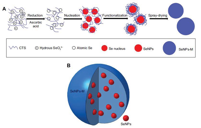
Bai et al. (2017) synthesised trigonal-phase selenium nanoparticles of ~35 nm by physical method from selenium-nanoparticle-loaded chitosan microspheres. In the preparation method, 10 mL of selenite aqueous solution containing 0.4 g of sodium selenite was slowly added to the 100 mL of 1% (w/w) acetic acid containing 0.5 g of chitosan and 1.6 g of ascorbic acid and vigorously stirred at 600–800 rpm. Then the mixture solution was dialysed against 1% (w/w) of acetic acid for 6 hrs to remove the excess ascorbic acid and other by-products. After that, the colloid was well mixed with another chitosan solution, the fi nal concentrations of selenium and chitosan being 0.09% (w/w) and 2.5% (w/w) respectively. Finally, the spray-drying process was applied to evaporate the moisture of the new mixture by the spray dryer tool. This preparation process is shown in Figure 4. The result showed that selenium-nanoparticle-loaded chitosan microspheres had powerful antioxidant activities, as evidenced by a dramatic increase of both selenium retention and the levels of glutathione peroxidase, superoxide dismutase, and catalase (Bai et
al., 2017). Basically, chitosan with low molecular weight has specifi c biological activities such as antibacterial activity, anticancer activity, and immune-boosting effects (Dodane & Vilivalam, 1998).
Figure 4. The preparation process (A) and the expected structure (B) of selenium-nanoparticle-loaded chitosan microspheres (SeNPs-M) (Bai et al., 2017)
Actually, ascorbic acid is a widely used reducing agent for the synthesis of selenium, gold, and silver nanoparticles (Qin et al., 2010; Sun et al., 2009). Basically, ascorbic acid is a vitamin participating in several biochemical reactions and a naturally available antioxidant. It has high water solubility, biodegradability, and low toxicity (Sun et al., 2009) compared with some chemical-reducing agents. Particularly, a spherical shape with an average diameter ranging between 15 and 18 nm of selenium nanoparticles was produced by ascorbic acid. In this study, selenium nanoparticles were prepared by the following procedure, a stock of aqueous solution of 100 mM sodium selenite and 50 mM ascorbic acid mixed in 1:4 ratios under magnetic stirring at ambient temperature for 30 min. Then the solution was centrifuged at 3,000 rpm. The authors mentioned that chemically synthesised selenium nanoparticles could be a potential antibacterial agent to treat humans affected by bacterial diseases caused by major pathogenic bacteria such as Staphylococcus aureus, Escherichia coli, and Pseudomonas aeruginosa. The inhibition of synthesised nano-selenium against pathogens was similar to the effect of ampicillin (Angamuthu et al., 2019). Another study used ascorbic acid
with various ratios and the stirring effect on the reduction of selenium. A stock solution of 100 mM sodium selenite and 50 mM ascorbic acid were mixed in various ratios (1:1, 1:2, 1:3, 1:4, 1:5, 1:6). Ascorbic acid was added dropwise to the sodium selenite solution under magnetic stirring at different rpms (200, 600, 1,000) at room temperature for 30 min. As a result, they suggested ratios of 1:3 and 1:4 of ascorbic acid and sodium selenite under stirring at 1,000 rpm for 30 min (Malhotra et al., 2014).
With Undaria pinnatifi da, polysaccharides and ascorbic acid produced selenium nanoparticles. In the procedure, 1 mL of 0.1% U. pinnatifi da polysaccharide solution was mixed with 8 mL 60 mM of ascorbic acid under magnetic stirring, and then 1 mL 30 mM of sodium selenite solution was slowly added. After reaction for 5 min under sonication conditions, the solutions were purifi ed with Milli-Q water. The produced selenium nanoparticles with IC50 values ranging from 3.0 to 14.1 μm showed the inhibition effect against human cancer cells such as A375, CNE2, Hep G2, and MCF-7 (Chen et al., 2008).
Similarly, ascorbic acid and polyvinyl alcohol (PVA) or chitosan (CS) as stabilizers synthesised selenium nanoparticles with an average diameter of 66.55 ± 8.46 and 48.52 ± 2.77 nm. In the preparation process, 50 mM of sodium selenite and PVA 0.1% or CS 1% solutions were mixed under magnetic stirring conditions at room temperature for 5 min. Then, 50 mM of ascorbic acid was added dropwise to mixture solutions and mixed with a magnetic stirrer for 30 min. As a result, the synthesised selenium nanoparticles showed dose-dependent antibacterial activity, but PVA-coated selenium nanoparticles exhibited signifi cant effects against S. epidermidis (MIC 125 ppm) and S. aureus (MIC 125 ppm). The IC50 values of the selenium nanoparticles were 26.56 and 530 ppm for PVA-coated selenium nanoparticles and CS-coated selenium nanoparticles respectively (Boroumand et al., 2019).
6. Characterization techniques
In these studies, the characteristics of the selenium nanoparticles were analysed by UV-Visible Spectrophotometry, X-ray diffraction analysis (XRD), Fourier transform resonance spectroscopy (FT-IR) analysis, Dynamic Light Scattering Particle Size Analyser (DLS), Scanning Electron Microscope (SEM), Energy Dispersive X-Ray (EDS), and Transmission Electron Microscope (TEM) techniques.
UV-Vis spectrum is the most basic and important technique for the identifi cation and characterization of nanoparticles. UV-Vis spectroscopy determines the “absorption maxima” of nanoparticles depending on the concentration of the precursor and other components of reaction mixtures. Biological-mediated selenium nanoparticles indicated an absorption peak at 226–590 nm, whereas
nanoparticles synthesised from bacterial strains of Bacillus megaterium, and Bacillus cereus exhibited maximum absorption at 200–300 nm and 590 nm respectively (Mishra et al., 2011; Dhanjal & Cameotra, 2010). Chemically synthesised selenium nanoparticles indicated an absorption peak at 270–580 nm, whereas lactose nanoparticles showed absorption maxima at 270 nm, and a high concentration of L-cysteine-induced nanoparticles exhibited absorption maxima at 580 nm (Cavalu et al., 2018; Li et al., 2010).
X-ray diffraction analysis (XRD) was used to examine the composition and phase of resultant samples of selenium nanoparticles. Basically, selenium in its nanoscale form exhibits a standard XRD pattern (23, 30, 43), which confi rms its nanoscale character, and it is similar to selenium nanoparticles originated from all different sources. The XRD analysis of biosynthesised selenium nanoparticles showed a clear structure. The XRD pattern was noisy and broader, with no sharp Bragg refl ections. Thus, the data indicates the amorphous nature of the synthesised selenium nanoparticles (Kora & Rastogi, 2016). Also, the diffraction peaks at 2θ = 23.6, 29.9, 41.4, 43.8, 45.4, 51.8, 55.9, 61.8, 65.3, and 68.3 were attributed to the (100), (101), (110), (102), (111), (201), (003), (202), (210), and (211) refl ections of the pure hexagonal phase of selenium crystal. The lattice parameters were as follows: a = 4.366 A° and c = 4.9536 A° (JCPDS 06-0362) (Srivastava & Mukhopadhyay, 2015b). For chemically synthesised selenium nanoparticles, the XRD pattern of the selenium nanoparticles indicated a broad and intense peak at about 2θ = 23°, which suggested that the nanoparticles were not crystalline (Cavalu et al., 2018). Diffraction peaks were observed at 2θ = 23, 29, 41, 43, 45, 51, 55, 61, and 65°, corresponding to the crystalline planes (100), (101), (110), (102), (111), (201), (112), and (103). All the peaks linked with the trigonal phase of selenium nanoparticles. The lattice constants of a = 4.3662 A° and c = 4.9521 A° are consistent with the standard values for bulk selenium with a = 4.3662 A° and c = 4.9536 A°, in accordance with JCPDS fi le No. 73-0465 (Vieira et al., 2017).
Fourier transform infrared spectroscopy (FT-IR) was used to analyse the surface interaction between synthesised nanoparticles and other molecules that took part in the synthesis and stabilization of nanoparticles. For example, in the case of FT-IR of chemically synthesised selenium nanoparticles, the spectrum has vibrational and stretching functions at wavenumbers 2919.69, 1630.51, 1380.78, and 1076.08 cm–1 corresponding to C–H, C=C, O–H, and C–O respectively. The band at 2,361 cm–1 is the C–H stretch of aryl acid. The strong band found at 1,654 cm–1 is characteristic of C=C stretch of an aromatic ring, N–H bending of amine, and a C=O stretch of polyphenols (Angamuthu et al., 2019).
The dynamic light scattering (DLS) technique was used to measure the hydrodynamic effective diameters of produced nanoparticles. Scanning electron microscopy (SEM) and transmission electron microscopy (TEM) with energy dispersive analysis (EDS) are well-known techniques to determine the structure,
morphology, and size of prepared nanoparticles. SEM and TEM analysis showed that biosynthesised nanoparticles exhibited spherical nanospheres with a size of 100–500 nm, showing that selenium nanospheres were located intracellularly as well as extracellularly (Eszenyi et al., 2011) and were also present in aggregates connected to the bacterial cell mass (Dhanjal & Cameotra, 2010).
7. Conclusions
The present review collected the production methods of selenium nanoparticles by biological and chemical ways as well as their properties and bioactivities. In all of the cases, selenium nanoparticles were synthesised from the reduction of sodium selenite by various types of reducing agents such as Gram-positive and Gram-negative bacterial strains, fungi, plant extracts, and pure chemical compounds. The biosynthesis usually resulted in amorphous spherical selenium nanoparticles, and chemical methods are able to synthesise selenium nanoparticles in multiple structures, depending on the reducing and stabilizer agents, their concentrations, and heat treatment. Biological sources and chemical-mediated selenium nanoparticles have shown dose-dependent antibacterial, antifungal, and anti-cancer activities.
Acknowledgements
This research was supported by the Stipendium Hungaricum Scholarship Programme.
References
[1] Alam, H., Khatoon, N., Raza, M., Ghosh, P. C., Sardar, M., Synthesis and characterization of nano selenium using plant biomolecules and their potential applications. BioNanoScience, 9. 1. (2019) 96–104. https://doi. org/10.1007/s12668-018-0569-5.
[2] An, C., Wang, S., Diameter-selected synthesis of single crystalline trigonal selenium nanowires. Materials Chemistry and Physics, 101. 2–3. (2007) 357–361. https://doi.org/10.1016/j.matchemphys.2006.06.011.
[3] Andreoni, V., Luischi, M. M., Cavalca, L., Erba, D., Ciappellano, S., Selenite tolerance and accumulation in the Lactobacillus species. Annals of Microbiology, 50. (2000) 77–88.
[4] Angamuthu, A., Venkidusamy, K., Muthuswami, R. R., Synthesis and characterization of nano-selenium and its antibacterial response on some important human pathogens. Current Science, 116. 2. (2019) 285. https:// doi.org/10.18520/cs/v116/i2/285-290.
[5] Anu, K., Singaravelu, G., Murugan, K., Benelli, G., Green-synthesis of selenium nanoparticles using garlic cloves (Allium sativum): Biophysical characterization and cytotoxicity on vero cells. Journal of Cluster Science, 28. 1. (2017) 551–563. https://doi.org/10.1007/s10876-016-1123-7.
[6] Bai, K., Hong, B., He, J., Hong, Z., Tan, R., Preparation and antioxidant properties of selenium nanoparticles-loaded chitosan microspheres. International Journal of Nanomedicine, 12. (2017) 4527–4539. https://doi.org/10.2147/IJN.S129958.
[7] Barnaby, S., Sarker, N., Dowdell, A., Bannerjee, I., The spontaneous formation of selenium nanoparticles on gallic acid assemblies and their antioxidant properties. The Fordham Undergraduate Research Journal, 1. 1. (2013) 3.
[8] Boroumand, S., Safari, M., Shaabani, E., Shirzad, M., Faridi-Majidi, R., Selenium nanoparticles: Synthesis, characterization and study of their cytotoxicity, antioxidant and antibacterial activity. Materials Research Express, 6. 8. (2019) 0850d8. https://doi.org/10.1088/2053-1591/ab2558.
[9] Cavalu, S., Kamel, E., Laslo, V., Fritea, L., Costea, T., Antoniac, I. V., Vasile, E.,
Antoniac, A., Semenescu, A., Mohan, A., Saceleanu, V., Vicas, S., Eco-friendly, facile and rapid way for synthesis of selenium nanoparticles production, structural and morphological characterization. Revista de Chimie, 68. 12. (2018) 2963–2966. https://doi.org/10.37358/RC.17.12.6017.
[10] Chapman, J., Sullivan, T., Regan, F., Nanoparticles in anti-microbial materials:
Use and characterisation. RSC Pub. (2012).
[11] Chen, H., Yoo, J.-B., Liu, Y., Zhao, G., Green synthesis and characterization of Se nanoparticles and nanorods. Electronic Materials Letters, 7. 4. (2011) 333–336. https://doi.org/10.1007/s13391-011-0420-4.
[12] Chen, T., Wong, Y.-S., Zheng, W., Bai, Y., Huang, L., Selenium nanoparticles fabricated in Undaria pinnatifi da polysaccharide solutions induce mitochondria-mediated apoptosis in A375 human melanoma cells. Colloids and
Surfaces B: Biointerfaces, 67. 1. (2008) 26–31. https://doi.org/10.1016/j. colsurfb.-2008.07.010.
[13] Cogun, H. Y., Fırat, Ö., Fırat, Ö., Yüzereroǧlu, T. A., Gök, G., Kargin, F.,
Kötemen, Y., Protective effect of selenium against mercury-induced toxicity on hematological and biochemical parameters of Oreochromis niloticus.
Journal of Biochemical and Molecular Toxicology, 26. 3. (2012) 117–122. https://doi.org/10.1002/jbt.20417.
[14] Cremonini, E., Zonaro, E., Donini, M., Lampis, S., Boaretti, M., Dusi, S.,
Melotti, P., Lleo, M. M., Vallini, G., Biogenic selenium nanoparticles:
Characterization, antimicrobial activity and effects on human dendritic cells and fi broblasts. Microbial Biotechnology, 9. 6. (2016) 758–771. https://doi. org/10.1111/1751-7915.12374.
[15] Cui, D., Liang, T., Sun, L., Meng, L., Yang, C., Wang, L., Liang, T., Li, Q.,
Green synthesis of selenium nanoparticles with extract of hawthorn fruit induced HepG2 cells apoptosis. Pharmaceutical Biology, 56. 1. (2018) 528–534. https://doi.org/10.1080/13880209.2018.1510974.
[16] Dhanjal, S., Cameotra, S. S., Aerobic biogenesis of selenium nanospheres by
Bacillus cereus isolated from coalmine soil. Microbial Cell Factories, 9. 1. (2010) 52. https://doi.org/10.1186/1475-2859-9-52.
[17] Dodane, V., Vilivalam, V. D., Pharmaceutical applications of chitosan. Pharmaceutical Science & Technology Today, 1. 6. (1998) 246–253. https://doi. org/10.1016/S1461-5347(98)00059-5.
[18] Estevam, E. C., Griffi n, S., Nasim, M. J., Denezhkin, P., Schneider, R., Lilischkis,
R., Dominguez-Alvarez, E., Witek, K., Latacz, G., Keck, C., Schäfer, K.-H.,
Kieć-Kononowicz, K., Handzlik, J., Jacob, C., Natural selenium particles from Staphylococcus carnosus: Hazards or particles with particular promise?
Journal of Hazardous Materials, 324. (2017) 22–30. https://doi.org/10.1016/j. jhazmat.2016.02.001.
[19] Eszenyi, P., Sztrik, A., Babka, B., Prokisch, J., Elemental, nano-sized (100–500 nm) selenium production by probiotic Lactic acid bacteria. International
Journal of Bioscience, Biochemistry and Bioinformatics, 1. 2. (2011) 148–152. https://doi.org/10.7763/IJBBB.2011.V1.27.
[20] Fardsadegh, B., Jafarizadeh-Malmiri, H., Aloe vera leaf extract mediated green synthesis of selenium nanoparticles and assessment of their in vitro antimicrobial activity against spoilage fungi and pathogenic bacteria strains.
Green Processing and Synthesis, 8. 1. (2019) 399–407. https://doi.org/10.1515/ gps-2019-0007.
[21] Fardsadegh, B., Vaghari, H., Mohammad-Jafari, R., Najian, Y., Jafarizadeh-
Malmiri, H., Biosynthesis, characterization and antimicrobial activities assessment of fabricated selenium nanoparticles using Pelargonium zonale leaf extract. Green Processing and Synthesis, 8. 1. (2019) 191–198. https:// doi.org/10.1515/gps-2018-0060.
[22] Fernandes, A. P., Wallenberg, M., Gandin, V., Misra, S., Tisato, F., Marzano,
C., Rigobello, M. P., Kumar, S., Björnstedt, M., Methylselenol formed by spontaneous methylation of selenide is a superior selenium substrate to the thioredoxin and glutaredoxin systems. PloS One, 7. 11. (2012) e50727. https://doi.org/10.1371/journal.pone.0050727.
[23] Fernández-Llamosas, H., Castro, L., Blázquez, M. L., Díaz, E., Carmona, M.,
Biosynthesis of selenium nanoparticles by Azoarcus sp. CIB. Microbial Cell
Factories, 15. 1. (2016) 109. https://doi.org/10.1186/s12934-016-0510-y.
[24] Fritea, L., Laslo, V., Cavalu, S., Costea, T., Vicaş, I. S., Green biosynthesis of selenium nanoparticles using parsley (Petroselinum crispum) leaves extract.
Studia Universitatis Vasile Goldis Arad, Seria Stiintele Vietii, 27. (2017) 203–208.
[25] Galbraith, M. L., Vorachek, W. R., Estill, C. T., Whanger, P. D., Bobe, G., Davis,
T. Z., Hall, J. A., Rumen microorganisms decrease bioavailability of inorganic selenium supplements. Biological Trace Element Research, 171. 2. (2016) 338–343. https://doi.org/10.1007/s12011-015-0560-8.
[26] Garousi, F., The essentiality of selenium for humans, animals, and plants, and the role of selenium in plant metabolism and physiology. Acta Universitatis
Sapientiae, Alimentaria, 10. 1. (2017) 75–90. https://doi.org/10.1515/ ausal-2017-0005.
[27] Giadinis, N. D., Loukopoulos, P., Petridou, E. J., Panousis, N., Konstantoudaki,
K., Filioussis, G., Tsousis, G., Brozos, C., Koutsoumpas, A. T., Chaintoutis, S.
C., Karatzias, H., Abortions in three beef cattle herds attributed to selenium defi ciency. Pakistan Veterinary Journal, 36. 2. (2016) 145–148.
[28] Gunti, L., Dass, R. S., Kalagatur, N. K., Phytofabrication of selenium nanoparticles from Emblica offi cinalis fruit extract and exploring its biopotential applications:
Antioxidant, antimicrobial, and biocompatibility. Frontiers in Microbiology, 10. (2019). https://doi.org/10.3389/fmicb.2019.00931.
[29] Hao, P., Zhu, Y., Wang, S., Wan, H., Chen, P., Wang, Y., Cheng, Z., Liu, Y.,
Liu, J., Selenium administration alleviates toxicity of chromium(VI) in the chicken brain. Biological Trace Element Research, 178. 1. (2017) 127–135. https://doi.org/10.1007/s12011-016-0915-9.
[30] Kessi, J., Hanselmann, K. W., Similarities between the abiotic reduction of selenite with glutathione and the dissimilatory reaction mediated by
Rhodospirillum rubrum and Escherichia coli. Journal of Biological Chemistry, 279. 49. (2004) 50662–50669. https://doi.org/10.1074/jbc.M405887200.
[31] Kora, A. J., Bacillus cereus, selenite-reducing bacterium from contaminated lake of an industrial area: A renewable nanofactory for the synthesis of selenium nanoparticles. Bioresources and Bioprocessing, 5. 1. (2018) 30. https://doi.org/10.1186/s40643-018-0217-5.
[32] Kora, A. J., Rastogi, L., Biomimetic synthesis of selenium nanoparticles by Pseudomonas aeruginosa ATCC 27853: An approach for conversion of selenite. Journal of Environmental Management, 181. (2016) 231–236. https://doi.org/10.1016/j.jenvman.2016.06.029.
[33] Kora, A. J., Rastogi, L., Bacteriogenic synthesis of selenium nanoparticles by Escherichia coli ATCC 35218 and its structural characterisation. IET
Nanobiotechnology, 11. 2. (2017) 179–184. https://doi.org/10.1049/ietnbt.2016.0011.
[34] Lampis, S., Zonaro, E., Bertolini, C., Bernardi, P., Butler, C. S., Vallini, G.,
Delayed formation of zero-valent selenium nanoparticles by Bacillus mycoides
SeITE01 as a consequence of selenite reduction under aerobic conditions.
Microbial Cell Factories, 13. 1. (2014) 35. https://doi.org/10.1186/1475-285913-35.
[35] Li, Q., Chen, T., Yang, F., Liu, J., Zheng, W., Facile and controllable onestep fabrication of selenium nanoparticles assisted by L-cysteine. Materials Letters, 64. 5. (2010) 614–617. https://doi.org/10.1016/j.matlet.2009.12.019.
[36] Liang, T., Qiu, X., Ye, X., Liu, Y., Li, Z., Tian, B., Yan, D., Biosynthesis of selenium nanoparticles and their effect on changes in urinary nanocrystallites in calcium oxalate stone formation. 3 Biotech, 10. 1. (2019) 23. https://doi. org/10.1007/s13205-019-1999-7.
[37] Ma, J., Liu, X., Wu, Y., Peng, P., Zheng, W., Controlled synthesis of selenium of different morphologies at room temperature. Crystal Research and Technology, 43. 10. (2008) 1052–1056. https://doi.org/10.1002/crat.200800058.
[38] Malhotra, S., Jha, N., Desai, K., A superfi cial synthesis of selenium nanospheres using wet chemical approach. International Journal of Nanotechnology and
Application (IJNA), 3. 4. (2014) 7–14.
[39] Medina Cruz, D., Mi, G., Webster, T. J., Synthesis and characterization of biogenic selenium nanoparticles with antimicrobial properties made by Staphylococcus aureus, methicillin-resistant Staphylococcus aureus (MRSA), Escherichia coli, and Pseudomonas aeruginosa. Journal of Biomedical Materials Research. Part A, 106. 5. (2018) 1400–1412. https://doi.org/10.1002/jbm.a.36347.
[40] Menon, S., Shrudhi Devi, K. S., Agarwal, H., Shanmugam, V. K., Effi cacy of biogenic selenium nanoparticles from an extract of ginger towards evaluation on anti-microbial and anti-oxidant activities. Colloid and Interface Science Communications, 29. (2019) 1–8. https://doi.org/10.1016/j.colcom.2018.12.004.
[41] Mishra, R. R., Prajapati, S., Das, J., Dangar, T. K., Das, N., Thatoi, H.,
Reduction of selenite to red elemental selenium by moderately halotolerant
Bacillus megaterium strains isolated from Bhitarkanika mangrove soil and characterization of reduced product. Chemosphere, 84. 9. (2011) 1231–1237. https://doi.org/10.1016/j.chemosphere.2011.05.025.
[42] Mulla, N. A., Otari, S. V., Bohara, R. A., Yadav, H. M., Pawar, S. H., Rapid and size-controlled biosynthesis of cytocompatible selenium nanoparticles by Azadirachta indica leaves extract for antibacterial activity. Materials
Letters, 264. (2020) 127353. https://doi.org/10.1016/j.matlet.2020.127353.
[43] Prasad, K. S., Patel, H., Patel, T., Patel, K., Selvaraj, K., Biosynthesis of Se nanoparticles and its effect on UV-induced DNA damage. Colloids and
Surfaces B: Biointerfaces, 103. (2013) 261–266. https://doi.org/10.1016/j. colsurfb.-2012.10.029.
[44] Prasad, K. S., Selvaraj, K., Biogenic synthesis of selenium nanoparticles and their effect on As(III)-induced toxicity on human lymphocytes. Biological
Trace Element Research, 157. 3. (2014) 275–283. https://doi.org/10.1007/ s12011-014-9891-0.
[45] Prokisch J., Zommara M., Process for producing elemental selenium nanospheres (Patent No. US 8003071 B2) (2011).
[46] Qin, Y., Ji, X., Jing, J., Liu, H., Wu, H., Yang, W., Size control over spherical silver nanoparticles by ascorbic acid reduction. Colloids and Surfaces A:
Physicochemical and Engineering Aspects, 372. 1–3. (2010) 172–176. https:// doi.org/10.1016/j.colsurfa.2010.10.013.
[47] Ramamurthy, Ch., Sampath, K. S., Arunkumar, P., Kumar, M. S., Sujatha, V.,
Premkumar, K., Thirunavukkarasu, C., Green synthesis and characterization of selenium nanoparticles and its augmented cytotoxicity with doxorubicin on cancer cells. Bioprocess and Biosystems Engineering, 36. 8. (2013) 1131–1139. https://doi.org/10.1007/s00449-012-0867-1.
[48] Ramya, S., Shanmugasundaram, T., Balagurunathan, R., Biomedical potential of actinobacterially synthesised selenium nanoparticles with special reference to anti-biofi lm, anti-oxidant, wound healing, cytotoxic and antiviral activities. Journal of Trace Elements in Medicine and Biology: Organ of the Society for Minerals and Trace Elements (GMS), 32. (2015) 30–39. https:// doi.org/10.1016/j.jtemb.2015.05.005.
[49] Sasidharan, S., Sowmiya, R., Balakrishnaraja, R., Biosynthesis of selenium nanoparticles using Citrus Reticulata peel extract. World Journal of
Pharmaceutical Research, 4. (2015) 1322–1330.
[50] Satgurunathan, T., Bhavan, P., Komathi, S., Green synthesis of selenium nanoparticles from sodium selenite using garlic extract and its enrichment on Artemia nauplii to feed the freshwater prawn Macrobrachium rosenbergii post-larvae. Research Journal of Chemistry and Environment, 21. 10. (2017) 1–12.
[51] Shi, L., Xun, W., Yue, W., Zhang, C., Ren, Y., Shi, L., Wang, Q., Yang, R., Lei,
F., Effect of sodium selenite, Se-yeast and nano-elemental selenium on growth performance, Se concentration and antioxidant status in growing male goats.
Small Ruminant Research, 96. 1. (2011) 49–52. https://doi.org/10.1016/j. smallrumres.2010.11.005.
[52] Shoeibi, S., Mashreghi, M., Biosynthesis of selenium nanoparticles using
Enterococcus faecalis and evaluation of their antibacterial activities. Journal of Trace Elements in Medicine and Biology, 39. (2017) 135–139. https://doi. org/10.1016/j.jtemb.2016.09.003.
[53] Sowndarya, P., Ramkumar, G., Shivakumar, M. S., Green synthesis of selenium nanoparticles conjugated Clausena dentata plant leaf extract and their insecticidal potential against mosquito vectors. Artifi cial Cells, Nanomedicine, and Biotechnology, 45. 8. (2017) 1490–1495. https://doi.org/10.1080/21691401.2016.1252383.
[54] Sribenjarat, P., Jirakanjanakit, N., Jirasripongpun, K., Selenium nanoparticles biosynthesised by garlic extract as antimicrobial agent. Science, Engineering and Health Studies. (2020) 22–31. https://doi.org/10.14456/sehs.2020.3.
[55] Srivastava, N., Mukhopadhyay, M., Biosynthesis and structural characterization of selenium nanoparticles mediated by Zooglea ramigera. Powder Technology, 244. (2013) 26–29. https://doi.org/10.1016/j.powtec.2013.03.050.
[56] Srivastava, N., Mukhopadhyay, M., Biosynthesis and structural characterization of selenium nanoparticles using Gliocladium roseum. Journal of Cluster Science, 26. 5. (2015a) 1473–1482. https://doi.org/10.1007/s10876-014-0833-y.
[57] Srivastava, N., Mukhopadhyay, M., Green synthesis and structural characterization of selenium nanoparticles and assessment of their antimicrobial property. Bioprocess and Biosystems Engineering, 38. 9. (2015b) 1723–1730. https://doi.org/10.1007/s00449-015-1413-8.
[58] Sun, K., Qiu, J., Liu, J., Miao, Y., Preparation and characterization of gold nanoparticles using ascorbic acid as reducing agent in reverse micelles.
Journal of Materials Science, 44. 3. (2009) 754–758. https://doi.org/10.1007/ s10853-008-3162-4.
[59] Tejo Prakash, N., Sharma, N., Prakash, R., Raina, K. K., Fellowes, J., Pearce,
C. I., Lloyd, J. R., Pattrick, R. A. D., Aerobic microbial manufacture of nanoscale selenium: Exploiting nature’s bio-nanomineralization potential.
Biotechnology Letters, 31. 12. (2009) 1857–1862. https://doi.org/10.1007/ s10529-009-0096-0.
[60] Torres, S. K., Campos, V. L., León, C. G., Rodríguez-Llamazares, S. M., Rojas,
S. M., González, M., Smith, C., Mondaca, M. A., Biosynthesis of selenium nanoparticles by Pantoea agglomerans and their antioxidant activity. Journal of Nanoparticle Research, 14. 11. (2012) 1236. https://doi.org/10.1007/ s11051-012-1236-3.
[61] Tóth, R. J., Csapó, J., The role of selenium in nutrition – A review. Acta
Universitatis Sapientiae, Alimentaria, 11. 1. (2018) 128–144. https://doi. org/10.2478/ausal-2018-0008.
[62] Tugarova, A. V., Kamnev, A. A., Proteins in microbial synthesis of selenium nanoparticles. Talanta, 174. (2017) 539–547. https://doi.org/10.1016/j. talanta.2017.06.013.
[63] Vetchinkina, E., Loshchinina, E., Kursky, V., Nikitina, V., Reduction of organic and inorganic selenium compounds by the edible medicinal basidiomycete
Lentinula edodes and the accumulation of elemental selenium nanoparticles in its mycelium. Journal of Microbiology (Seoul, Korea), 51. 6. (2013) 829–835. https://doi.org/10.1007/s12275-013-2689-5.
[64] Vieira, A., Stein, E., Andreguetti, D., Cebrián-Torrejón, G., Doménech-
Carbó, A., Colepicolo, P., Ferreira, A. M., “Sweet chemistry”: A green way for obtaining selenium nanoparticles active against cancer cells. Journal of the Brazilian Chemical Society, 28. 10. (2017) 2021–2027. https://doi. org/10.21577/0103-5053.20170025.
[65] Visha, P., Nanjappan, K., Jayachandran, S., Selvaraj, P., Elango, A., Kumaresan, G., Biosynthesis and structural characteristics of selenium nanoparticles using Lactobacillus acidophilus bacteria by wet sterilization process. International Journal of Advanced Veterinary Science and Technology, 4. 1. (2015) 178–183.
[66] Vyas, J., Rana, S., Antioxidant activity and green synthesis of selenium nanoparticles using Allium sativum extract. International Journal of Phytomedicine, 9. 4. (2017) 634–641. https://doi.org/10.5138/09750185.2185.
[67] Vyas, J., Rana, S., Biosynthesis of selenium nanoparticles using aloe vera leaf extract. International Journal of Advanced Research, 6. 1. (2018a) 104–110. https://doi.org/10.5281/zenodo.1173991.
[68] Vyas, J., Rana, S., Synthesis of selenium nanoparticles using Allium sativum extract and analysis of their antimicrobial property against gram positive bacteria. The Pharma Innovation Journal, 7. 9. (2018b) 262–266.
[69] Wang, H., Chen, B., He, M., Yu, X., Hu, B., Selenocystine against methyl mercury cytotoxicity in HepG2 cells. Scientifi c Reports, 7. 1. (2017) 147. https://doi.org/10.1038/s41598-017-00231-7.
[70] Wang, R. R., Pan, X. J., Peng, Z. Q., Effects of heat exposure on muscle oxidation and protein functionalities of pectoralis majors in broilers. Poultry
Science, 88. 5. (2009) 1078–1084. https://doi.org/10.3382/ps.2008-00094.
[71] Wang, T., Yang, L., Zhang, B., Liu, J., Extracellular biosynthesis and transformation of selenium nanoparticles and application in H2O2 biosensor.
Colloids and Surfaces B: Biointerfaces, 80. 1. (2010) 94–102. https://doi. org/10.1016/j.colsurfb.2010.05.041.
[72] Xiong, Y., Xia, Y., Shape-controlled synthesis of metal nanostructures: The case of palladium. Advanced Materials, 19. 20. (2007) 3385–3391. https:// doi.org/10.1002/adma.200701301.
[73] Xu, C., Guo, Y., Qiao, L., Ma, L., Cheng, Y., Roman, A., Biogenic synthesis of novel functionalized selenium nanoparticles by Lactobacillus casei ATCC 393 and its protective effects on intestinal barrier dysfunction caused by enterotoxigenic Escherichia coli K88. Frontiers in Microbiology, 9. (2018) 1129. https://doi.org/10.3389/fmicb.2018.01129.
[74] Yazdi, M. H., Mahdavi, M., Varastehmoradi, B., Faramarzi, M. A., Shahverdi, A. R., The immunostimulatory effect of biogenic selenium nanoparticles on the 4T1 breast cancer model: An in vivo study. Biological Trace Element Research, 149. 1. (2012) 22–28. https://doi.org/10.1007/s12011-012-9402-0.
[75] Zare, B., Babaie, S., Setayesh, N., Shahverdi, A. R., Isolation and characterization of a fungus for extracellular synthesis of small selenium nanoparticles.
Nanomedicine Journal, 1. 1. (2013) 13–19. https://doi.org/10.7508/ nmj.2013.01.002.
[76] Zhang, J., Wang, X., Xu, T., Elemental selenium at nano size (nano-Se) as a potential chemopreventive agent with reduced risk of selenium toxicity:
Comparison with Se-methylselenocysteine in mice. Toxicological Sciences, 101. 1. (2008) 22–31. https://doi.org/10.1093/toxsci/kfm221.
[77] Zhang, W., Chen, Z., Liu, H., Zhang, L., Gao, P., Li, D., Biosynthesis and structural characteristics of selenium nanoparticles by Pseudomonas alcaliphila. Colloids and Surfaces B: Biointerfaces, 88. 1. (2011) 196–201. https://doi.org/10.1016/j.colsurfb.2011.06.031.
