24 minute read
Ç. Öktem 1 , E. Ö. Öktem 2, A. Kurt 3, R. Kilic 3, B. E. Sahin 4, A. Yetis 4, Y. Dadali 5


Choroidal thickness in asymp tomatic patients with carotid artery stenosis
Choroidální tloušťka u asymp tomatických pa cientů se stenózou karotidy
Abstract Aim: To measure the choroidal thickness (CT) with enhanced-depth imag ing optic coherence tomography (EDI-OCT) in patients with internal carotid artery (ICA) stenosis and to investigate the relationship between the CT and ICA stenosis. Material and methods: We included 36 eyes of 25 asymp tomatic patients with 50% or higher ICA stenosis and 36 eyes of 21 healthy controls in the study. The CT was measured with EDI-OCT from a total of 6 points in both groups. The results were compared statistical ly between the groups. Results: There were no signifi cant diff erences between patients with asymp tomatic ICA stenosis and non-stenotic healthy individuals at subfoveal CT (P = 0.085), at 500 μm nasal to the fovea (P = 0.076), at 1,000 μm nasal to the fovea (P = 0.052), at 500 μm temporal to the fovea (P = 0.182), at 1,000 μm temporal to the fovea (P = 0.115) and at 1,500 μm temporal to the fovea (P = 0.174). Additional ly, no signifi cant diff erence was observed in CT values measured from 6 points between the stenotic side and the non-stenotic side in 14 patients with unilateral ICA stenosis (P > 0.05 for all points). Conclusion: The CT may not alter in asymp tomatic ICA stenosis compared with healthy non-stenotic individuals. However, more studies are needed to cor roborate our fi ndings.
Souhrn Cíl: Měřit choroidální tloušťku (ChT) metodou optické koherentní tomografie se zlepšeným hloubkovým zobrazováním (enhanced-depth imaging optic coherence tomography; EDI-OCT) u pacientů se stenózou a. carotis interna (ACI) a zkoumat vztah mezi ChT a stenózou ACI. Materiál a metody: Do studie jsme zařadili 36 očí 25 asymptomatických pacientů s 50% nebo vyšší stenózou ACI a 36 očí 21 zdravých kontrol. ChT byla měřena metodou EDI-OCT z celkem 6 bodů u obou skupin. Výsledky byly statisticky porovnávány mezi skupinami. Výsledky: Mezi pacienty s asymptomatickou stenózou ACI a zdravými jedinci bez stenózy nebyly signifi kantní rozdíly v subfoveální ChT (p = 0,085), v 500 μm nasálně k fovee (p = 0,076), v 1 000 μm nasálně k fovee (p = 0,052), v 500 μm temporálně k fovee (p = 0,182), v 1 000 μm temporálně k fovee (p = 0,115), v 1 500 μm temporálně k fovee (p = 0,174). Navíc nebyl pozorován signifi kantní rozdíl v hodnotách ChT naměřených z 6 bodů mezi stenotickou stranou a nestenotickou stranou u 14 pacientů s jednostrannou stenózou ACI (p > 0,05 pro všechny body). Závěr: Choroidální tloušťka se nemusí měnit u asympromatické stenózy ACI v porovnání se zdravými jedinci bez stenózy. Jsou však zapotřebí další studie pro potvrzení našich výsledků.
The Editorial Board declares that the manuscript met the ICMJE “uniform requirements” for biomedical papers. Redakční rada potvrzuje, že rukopis práce splnil ICMJE kritéria pro publikace zasílané do biomedicínských časopisů.
Ç. Öktem 1 , E. Ö. Öktem 2 , A. Kurt 3 , R. Kilic 3 , B. E. Sahin 4 , A. Yetis 4 , Y. Dadali 5
1 Department of Ophthalmology, Alaaddin Keykubat University Alanya Education and Research Hospital, Antalya, Turkey 2 Department of Neurology, Alaaddin Keykubat University Alanya Education and Research Hospital, Antalya, Turkey 3 Department of Ophthalmology, Ahi Evran University Education and Research Hospital, Kirsehir, Turkey 4 Department of Neurology, Ahi Evran University Education and Research Hospital, Kirsehir, Turkey 5 Department of Radiology, Ahi Evran University Education and Research Hospital, Kirsehir, Turkey
Çağlar Öktem, MD Department of Ophthalmology Alaaddin Keykubat University Alanya Education and Research Hospital 074 00 Antalya Turkey e-mail: cglroktm@gmail.com
Accepted for review: 22. 5. 2019 Accepted for print: 18. 11. 2019
Key words internal carotid artery – choroid – optic coherence tomography – stenosis – thickness
Klíčová slova vnitřní karotida – cévnatka – optická koherentní tomografi e – stenóza – tloušťka
Introduction Carotid artery dis ease (CAD) is a major cause of morbidity and mortality that usual ly develops due to atherosclerosis. It is characterized by internal carotid artery (ICA) stenosis or occlusion that can lead to cerebral or retinal ischaemia. CAD-related eye fi ndings include amaurosis fugax, ischaemic optic neuropathy, ocular ischaemic syndrome (OIS), retinal embolism, retinal and iris neovascularization, venous stasis retinopathy and fundus cholesterol (Hol lenhorst) plaques [1–3]. These ocular findings are thought to occur due to thromboembolic or haemodynamic mechanisms. The ICA becomes progres sively nar rower due to thromboembolic mechanism in atherosclerosis. The arterial thrombi on the atheromatous plaque break off and cause occlusion and ischaemia in the distal ves sels. On the other hand, inadequate perfusion due to chronic ICA stenosis or occlusion may lead to retinal ischaemia due to the haemodynamic mechanism [1,2,4].
Orbital vascularization is mainly from the ophthalmic artery (OA), which is the first major branch of the ICA. The OA is the origin of the central retinal artery, posterior ciliary artery (PCA), lacrimal artery and muscular artery branches within the orbit. These branches make anastomoses, especial ly with the branches of the maxil lary artery and other external system arteries. A small part of the orbital blood fl ow is provided by the orbital branch of the middle meningeal artery and the infraorbital artery originat ing from the external carotid artery [5,6].
The choroid, located between the retina and the sclera, has one of the highest blood fl ow rates in the human body, receiv - ing 70% of all the blood fl ow of the eye [7,8]. This vascular structure consists mainly of the posterior ciliary branches of the OA and the plexus of these branches [7]. Short PCAs supply the posterior choroid and peripapillary region while the anterior parts of the choroid are supplied by the long PCAs and anterior ciliary arteries, which are the terminal branches of the muscular artery that also derives from the OA [9]. Therefore, the choroid receives approximately 85% of the OA blood [5,8]. Accord ing to these anatomical vascular relations, ICA stenosis may aff ect the choroidal perfusion due to hypoperfusion in branches of the OA, which originates from the ICA.
Until recently, the information available on choroidal thicknes s (CT) was based on histopathological studies conducted on cadavers. However, now it is pos sible to obtain in vivo sections of the choroid using the enhanced depth imag ing optic coherence tomography (EDI-OCT), which was developed recently [10–12]. The aim of our study was to evaluate the CT in patients with asymptomatic ICA stenosis and to investigate the relationship between the degree of stenosis and the CT.
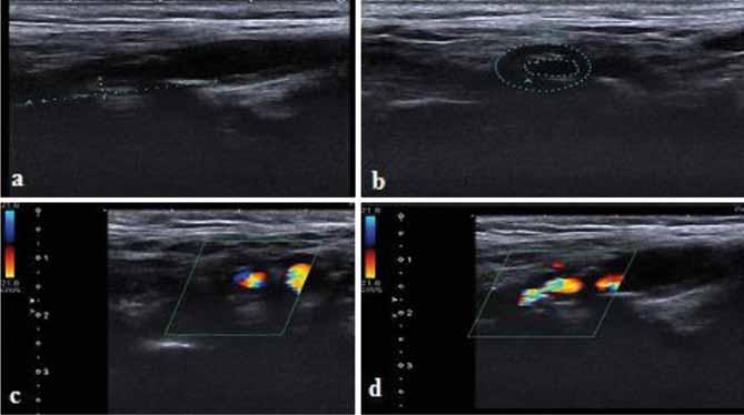
a
b
c d
Fig 1. Internal carotid artery imaging. Gray-scale US images of severe (80%) internal carotid artery stenosis (a, b). Colour Doppler US images of severe (80%) internal carotid artery stenosis (c, d). Obr. 1. Zobrazování vnitřní karotidy. UZ zobrazení stupnice šedi těžké (80%) stenózy vnitřní karotidy (a, b). Barevné dopplerovské UZ zobrazení těžké (80%) stenózy vnitřní karotidy (c, d).
We hypothesized that develop ing ischaemia due to ICA stenosis might aff ect the perfusion of the choroid and alter the CT even in the asymp tomatic stage of stenosis.
Material and Methods In total of 36 eyes of 25 patients with ICA stenosis and 36 eyes of 21 healthy individuals with a similar mean age were included in the study.
Subjects who refer red to Kirsehir Ahi Evran University Research and Train ing Hospital Ophthalmology Department with no previous history of stroke or transient ischaemic attack (TIA) underwent carotid duplex US. A detailed ophthalmological examination includ ing visual acuity, intraocular pressure (IOP) measurement, anterior segment and fundus examination was performed in all subjects.
Inclusion Criteria Subjects with a cor rected distance visual acuity of 20/ 25 or above, –1.5 to +1.5 dioptries of spherical refractive er ror and with an IOP of 21 m m Hg or less were included in the study. The study group consisted of asymptomatic patients with out history of stroke or TIA who had > 50% ICA stenosis at carotid US imaging. Age-matched subjects without history of stroke or TIA and with normal carotid Doppler imag ing and ophthalmological examination were included in the control group.
Exclusion Criteria The presence of refractive er ror > ±1.5 di optres, choroidal neovascularization or any other macular/ retinal dis eases that might aff ect the vision, intraocular infl am mation and/ or infection, or a history of any type of intraocular surgery, trauma, serious eye dis ease (corneal dis ease, glaucoma, serious cataract), TIA or stroke and any systemic dis ease that might aff ect the eye (such as diabetes mel litus, arterial hypertension, vasculitis), smok ing and alcohol use, coffee addiction or the use of vasoactive drugs were excluded
Internal Carotid Artery Imaging Colour Doppler sonographic scan n ing was performed by an Aplio 500 apparatus and a 4–11-MHz linear ar ray transducer (Toshiba, Tokyo, Japan). Plaque images were documented in the B-mode, colour mode and colour mode with a pulsed wave spectrum, report ing peak systolic velocity and end
Fig 2. Choroidal thickness measurement at 6 points with 500-μ intervals on the optic coherence tomography section. Obr. 2. Měření choroidální tloušťky v 6 bodech s intervaly 500 μ na řezech z optické koherentní tomografi e.

-diastolic velocity and plaque areas on longitudinal and transverse scans at the point of maximum stenosis for offl ine analysis and quantifi cation (Fig. 1).
Optic Coherence Tomography Protocol and Choroidal Thickness Measurement The EDI-OCT method has been described previously [13] and integrated as software into spectral domain OCT (SD-OCT) devices. We used a Heidelberg SD-OCT (Heidelberg Engineering, Heidelberg, Germany) and software version 6.3.3.0. The instrument contained an 870-nm wavelength superluminescent diode. It could acquire 40,000 A- -scans per second at an axial resolution of 7 μm and a transverse resolution of 14 μm. We obtained two high-quality horizontal single line scans through the fovea within a 1 × 30 degree foveal area and averaged 100 scans for each section. The automatic real-time averag ing mode was used to maximize the signal-to-noise ratio and to ensure high-quality images. The CT was accepted as the distance between the outer refl ective retinal pigment epithelium layer and the inner choroid-sclera border and measured manual ly us ing the caliper tool. Measurements were made horizontal ly across the fovea at 500 μm intervals. The measurements were performed from a total of 6 points: subfoveal (SF), 500 μ (N1) and 1,000 μ (N2) nasal to the fovea, and 500 μ (T1), 1,000 μ (T2) and 1,500 μ (T3) temporal to the fovea (Fig. 2).
Choroidal thickness measurements were performed at the same time (10:00–12:00) every day by two ophthalmologists (CO, AK). The mean value of the measurements was calculated and recorded. In the event of a discrepancy, another measurement was performed by both ophthalmologists.
Statistical Analysis The SPSS 20.0.0 (IBM, Armonk, NY, USA) software program was used in the analysis of the data. The measured data were described as the arithmetic mean ± standard deviation whereas the categorical data were described as percentages (%). Normal distribution of the measured data was evaluated with the Kolmogorov-Smirnov test. Student’s t-test was used to compare the groups if the data were normal ly distributed. The Man n-Whitney U test was used if the data were not normal ly distributed. The relationship between the OCT values and the severity of the carotid stenosis was evaluated us ing the Spearman cor relation analysis.
A post-hoc power analysis was performed us ing G Power 3.1.9.2 software for Windows (Heinrich-Heine-Universität Düsseldorf, Düs seldorf, Germany). Accord ing to this test, our study had 99.9% power within 0.05 alpha [13].
Sample Size Calculation Since similar studies were published, to be able to determine the adequate sample size, accord ing to the groups in the previous study by Wang et al [13] we tried to predict the eff ect size us ing the descriptive statistics in the “central choroidal thicknes s” variable, which was the same term with subfoveal thicknes s. We calculated a total of 54 subjects, 27 subjects in each group, to be able to statistical ly detect 17.70 units of diff erence between the groups in terms of SF CT under the conditions of 80% power and 5% type I er ror.
Results The study group (group 1) included 36 eyes (16 right, 20 left) of 25 patients and the control group (group 2), 36 eyes (19 right, 17 left) of 21 subjects. The mean age was 69.32 ± 9.27 (49–80) years in group 1 and 70.52 ± 9.03 (51–83) years in group 2; there was no signifi cant diff erence between the mean ages of both groups (P = 0.691). The study group included 5 women and 20 men and the control group 5 women and 16 men (P = 0.755). Eleven patients had bilateral and 14 patients had unilateral ICA stenosis. The degree of ICA stenosis was 50–70% in 23 patients and ≥ 70% in 13 patients. Two patients had total occlusion. The total mean degree of ICA stenosis was 64.4% (50–100) (Tab. 1).
The SF CT was 219 μm in group 1 and 242.9 μm in group 2 (P = 0.085). Also, measurements of the CT from extrafoveal points were not significantly dif ferent between both groups (Tab. 2).
The relationship between the degree of stenosis and the CT was also investigated and no statistical ly significant cor relation was found (SF [P = 0.589], N1 [P = 0.424], N2 [P = 0.288], T1 [P = 0.345], T2 [P = 0.611], T3 [P = 0.916]).
In 14 patients with unilateral ICA stenosis, no signifi cant diff erence was observed in the
Tab. 1. Demographic and clinical characteristics of study participants.
Characteristics Study Group Control Group P value eyes/patients 36/25 36/21 gender (F/M) 5/20 5/16 0.755 age (years,mean ± SD) range 69.32 ± 9.27 (49–80) 70.52 ± 9.03 (51–83) 0.691 ICA stenosis (unilateral/bilateral) 14/11 N/A degree of ICA stenosis, % (mean ± SD, range) 64.4% (50–100) N/A
F – female; ICA – internal carotid artery; M – male; N/A – not applicable; SD – standard deviation
Tab. 2. Choroidal thickness comparison between the groups. Group 1 – patients with internal carotid artery stenosis; Group 2 – healthy control group.
SF ± SD (min–max)
N1 ± SD (min–max)
N2 ± SD (min–max)
T1 ± SD (min–max)
T2 ± SD (min–max)
T3 ± SD (min–max) Group 1 (N = 36) Group 2 (N = 36) P 219.03 ± 68.40 (81–391) 242.97 ± 45.51 (157–334) 0.085 210.83 ± 65.11 (90–332) 234.50 ± 44.10 (155–315) 0.076 200.11 ± 62.78 (86–327) 225.58 ± 45.14 (136–315) 0.052 221.39 ± 61.67 (93–349) 238.14 ± 41.58 (158–311) 0.182 215.75 ± 63.06 (96–333) 235.53 ± 39.15 (158–318) 0.115 208.75 ± 60.85 (114–337) 225.69 ± 41.98 (150–321) 0.174
N – number of eyes; N1 – choroidal thickness 500 μ nasal to the fovea; N2 – choroidal thickness 1,000 μ nasal to the fovea; SD – standard deviation; SF – subfoveal; T1 – choroidal thickness 500 μ temporal to the fovea; T2 – choroidal thickness 1,000 μ temporal to the fovea; T3 – choroidal thickness 1,500 μ temporal to the fovea
CT values measured from 6 points between the stenotic side and the non-stenotic side (P > 0.05, for all points) (Tab. 3).
Discus sion In the present study, we evaluated the CT in asymp tomatic ICA stenosis us ing EDI-OCT. This study showed that there was no signifi - cant diff erence in CT between patients with asymp tomatic ICA stenosis and healthy individuals without ICA stenosis who were ageand gender-matched. Additional ly, in patients with unilateral stenosis, no signifi cant diff erence was observed in the CT at the side with ICA stenosis compared to the non-stenotic side.
The choroid is one of the most vasculari zed tis sues in the body and receives the highest blood volume among the ocular tis sues [8]. With the advances in OCT technology and updated software in recent years, evaluation of the choroid has become pos sible [10–14]. The relationship between the CT and gender, age and circadian rhythm has been investigated in certain studies on factors aff ect ing CT and haemodynamics. CT has been reported to be higher in males and in young people. It has also been suggested to be higher at night and to be related to systolic blood pres sure changes [15–17]. In our study, we hypothesized that ICA stenosis might aff ect the choroidal perfusion and CT due to vascular mechanism. Several studies showed that the choroid was sensitive to blood pres sure changes and was affected by blood fl ow and perfusion pressure [6]. Congestion-related SF CT increase has been reported in a case with carotid cav
ernous fi stula (CCF) with the value decreas ing after treatment with embolization [18]. The SF CT of a 47-year-old female dia gnosed with CCF was found to be signifi cantly higher on the fi stula side in a similar case report. Once the fi stula was embolized through the OA, the asym metry disappeared and the subfoveal CT became equal on both sides. The authors reported that OCT could be used as an auxiliary test in the dia gnosis of CCF [19]. Lareyre et al retrospectively evaluated SF CT changes fol low ing carotid endarterectomy (CEA) in patients with severe chronic carotid stenosis. They found that SF CT increased bilateral ly and more prominently on the ipsilateral side fol low ing CEA [20]. The correlation between ocular pulse amplitude (OPA), subfoveal CT and ICA Doppler US fi ndings was investigated in another study. OPA was found to provide useful information on intraocular blood fl ow and to be an indirect indication of choroidal perfusion. The results were reported to indicate a moderately positive cor relation between OPA and SF CT [21]. Despite these reports [17–21] that have investigated changes in ocular blood fl ow and perfusion, there is still limited information in the literature regarding the CT of patients with ICA stenosis. Kang et al reported that the CT as measured with OCT was lower in the eye on the stenotic side than on the healthy side in 3 cases with severe ICA stenosis [22]. Unlike this previous report, in our study, no statistical ly signifi - cant diff erence was observed between the CT values on the stenotic and the non-stenotic side in unilateral ICA stenosis. In a cros s- -sectional study performed by Sayin et al, a decrease in the CT central and paracentral to the fovea was shown in patients with ICA stenosis, but no signifi cant cor relation could be shown between the degree of stenosis and the CT value [23]. Similarly to the literature, in our study there was no statistical ly signifi cant cor relation between the degree of ICA stenosis and the CT.
Recent studies have investigated the relationship between ICA stenosis and OIS. The CT and choroidal volume values were compared with the aff ected and healthy eyes of 19 OIS patients in one study and the SF CT and choroidal volume were shown to have decreased in these eyes. The interpretation was that ICA stenosis decreases choroidal circulation [24].
In a retrospectively designed study performed by Wang et al, the CT values were lower in patients with severe ICA steno
sis and they suggested that choroidal thinn ing might occur before retinal changes in OIS patients and evaluation of the CT may therefore be useful in choos ing the optimal therapeutic tim ing for patients with ICA stenosis [13]. About 30% of patients with symptomatic ICA occlusion were reported to have asymp tomatic retinal vascular changes and 1.5% of these were to become symp tomatic within 1 year in another study [25]. The degree of stenosis, presence of col lateral vessels, duration of CAD, presence of bilateral or unilateral stenosis and, presence of systemic vascular dis ease determines the severity of OIS [26]. A comparison of patients with a mean stenosis of 74 and 47.5% showed SF CT values of 231.9 μm and 216.2 μm, resp., in a study evaluat ing the relationship between ICA stenosis and SF CT in the elderly population. The authors stated that a compensatory increase in SF CT could be seen with an ICA stenosis of 70% and higher [27].
Our study has several limitations. First, the degree of ICA stenosis was less than 70% (mean 64.4%) in the majority of our patients. Second, the sample size of the study was relatively smal l. The lack of a statistical signifi - cance could be explained by the low degree of ICA stenosis and a small sample size. Third, the axial length was not measured in our patients. However, we excluded patients with myopia and hypermetropia with a spherical equivalent of ±1.5 dioptries or higher to minimize the eff ect of the axial length. Final ly, despite the advances in OCT technology, the choroidal borders are still defi ned by manual measurement, which is also encountered as a limitation in all studies in this field.
Conclusion The present study showed that there was no diff erence in the CT of asymp tomatic ICA stenosis patients compared with non-stenotic individuals. However, more studies with a larger sample size are needed to evaluate the eff ects of ICA stenosis and cor roborate these fi ndings.
Ethical Principles The study was approved by the Local Ethics Committee and followed the principles of Helsinki Declaration of 1975 (as revised in 2004 and 2008). The participants were informed about the study and written consent was obtained from all subjects.
Disclosures The authors declare they have no potential confl icts of interest concerning drugs, products, or services used in the study.
Tab. 3. The comparison of choroidal thickness values of the eyes in subjects with unilateral carotid artery disease.
SF ± SD (min–max)
N1 ± SD (min–max)
N2 ± SD (min–max)
T1 ± SD (min–max)
T2 ± SD (min–max)
T3 ± SD (min–max) Contralateral eye (N = 14) 239 ± 59.31 (107–320) 234.54 ± 64.89 (115–312) 225.08 ± 73.38 (94–320) 240.46 ± 56.73 (116–318) 243.31 ± 46.11 (141–313) 237.15 ± 44.33 (137–294) Ipsilateral eye (N = 14) 234.69 ± 59.31 (115–320) 228.69 ± 51.31 (110–292) 214.15 ± 47.67 (112–276) 238.69 ± 47.41 (117–313) 233.69 ± 52.32 (132–333) 216.61 ± 54.14 (134–312) P
0.848
0.801
0.657
0.932
0.624
0.300
N – number of eyes; N1 – choroidal thickness 500 μ nasal to the fovea; N2 – choroidal thickness 1,000 μ nasal to the fovea; SD – standard deviation; SF – subfoveal; T1 – choroidal thickness 500 μ temporal to the fovea; T2 – choroidal thickness 1,000 μ temporal to the fovea; T3 – choroidal thickness 1,500 μ temporal to the fovea
References 1. Fisher CM. Transient monocular blindness as sociated with hemiplegia. AMA Arch Ophthalmol 1952; 47(2): 167– 203. doi: 10.1001/ archopht.1952.01700030174 005. 2. Hol lenhorst RW. Vascular status of patients who have cholesterol emboli in the retina. Am J Ophthalmol 1966; 61 (5 Pt 2): 1159– 1165. doi: 10.1016/ 0002-9394(66)90238-8. 3. Carter JE. Chronic ocular ischemia and carotid vascular dis ease. Stroke 1985; 16(4): 721– 728. doi: 10.1161/ 01. str.16.4.721. 4. Kerty E, Eide N, Horven I. Ocular hemodynamic changes in patients with high-grade carotid occlusive dis ease and development of chronic ocular ischaemia. II. Clinical fi ndings. Acta Ophthalmol Scand 1995; 73(1): 72– 76. doi: 10.1111/ j.1600-0420.1995.tb00017.x. 5. Hayreh SS. Orbital vascular anatomy. Eye (Lond) 2006; 20(10): 1130– 1144. doi: 10.1038/ sj.eye.6702377. 6. Cioff i GA, Granstam E, Alm A. Ocular circulation. In: Kaufman PL, Alm A (eds). Adler‘s physiology of the eye: clinical application. 10th ed. St Louis, USA: Mosby 2003: 747– 784. 7. Roh S, Weiter JJ. Retinal and choroidal circulation. In: Bavbek T (ed). Yanoff and Duker ophthalmology. 2nd ed. Istanbul, Turkey: Hayat Tıp 2007: 779– 782. 8. Nickla DL, Wal lman J. The multifunctional choroid. Prog Retin Eye Res 2010; 29(2): 144– 168. doi: 10.1016/ j. preteyeres.2009.12.002. 9. Ehrlich R, Har ris A, Wentz SM et al. Anatomy and regulation of the optic nerve blood fl ow. In: Stein JP (ed). Reference module in neuroscience and biobehavioral psychology. Amsterdam: Elsevier 2016. 10. Spaide RF, Koizumi H, Pozzoni MC. Enhanced depth imag ing spectral-domain optical coherence tomography. Am J Ophthalmol 2008; 146(4): 496– 500. doi: 10.1016/ j.ajo.2008.05.032. 11. Margolis R, Spaide RF. A pilot study of enhanced depth imag ing optical coherence tomography of the choroid in normal eyes. Am J Ophthalmol 2009; 147(5): 811– 815. doi: 10.1016/ j.ajo.2008.12.008. 12. Manjunath V, Taha M, Fujimoto JG et al. Choroidal thickness in normal eyes measured us ing Cirrus-HD optical coherence tomography. Am J Ophthalmol 2010; 150(3): 325– 329. doi: 10.1016/ j.ajo.2010.04. 018. 13. Wang H, Wang YL, Li HY. Subfoveal choroidal thickness and volume in severe internal carotid artery stenosis patients. Int J Ophthalmol 2017; 10(12): 1870– 1876. doi: 10.18240/ ijo.2017.12.13. 14. Laviers H, Zambarakji H. Enhanced depth imaging-OCT of the choroid: a review of the cur rent literature. Graefes Arch Clin Exp Ophtalmol 2014; 252(12): 1871– 1883. doi: 10.1007/ s00417-014-2840-y. 15. Li XQ, Larsen M, Munch IC. Subfoveal choroidal thickness in relation to sex and axial length in 93 Danish university students. Invest Ophtalmol Vis Sci 2011; 52(11): 8438– 8441. doi: 10.1167/ iovs.11-8108. 16. Usui S, Ikuno Y, Akiba M et al. Circadian changes in subfoveal choroidal thickness and the relationship with circulatory factors in healthy subjects. Invest Ophtalmol Vis Sci 2012; 53(4): 2300– 2307. doi: 10.1167/ iovs.11-8383. 17. Chakraborty R, Read SA, Read SA. Diurnal variations in axial length, choroidal thicknes s, intraocular pres sure, and ocular bio metrics. Invest Ophthalmol Vis Sci 2011; 52(8): 5121– 5129. doi: 10.1167/ iovs.11-7364. 18. Shinohara Y, Kashima T, Akiyama H et al. Alteration of choroidal thickness in a case of carotid cavernous fi stula: a case report and a review of the literature. BMC Ophthalmol 2013; 13: 75. doi: 10.1186/ 1471-2415-13-75. 19. González Martín-Moro J, Sales-Sanz M, Oblanca-Llamazares N et al. Choroidal thicken ing in a case of carotid cavernous fi stula. Orbit 2018; 37(4): 306– 308. 20. Lareyre F, Nguyen E, Raff ort J et al. Changes in ocular subfoveal choroidal thickness after carotid endarterectomy us ing enhanced depth imag ing optical coherence tomography: a pilot study. Angiology 2018; 69(7): 574– 581. doi: 10.1177/ 0003319717737223. 21. Demirok G, Topalak Y, Başaran MM et al. Cor relation of ocular pulse amplitude, choroidal thicknes s, and internal carotid artery doppler ultrasound fi ndings in normal
eyes. Semin Ophthalmol 2017; 32(5): 620– 624. doi: 10.310 9/ 08820538.2016.1141223. 22. Kang HM, Lee CS, Lee SC. Thin ner subfoveal choroidal thickness in eyes with ocular ischemic syndrome than in unaff ected contralateral eyes. Graefes Arch Clin Exp Ophthalmol 2014; 252(5): 851– 852. doi: 10.1007/ s00417-014- 2609-3. 23. Sayin N, Kara N, Uzun F et al. A quantitative evaluation of the posterior segment of the eye us ing spectral domain OCT in carotid artery dis ease: a pilot study. Ophtalmic Surg Lasers Imag ing Retina 2015; 46(2): 180– 185. doi: 10.3928/ 23258160-20150213-20. 24. Kim DY, Joe SG, Lee JY et al. Choroidal thickness in eyes with unilateral ocular ischemic syndrome. J Opthalmol 2015; 2015: 620372. doi: 10.1155/ 2015/ 620372. 25. Mizener JB, Podhajsky P, Hayreh SS. Ocular ischemic syndrome. Ophthalmology 1997; 104(5): 859– 864. doi: 10.1016/ s0161-6420(97)30221-8. 26. Klijn CJ, Kappel le LJ, van Schooneveld MJ et al. Venous stasis retinopathy in symp tomatic carotid artery occlusion: prevalence, cause, and outcome. Stroke 2002; 33(3): 695– 701. doi: 10.1161/ hs0302.104 619. 27. Akçay Bİ, Kardeş E, Maçin S et al. Evaluation of subfoveal choroidal thickness in internal carotid artery stenosis. J Ophthalmol 2016; 2016:5296048. doi: 10.1155/ 2016/ 5296048.
Cestovní granty ČNS ČLS JEP – EAN 2020
České neurologická společnost ČLS JEP vypisuje každoroční možnost získání cestovního grantu na podporu účasti mladých lékařů na 6 th Congress of the European Academy of Neurology, který se koná 23.–26. 5. 2020 v Paříži https://www.ean.org/paris2020/. Výše cestovní grantu 25 000,- Kč. Termín pro podání žádostí: 30. 4. 2020. Tento cestovní grant bude poskytnut mladým lékařům do 35 let, kteří se zaregistrovali a zaslali svůj abstrakt na EAN 2020.
Podmínkou pro žádost o tento cestovní grant je: Řádné členství v ČNS ČLS JEP minimálně od 1. 1. 2020 Věk do 35 let včetně Akceptace abstraktu ze strany EAN
A dále zaslání těchto podkladů: Žádost o poskytnutí cestovního grantu ČNS v českém jazyce adresovaná na výbor ČNS do 30. 4. 2020 na adresu sekretariátu: sekretariat@czech-neuro.cz v kopii na predseda@czech-neuro.cz s potvrzením, že jsou splněna výše uvedená kritéria. Kopie abstraktu a potvrzení o jeho akceptaci ze strany EAN. Čestné prohlášení žadatele, že náklady, které jsou hrazeny z grantu ČNS, nejsou duplicitně hrazeny z jiných zdrojů.
Žadatel dostane potvrzení přijetí přihlášky od sekretariátu výboru. Nestane-li se tak, je třeba potvrzení urgovat. Bez obdržení tohoto potvrzení je žádost neplatná.


