9 minute read
Microrobots: Bridging The Neuronal Gap, One Micron At A Time Natalie Slosar
Microrobots: bridging the neuronal gap, one Micron at a tiMe Microrobots: bridging the neuronal gap, one Micron at a tiMe
BY NATALIE SLOSAR
Advertisement
Weighing in at a little over three pounds, the mass of jellylike tissue residing in our skulls is nearly everything that makes us human. It is here that we process sights and sounds, feel emotions such as love or hate, or recall a friend’s favorite ice cream flavor. Despite its simple, globular appearance, our brain is the most complex organ nature has created—it contains 86 billion nerve cells that must migrate to their proper location, differentiate into the correct type of neuron, and eventually die naturally.1 After differentiation, each of these neurons begins to make contact with thousands of others through junctions called synapses.2 Thanks to these constantly changing connections, thoughts are formed, memories are stored, habits and personalities are shaped.
Understanding the functions of the human brain—the crown jewel of the human body—is perhaps the most daunting task faced by modern biology. For centuries the intricate network of electrically pulsating nerve cells baffled scientists. But after Sir Charles Sherrington proposed the concept of synapses (1932) and Alan Hodgkin showed how neurons communicate electrochemically (1963), neuroscience research exploded.3 Over the past ten years, neurogenetics, brain mapping, and the discovery of a malleable brain have significantly advanced our collective knowledge of the structure and function of parts of the brain.4 But the exact systems governing specific synaptic connections are yet to be fully understood.
A recent and increasingly popular way to model synaptic connections is to grow a brain from nerve cells. Prior research has shown that it is possible to grow a neural cell network on a plate in the lab, but we have yet to mimic the brain’s function completely.5 A fully functional neural network requires selective neural connections at specified locations—a difficult task, given that the soma, or cell body, of a typical central nervous system neuron is less than 18 micrometers in diameter.6 To further complicate matters, once this network is established, scientists need a method of measuring the neuronal activity at synapses in order to determine how neurons communicate.7 In simplified terms: scientists require a way to position neurons, make them grow in a desired direction, and measure their connectivity.
Scientists often run into obstacles engineering their hodgepodge of petri dish neurons to grow and interconnect the way they intend. Multiple groups have attempted

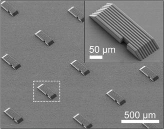
Figure 1. Pictured here are the microrobots, with small grooves and gently sloping sides to
aid neuronal growth. Licensed under CC BYNC 4.0.
to manipulate the patterns and direction in which nerve cells grow, using complicated grids, linear and circular micropatterns, and microplates to direct neuronal growth.8,9 Unfortunately, neurons are often unable to approach these small and complex sites, and many of these operations limit neuronal cell growth, leaving scientists with no way of effectively studying how neurons interconnect and communicate.10 Last month, however, a research group based out of the Daegu Gyeongbuk Institute of Science & Technology proposed an ingenious solution—microrobots.11
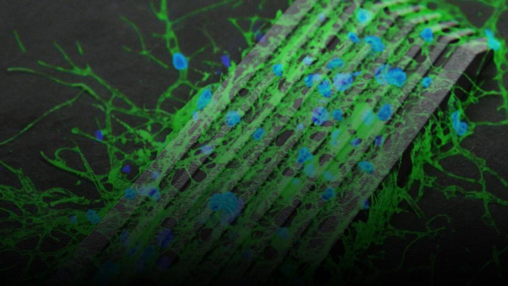
These microrobots essentially function as moveable bridges. Their job is to move in between groups of neuron clusters, bridge the gap, and measure the connectivity between the neurons growing on it. As seen in Figure 1, the microrobots are small— about 300 micrometers—and are lined with slender horizontal grooves.7 These grooves are the same width as a neurite—a special term for axons or dendrites, the projections of a nerve cell. Pulling from a 2016 study on directing neuronal growth, the South Korean research group realized that channels the same width of neurites could guide neuron growth in a desired direction.10 Neurons would grow more easily on a substrate with these microgrooves than on one without. The microrobots were made with metal-oxide nanoparticles, so they could be manipulated by rotating magnetic fields. They also feature gently sloping sides that allow neurites to grow smoothly from the microrobot to the surrounding substrate and vice versa.7
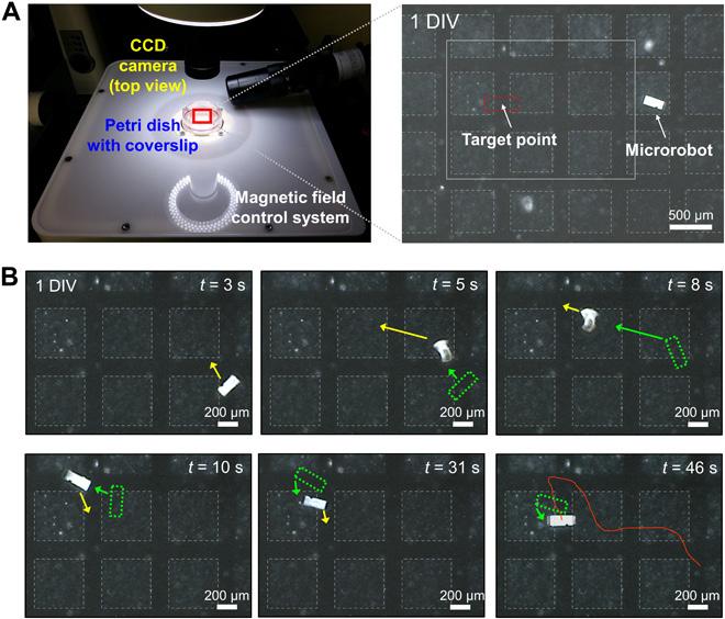
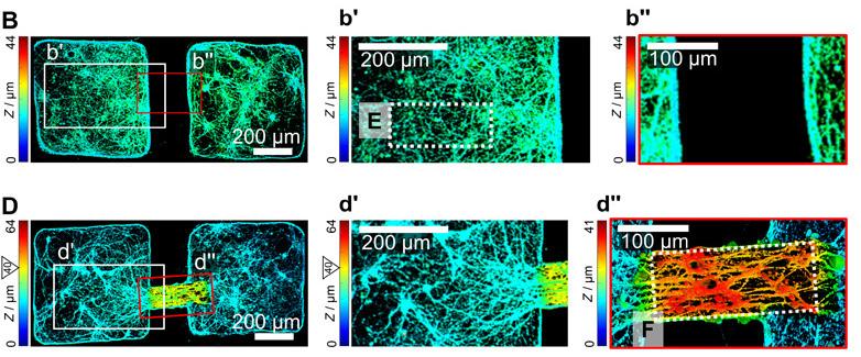
The researchers then placed the microrobot and neuronal clusters on an MEA (multielectrode array) chip, a device that can measure axonal signal transmission.11 This allowed the researchers to access the activity of individual nerve cells and simultaneously record the electrical activity from thousands of cells. As seen in Figure 2, the researchers then manipulated the microrobot to “swim” to the space between two neuronal clusters. As hoped, the neurons in the two cell clusters began growing on the perfectly sized grooves of the robot. By recording the electrical activity on the microrobot with the MEA chip, the researchers deduced that the two neuron groups joined together and began interacting with each other. These results, as shown in Figure 3, were incredibly exciting—the
researchers saw that it was possible to connect separate neuronal clusters in vitro both functionally and morphologically to study how they communicated upon connecting.
This group has introduced a completely new way of studying neuronal networks: forcing neurons to grow on a carefully controlled substrate that can measure electrical activity between the neurons. By studying the way neuron groups grow and connect, we simplify the complexities of the brain and gain an in vitro insight into what is happening in vivo. Coupling human neurons with this microrobot may yield

Figure 2. In this experiment, the researchers sought to manipulate the microrobot to bridge two cell groups (boxes outlined in faint dashed
lines). The microrobot was controlled wirelessly by magnetic fields to “swim” to the target point. The green dashed boxes indicate the position of the microrobot at different timestamps. Licensed under CC BY-NC 4.0.
accurate and highly applicable results for the study of neurological disease. With a greater understanding of these signaling mechanisms, scientists may develop more precise therapies to target the fundamental causes of disease. For instance, individuals afflicted with Parkinson’s Disease are shown to have abnormally weak synaptic connections in regions of the brain essential for voluntary movement. This results in impaired brain plasticity, or an inability of neuronal networks in the brain to change through growth and reorganization, as well as a decrease in the release of the neurotransmitter dopamine.13,14 Low dopamine causes many of the symptoms of Parkinson’s, such as tremors and slow movement. Using microrobots to mimic corticostriatal synaptic connections would give us a greater insight into the causes behind the abnormal connections, as well as a platform to develop and test therapies.7 And the same process could apply to numerous other neurological disorders, such as Alzheimer’s Disease or schizophrenia. Instead of testing out a drug on animals and merely speculating its effects by studying brain scans or behavioral changes, imagine analyzing its direct effects on human neurons. Imagine being able to see how a disease or therapy changes the speed at which neurons grow, how they grow, and how they communicate. The possibilities are endless for neurological disease prevention and treatment research—all thanks to these tiny robots.
REFERENCES
1. Azevedo, F. A. C., Carvalho, L. R. B.,
Grinberg, L. T., Farfel, J. M., Ferretti, R.
E. L., Leite, R. E. P., Filho, W. J., Lent,
R., & Herculano‐Houzel, S. (2009).
Equal numbers of neuronal and nonneuronal cells make the human brain an isometrically scaled-up primate brain. Journal of Comparative
Neurology, 513(5), 532–541. https://doi. org/10.1002/cne.21974 2. National Institute of Neurological Disorders and Stroke. (2019, December 16). Brain basics: The life and death of a neuron. National Institutes of Health. https://www.ninds.nih.gov/Disorders/
Patient-Caregiver-Education/life-anddeath-neuron 3. Queensland Brain Institute. (2019,
January). Understanding the brain: A brief history. The Brain Series: Intelligent
Machines, 4, https://qbi.uq.edu.au/ intelligentmachines 4. Calderone, J. (2014, November 6). 10
Big Ideas in 10 Years of Brain Science.
Scientific American. https://www. scientificamerican.com/article/10-bigideas-in-10-years-of-brain-science/ 5. Simmons, H. (2019, February 26). Labgrown neurons. News-Medical.Net. https://www.news-medical.net/lifesciences/Lab-Grown-Neurons.aspx 6. Chudler, E. H. (n.d.). Brain Facts and
Figures. University of Washington. https://faculty.washington.edu/chudler/ facts.html 7. Kim, E., Jeon, S., An, H.-K., Kianpour,
Figure 3. The results of using the microrobot to bridge two neural clusters are seen in graph D, while graph B represents the control group.
Researchers used a type of micrographs called height coding fluorescence to analyze the differences between a plain surface and one with a microrobot. Licensed under CC BY-NC 4.0.

M., Yu, S.-W., Kim, J., Rah, J.-C., & Choi, H. (2020). A magnetically actuated microrobot for targeted neural cell delivery and selective connection of neural networks. Science
Advances, 6(39), eabb5696. https://doi. org/10.1126/sciadv.abb5696 8. Greene, A. C., Washburn, C. M.,
Bachand, G. D., & James, C. D. (2011).
Combined chemical and topographical guidance cues for directing cytoarchitectural polarization in primary neurons. Biomaterials, 32(34), 8860–8869. https://doi.org/10.1016/j. biomaterials.2011.08.003 9. Roth, S., Bugnicourt, G., Bisbal, M.,
Gory‐Fauré, S., Brocard, J., & Villard,
C. (2012). Neuronal architectures with axo-dendritic polarity above silicon nanowires. Small, 8(5), 671–675. https:// doi.org/10.1002/smll.201102325 10. Magdesian, M. H., Lopez-Ayon, G.
M., Mori, M., Boudreau, D., Goulet-
Hanssens, A., Sanz, R., Miyahara,
Y., Barrett, C. J., Fournier, A. E.,
Koninck, Y. D., & Grütter, P. (2016).
Rapid mechanically controlled rewiring of neuronal circuits.
Journal of Neuroscience, 36(3), 979–987. https://doi.org/10.1523/
JNEUROSCI.1667-15.2016 11. Cai, L., Zhang, L., Dong, J., & Wang,
S. (2012). Photocured biodegradable polymer substrates of varying stiffness and microgroove dimensions for promoting nerve cell guidance and differentiation. Langmuir, 28(34), 12557–12568. https://doi.org/10.1021/ la302868q 12. Bakkum, D. J., Frey, U., Radivojevic,
M., Russell, T. L., Müller, J., Fiscella,
M., Takahashi, H., & Hierlemann,
A. (2013). Tracking axonal action potential propagation on a high-density microelectrode array across hundreds of sites. Nature Communications, 4(1), 2181. https://doi.org/10.1038/ ncomms3181 13. Gerfen, C. R., & Surmeier, D. J. (2011).
Modulation of striatal projection systems by dopamine. Annual Review of Neuroscience, 34(1), 441–466. https://doi.org/10.1146/annurevneuro-061010-113641 14. Akopian, G., & Walsh, J. P. (2006).
Pre- and postsynaptic contributions to age-related alterations in corticostriatal synaptic plasticity. Synapse, 60(3), 223–238. https://doi.org/10.1002/syn.20289
IMAGE REFERENCES
1. Banner: Choi, H. (2020). Nerve cells (colored blue and green in this microscope image) grow along thin grooves of a microrobot that scientists control with magnetic fields [Microscope
Image]. Retrieved from https://www. sciencenews.org/article/magneticrobots-nerve-cells-connections-braininjury 2. Figure 1: Kim, E., Jeon, S., An, H.-K.,
Kianpour, M., Yu, S.-W., Kim, J., Rah,
J.-C., & Choi, H. (2020). Schematic illustration and fabrication process of a magnetically actuated microrobot for neural networks (D) [Microscope image]. Retrieved from https://doi. org/10.1126/sciadv.abb5696 3. Figure 2: Kim, E., Jeon, S., An, H.-K.,
Kianpour, M., Yu, S.-W., Kim, J., Rah,
J.-C., & Choi, H. (2020). Magnetic manipulation of the microrobot on a glass substrate with an array of neural clusters [Microscope image]. Retrieved from https://doi.org/10.1126/sciadv. abb5696 4. Figure 3: Kim, E., Jeon, S., An, H.-K.,
Kianpour, M., Yu, S.-W., Kim, J., Rah,
J.-C., & Choi, H. (2020). Hippocampal neural connections between neural clusters with and without the microrobot at 17 DIV (B, D) [Microscope image].
Retrieved from https://doi.org/10.1126/ sciadv.abb5696


