14 minute read
Reactivating Regeneration: The Power of Neural Crest Cells in Repairing Damaged Tissues (Dr. Megan Martik)
from Flux
reactivatingregeneration
The Power of Neural Crest Cells in Repairing Damaged Tissues
Advertisement
INTERVIEW WITH DR. MEGAN MARTIK
Megan Martik, PhD, joined UC Berkeley in July 2021 as an assistant professor of genetics, genomics, and development in the department of Molecular and Cell Biology. The Martik lab studies the neural crest–a multipotent and migratory cell population that differentiates into various tissues. They use developmental biology approaches to understand how gene regulation impacts neural crest differentiation, disease, evolution, and adult regeneration. Some of her current projects include studying how neural crest cells contribute to adult tissue regeneration and vertebrate evolution, as well as studying the dysregulation of neural crest gene regulatory networks in neural crest-derived cancers. Her continued work in these areas has implications for human disease treatment and regenerative medicine.
BY LEXIE EWER, GRACE GUAN, LUKE LYONS, AND ESTHER LIM

BSJ: Your research centers around elucidating the role of neural crest cells in developing embryos. What drew you to research this cell population in particular?
MM: The neural crest is a stem cell population that is important during embryonic development. The neural crest is fascinating because it can make many different derivatives in the adult body plan—ranging from the craniofacial skeleton to cardiovascular derivatives, pigmentation in the skin, and many aspects of the peripheral nervous system. I became interested in this population, in particular, because we can unravel the different regulatory programs that control cell state transitions from multipotency to differentiated cell types, then use those programs as tools to understand other complex biological problems. For example, we discover how tissues can regenerate, how the cell types facilitate vertebrate evolution, and how organs become dysregulated to give rise to diseases or cancers.
BSJ: You recently published a paper which revealed that neural crest cells aid in the development of cardiomyocytes in the ventricles of the heart in birds and mammals. What is the significance of your findings?
MM: Before our paper, it had been shown that the neural crest could give rise to cardiomyocytes in zebrafish hearts. Those previous papers showed that this contribution existed, but also that when you ablate this population, it would give rise to a poorly functioning adult heart. The adult zebrafish without a neural crest component suffered from severe hypertrophic cardiomyopathy, and would not survive when exposed to stress tests. Due to limitations in tools, there was never a description of this population of cells to amniote (like mammals and chickens) hearts. We were the first
to show, using new techniques, that neural crest cells were actually contributing a similar proportion of cells to cardiomyocytes in the heart. This is important because not only do we show that they represent a significant population of cardiomyocytes in the adult heart, but we also show that this population of cells is very important for cardiac regeneration. Knowing that chickens and mammals could have this population of cells in their heart got us one step closer to understanding how we can tinker with these programs that are controlling regeneration.
BSJ: The primary technique that you employed to determine neural crest cell involvement with cardiomyocytes was retroviral lineage analysis. What advantage does lineage tracing have over other developmental and molecular biology approaches?
MM: I should talk a little bit about the history of how people have lineage-traced using chicken embryos before. Classical embryology experiments, including those that are foundational to understanding neural crest contributions to different lineages, use two different approaches. One approach is to make chimeric embryos by tissue grafting, for example, from quail to chick
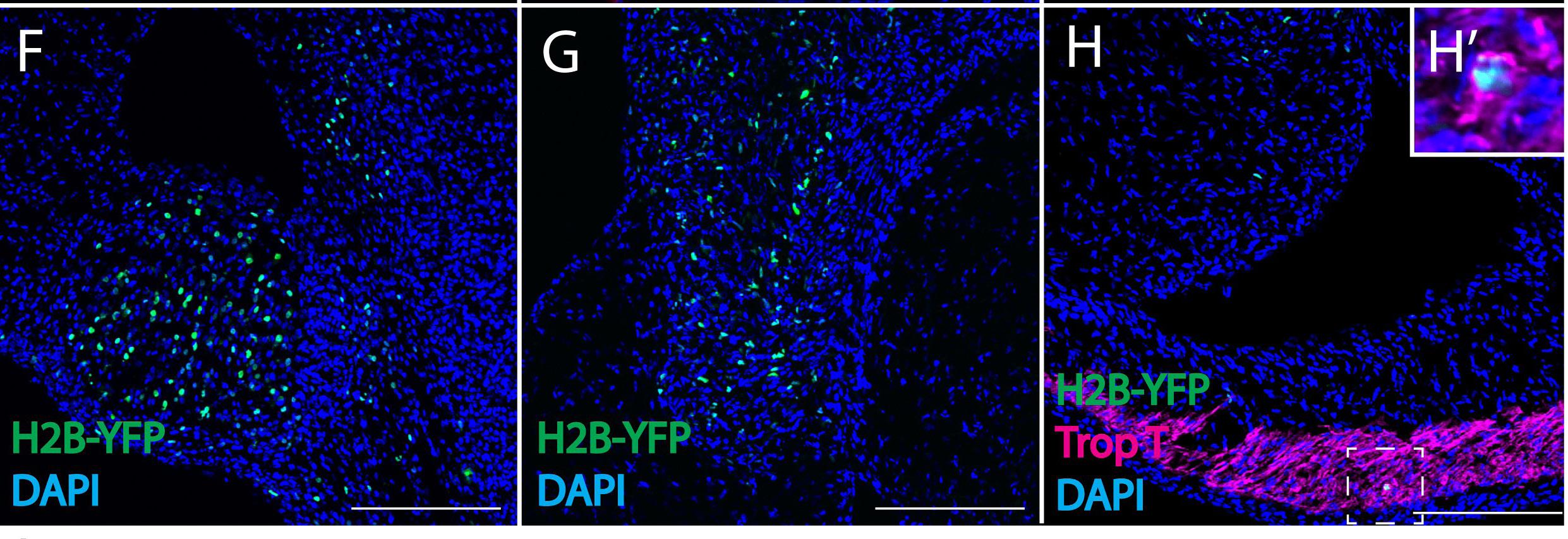
Figure 1: Retrovirally-labeled chick cardiac neural crest cells reveal novel derivatives. A replication incompetent avian retrovirus encoding H2B-YFP, a fusion protein that fluorescently labels cell nuclei, was injected into the lumen of the hindbrain—the origin of cardiac neural crest cells in a chick embryo. By embryonic day 6, the cardiac crest cells (labeled in green in the above panels), migrated to the heart and settled in the pharyngeal arch arteries (F), aorticopulmonary septum (G), and the outflow tract and ventricles (H). Panel H also displays that cardiac neural crest cells express the myocardial marker Trop T (labeled in magenta), indicating that the neural crest contributes to cardiomyocytes.
or chick to chick, then trace where the donor graft migrates in the host embryo. That is a beautiful, elegant technique, but it comes with many problems because you have a graft that has been put into a host environment that needs to heal. You lose a lot of information due to cells that die around the incision or cells that are not receptive to taking new cells on. The second approach involves lineage tracing done with a lipophilic dye called DiI that is used to trace cells over developmental time. However, as cells proliferate, this dye will dilute over time. This results in a loss of ability to do long-term analysis. In developmental biology, some research organisms such as mice and fish can be genetically manipulated to make transgenic lines to permanently lineage trace cells from early in development into adulthood. With chickens, we cannot make transgenic lines easily due to various restrictions, such as housing the chickens. In our paper, we use retroviral lineage analysis, which uses viruses to permanently label different cell populations. In chickens, we are able to specifically inject these retroviruses into the cells that give rise to the neural crest. Then, we track over time what cells they give rise to. This is similar to the dye injections, but now with a permanent label because the virus will integrate a fluorophore lineage tracer directly into the host genome. Each replication does not dilute the tracer over time allowing for long-term analysis which had not been used before. That is why people had not seen this contribution of cardiomyocytes previously.
BSJ: Why is it important that the cells involved in regeneration are embryonic-derived? What is known about the mechanism zebrafish use to reactivate neural crest cell genes?
MM: There are two different lineages to cardiomyocytes in the adult heart and both are embryonic-derived—the mesoderm and the neural crest. It is interesting because they activate their own gene regulatory programs during development. Yet, they both give rise to physiologically indistinguishable cardiomyocytes in the adult, until injury. After an injury, the neural crest-derived cardiomyocytes will reactivate these developmental programs, and we are not sure how that works yet. Figuring out what that trigger is to facilitate this reactivation is crucial to understanding how we can hijack this information for any type of therapeutic approach in the future.
BSJ: Given the contribution of neural crest cells in cardiomyocyte regeneration, you mention the possibility of new therapeutic approaches these results indicate. What information is necessary to translate this research into a human therapeutic?
MM: The first thing is understanding how nature has already figured this out. How does it work in something that can regenerate, like zebrafish? Once we have the information about the programs that are being reactivated, we need to know how the programs are being reactivated. Given this contribution of cells exists in mammalian hearts, this implies whatever regeneration is happening in zebrafish that is not happening in mice, or possibly humans, can be switched on. It is just a program, a regulatory logic that needs to be driven, and not a cell population that does not exist. In terms of therapeutics, we have to understand how it works, figure out what that trigger is, and go into mammalian systems to determine how we can reactivate this, either by activating or suppressing the trigger in order to drive a regenerative process. In terms of actual therapeutics, if it is a genetic switch, we can use CRISPR-based therapeutics to facilitate some kind of reactivation after injury.
BSJ: Are neural crest cells likely to be involved in regeneration of any other organ systems?
MM: This is something I think so much about, because neural crest cells are multipotent, and they give rise to so many differentiated derivatives in the adult body plan. You can imagine the neural crest as a special population in the adult

Figure 2: Cardiac neural crest contributes to zebrafish heart regeneration. A transgenic line of zebrafish is used to fluorescently turn cells green upon transcription of Sox10, a gene which is expressed in migrating cardiac neural crest cells but is down-regulated once these cells reach the heart. In an uninjured zebrafish heart, there is very little expression of Sox10. However, 7 days post amputation (dpa) of approximately 20% of the zebrafish heart, Sox10 is reactivated, shown by the presence of green cells in the middle panel. After 21dpa, the heart regenerated and green cells are seen surrounding the site of injury, indicating that cardiac neural crest cells reactivate transcriptional programs to contribute to heart regeneration.
heart. For instance, it holds some kind of “stemness” to be able to dedifferentiate, proliferate, and tell other cells around them that it is time to respond to injury. That same process could be happening in other organ systems that the neural crest contributes to, like the craniofacial skeleton. In terms of differentiated derivatives of the neural crest, I do think that they hold a special ability to regenerate different organs, and that is something we definitely want to look at in the future. However, sea lampreys, a basal vertebrate, lack the full complement of neural crest derivatives. They do not have a jaw and do not have a lot of other derivatives, like organized sympathetic ganglia. The neural crest is really interesting to study in terms of evolution because, throughout vertebrate evolution, we see the increasing complexity of these different derivatives and their regulatory networks.
BSJ: Since the publication of this article, what new information has been discovered concerning the connection between neural crest cells and cardiomyocytes?
MM: In the lab, we have made a lot of headway on what programs are controlling multipotency to differentiate cardiomyocytes. Since differentiation during development and regeneration are very similar programs, learning more about these differentiation programs allows us to learn more about how they control regeneration, and vice versa. Also, in terms of regeneration, we have seen that if you use genetic tools to ablate these cells after injury, the hearts are not able to regenerate. We assumed this was going to be the case, but it is nice to see that whenever you do not have neural crest cells after injury, the heart cannot regenerate. Now, we are starting to look at what the cells are doing besides reactivating genes. What are they doing in order to drive this regeneration process, and how are they responding to an injury?
BSJ: In another one of your papers, you discussed the evolution of the “new head” and its relation to neural crest circuits. What can researching the neural crest tell us about vertebrate evolution?
MM: The neural crest as a whole population is a vertebrate innovation, meaning they are exclusive to vertebrates.
BSJ: Could you briefly explain to our readers what the new head hypothesis is?
MM: About 40 years ago, the new head hypothesis proposed that, with the advent of the neural crest and the cranial placodes, the craniofacial skeleton and highly complex sensory system arose, which allowed the elaboration and expansion of the vertebrate brain. With these complexities, invertebrate chordates, which were predominantly filter feeding animals, became active predators. That is crucial for the success of vertebrates as a lineage. Because this is a neural crest-derived feature, the neural crest is thought to be intimately linked with vertebrate evolution.
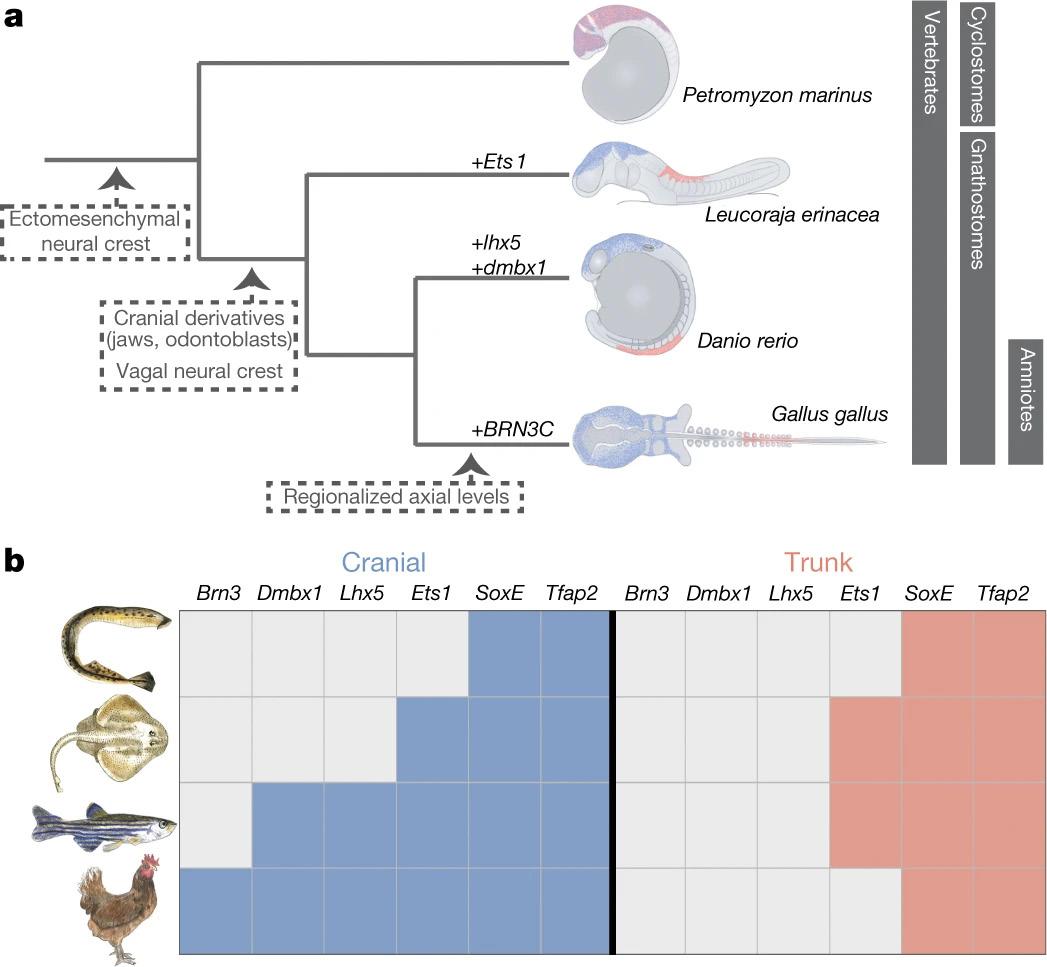
BSJ: You posit an alteration to the new head hypothesis, stating that the neural crest portion of the new head arose via continued regulatory modifications rather than all-at-once at the base of the vertebrate lineage. What implications do these findings have for the study of neural crest evolution?
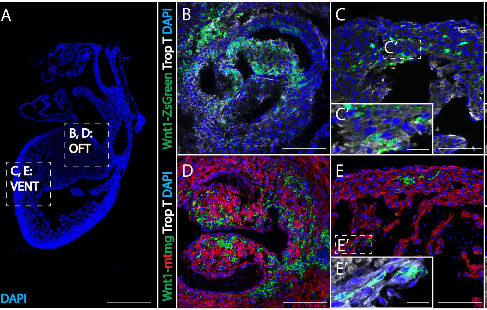
Figure 3: Evidence of neural-crest derived cells in mouse myocardium. Figure 3A is an overview of a mouse heart showing locations of images B, C, D, E (cell nuclei labeled with DAPI-blue). Two transgenic mice lines were used: a Wnt1-ZsGreen line (B, C) and an improved Wnt1-mtmg line (D, E). Figures 3B and 3D compare the outflow tracts of the myocardium, while Figure 3C and 3E focus on the ventricles. Cells shown in green represent cardiac neural crest cells, indicating that both mice lines contain neural crest cells in the heart. Dashed box C’ and E’ shows enhanced magnification to highlight dual-staining between myocardial marker Trop T (in gray) and green neural crest cells.
MM: The paper is expanding upon what we know about this new head hypothesis. Instead of the neural crest arising as a whole set of derivatives at the base of vertebrates, we are essentially saying they arose from a homogenous, very rudimentary population. The complexity we now see arose via continued evolution and regulatory modifications. It is not really the case to think of all vertebrates having this new head or full set of derivatives. What happens is that throughout vertebrate evolution, we get more complexity.
BSJ: Can you elaborate further on what mechanisms you believe are responsible for these neural crest regulatory modifications within jawed vertebrates?
MM: The short of it is no, but I can speculate. With these modifications, it is on the cis-regulatory level, meaning it is modifications in enhancer sequences and elements that control the activation of gene expression. Looking deeper at this regulatory DNA compared to gnathostomes (jawed vertebrates), we can get an understanding of which features have been acquired with the regulatory genome. This helps us to understand more about how novel gene expression and regulatory circuits are being acquired to give rise to these complex derivatives. But I do not have an answer.
BSJ: Your lab is also interested in researching the gene regulatory circuitry underlying the formation of neuroblastomas. Can you describe some of the projects you have planned to research this? MM: We want to know how this multipotent stem cell be comes a differentiated sympathetic nervous system de rivative, as the sympathetic nervous system is where neuroblastomas arise. More often than not, it is because of a dysregulation of the events culminating in these differentiated cell types. We want to understand the trajectory of neural crest cells to sympathetic neurons, chromaffin cells, and other derivatives of the sympathetic nervous system. With that information, we can then understand how those programs that are controlling differentiation become dysregulated to give rise to neuroblastomas. While we are doing that kind of research, neuroblastoma formation requires us to develop better animal models to recapitulate this tumorigenesis phenotype. We use zebrafish in the lab as our main model organism for understanding tumorigenesis. Hopefully, all of this will culminate in the ability to make therapeutics, maybe patient-specific therapeutics, by modeling tumor formation in zebrafish.
BSJ: How do you see the field of regenerative medicine evolving in the near future, and how will your lab be a part of the effort to translate regeneration from one animal to another?
MM: The first thing is figuring out how this works in something that can regenerate. That is what we are really focused on now. Our immediate next steps are understanding what is not being reactivated and what is not happening in humans and mammals that is happening in zebrafish. Using the mouse as a model system, as well as human-derived cell culture and organoids, we can
model these things in human-derived tissues and see, using genome wide screens, what is not being reactivated after injury. Following this, we can use CRISPR-based approaches to reactivate these regenerative genes in the mammalian system. As we unveil candidates that are necessary for this reactivation, it becomes very easy to stimulate them with tools that already exist. I think we are on the precipice of developing translational strategies because the tools and technologies that we have now allow us to uncover these things rather quickly.
REFERENCES
1. Headshot: [Photograph of Megan Martik]. Image reprinted with permission. 2. Figures 1, 2, and 3: Tang, W., Martik, M. L., Li, Y., & Bronner, M. E. (2019). Cardiac neural crest contributes to cardiomyocytes in amniotes and heart regeneration in zebrafish. ELife, 8. https://doi.org/10.7554/elife.47929 3. Figure 4: Martik, M. L., Gandhi, S., Uy, B. R., Gillis, J. A.,
Green, S. A., Simoes-Costa, M., & Bronner, M. E. (2019).
Evolution of the new head by gradual acquisition of neural crest regulatory circuits. Nature, 574(7780), 675–678. https://doi.org/10.1038/s41586-019-1691-4 Figure 4: Evolution of neural crest subpopulations. Figure 4a illustrates the evolution of neural crest (NC) subpopulations, and the similarities between vertebrate NC and amniote trunk subpopulations. Early vertebrate evolution led to the division of NC into cranial and trunk groups. Figure 4b shows the molecular differences between lamprey and gnathostome NC, namely that lamprey lack differentiation between cranial and trunk NC.
