13 minute read
Prognostic Potential of Extracellular Vesicles: Noninvasive Monitoring of Chemotherapeutic Resistance Devel opment
from Flux
Prognostic Potential of Extracellular Vesicles: Noninvasive Monitoring of Chemotherapeutic Resistance Development
1Jennifer C. Hall, 2Thomas R. Carey, 3Lydia L. Sohn Research Sponsor: 3Lydia L. Sohn
Advertisement
ABSTRACT
Chemotherapy remains the most common modality of cancer treatment, used both independently and in combination with other systemic or localized therapies. It has been shown that patient response to chemotherapeutics is a potent predictor of prognosis and over 90% of cancer patient mortalities are related to drug resistance. Resistance to chemotherapy can develop during the course of treatment when tumor cells become less sensitive to therapy and is particularly dangerous to patient survival due to the difficulty of detection. As such, it has become essential to develop efficient methods of monitoring changes in patients’ response to treatment. Our research demonstrates the potential of extracellular vesicles (EVs) in rapid, noninvasive monitoring of tumor response to chemotherapeutics. Through analysis of EV mRNA cargo, trends in gene expression observed in cells are shown to be conserved in their derived EVs. To determine tumor response to treatment, we devised a model system to mimic interactions between tumor cells, chemotherapeutic(s), and gene expression alterations that confer resistance. We isolated EVs from MCF7/wt and MCF7/ADR cell culture supernatant to model EVs derived from doxorubicin-sensitive and resistant tumors, respectively. We then extracted mRNA from EVs and quantified the expression of top2a. We observed downregulation of top2a, which confers resistance to doxorubicin, in MCF7/ADR (doxorubicin resistant) EVs relative to MCF7/ wt EVs. Our findings establish the feasibility of using mRNA in tumor-derived EVs to assess drug sensitivity of tumors via liquid biopsy.
Major, Year, Departmental: 1Class of 2021, Molecular and Cellular Biology Neurobiology Emphasis, Department of Molecular and Cellular Biology, University of California, Berkeley; 2UC Berkeley–UC San Francisco Graduate Program in Bioengineering, University of California, Berkeley; 3Department of Mechanical Engineering, University of California, Berkeley, 5118 Etcheverry Hall, Berkeley, CA 94720, US
INTRODUCTION
Cancer treatment often employs an array of modalities utilized independently, in sequence, or concurrently. Treatment modalities include surgery, radiation, and systemic therapies such as chemotherapy or immunotherapy. Chemotherapy remains the most common form of cancer treatment in the US (Figure 1A).1 Patient response to administered chemotherapeutics is a strong predictor of prognosis, and over 90% of cancer patient mortalities are related to drug resistance.2,3
Chemotherapeutic resistance is categorized as either intrinsic or developed. Intrinsic resistance inhibits a drug’s mechanism of action and exists in tumor cells prior to exposure. Developed resistance occurs when tumors evolve decreased sensitivity after initial or prolonged exposure. In conjunction with the difficulty of detection, this makes developed resistance particularly threatening to positive patient outcomes. At present, determination of tumor response to chemotherapy treatment typically requires invasive tumor biopsies. This demonstrates the need for more efficient, noninvasive mechanisms for monitoring tumor response to treatment frequently throughout the treatment course.
As a result of the frequency of chemotherapy use and the implications of losses in sensitivity to treatment, much research has gone into elucidation of the mechanisms and gene expression alterations conferring chemotherapeutic resistance (Figure 1B). Similarly, pathways and gene targets of many chemotherapeutics are well understood. For example, the chemotherapy drug doxorubicin acts by inhibiting topoisomerase-IIα (TOP2A) function within the nucleus of tumor cells (Figure 1C), and decreases in TOP2A gene expression are indicative of doxorubicin resistance. Broadly, altered gene expression within drug-resistant tumor cells provides clear ev-
Figure 1. Chemotherapeutics remain the most prescribed cancer treatment and accordingly, mechanisms of drug action and drug
resistance are well-characterized for many drugs. Chemotherapy utilized in breast cancer treatment (A) demonstrates its prevalence, particularly in late-stage cases. This prevalence has led to research into the different mechanisms of drug resistance (B). Likewise, much research has further illuminated the different mechanisms of action of given drugs. In a specific example (C), doxorubicin is shown entering the cell and nucleus to inhibit the TOP2A enzyme.

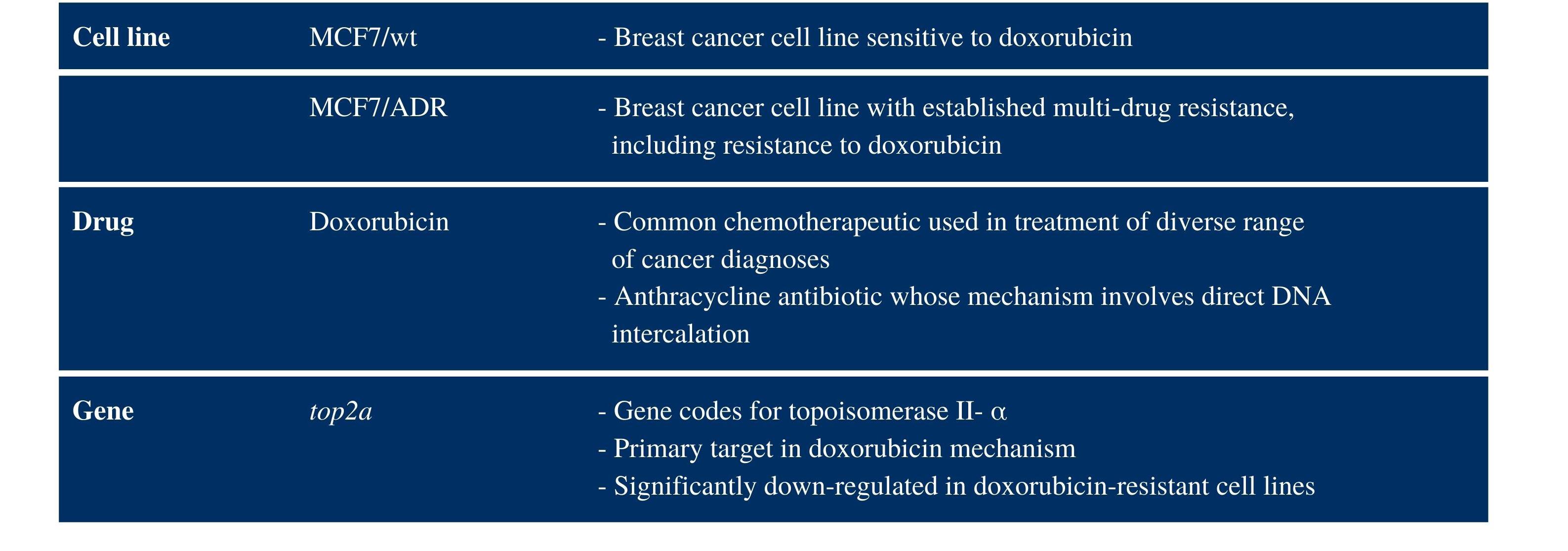
Table 1. A model system was devised to measure gene expression alterations associated with developed chemotherapeutic resistance.
MCF7/wt and MCF7/ADR cell lines were used to model doxorubicin-sensitive and resistant tumors, respectively. TOP2A is an enzyme that acts in DNA repair and has been shown to be downregulated in doxorubicin resistant cells.
idence of resistance when compared to that of drug-sensitive tumor cells.4 However, this method is limited in its clinical potential because probing for altered gene expression in tumor cells fails to eliminate the need for invasive biopsy procedures.
To address this limitation, we put forth a method for the efficient and noninvasive monitoring of altered gene expression through the examination of mRNA cargo within extracellular vesicles (EVs) derived from tumor cells. EVs are lipid bilayer-delimited nanoparticles secreted by cells and heralded as a primary mechanism for cell-tocell communication. As a result of their formation via exocytosis or membrane budding, EVs display protein surface markers and contain mRNA cargo conserved from their cell of origin. In addition, it has been shown that EVs travel throughout the body within biofluids.5 These characteristics of EVs highlight their potential application in a method for noninvasively monitoring tumor cell sensitivity to cancer treatment.
For our research, a model system was devised to mimic the development of chemotherapeutic resistance in tumor cells (Table 1). The system was comprised of a chemotherapy drug, sensitive and resistant tumor cells, and a target gene known to be differentially expressed in association with drug resistance. We utilized the common anthracycline antibiotic, doxorubicin, as the model chemotherapeutic. We used the MCF7 (MCF7/wt) breast cancer cell line to model drug-sensitive tumor cells, and we used the MCF7/ ADR cell line—known to be multi-drug resistant—to model tumor cells with developed drug-resistance. We then isolated EVs from the cell culture supernatant of the drug-sensitive and drug-resistant cell lines. Finally, we selected top2a as the target gene because it is highly downregulated in doxorubicin-resistant MCF7 cells, top2a codes for TOP2A, an enzyme involved in DNA repair, and is a primary target in the doxorubicin mechanism.4
With this model, we demonstrated mRNA within tumor-derived EVs reflects the gene expression alterations observed in tumor cells associated with developed resistance to doxorubicin. This finding indicates the highly impactful clinical motivation and potential of our research. RESULTS
Using RT-qPCR, we measured top2a expression in both cells and EVs derived from the MCF7/ADR and MCF7/wt cell lines. In MCF7/ADR cells, we observed a 3.58-fold downregulation relative to MCF7/wt cells; in MCF7/ADR EVs, we observed a 24.29-fold downregulation relative to MCF7/wt EVs (Figure 2). We normalized top2a expression in cells and EVs derived from both cell lines to baseline expression of the glyceraldehyde-3-phosphate dehydrogenase (gapdh) housekeeping gene.
These data strongly support the notion that gene expression alterations in cells can be detected through quantification of the
A.
10 9 8
value Δ Ct
7 6 5 4 3 2 1 0
ΔCt values of MCF7/wt and MCF7/ADR cells
MCF7/wt cells MCF7/ADR cells
B.
10 9 8
value Ct Δ
7 6 5 4 3 2 1 0
ΔCt values of MCF7/wt and MCF7/ADR EVs
MCF7/wt EVs MCF7/ADR EVs
C.
down regulation Fold -
40
35
30
25
20
15
10
5
0
Fold-down regulation of TOP2A for MCF7/ADR cells and EVs
Cells EVs
Figure 2. Relative expression of top2a in doxorubicin-resistant tumor cells and their derived EVs compared to MCF7/wt cells and
EVs, respectively. Comparison of the ΔCt values (normalized to gapdh) of (A) cells and (B) EVs reveals decreased expression of top2a in both MCF7/ADR cells and EVs. (C) A 3.58-fold downregulation was observed in MCF7/ADR cells compared to MCF7/wt cells. A 24.29fold downregulation was observed in MCF7/ADR EVs compared to MCF7/wt EVs.
mRNA cargo of their derived EVs. In particular, decreased top2a expression in MCF7/ADR cells agrees with established trends observed in MCF7 cells with developed doxorubicin resistance and further underscores the potential of using EVs to noninvasively monitor tumor resistance development.4
DISCUSSION
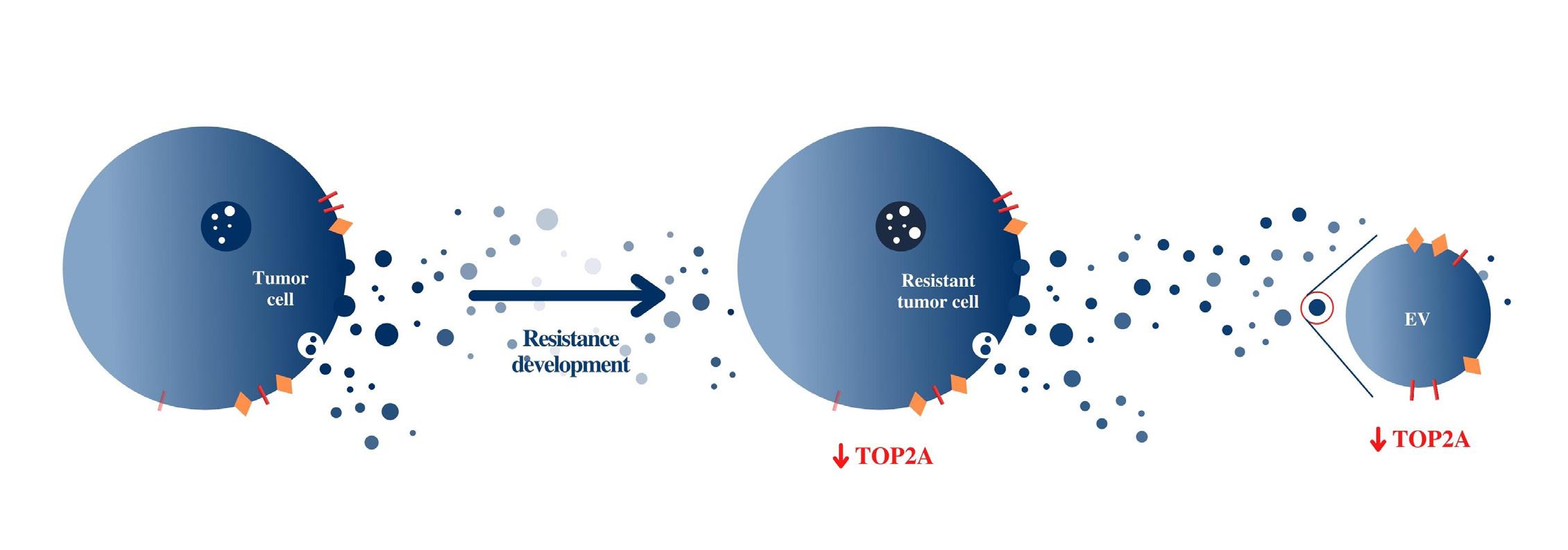
Through our research, we have presented a novel method for noninvasively monitoring gene expression alterations in tumor cells through quantification of a target gene in the mRNA cargo of their derived EVs. Observation of differential expression of the target gene, top2a, demonstrates the conservation of trends in gene expression from cells to EVs (Figure 3).
Our findings illuminate clear next steps for the implementation of this method into a clinical setting. In addition to tumor-derived EVs, biofluids contain abundant healthy-cell-derived EVs, as well as cells, proteins, and free oligonucleotides. Accordingly, future work should improve upon the current EV isolation protocols to more effectively address complex biofluid samples. Coupled with the properties of EVs, including their ability to travel great distances from the site of their genesis and their conserved biomarkers derived from their cell of origin, it is possible to select for tumor-derived EVs among samples comprised of EVs secreted from a diverse range of cell types.5,6
A potential mechanism for this selection is immuno-capture where antibody-functionalized microbeads bind EVs based on surface markers differentially expressed on tumor cells. This is possible because these surface markers are inherited by EVs secreted from tumor cells and may be used to differentiate them from other EV populations.
Implementation of a two-step immuno-capture procedure has previously been shown to increase selectivity.6 The primary step aims to diminish background and consists of negatively selecting for EVs from non-tumor cell types with a first round of microbeads coated with surface markers known to be generally expressed. This is then followed by a secondary step which utilizes functionalized microbead interactions with known tumor surface markers yielding positive selection of tumor-derived EVs. Captured EVs can be lysed on the bead to release their cargo, enabling subsequent isolation of their mRNA and quantification of target genes.
The addition of tumor-derived EV selection furthers the clinical potential of utilizing EVs in liquid biopsy applications. Additional future directions include developing a streamlined protocol incorporating isolation/selection of tumor-derived EVs, extraction of their mRNA cargo, and quantification of target genes that confer drug resistance. All together, this would fulfill the need for an efficient, noninvasive method of determining patient prognosis through the rapid evaluation of response to chemotherapy.
As long as drug therapies remain a prominent tool for limiting disease progression, there will likewise remain the need to verify tumor sensitivity to prescribed drugs. To assess this, we have presented a novel method to noninvasively monitor tumor response to chemotherapeutics through examination of tumor-derived EV mRNA. Our findings highlight the potential use of EVs in liquid biopsy applications that may be performed at frequent timepoints throughout a cancer treatment course to support improved patient outcomes.
METHODS Cell culture and growth conditions
The MCF7/wt and MCF7/ADR cell lines were obtained from the UCB Cell Culture Facility supported by the University of California, Berkeley. Both cell lines were cultured in an attached monolayer in DMEM media (Thermo Fisher Scientific, USA) supplemented with 10% exosome depleted fetal bovine serum (Thermo Fisher Scientific, USA) and 1% penicillin-streptomycin (Roche Molecular Systems, USA). Additionally, cells were grown in either T25, T75, or T175 attached type, filter-cap culture flasks (Thermo Fisher Scientific, USA). Cells were incubated in a 37 °C humidified atmosphere with 5% CO2.
EV isolation
EVs were isolated from cell culture supernatant using a membrane-affinity-based commercial isolation method (ExoEasy Maxi, Qiagen, Germany). Briefly, cell culture supernatant was clarified by centrifugation at 3000 rcf for 15 minutes, and the resulting supernatant was mixed with a binding buffer and added to the spin column.

Figure 3. Conservation of gene expression from cells to EVs. Decreased top2a expression was observed in EVs derived from cell types in which top2a expression was similarly decreased. This demonstrates the conservation of trends in gene expression from cells to their derived EVs.
RNA isolation
RNA from both EVs and cells was isolated using a phenol-chloroform method with the RNeasy® Mini Kit (Qiagen, Germany) according to the manufacturer’s protocol. An additional RNase-free DNase step was performed according to manufacturer’s protocol and utilizing an RNase-free DNase kit (Qiagen, Germany). This step was carried out to ensure total elimination of genomic DNA. Finally, to ensure purity of RNA, optical density was measured using a NanoDrop® spectrophotometer at 260 and 280 nm and 260/280 ratios were compared to published qualifications of purity (Thermo Fisher Scientific, USA).
Gene quantification
The top2a gene was quantified in the isolated RNA of both cells and EVs using a 48-well RT qPCR assay. Each well containing samples also contained the components necessary for the PCR reaction (New England BioLabs, USA) as well as a primer for either top2a or gapdh. RT-qPCR was performed following the manufacturer’s protocols. The gapdh housekeeping gene was used to normalize top2a expression. top2a and gapdh primers (Integrated DNA Technologies, USA) were used at an in-well concentration of 40 nM.7 This primer concentration was found to minimize primer dimer formation observed in initial testing. Initial testing also produced an empirical top2a primer efficiency value of 99.43% which translates to an amplification value of 1.99 used in gene expression fold-change calculations.
Data analysis
Data analysis was performed using the delta delta cycle threshold method (ΔΔCt) and an amplification factor of 1.99. Fold-increases or -decreases in gene expression were calculated using the following equation: fold-increase or decrease = (amplification value)- ΔΔCt
REFERENCES 1. American Cancer Society. (2019). Cancer treatment & survivorship facts & figures. https://www.cancer.org/research/ cancer-facts-statistics/survivor-facts-figures.html 2. Pierga, J. Y., Robain, M., Jouve, M., Asselain, B., Diéras, V.,
Beuzeboc, P., Palangié, T., Dorval, T., Extra, J. M., Scholl,
S., & Pouillart, P. (2001). Response to chemotherapy is a major parameter-influencing long-term survival of metastatic breast cancer patients. Annals of Oncology,, 12(2), 231–237. https://doi.org/10.1023/a:1008330527188 3. Bukowski, K., Kciuk, M., & Kontek, R. (2020). Mechanisms of multidrug resistance in cancer chemotherapy. International Journal of Molecular Sciences, 21(9), 3233. https:// doi.org/10.3390/ijms21093233 4. AbuHammad, S., & Zihlif, M. (2013). Gene expression alterations in doxorubicin resistant MCF7 breast cancer cell line. Genomics, 101(4), 213–220. https://doi.org/10.1016/j. ygeno.2012.11.009 5. Zhang, J., Nguyen, L. T. H., Hickey, R., Walters, N., Wang,
X., Kwak, K. J., Lee, L. J., Palmer, A. F., & Reátegui, E.
(2021) Immunomagnetic sequential ultrafiltration (iSUF) platform for enrichment and purification of extracellular vesicles from biofluids. Scientific Reports, 11. https://doi. org/10.1038/s41598-021-86910-y 6. Ko, J., Hemphill, M., Gabrieli, D., Wu, L, Yelleswarapu,
V., Lawrence, G., Pennycooke, W., Singh, A., Meaney, D.
F., & Issadore, D. (2016).Smartphone-enabled optofluidic exosome diagnostic for concussion recovery. Scientific Reports, 6, https://doi.org/10.1038/srep31215 7. Szlachta, K., Manykyan, A., Raimer, H. M., Singh, S., Salamon, A., Guo, W., Lobachev, K. S., & Wang, Y. (2020).
Topoisomerase II contributes to DNA secondary structure-mediated double-stranded breaks. Nucleic Acids
Research, 4812), 6654–6671, https://doi.org/10.1093/nar/ gkaa483 8. Burgess, D. J., Doles, J., Zender, L., Xue, W., Ma, B., Mc-
Combie, W. R., Hannon, G. J., Lowe, S. W., & Hemann, M.
T. (2008). Topoisomerase levels determine chemotherapy response in vitro and in vivo. Proceedings of the National Academy of Sciences, 105(26), 9053–9058. https://doi. org/10.1073/pnas.0803513105 9. Thorn, C. F., Oshiro, C., Marsh, S., Hernandez-Boussard,
T., McLeod, H., Klein, T. E., & Altman, R. B. (2011). Doxorubicin pathways: pharmacodynamics and adverse effects.
Pharmacogenetics and Genomics, 21(7), 440–446. https:// doi.org/10.1097/FPC.0b013e32833ffb56 10. An, X., Xu, F., Luo, R., Zheng, Q., Lu, J., Yang, Y., Qin, T.,
Yuan, Z., Shi, Y., Jiang, W., & Wang, S. (2018). The prognostic significance of topoisomerase II alpha protein in early stage luminal breast cancer. BMC Cancer, 18(1). https://doi. org/10.1186/s12885-018-4170-7 11. Stevic, I., Buescher, G., & Ricklefs, F. L. (2020).Monitoring therapy efficiency in cancer through extracellular vesicles.
Cells, 9(1), 130. https://doi.org/10.3390/cells9010130 12. Kosaka, N., Yoshioka, Y., Fujita, Y., & Ochiya, T. (2016).
Versatile roles of extracellular vesicles in cancer. The Journal of Clinical Investigation, 126(4), 1163–1172. https:// doi.org/10.1172/JCI81130 13. Choi, D. S., Lee, J., Go, G., Kim, Y., & Gho, Y. S.. (2013).
Circulating extracellular vesicles in cancer diagnosis and monitoring. Molecular Diagnosis & Therapy, 17, 265–271. https://doi.org/10.1007/s40291-013-0042-7 14. Redzic, J. S., Ung, T. H., & Graner, M. W. (2014). Glioblastoma extracellular vesicles: reservoirs of potential biomarkers.
Pharmacogenomics and Personalized Medicine, 7, 65–77. https://doi.org/10.2147/PGPM.S39768 15. Zhou, S., Yang, Y., Wu, Y., & Liu, S. (2021). Review: Multiplexed profiling of biomarkers in extracellular vesicles for cancer diagnosis and therapy monitoring. Analytica Chimica Acta, 1175. https://doi.org/10.1016/j.aca.2021.338633


