15 minute read
Pain Versus Itch: the Role of S1P (Dr. Diana Bautista
Pain Versus Itch: The Role of S1P
Interview with Professor Diana Bautista
Advertisement
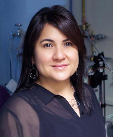
Professor Diana Bautista 1
By Shevya Awasthi, Doyel Das, Emily Harari, Ananya Krishnapura, Michael Xiong, and Rosa Lee
Dr. Diana Bautista is an Associate Professor of Cell and Developmental Biology and Affiliate of the Division of Neurobiology in the Department of Molecular and Cell Biology and the Helen Wills Neuroscience Institute at the University of California, Berkeley. Professor Bautista’s research focuses on using molecular, cellular, and physiological approaches to investigate how humans perceive and distinguish between pain and itch. In this interview, we discuss her findings on the importance of the sphingosine 1-phosphate (S1P) signaling pathway in mechanosensation.
BSJ : Before we talk about how the body perceives the world, how have you perceived the world throughout your life? Did you see yourself studying somatosensation early on in your academic career? What about your path to Berkeley has surprised you?
DB : I started out as a fine arts major. I took a number of years to figure out that I was more interested in studying science as an academic pursuit and potentially as a career. As an undergraduate, when I was working in the lab, I became more interested in science and experiments. I worked in a lab that was studying basic mechanisms of phototransduction, vision, and how light gets converted into an electrical signal. I just really liked neuroscience and the idea that you can take something that everybody can relate to and experience and try to understand it at a molecular and cellular level—like how we perceive light. To me, that was really exciting, that something so fundamental could be looked at. As an undergraduate, in real time, I was able to shine light on a photoreceptor and record the electrical signal. That was really amazing to me. I didn’t know any scientists growing up, so it was an introduction to science and doing research. Working in that lab as a work-study student, I found out about graduate school and how you can get paid to go to graduate school and do science, and that work inspired me to go into biology and neuroscience. I didn’t study somatosensation until I was a postdoc. I studied ion channels when I was in graduate school at Stanford, but then I wanted to go back to studying sensory systems. So, I went back to that initial love of processing real world signals by the nervous system for my postdoc at UCSF.
BSJ : So going from ion channels to somatosensation was a natural next step.
DB : Yeah, it was a way to still look at ion channels and excitability in the nervous system, but then bring it back to how we interact with the outside world. For me, it’s still really interesting.
BSJ : What does it mean to activate a cell in response to neuronal stimulation?
DB : For you to see light or hear a sound or feel a touch, your nervous system has to become activated. Your nervous system communicates with your body through electrical signals, so when we say neurons get activated, we mean that there’s some type of trigger that causes an electrical signal to run through that neuron. The activation could be a variety of different things, but we are particularly interested in how neurons get activated by physical stimuli and the outside world. Consequently, we study touch and pain, such as the brush of a feather or the prick of a pin. These are the mechanical forces that activate neurons and trigger electrical signals directly.
BSJ : What is the difference between noxious tactile stimuli and innocuous tactile stimuli?
DB : Noxious stimuli are stimuli that are capable of causing tissue damage, while innocuous stimuli generally do not. An innocuous stimulus isn’t as painful or irritating as a noxious stimulus. If I poked you with a pin, you would probably say that it’s a noxious stimulus, whereas if I brushed you with a feather, it would be gentle. You know the weight of your clothes is innocuous because when you put your clothes on, you might feel them, but then you just don’t really think about them the rest of the day. We’re interested in how cells respond to both noxious and innocuous signals like that, and how under conditions of disease, our responses change.
BSJ : You investigated the role of sphingosine 1-phosphate (S1P) and S1P Receptor 3 (S1PR3) in mechanical pain. What are S1P and S1PR3? Overall, how did the responses of S1PR3 knockout mice differ from those of wild-type and heterozygous mice?
DB : We are very interested in the molecules that allow us to detect the difference between noxious and innocuous stimuli. We did a screen for candidate genes that encode proteins that are involved in mechanotransduction, the act of converting a mechanical force into an electrical signal, and S1PR3 was one of our candidate molecules. It turns out that this is a G-proteincoupled receptor that binds to a signaling lipid. Our hypothesis was that when this protein gets activated, it opens an ion channel that generates an electrical signal and modulates how we experience touch. However, we didn’t know if it modulates noxious or innocuous touch, so we obtained S1PR3 knockout mice. Nobody had looked at this animal in terms of its ability to detect touch, so we decided to do the same types of experiments that a neurologist might do to assess sensitivity and patience. We could do the same thing in mice that either have a normal functioning gene or have a gene mutated, such that it no longer is expressed. We can touch the mouse’s paw with a gentle probe or with a probe that applies more force, and we can ask, how much force does it take for the paw to withdraw? We would predict that the wild-type mice would have a regular response. Then, we can compare them to the knockout mice that have no functional copies of these genes being expressed. We can also compare them to the heterozygous mice that have one functional copy of the gene and display an intermediate phenotype. We found that the heterozygous phenotype looks just like the wild type, but that the mutant mice were very different. They didn’t respond normally.
BSJ : You also explored this effect through pharmacological means. Why did you inject mice with both S1PR3- and SIPR1-selective antagonists?
DB : We wanted to see how specific the S1PR3 receptor is. We injected some mice with S1PR3 antagonist to see if it affected touch. We also injected the S1PR1 antagonist as a control experiment, because some people have suggested that S1PR1 might be important as well. However, we didn’t see any effect by S1PR1 when we looked at mechanical pain. But when we used the S1PR3 antagonist, we did see an effect. Scientists always want to test things in multiple ways. Maybe the mouse had a developmental defect that didn’t really have to do with sensing, such that it could no longer process information normally. So being able to put in an acute pharmacological agent and see the same effect really suggests that it’s an active process. Another cool thing is that these S1PR3 inhibitors had been tested in humans. Not for pain, but for other unrelated things, so we knew that they were safe. It was exciting to use a drug that was potentially safe to use in humans.
BSJ : When S1P levels of mice were decreased by injection of the inhibitor SKI II, you found that they had reduced mechanical sensitivity. Were you able to recover mechanical sensitivity by injecting exogenous S1P into these mice?
DB : We used an enzyme, the SKI II inhibitor, which blocks production of S1P. We saw reduced sensitivity, suggesting that active production of this lipid, S1P, mediates this response. We also injected different amounts of exogenous S1P directly back in after we got rid of the normal S1P that’s being produced. Then, we could generate a dose response of how the response changes at different concentrations of S1P.

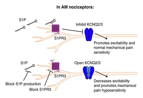
Figure 1: Schematic for S1P-mediated AM activation. Under baseline conditions, S1P mediates the normal response to mechanical stimuli via AMs. 2
BSJ : What did that show you about S1P? How does it operate at baseline levels, and how does it activate S1PR3 for functional mechanical sensitivity?
DB : There seemed to be a range of S1P levels in the skin that give rise to different types of pain. Under normal conditions, it seems to be important for normal sensitivity to mechanical pain: you bump your knee, or you touch a cactus, and you move away. If you turn the S1P down, then you lose sensitivity, similar to diabetic neuropathy, where you don’t feel as well. If you put really high levels in—abnormally high—you become hypersensitive to pain. It was a continuum. It’s often called the rheostat, where you get a continuum of different phenotypes. So, at baseline levels, you have normal responses to pain, which are important protective reflexes. But if you turn it down or up, then you get these really extreme conditions that aren’t good. For example, loss of tactile sensitivity in diabetic neuropathy causes repetitive injuries that cause tissue damage, and eventually, in many patients, amputations. High levels of S1P could cause hypersensitivity to pain, which is associated with lots of different chronic conditions. If you actually go to the literature, people have measured high levels of S1P in patients with really severe arthritis pain, suggesting that elevated S1P might be a disease related to different pain diseases.
BSJ : Your study largely employs a type of neuron called the dorsal root ganglion (DRG). What is the significance of these neurons in the body?
DB : The DRG neurons in both humans and mice are a distinct cluster of neurons alongside your spinal cord that innervate your body from the neck down. They allow you to feel temperature and mechanical sensations. If you get poison oak when hiking, these are the neurons that get activated that make you itchy. They also innervate the gut and the lungs and are involved in interoception—detection of stimuli within your body. When you get a stomach ache, or if you have asthma or lung irritation from smoke, these are the neurons that signal. They are the sensory system for the outside world as well as for the inside of the body.
BSJ : In a similar vein, what are mechanonociceptors and nociceptors, and what distinguishes A mechanonociceptors (AMs) from C nociceptors?
DB : Your somatosensory neurons come in different flavors. Half are dedicated to gentle touch and proprioception, which is the awareness of limb position. These neurons are all mechanosensitive. They are the ones that innervate the skin and allow you to feel the brush of a feather or vibration. When you touch something, they’ll let you feel that there’s an edge and distinguish what the shapes are. The ones in your muscles are also sensing muscle tension. Half of your DRG somatosensory neurons are dedicated to those senses. The other half are dedicated to pain. There are fast pain neurons—when you stub your toe, if you count to three, you’re going to feel that “ouch”! Those are the A deltas that mediate noxious mechanical pain. Then you have
C fiber nociceptors, and those are a little bit slower. If you have a cut, you might notice that there’s a red weal of sensitivity that surrounds the cut. That’s mediated by the slower-pain C fibers. These are the same ones that get activated if you squirt lemon in your eye and it feels painful. They detect noxious mechanical pressure, temperature, and chemicals. The C fibers also respond to capsaicin, menthol, wasabi, and Szechuan peppercorn. So AMs and C nociceptors have somewhat overlapping and distinct functions in pain.
BSJ : How did you determine the localization of S1PR3 in C nociceptors and AMs?
DB : Prurireceptors are itch receptors. The name comes from “pruritis,” which means itchy. A subset of the C fibers that were previously classified as just pain receptors also mediate itch. There are a lot of inflammatory mediators that trigger itch in some conditions and trigger pain in others. One common example is histamine. If you get a mosquito bite, that makes you itchy—wherever histamine is released in your skin, it preferentially activates itch receptors. But in some chronic pain conditions, histamine can become elevated in a different part of the skin, where it activates pain neurons. We found that depending on where S1P gets elevated in different disease conditions, it can trigger itch instead of pain.
DB : We used a variety of different techniques. We had RNA sequencing data, so we knew what types of transcripts were expressed in different subsets of neurons. We also used antibodies against the protein to see what types of cell subsets it’s expressed in, and we stained with other markers that we know of for these different subtypes of pain versus touch. We also used in situ hybridization to fluorescently label the mRNA. So, we looked at mRNA in two different ways and looked at proteins using fluorescence.
BSJ : You found that high levels of S1P activate C nociceptors and not AMs. What does this finding suggest about their respective roles in mechanosensation?
BSJ : Could you briefly describe for our readers what TRP channels are? What is the difference between TRPA and TRPV channels?
DB : TRP channels stands for transient receptor potential ion channels. It’s a family of ion channels that has about 30 members broadly expressed throughout the body. TRPV1 is a particular ion channel that is activated by capsaicin (the active component in chili peppers), heat, and different inflammatory mediators. It plays an important role in pain and temperature sensation. In a subset of cells, it can also contribute to histamine-dependent itch signaling. So TRPV1 is the heat and capsaicin receptor. If you delete that receptor, there’s
DB : It’s a little complicated, because we found that, under baseline levels, AMs were activated. 100 nanomolar is the typical concentration of S1P in the body under normal conditions—a very low level of S1P. The AM nociceptors were playing an important homeostatic role in keeping excitability in check and maintaining normal sensitivity (Fig. 1). Then we elevated S1P concentration to that of disease levels, around one micromolar. We saw a preferential activation of C nociceptors. Normally, C nociceptors are not active, but under these conditions, they activated without any stimuli, which is a concern.
BSJ : You found that S1PR3 is implicated in our perception of itch in addition to mechanical pain. Now that we’ve discussed nociceptors, what is a prurireceptor?

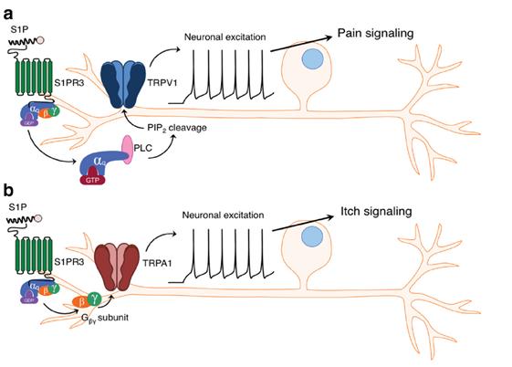
Figure 2: Schematic for S1P-mediated activation of the pain pathway versus the itch pathway. The subset of neurons co-expressing TRPV1 and S1PR3 mediate the pain response, while the subset of neurons co-expressing TRPA1 and S1PR3 mediate the itch response. 3
a deficit in thermal pain and an inability to detect capsaicin. The TRPA1 ion channel is a relative of TRPV1, but it is activated by many different inflammatory mediators both found in the body and the environment—for example, reactive oxygen species, markers of inflammation, or disease-activated markers in the body. It’s also activated by wasabi. When you feel that pungent wasabi next time you eat sushi, it’s because you’re activating the TRPA1 ion channel on the free nerve endings in your mouth. However, it’s very short-lived, so you don’t trigger a big inflammatory response when you just eat sushi. You would need high levels of TRPA1 activation to cause pain and inflammation.
BSJ : What differences did you find between S1P signaling via S1PR3 in prurireceptors versus nociceptors? What role do the TRPA1 and TRPV1 channels play in our perception of pain versus itch?
DB : We found that there are many different kinds of neurons that express TRPA1 and TRPV1. There’s a subset of them that are co-expressed with S1PR3. The subset of TRPA1-S1PR3 neurons are a small population that trigger the itch pathway when activated. The subset of TRPV1-S1PR3 neurons gives rise to inflammatory pain when activated (Fig. 2). These different subsets get activated in different parts of the skin under varying conditions.
BSJ : You collaborated with scientists to generate a synthetic, “photoswitchable” analogue of S1P called PhotoS1P. What does it mean to be “photoswitchable”?
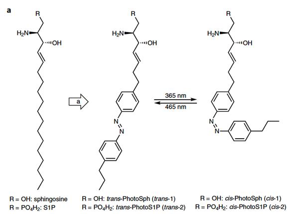
DB : We had a really cool collaboration with Dirk Trauner’s lab. They are chemists who are interested in a lot of interesting biological molecules, including signaling lipids like S1P. They engineered the lipid itself so that it had an extra chemical moiety that was very sensitive to light. This moiety keeps the signaling molecule in a locked conformation under normal light. If you hit the molecule with a certain wavelength of light, it causes a conformational change that now makes that lipid accessible and able to signal (Fig. 3). We injected this engineered S1P into mice and put it into cells, and it didn’t do anything until we zapped it with light. It then sprang into action and could trigger neuronal activation or reflexive behaviors in the animal very rapidly, as if we were poking it.
BSJ : How does the stability of Photo-S1P make it easier to control for sphingolipid metabolism, and what implication does this have for future academic and clinical studies?

Figure 3: Structure of PhotoS1P. On the left is PhotoSph, the precursor of PhotoS1P. The two molecules to the right demonstrate photoswitching between the cis and trans isomers of PhotoS1P, induced by 365 nm or 465 nm light. 4
DB : Right now, it’s a really amazing tool. Lipids are notoriously hard to control. They’re produced all the time. There aren’t a lot of specific tools to manipulate them. So it was really amazing to be able to turn on and off whatever amount of S1P we wanted to. That is not something that’s easy to do. S1P is becoming a very important signaling lipid in the nervous system. It’s not just in the pain pathway, where abnormally high levels are linked to disease. S1P is also thought to be elevated in the hippocampus and in Alzheimer’s patients, so I think it could be a really important tool to figure out all of the diverse roles that this very important signaling lipid has under normal and disease conditions.
REFERENCES
1. Diana Bautista [Photograph]. Retrieved from https:// vcresearch.berkeley.edu/faculty/diana-bautista. 2. Hill, et al. (2018). The signaling lipid sphingosine 1-phosphate regulates mechanical pain. eLife, 7:e33285. doi:10.7554/ eLife.33285 3. Hill, et al. (2018). S1PR3 mediates itch and pain via distinct TRP channel-dependent pathways. The Journal of Neuroscience, 38(36), 7833-7843. doi: 10.1523/JNEUROSCI.1266-18.2018 4. Morstein, et al. (2019). Optical control of sphingosine-1- phosphate formation and function. Nature Chemical Biology, 15, 623–631. doi:10.1038/s41589-019-0269-7


