7 minute read
Regrowing Ourselves: Possibilities of Regenerative Medicine Jessica Jen
REGROWING OURSELVES:
POSSIBILITIES OF REGENERATIVE MEDICINE
Advertisement
BY JESSICA JEN
The myth of Prometheus is most famously identified by long-term suffering involving an eagle and Prometheus’ liver—a liver that merits its own story for its ability to regenerate. This remarkable organ is one of the earliest and most prominent illustrations of regenerative capabilities, although it is highly unlikely that the creators of the myth actually understood the biological reality of hepatic regrowth at the time. In the ancient Greek myth, Prometheus was doomed to eternal punishment after stealing fire from the gods to give to humans. He was chained to a rock where an eagle consumed his liver daily, which then regrew during the night for the next day’s meal. While overnight organ regrowth in humans is still quite a stretch by current standards—Prometheus’ immortality may have represented an accelerated timeline—the concepts behind Prometheus’ exceptional liver remain intriguing. Yet, since regrowth is a much slower process in us mortal organisms, we have found other ways to compensate for our less spectacular regenerative capabilities.
Regenerative medicine and tissue engineering are promising applications of cell repair and regrowth. While the specifics of cell and tissue repair differ between species and biological context, understanding the mechanisms behind these processes provides opportunities to manipulate cell repair and regeneration in a variety of manners.
REGENERATION
Some tissues completely regenerate after trauma, such as salamander limbs, certain human oral tissues, and the liver. 1 After tissues sustain trauma, surviving cells repair themselves, multiply, differentiate, and communicate with one another to regrow any missing or dead tissue. However, not all cells possess the ability to regenerate quickly. If neuronal, cardiac, or muscle cells are damaged by external trauma, they must first reseal their membranes and then proceed to repair themselves internally, a process that typically takes longer than Prometheus’ overnight organ regrowth. 2 This energy-intensive process partially explains why degenerative diseases involving these types of cells are so harmful. Similar repair processes can be observed in single-celled organisms and have been studied in great detail to investigate how such cellular tactics might be used in therapeutic applications. 3 Rather surprisingly, more extreme damage in the form of cell death and aging cause effects that lead to regeneration. During cell death, the dying cell releases signals that initiate cell replication. 4 In the cnidarian genus Hydra, large-scale apoptosis of head cells triggers surrounding cells to regenerate the same head tissue that is nearing the end of its functionality. Interestingly, cell injury without death does not induce regeneration in this scenario.
Furthermore, aging affects some cells’ abilities to regenerate (as seen in muscle and hematopoietic stem cells) while in other organisms, age does not influence regenerative capacity. 5 Overall, we see that the downstream regenerative effects of cell injury, aging, and death all work via differing pathways and often do not cause the same levels and types of regrowth in species.
TISSUE ENGINEERING
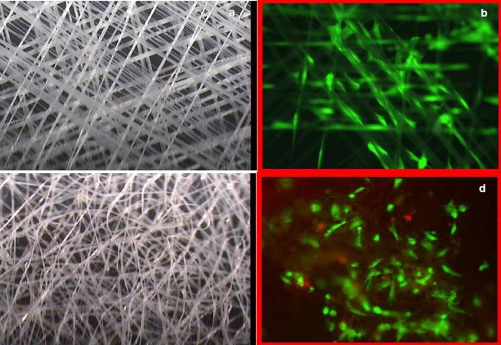
While regeneration uses the body’s own healing processes, it can also be encouraged by outside intervention. Regenerative medicine can restore damaged tissues by inducing the body’s intrinsic repair mechanisms, introducing engineered tissue, or combining both techniques. 6 Tissue engineering is one form of regenerative medicine that assembles cells and molecules into working tissues. Using a combination of scaffolds, growth factors, and additional biological materials, this technique works to create an extracellular matrix-like structure for new tissue to develop(Fig. 1). 7 Scaffolds employ a variety of materials and functions to mimic the conditions of the extracellular matrix. 6 In order to repair, scaffolds can incite the immune response to promote vascular tissue growth, arrange cells, or merely offer mechanical support. Tissue-derived extracellular matrix scaffolds are showing increasing promise as they contain the necessary composition to support cells and do not lead to inflammation like synthetic materials do. 8 These scaffolds are generated by decellularizing a part of the original tissue, leaving only tubular and mechanical structures behind. These “cleaned” scaffolds often require modification, which some researches have achieved by including parallel pore channels inside fabricated extracellular matrix scaffolds. The ability to specify channel size, shape, and arrangement based on the target tissue is a major benefit of this scaffold style and allows researchers a

Figure 1: Electroactive scaffolds. Visual comparison of electroactive scaffolds that provide electrical stimulation in addition to structure. Scaffold varieties provide structure, biological materials, and growth factors to encourage cellular growth. large degree of flexibility in the types of tissues that their scaffold is able to support.
Traditionally, such scaffold develoment and testing has been performed in ex vivo environments with patchy clinical success, but recently, newer forms of tissue engineering have begun to use the body’s own systems of repair without any additional scaffolding to optimize regrowth. One promising biological structure is that of bone, which during embryonic development, utilizes mechanical characteristics coupled with the immune response to heal.
LIMITATIONS, ADVANCEMENTS, AND FUTURE STEPS
Although the basic mechanisms behind healing and regenerative processes are consistent throughout the contexts in which they are studied, their specifics are yet to be completely understood. Not all dying cells trigger neighboring cells to replicate, nor are all tissues capable of complete regeneration. 4 Current research indicates that the scale and type of cell death are major factors in determining the body’s response to mass apoptosis. A deeper understanding of these processes in nature will likely provide opportunities in bioengineering and regenerative medicine at the cellular and tissue levels. Many applications of regenerative medicine have been identified, but are not yet scalable to a level practical for human usage.
There has been progress, however. For example, researchers have recently created a microfluidic guillotine that precisely and efficiently bisects cells. 9 The guillotine uses tiny amounts of fluid to push cells within narrow channels onto a fixed blade, then forces the bisected cell sections on separate paths. This device allows more cells to be studied under controlled situations, and is a major improvement from the traditional method of manually bisecting cells with a glass needle under a microscope. Technique developments like this make it easier for researchers to investigate the cellular mechanisms of regeneration by providing a clear view into what is going on inside the cell.
Understanding the mechanisms behind healing and regeneration at the cell and tissue levels will lead to greater opportunities for advancement in regenerative medicine. Granted, there are limits on regeneration’s capabilities, but tissue engineering is just one form of regenerative medicine with many promising paths and a multitude of applications. Technological advancements in scaffold development and experimental techniques offer new ways for us to push the limits on regrowth. So while his liver’s impressive regrowth did not improve Prometheus’ own situation (indeed it prolonged his pain), it is an inspiring paragon of what our bodies might be capable of with some assistance from regenerative medicine.
REFERENCES
1. Pritchard, M. T. & Apte, U. (2015). Models to study liver regeneration. In U. Apte (Ed.), Liver regeneration: Basic mechanisms, relevant models and clinical applications (1st ed., pp. 15- 40). Academic Press. 2. Abreu-Blanco, M. T., Verboon, J. M., & Parkhurst, S. M. (2011). Single cell wound repair: Dealing with life’s little traumas. BioArchitecture, 1(3), 114-121. https://doi.org/10.4161/ bioa.1.3.17091 3. Tang, S. K. Y. & Marshall, W. F. (2017). Self-repairing cells: How single cells heal membrane ruptures and restore lost structures. Science, 356(6342), 1022-1025. https://doi.org/10.1126/ science.aam6496 4. Vriz, S., Reiter, S., & Galliot, B. (2014). Cell death: A program to regenerate. Current Topics in Developmental Biology (Vol. 108, pp. 121-151). Academic Press. https://doi.org/10.1016/B978- 0-12-391498-9.00002-4 5. Sousounis, K., Baddour, J. A., & Tsonis, P. A. (2014). Aging and regeneration in vertebrates. Current Topics in Developmental Biology (Vol. 108, pp. 218-233). Academic Press. https:// doi.org/10.1016/B978-0-12-391498- 9.00008-5 6. Mao, A. S & Mooney, D. J. (2015). Regenerative medicine: Current therapies and future directions. Proceedings of the National Academy of Sciences of the United States of America, 112(47), 14452-14459. https://doi.org/10.1073/ pnas.1508520112 7. Tonnarelli, B., Centola, M., Barbero, A., Zeller, R., & Martin, I. (2014). Re-engineering development to instruct tissue regeneration. Current Topics in Developmental Biology (Vol. 108, pp. 320-334). Academic Press. https://doi.org/10.1016/B978-0-12- 391498-9.00005-X 8. Zhu, M., Li, W., Dong, X., Yuan, X., Midgley, A. C., Chang, H., … Kong, D. (2019). In vivo engineered extracellular matrix scaffolds with instructive niches for oriented tissue regeneration. Nature Communications, 10(4620). https://doi.org/10.1038/ s41467-019-12545-3 9. Blauch, L. R., Gai, Y., Khor, J. W., Sood, P., Marshall, W. F., & Tang, S. K. Y. (2017). Microfluidic guillotine for single-cell wound repair studies. Proceedings of the National Academy of Sciences of the United States of America, 114(28), 7283-7288. https://doi. org/10.1073/pnas.1705059114
IMAGE REFERENCES
10. Electroactive Scaffold: Three-dimensional scaffold that mimics native biological environment. Retrieved from https://techology.nasa.gov/patent/ LAR-TOPS-200 11. iPS-derived cardiomyocytes. (2018, April 30). Retrieved from https:// www.flickr.com/photos/nih- gov/40906400815/ “Tissue-derived extracellular matrix scaffolds are showing increasing promise as they already contain the necessary composition to support cells, and do not lead to inflammation like synthetic materials do.”


