17 minute read
Dual Imaging: a New Frontier in MRI (Dr. Ashok Ajoy
from ORIGINS
Dual Imaging: A New Frontier in MRI
INTERVIEW WITH DR. ASHOK AJOY BY ANDREW DELANEY, LEXIE EWER, AND ESTHER LIM
Advertisement
Ashok Ajoy, PhD, is an assistant professor of chemistry at the University of California, Berkeley. His research team focuses on utilizing physical chemistry to develop “quantum-enhanced” NMR and MRI technologies, pushing past the current resolution and signal limitations. Beyond his research, Dr. Ajoy is very enthusiastic about his students and emphasizes the importance of the contributions made by his graduate and undergraduate researchers. Having become a professor during the SARS-CoV-2 pandemic, he is especially grateful for his students and expressed that the multiple papers published by his lab are due to the hard work of everyone on his team. Sophie Conti, one of Dr. Ajoy’s research assistants who works on the nitrogen vacancy center magnetometry in microfluidics project, said of the Ajoy lab, “I’ve really loved working in the Ajoy lab thus far because of the supportive community and amazing opportunities for learning. I think our lab is really unique in that undergraduates are really encouraged and supported by the other lab members to further their own learning and research if it interests them.” In this interview, we explore how the use of diamond microparticles can enhance MRI and optical imaging, resulting in a form of dual imaging that has revolutionary impacts for the fields of medicine, biology, and the physical sciences. BSJ: Can you explain the concept of hyperpolarization in imaging, including how it is achieved and how it increases the clarity of a magnetic resonance image (MRI)?
AA: MRI is an extremely versatile and powerful imaging tool, which has become so mature that now a doctor can just press a button and get an image. In reality, you are imaging water in the human body, and within the water, you are imaging the proton nuclear spin. In each hydrogen atom of water, there is a nucleus which contains a proton, and this proton has a spin which is essentially a little magnet itself. The tunnel that you go into in an MRI machine is a superconducting magnet, which is 100,000 times stronger than a weak refrigerator magnet. When you go into this magnetic field, the proton spins align in accordance with the magnet. Then, you tip the protons, and the spins start processing to generate a signal. This alignment is called polarization, and it depends linearly on the strength of the magnetic field. It is an amazing quantum mechanical effect that generates a signal which leads to the images, but the amount of alignment of the spins in a magnetic field is very low and inefficient. That is because the nuclei are in the center of the atom, and they cannot interact very strongly with their environment. If you increase the magnetic field, you will get more and more polarization, but from one Tesla to 20 Tesla, there is only a twenty-fold increase. Additionally, a 20 Tesla magnet is very expensive and very large. You cannot put a human being into it due to the physiological effects of high magnetic fields. As such, there is a big push in the community to improve the polarization and to see if we can generate this alignment of the nuclei in a magnetic field without a magnet; for instance, by shining a laser on a sample. Hyperpolarization is the process of aligning spins without using a magnet or aligning them much more than you could with a magnet. In our paper, we showed that for a special class of materials—diamonds—shining laser light on the material can align these spins about a million times more than in a normal magnetic field. You only need slightly more than the power of a laser pointer to accomplish this. This is remarkable because now the MRI signals will be significantly brighter and faster. Additionally, this laser-assisted hyperpolarization has many other advantages. Devices using this technology can potentially be made at a very low cost, and once you make a device out of it, you can get very high spatial resolution in addition to the improvement in signal.
The current frontier of MRI asks whether you can make an MRI machine that images down to the molecular scale since current MRI is in a millimeter to centimeter length scale. You can see full three-dimensional imaging through a person, cell, or tissue. But it still has very poor resolution and signal, so many people are interested in trying to improve MRI on both of these fronts.

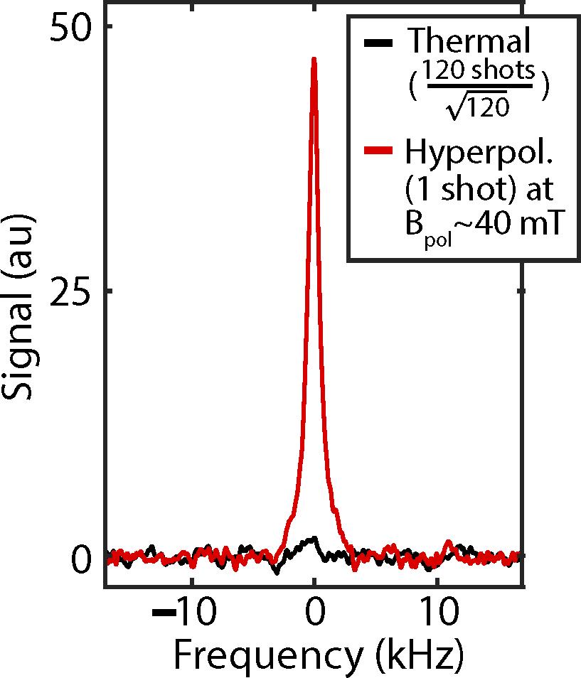
Figure 1: Increased Signaling of Hyperpolarization. Graph illustrates the increase in signal intensity of hyperpolarized imaging (red line) as compared to magnetic resonance imaging (black line), with a 7 Tesla magnet. For reference, the magnets commonly used in clinical imaging are 1.5 T and 3 T.
BSJ: You mentioned in your paper that we may be able to study other hyperpolarized solids, such as silicon and silicon carbide, using a similar mechanism to how you studied diamond microparticles. What is the significance of studying other hyperpolarized solids in this manner?
AA: To understand this, we have to step back and ask, “If you can shine a laser’s light on something, will it be polarized?” For example, if you take a bath of water and shine laser light on it, the spins of the water molecules are not going to align because optically there is no transition that these spins are reacting to. There are special materials, however, which you can excite with light and polarize; for instance, diamond. The advantage is that once the spins of diamond are aligned, you can transfer this alignment to other molecules that come into contact with it. Therefore, we became interested in asking: if you can make a diamond sponge in which you shine light on the diamond to align its spins and flow water through it, then the water will come out polarized. If someone drinks the water and then goes into an MRI machine, will the image be hundreds of thousands of times brighter? This is a frontier direction, and it is still not clear whether diamond is the only material—or the best material—for this task. Other materials have been discovered, and many people are interested in first
Figure 2: Nitrogen vacancy centers and hyperpolar-
ization. Top diagram depicts the chemical structure of the diamond nanoparticles. As shown, the lattice of 13C nuclei contains infrequent nitrogen atoms adjacent to lattice vacancies. These nitrogen vacancy centers result in the distinct purple color of the diamond and its disposition to be hyperpolarized. Bottom diagram is a schematic of a hyperpolarization experiment, displaying the laser (green line) used to hyperpolarize the diamond nanoparticles and align their spins.
exploring a large number of these molecules and making scaffolds out of such porous materials, so that they can bring analytes—water, carbon dioxide, or anything else in contact with them—and spin-polarize them.
BSJ: You also mentioned that the imaging of microdiamonds may lead to further applications for physical, chemical, and biological analysis. Could you elaborate more on how microdiamond imaging will lead to developments in each of these fields?
AA: We showed that it is particularly easy to hyperpolarize nanodiamonds or microdiamonds. Interestingly, it turns out that these particles also fluoresce. Before we got into this field, many people used diamond fluorophores as biological markers. Due to a point defect in diamond, called a nitrogen vacancy center, when you shine a small amount of laser light on the diamond, it will fluoresce forever. Diamond nanoparticles were used as fluorescent tags for many years, and now, we show that by shining light on them, the spins can be spin-polarized. Now, you can image the diamond nanoparticles optically and through MRI, which is the “dual-mode imaging” we describe in the paper. You may ask, what is the big deal? It turns out that optical imaging is amazing, and all the things that we see around us are
mostly optical images, which are fast and cheap. The detectors are also very good. However, optical imaging has one major problem, which is that it works poorly in scattering environments. Say you have a bottle of milk, you put something inside it, and you try to image through it. You cannot do it, because optical imaging normally has a spatial resolution that is set by the wavelength of light. All of microscopy uses the fact that optical imaging has very high spatial resolution, but, if you look at these optical images, very rarely will you see an image that is taken in a centimeter of tissue or a centimeter deep in something that is scattering. There is a big challenge in the community about deep tissue imaging. How do you image not just in one slice of cell or tissue but actually reasonably deep? It turns out that MRI is completely immune to scattering. This is because what you are seeing in an MRI is actually the nuclear spins. You can image through solid, opaque things such as bone, blood, and fatty tissue and be able to see through the person. The technical reason for this is very interesting: MRI does not image in real space. Actually, the image is in the conjugate space to real space, called k-space. It is somewhat similar to X-ray imaging where you image in a Fourier reciprocal space. These two spaces are connected with a Fourier transform. The big picture is that these diamond particles can act as fluorescent tags to indicate a particular place in a cell to image optically. We have now learned that you can image them through MRI. We are hoping to use this facility to image slightly deeper. For example, you can tag things in the body or parts of the cell and try to do some chemical imaging in the tissue. We are still not there yet, but it is a potential approach that could allow you to do deep tissue imaging.
BSJ: What are the advantages and drawbacks of single imaging, via visible-wavelength optics, and MRI?
AA: Optical imaging has a resolution that is set by the wavelength of light, which is called the Rayleigh limit. Ultimately, optical imaging is very fast, cheap, and high resolution. Wherever you can use optical imaging, you should. In some cases, however, the resolution of optical imaging is not good enough. For instance, if you want to see what happens inside a molecule, then the resolution of optical imaging is sub-par. This is because the resolution of optical imaging is around half a micron, and, within a cell or in molecules, the objects are a few nanometers across. While, generally, for biological imaging, optical imaging is the workhorse, it suffers from the issue that in a scattering medium, you attenuate very largely and you lose resolution. For that reason, if you try to image through skin, you have to use something like ultrasound or MRI. But MRI suffers from low resolution and low sensitivity. It is also expensive because it needs big magnets. You can trace the reason why the signal resolution is low to the fact that the polarization is low. So, if you can polarize the spins without a magnet, then you can increase the signal by several fold, approaching a million fold, and you can therefore increase the resolution by the same factor. The advantage of using these small diamond microparticles

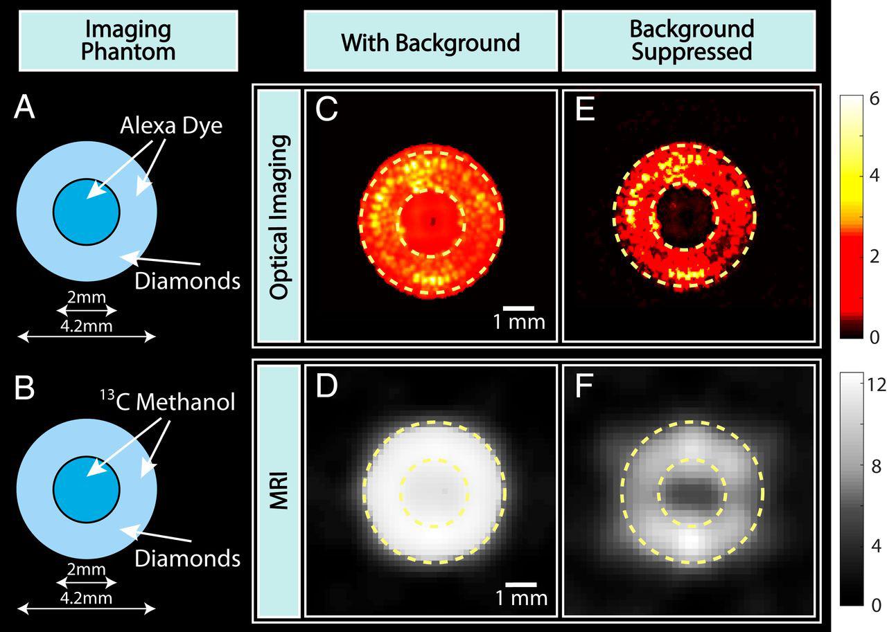
Figure 3: Background Suppression. To mimic the background that is present in optical imaging and MRI, Alexa dye, and 13C methanol were deposited in the center of a ring of diamond microparticles in a phantom. Upon imaging, the circle of diamond microparticles is indistinguishable from the background; however, background subtraction can be used to increase the contrast of the image and allow the diamond phantom to be seen.
is that you can increase their polarization with light, and then you can image them through MRI very efficiently.
BSJ:You used multiple regimes to manipulate the nanoparticles in your experiment. What are the differences between these regimes, and why are each of these regimes needed?
AA: We ask the question: if you can image an object through optics or MRI, could you image through both? It turns out you can. Optical imaging is real space imaging, which means if I look there, I can see that point. On the other hand, MRI images are in Fourier reciprocal space which means that each point that you get has information of all the real space pixels. Ultimately, this means that the MRI image that you get must then go through a Fourier transformation to obtain the real image. The image that you see is not what you are actually sampling. You are actually sampling in k-space. For instance, when k is equal to zero, the first k-pixel is the mean of the image, and the second pixel gives you the first wavelength. This tells us the spatial wavelengths that make up an object. To know what your MRI image is in real space, you use a Fourier transform, which is a mathematical linearity transformation widely used in computer science, physics, and electrical engineering. Essentially, you reconstruct the structure of a molecule, and we get it in real space. However, things get very complicated if you are trying to image things that are sparse. Say you are trying to image stars in the sky, and the light is very low. Normally, you will focus your detector on only one part of the night sky and try to increase the exposure time. But, by definition, there are very few objects. So, most of the time, you are focusing your camera into parts of the sky where there is nothing. Optical imaging does not work well when the number of photons are low and images are sparse. But, if you imagine k-space, for instance, wherever you look, every pixel of k-space will give you information about all the stars. Because it is a Fourier reciprocal, you can use this property to combine both real and k-space images and improve the speed of imaging. This has never been done before, as far as I am aware. We have not done it in this paper yet, but we postulate that this can be done, and we calculate that we can accomplish that incredible speed in the future.
BSJ: What is the importance of 13C and nitrogen vacancy centers on the imaging of diamond microparticles?
AA: Nitrogen vacancy centers are essentially defective diamonds. Diamond is made up of carbon atoms and the lattice. If you remove one carbon atom, replace it with nitrogen, and have a vacancy in the neighboring site, that is a nitrogen vacancy center. This property was discovered by accident by the diamond industry. Colored diamonds are often deemed to be much more expensive than normal diamonds, and this color is achieved by bombarding diamonds with nitrogen atoms. For example, we have diamonds that are deep purple in our lab. It is because of these defects and nitrogen atoms inside that causes the diamond particles to have very nice spin properties. The nitrogen vacancy centers also spin, and they can be polarized when you shine light on them. Then, we transfer the spin alignment to the neighboring nuclei, which in this case is carbon, and that is how we get these images. I should emphasize that diamond is not the only material of this class. Although diamond is expensive, it is good because it is relatively inert. Other molecules that have similar properties to diamond are often carcinogenic, such as pentacene and anthracene. There is a growing effort to try to discover more molecules that, like diamond, have the property of a spin-photon coupling.
BSJ: Since their inception in 1977, MRI machines have revolutionized the diagnosis of many conditions in medicine. In what ways do you expect studies in this area to impact what can be visualized in animals?
AA: We do not intend to ever put diamonds into people. Instead, we want to use diamond as a conduit to transfer spin polarization from the diamond into something else. But, for imaging cells, or in vitro samples, there is still a place for it. For live animal imaging, or human imaging, it is mostly that you are going to create porous materials where you trap water. These materials could be diamonds or some of these other molecular centers. You shine light onto this material, the spins in the water molecules get polarized, and you can take it into the body. We are interested in a future where we have a machine in which you flow water through it, you shine laser light into it to polarize the water, and you inject it into a person. The applications of this would be very far-reaching. Additionally, you can use molecular markers such as pyruvate for cancer imaging, or you can image metabolism to diagnose cancer since cancer cells metabolize faster. It is going to have a revolutionary impact because most people diagnosed with cancer are already in the late stage. But, what if you can devise an imaging technology that is selectively imaging cancer so that you can find it at an early stage? You can spot cancer before you normally would, which would make a world of difference. Hyperpolarized imaging is a key component of this because it can give you selective information. Currently, the many other forms of medical imaging require quite a significant dose of radiation, and there is only a certain limited number of such scans allowable. MRI has a very big advantage in that you can see things and it is radiation-free.
BSJ: Beyond its impact on healthcare and biology, how do you expect the development of dual imaging technology to contribute to our understanding of the physical sciences?
AA: I think one of our biggest contributions is demonstrating the usefulness of nuclear spins. Nuclear spins exist in everything; you and I are made up of atoms and the center of atoms are nuclei, many of which have spins. Usually, these spins are weak and difficult to observe, so people do not study them very much. However, we are interested in coupling these spins with light. Thus, we shine light on atoms and then make their nuclear spins “glow” so that you can then study them, and, through them, you can probe matter. Thus, we are very interested in making chemical sensing probes. This will report not just color, but nuclear magnetic resonance (NMR) information and chemical information. We are trying to imagine a sensor where you put it into a cell, and it will report on all the molecules around it. You can do this through NMR spectroscopy, which gives you very chemically specific information—a fingerprint of the
molecule from which you can extract information about the molecule—and the core of this is this hyperpolarization technology. There are many applications of this chemistry in technology.2
BSJ: Do you have anything else to add about your current research or upcoming projects?
AA: We recently published a paper showing some of these applications of spins, especially in the quantum sciences. It turns out that these nuclei are some of the most coherent quantum mechanical objects in the universe. This means that we were able to keep them processing for something like one thousand and ten oscillations. It is so surprising to us that these defective diamonds have these remarkable nuclear spins that are very coherent. We are interested in using these nuclear spins in various tasks in fundamental physics, such as quantum information science, quantum simulation, or quantum sensing. My point is, NMR and MRI are very central to many sciences because they create an image ultimately generated from spins, and spins are quantum mechanical objects. Thus, there is chemistry there, because the spins can report on their local environment. But, the spins themselves are quantum mechanical, so you can do a lot of fundamental physics. Furthermore, you can exploit these resources for quantum computing, which makes computers much faster. This is the amazing part of this research field.
REFERENCES
1. Lv, X., Walton, J. H., Druga, E., Wang, F., Aguilar, A., McKnelly,
T., Nazaryan, R., Liu, F. L., Wu, L., Shenderova, O., Vigneron,
D. B., Meriles, C. A., Reimer, J. A., Pines, A., & Ajoy, A. (2021).
Background-free dual-mode optical and 13C magnetic resonance imaging in diamond particles. Proceedings of the National Academy of Sciences, 118(21). https://doi.org/10.1073/ pnas.2023579118 2. Beatrez, W., Janes, O., Akkiraju, A., Pillai, A., Oddo, A., Reshetikhin, P., Druga, E., McAllister, M., Elo, M., Gilbert, B., Suter,
D., & Ajoy, A. (2021). Floquet prethermalization with lifetime exceeding 90 s in a bulk hyperpolarized solid. Physical Review
Letters, 127(17). https://doi.org/10.1103/physrevlett.127.170603
IMAGE REFERENCES
1. Headshot: [Photograph of Ashok Ajoy]. Ajoy Lab. https://chemistry.berkeley.edu/faculty/chem/ajoy. Image reprinted with permission. 2. Figure 1: Lv, X., Walton, J., Druga, E., Nazaryan, R., Mao, H.,
Pines, A., Ajoy, A., & Reimer, J. (2020). Imaging sequences for hyperpolarized solids. Molecules, 26(1), 133. https://doi. org/10.3390/molecules26010133 3. Figure 2: See reference #1. 4. Figure 3: Lv, X., Walton, J. H., Druga, E., Wang, F., Aguilar, A.,
McKnelly, T., Nazaryan, R., Liu, F. L., Wu, L., Shenderova, O.,
Vigneron, D. B., Meriles, C. A., Reimer, J. A., Pines, A., & Ajoy,
A. (2021). Background-free dual-mode optical and 13C magnetic resonance imaging in diamond particles. Proceedings of the National Academy of Sciences, 118(21). https://doi.org/10.1073/ pnas.2023579118


