
13 minute read
Cl INICA l upDAte tracheal intubation in cats


COlette JOll I ffe
Introduction
tracheal intubation provides several functions. It protects the patient’s airway from obstruction due to reduced muscle tone under anaesthesia; protects against aspiration of saliva, blood or gastric contents; allows administration of oxygen, volatile agent and positive pressure ventilation; and enables suction of the airway.
Poor technique during tracheal intubation can cause laryngospasm, laryngeal oedema, haemorrhage, vagal stimulation, arrhythmias and tracheal damage, ranging from transient inflammation to tracheal rupture. use of inappropriately small internal diameter tubes can increase resistance to flow of gas and the work of breathing, leading to hypoventilation. Small diameter tubes also increase the risk of tube obstruction by mucus, blood or other debris. Inappropriately large tubes can traumatise the airway. Cuffed tubes have been associated with tracheal trauma in cats including tracheal mucosal ischaemia, stenosis and tracheal rupture. t his article will discuss equipment and techniques used for tracheal intubation in cats.
Selection Of Tube Type
t here are many different endotracheal (E t ) tubes available for use in veterinary patients. t he material, diameter and shape of the tube required may depend on the patient and the procedure.
Tube Material
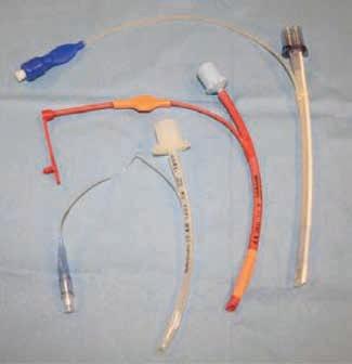
Endotracheal tubes may be made of rubber, silicone rubber or polyvinyl chloride (PVC) (Figure 1). Red rubber tubes and silicone tubes are re-usable and this is reflected in the price. Polyvinyl chloride tubes can be clear, siliconised, or ivory. t hese tubes are inexpensive as they are designed for single use, although it is common in veterinary practice to re-use them after cleaning. t he PVC tubes soften at body temperature and are reported to conform to the shape of the trachea. Generally tracheal intubation is technically easier using the stiffer tubes such as red rubber rather than the soft ivory PVC or silicone rubber tubes. Soft tubes are often straight rather than curved, which may also make intubation more challenging. However, softer tubes are less likely to cause damage to the larynx during insertion, and cause less pressure on the tracheal wall during anaesthesia as they conform better to the shape of the trachea. With good patient positioning and some practice, these tubes are easy to use.
Cuffed tubes
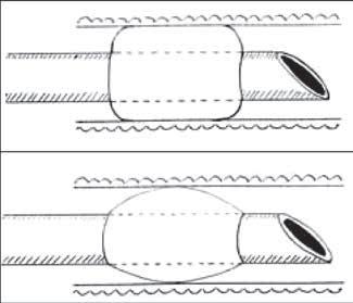
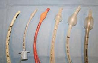
Endotracheal tubes may be cuffed or plain (uncuffed). Inflation of the cuff provides a seal between the tube and the tracheal mucosa, preventing pollution of the atmosphere with anaesthetic gases, aspiration of saliva or gastric contents, and facilitating the provision of positive pressure ventilation. However, the use of cuffed tubes in cats has been associated with tracheal injury including rupture. t he mucosal perfusion pressure of the trachea is between 20 and 30 mmHg, hence if the cuff exerts a pressure greater than this, ischaemia will occur. High pressure/ low volume cuffs are more likely to cause tracheal ischaemia, necrosis and subsequent stenosis when overinflated due to the small area of trachea over which the force is exerted. Low pressure/high volume cuffs exert the force over a larger area, resulting in a lower pressure on the tracheal mucosa (Figure 2). Cuffs of tubes manufactured from different materials have different properties (Figure 3).
Contact: Jo.Hart@dechra.com
Red rubber tubes have high pressure/ low volume cuffs due to the thickness of the cuff material which requires a high pressure for inflation. PVC tubes can have low pressure/high volume, intermediate, or high pressure/low volume cuffs because the cuff wall is thin and inelastic. However, when deflated, these cuffs do not conform well to the contours of the tube, which may necessitate the use of a smaller internal diameter tube (Figure 4). Also, they do not protect the airway against aspiration as effectively as high pressure/ low volume cuffs due to wrinkles in the cuff which may allow passage of fluids. Application of a water-based gel to the cuff reduces the risk of aspiration. t his phenomenon has not been investigated in cats where it is possible that the small size of the trachea and hence any wrinkles in the cuff may not be sufficient to allow liquids to pass. Silicone tube cuffs are usually medium pressure/ medium volume and the soft elastic material conforms well to the contour of the tube, enabling passage of a large diameter tube (Figure 4). low pressure/high volume cuffs tended to be more severe due to their longer length. most of the cats had undergone dental procedures. t he most likely cause of tracheal rupture in these cases was over-inflation of the cuff, possibly in an attempt to prevent aspiration of water and debris, although use of stylets and excessive movement of the tube during the procedure were also suggested as possible causative factors. A plain tube combined with a throat pack will provide adequate protection from aspiration during dental procedures, and it is recommended to disconnect the E t tube from the anaesthetic breathing system each time the patient is repositioned during the procedure to minimise traction and twisting of the E t tube within the trachea. occlusion of the lumen of the tube may occur due to over-inflation of the cuff, either by prolapse of the cuff over the end of the tube or by compression of the tube lumen (Figure 5). t he use of nitrous oxide as part of the carrier gas mixture can alter cuff pressure. Due to its low blood:gas solubility coefficient, nitrous oxide diffuses into the air-filled cuff and expands it further, resulting in increased pressure on the tracheal mucosa. t he magnitude of the expansion depends on the percentage of nitrous oxide delivered and the material of the cuff. Nitrous oxide diffuses more easily across red rubber than PVC tube cuffs. In human practice there are various devices designed to negate this effect. For veterinary patients, if using nitrous oxide, it may be wise to slightly deflate the cuff and reassess cuff pressure by the method described above after five to ten minutes of anaesthesia. Cuffed tubes can be used without cuff inflation if required.

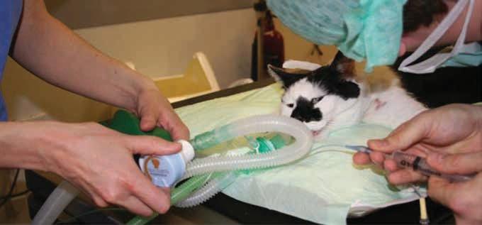
It has been well demonstrated that cuff pressure cannot be assessed by subjectively estimating the pressure or volume of the pilot balloon. Cuff pressure can be measured using a pressure manometer attached to the pilot balloon, but depending on the type of cuff, this may not reflect accurately the pressure exerted on the tracheal wall. t he most practical way to assess cuff inflation is to connect the tracheal tube to the breathing system and inflate the lungs with oxygen while listening for sounds of gas leakage around the E t tube (Figure 6). t he cuff can then be inflated slowly to the point where there is no audible leakage.
Plain Tubes
If an uncuffed or plain tube is used, the internal diameter of the tube t here have been reports of tracheal rupture in cats associated with the use of cuffed E t tubes, both high pressure/ low volume and low pressure/high volume cuffs. t he tears associated with is likely to be larger than that of a cuffed tube for the same size trachea, resulting in decreased resistance and work of breathing, and decreased risk of obstruction of the tube by mucus. t here is also less risk of tracheal rupture, although one reported case of tracheal rupture involved a cuffed tube which was not inflated. A close fitting plain tube is required to prevent pollution of the environment with anaesthetic gases and inhalation agents, especially if positive pressure ventilation is to be imposed. Aspiration of saliva, blood or debris from dental procedures can be prevented by packing the pharynx ideally with a throat pack or with dampened swabs. l ength of the tube t he optimal position of the E t tube is with the distal end in the mid to distal cervical region and the proximal end level with the incisor arcade. If the tube is too long there is a risk of bronchial intubation with resulting ventilation perfusion mismatching. In the event of tracheal rupture following intubation, the prognosis for survival following surgical repair is worse if the rupture is close to the carina. If the E t tube protrudes from the mouth this results in increased apparatus dead space with potential for decreased alveolar ventilation and carbon dioxide rebreathing. t here may be increased risk of kinking and movement of the tube.

For each patient the advantages and disadvantages of use of a cuffed tube should be assessed. For example, in a cat with megoesophagus the risk of regurgitation and aspiration may outweigh the risk of tracheal rupture and a cuffed tube should be considered.
Width of the tube t he internal diameter (ID) of the E t tube is marked on the tube and sometimes on the pilot balloon of the cuff. In general, use of the largest diameter tube that can be inserted without causing damage to the larynx or trachea (i.e. with no resistance) is advantageous. A wider tube will give less resistance to breathing and is less likely to become obstructed by mucus, lubricating gel or mucosa. For a given tracheal diameter, an appropriate plain tube is likely to have a wider ID than a cuffed tube, because of the bulk of the cuff itself. t his is less true of silicone rubber tubes, whose cuffs are very low profile when not inflated (Figure 4). An average 4 kg cat’s trachea will usually accommodate a 4.5 mm ID plain tube.
Bevel
E t tubes are bevelled to aid visualisation of the larynx and insertion of the tube. Some bevels have a hole opposite the aperture called a murphy eye (Figure 4). t his is a safety feature to enable passage of gases should the opening of the tube become lodged against the tracheal wall.
Preparation For Intubation
Before the cat is anaesthetised equipment for intubation should be prepared.
tubes t he size and conformation of the cat should be assessed visually to estimate the likely appropriate ID of the E t tube to be inserted. Gentle palpation of the trachea may also be useful. t here are no published guidelines on selection of tube size, this must be based on clinical judgement and experience. Several tubes of different internal diameters should be available. t he required length of the tube should be estimated, for example by measuring a tube against the cat. t he tube should be cut if necessary. If cuffed tubes are used, the cuffs should be inflated for a few minutes before induction of anaesthesia to check for leaks.
Laryngoscope
Although intubation can be performed without a laryngoscope, a laryngoscope or other light source should always be readily available in case of difficult intubation. A laryngoscope is useful to depress the base of the tongue to provide good illumination of the larynx. An appropriate short blade should be fitted and the light tested prior to induction of anaesthesia (Figure 7). tube ties tape or bandage may be pre-tied around the E t tubes to tie the tube in place after intubation. A dry swab may be useful to hold the tongue.

L Ocal Anaesthetic
Due to the sensitivity of the feline larynx, the mucosa should be desensitised with local anaesthetic prior to intubation to help avoid laryngospasm. Intubeaze (Dechra Veterinary Products) is the only spray bottle licensed for this use in New Zealand.
Lubrication
t he E t tube may be lubricated with a water based gel such as KY jelly or with local anaesthetic gel, although no products are licensed for this purpose.
skilled assistance t he most important requirement is a skilled assistant who can position the patient for the intubation procedure, secure the tube and assist with cuff inflation.
Intravenous access
Ideally an intravenous catheter should be placed to enable administration of incremental doses of injectable anaesthetic agent during the intubation procedure. Alternatively a syringe and needle can be taped in position for this purpose. Intravenous injection of incremental doses helps to ensure adequate depth of anaesthesia during intubation.
Technique
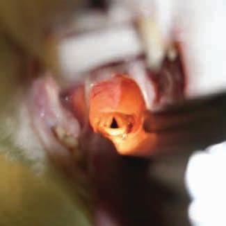
once anaesthetised, the patient should be placed in the preferred recumbency for tracheal intubation. t his may be sternal, left or right lateral or dorsal recumbency depending on the preference of the anaesthetist. sternal and lateral recumbency t he assistant should extend the patient’s head and neck by lifting the lips and grasping the cat behind the maxillary canine teeth with the thumb and forefinger of one hand. An alternative technique is to thread a piece of bandage behind the maxillary canines and use the bandage to extend the head (Figure 8). t he assistant or the anaesthetist then pulls the tongue forward by grasping it gently with fingers or a dry swab. If the tongue tip is within the mouth, the laryngoscope blade or a tongue depressor should be used to pull the tongue forward to avoid injury to fingers. enable visualisation of the larynx. t his technique can be performed without assistance, but there may be increased risk of regurgitation and aspiration. laryngoscopy t he larynx is visualised, ideally using a laryngoscope. For a right-handed person, the tongue is held in the left hand or by the assistant. t he laryngoscope is held in the right hand and positioned with the tip at the base of the tongue. Depression of the base of the tongue causes the epiglottis to rotate rostrally, enabling visualisation of the vocal folds (Figure 9). t he laryngoscope should not be used to pull the epiglottis forwards unless it is absolutely necessary, as touching the epiglottis and other laryngeal structures should be kept to a minimum to avoid iatrogenic damage. t he laryngoscope can then be held by the left hand leaving the right hand free for applying local anaesthetic and inserting the E t tube (Figure 10). laryngeal desensitisation t he larynx is usually desensitised using a lidocaine spray. It is possible to use injectable lidocaine 2% solution applied using a syringe and an intravenous catheter to spray the larynx, but this is not licensed, and care should be taken not to exceed the toxic dose. When using Intubeaze, the bottle must be held upright or the spray generated is inadequate. t his should be factored into the decision as to which recumbency to position the patient. For example, the (right handed) author’s preferred technique is to position the cat in left lateral recumbency, and operate the Intubeaze bottle with the right hand while holding the tongue and laryngoscope with the left hand. t he lidocaine needs 30 to 90 seconds to take effect before intubation is attempted. It is imperative that the cat is adequately anaesthetised before tracheal intubation is attempted. Attempts to intubate the trachea with the cat too lightly anaesthetised may result in excessive coughing and laryngospasm, potentially necessitating a tracheostomy. maintenance of intravenous access allows titration of the depth of anaesthesia to optimise intubation conditions. tracheal intubation


(a) the assistant holds the patient’s maxilla between thumb and forefinger. (b) the maxilla is held with tape.
Dorsal recumbency t he patient is positioned in dorsal recumbency, and the anaesthetist uses a laryngoscope to raise the mandible and push the tongue upwards to once the larynx is desensitised the E t tube is advanced through the rima glottidis between the vocal folds. If the arytenoid cartilages are moving with respiration, the tube should be advanced during maximal abduction. t he tube should not be forced through a closed glottis. t here should be no resistance to the passage of the tube. t he tube is then secured in place using a tie made of woven bandage or other suitable material which is usually tied behind the head or around the mandible caudal to the canines. once the tube is secure, it can be connected to the breathing system and oxygen supplied. If cuff inflation is required cuff pressure should be assessed by the method described above (Figure 6). It is recommended to secure the tube before cuff inflation to minimise movement of the inflated cuff within the trachea. once the cuff is inflated administration of anaesthetic agent (and nitrous oxide if used) can be started.

Preparation For A Potentially Difficult Intubation
Before anaesthesia, the risk of problems during tracheal intubation should be assessed for each patient. If the risk of complications is considered to be high, e.g. in patients with upper respiratory tract noise or suspected nasopharyngeal polyp, preparations for alternative intubation techniques and emergency procedures should be made (Figure 11).
needle passed between tracheal rings. t his can be attached to a size 3.5 ID 15 mm E t tube connector and connected to an anaesthetic breathing system. Apparatus for this procedure should be prepared prior to anaesthesia in high risk cases. A transtracheal needle can also be used for retrograde wireguided orotracheal intubation. Lastly, a sterile tracheostomy kit should be available.
E Xtubation
A skilled assistant must be available. Pre-oxygenation by mask or flow-by technique should be considered as this will increase haemoglobin saturation and provide more time for intubation before hypoxaemia occurs.
A laryngoscope is invaluable for aiding visualisation and access to the larynx. A blunt stylet may be useful to stiffen the E t tube. A stylet or a dog urinary catheter threaded through an E t tube can be passed through the larynx, and the tube guided over it once access to the trachea is achieved (Figure 12). Care must be taken to avoid iatrogenic damage to the larynx and trachea if a stiff stylet is used for this. Suction and throat swabs should be available if regurgitation or haemorrhage is likely. Suction can be achieved using a dog urinary catheter and a large syringe if a suction machine is not available.
If orotracheal intubation proves impossible, oxygen can be insufflated using a transtracheal 18 G hypodermic
Prior to extubation, the pharynx and larynx should be examined to detect the presence of debris, blood, gastric contents etc, particularly following dental procedures, gastroscopy, gastro-intestinal surgery, and in cases of megoesophagus. A laryngoscope may be useful for this. t he unwanted material can then be removed by swabbing or suction. Any throat packs or swabs should be removed. t he cuff must be deflated before extubation. Some anaesthetists recommend extubation early, when the ‘ear twitch’reflex returns, to minimise coughing and irritation of the larynx. o thers recommend extubation at the return of oral and pharyngeal reflexes. t iming of extubation may depend on the patient, e.g. brachycephalic patients should be extubated late to minimise the risk of upper respiratory tract obstruction. t he patient should be monitored for post-extubation upper respiratory tract obstruction until fully awake, although airway oedema may not develop for several hours. Patients considered at risk of obstruction should be closely monitored and equipment for emergency intubation should be prepared and readily available.
Conclusions
tracheal intubation is routine in feline patients. An understanding of possible complications and their prevention is paramount in reducing morbidity. As with any clinical technique, adequate preparation is the key to a trouble-free procedure.

Acknowledgements
t hanks to michelle Higman, Elizabeth Leece and Andy Sparkes for the photographs used. t his article was sponsored by Dechra Veterinary Products.
References
Al-Shaikh B, Stacey S. Essentials of Anaesthetic Equipment (2nd edn), Churchill livingstone, e dinburgh, uk , pp 55–71, 2002
Davey A, Moyle JTB, Ward C. Ward’s Anaesthetic Equipment (3rd edn), WB saunders Company ltd, l ondon, uk , pp 120–166, 1992
Hardie EM, Spodnick GJ, Gilson SD. tracheal rupture in cats: 16 cases (1983–1998). J Am Vet Med Assoc 214, 508–512, 1999
Hartsfield SM. Airway management and ventilation. In: lumb and Jones’ Veterinary Anaesthesia (3rd edn). Thurmon JC, tranquilli WJ, Benson GJ (eds). Williams and Wilkins, Baltimore, usA, pp 515–556, 1996
Mitchell SL, McCarthy R, Rudloff E et al . tracheal rupture associated with intubation in cats: 20 cases (1996–1998). J Am Vet Med Assoc 216, 1592–1595, 2000 Wong WT, Brock KA. tracheal laceration from endotracheal intubation in a cat. Vet Rec 134, 622–624, 1994 l


