Cuadernos de

HERPETOLOG ÍA VOLUMEN 36 - NUMERO 2 - SEPTIEMBRE 2022 ojs.aha.org.ar - aha.org.ar ISSN 1852 - 5768 (en línea) Revista de la Asociación Herpetológica Argentina
Asociación Herpetológica Argentina
Presidenta: María Laura Ponssa
Vicepresidenta: Vanesa Arzamendia
Secretaria: Marta Duré
Prosecretaria: Laura Nicoli
Tesorera: Ana Duport
Vocales Titulares: Gabriela Gallardo , Cristian Abdala
Vocal Suplente: Julián Faivovich
Junta Revisora de Cuentas: Darío Cardozo , Diego Barrasso
CUADERNOS de HERPETOLOGÍA
Una publicación semestral de la Asociación Civil Herpetológica Argentina (Paz Soldán 5100. Piso 1 Dpto 8. Ciudad Autónoma de Buenos Aires, Argentina). Incluye trabajos científicos relacionados con todos los aspectos de la investigación en Anfibios y Reptiles, abarcando tópicos como: sistemática, taxonomía, anatomía, fisiología, embriología, ecología, comportamiento, zoogeografía, etc. Comprende las siguientes secciones: Trabajos, Puntos de Vista, Notas, Novedades Zoogeográficas y Novedades Bibliográficas. Publica en formato digital online y en formato impreso artículos científicos originales asegurando a los autores un proceso de revisión por evaluadores externos sólido y trasparente más una alta visibilidad internacional de sus trabajos. Para los lectores, se garantiza el acceso libre a los artículos. Los idiomas aceptados son castellano, portugués e inglés.
Virginia Abdala
Instituto de Biología Neotropical (CONICET-UNT), Tucumán, Argentina.
Vanesa Arzamendia
Instituto Nacional de Limnología (CONICET-UNL), Facultad de Humanidades y Ciencias, Universidad Na cional del Litoral, Santa Fe, Argentina.
María Laura Ponssa
Unidad Ejecutora Lillo (CONICET-FML), Tucu mán, Argentina.
María Florencia Vera Candioti
Unidad Ejecutora Lillo (CONICET-FML), Tucu mán, Argentina.
Margarita Chiaraviglio
Instituto de Diversidad y Ecología Animal (CONI CET-UNC), Córdoba, Argentina.
Comité Científico
Gabriela Perotti
Instituto de Investigaciones en Biodiversidad y Medioambiente (CONICET–UNComa), San Carlos de Bariloche, Rio Negro, Argentina.
Juliana Sterli
Museo Paleontológico Egidio Feruglio (CONICET), Trelew, Chubut, Argentina.
Lee Fitzgerald
Department of Wildlife and Fisheries Sciences, Texas A&M University, College Station, Texas, EE.UU.
Darrel Frost
Division of Vertebrate Zoology, Herpetology, American Museum of Natural History, New York, EE.UU.
Célio F. B. Haddad
Departamento de Zoologia, Instituto de Biociências, UNESP, Rio Claro, São Paulo, Brasil.
Directores / Editores
Javier Goldberg / Diego Baldo
Taran Grant
Departamento de Zoologia, Instituto de Biociências, Universidade de São Paulo (USP), São Paulo, Brasil.
James A. Schulte II
Department of Biology, 212 Science Center, Clarkson University, Potsdam, NY, EE.UU.
Esteban O. Lavilla
Unidad Ejecutora Lillo (CONICET-FML), Tucu mán, Argentina.
Gustavo Scrocchi
Unidad Ejecutora Lillo (UEL, CONICET-FML), Tu cumán, Argentina.
Instituto de Diversidad y Ecología Animal, (IDEA, CONICET-UNC), Córdoba, Argentina / Laboratorio de Genética Evolutiva, Instituto de Biología Subtropical (CONICET – UNaM), Facultad de Ciencias Exactas Químicas y Naturales, Universidad Nacional de Misiones, Argentina
Editores asociados
María Gabriela Agostini
Instituto de Ecología, Genética y Evolución de Bue nos Aires (CONICET-UBA), Ciudad Autónoma de Buenos Aires, Argentina.
Luciana Bolsoni Lourenço
Departamento de Biologia Estrutural e Funcional, Ins tituto de Biologia, Universidade Estadual de Campinas (UNICAMP), Campinas, São Paulo, Brasil.
Claudio Borteiro
Sección Herpetología, Museo Nacional de Historia Natural de Montevideo, Montevideo.Uruguay.
David Buckley
Dpto de Biodiversidad y Biología Evolutiva, Museo Na cional de Ciencias Naturales, CSIC, Madrid, España.
Mario R. Cabrera
Departamento Diversidad Biológica y Ecología, FCEFyN, Universidad Nacional de Córdoba, Córdo ba, Argentina.
Clarissa Canedo
Departamento de Zoologia, IBRAG, UERJ, Mara canã, Rio de Janeiro, Brasil.
Santiago Castroviejo-Fisher
Laboratorio de Sistemática de Vertebrados, Pontifícia Universidade Católica do Rio Grande do Sul (PUCRS), Porto Alegre, Brasil.
Ana Lucia da Costa Prudente
Coordenação de Zoologia, Museu Paraense Emilio Goeldi, Belém, Estado do Pará, Brasil.
Julián Faivovich
Museo Argentino de Ciencias Naturales “Bernardino Ri vadavia”, Ciudad Autónoma de Buenos Aires, Argentina.
Thais Barreto Guedes
Universidade Estadual do Maranhão (UEMA), Caxias, Maranhão, Brasil.
Nora Ruth Ibargüengoytía
Instituto de Investigaciones en Biodiversidad y Medioambiente (CONICET-UNComa), San Carlos Bariloche, Argentina.
Adriana Jerez
Departamento de Biología, Universidad Nacional de Colombia, Bogotá, Colombia.
Claudia Koch
Alexander Koenig Research Museum, Bonn, Alema nia (ZFMK).
Julián N. Lescano
Instituto de Diversidad y Ecología Animal, (IDEA, CONICET-UNC), Córdoba, Argentina.
Antonieta Labra
1. ONG Vida Nativa, Chile. 2. Centre for Ecological and Evolutionary Synthesis, University of Oslo, Noruega.
Carlos A. Navas
Departamento de Fisiologia, Instituto de Biociências, Universidade de São Paulo (USP), São Paulo, Brasil.
Daniel E. Naya
Dpto de Ecología y Evolución, Facultad de Ciencias, Universidad de la República, Montevideo, Uruguay.
Laura Nicoli
Museo Argentino de Ciencias Naturales “Bernardino Rivadavia”, Ciudad Autónoma de Buenos Aires, Ar gentina.
Paulo Passos
Dpto de Vertebrados, Setor de Herpetologia, Museu Nacional, Universidade Federal do Rio de Janeiro, Brasil.
Paola Peltzer
Facultad de Bioquímica y Ciencias Biológicas, Uni versidad Nacional del Litoral, Santa Fe, Argentina.
Sebastián Quinteros Instituto de Bio y Geociencias del NOA (IBIGEO, CONICET-UNSa), Salta, Argentina.
Silvia Quinzio
Instituto de Diversidad y Ecología Animal, (IDEA, CONICET-UNC), Córdoba, Argentina.
Alex Richter-Boix
Evolutionary Biology Centre, Uppsala Universitet, Norbyvägen, Uppsala, Suecia.
Miguel Tejedo
Departamento de Ecología Evolutiva Estación Bioló gica de Doñana (EBD-CSIC), Sevilla, España.
Marcos Vaira
Instituto de Ecorregiones Andinas (CONICET-UN Ju), San Salvador de Jujuy, Argentina.
Soledad Valdecantos Instituto de Bio y Geociencias del NOA (IBIGEO, CONICET-UNSa), Salta, Argentina.
Cuadernos de
HERPETOLOG ÍA Revista de la Asociación Herpetológica Argentina Volumen 36 - Número 2 - Septiembre 2022
Trabajo
Cuad. herpetol. 36 (2): 125-154 (2022)
Dos nuevas especies del grupo Pristimantis boulengeri (Anura: Strabomantidae) de la cuenca alta del río Napo, Ecuador
Patricia Bejarano-Muñoz1, Santiago R. Ron2, María José Navarrete2,3, Mario H. Yánez-Muñoz1
1 Unidad de Investigación, Instituto Nacional de Biodiversidad, calle Rumipamba 341 y Av. de los Shyris, Casilla 17-07-8976. Quito, Ecuador.
2 Museo de Zoología, Escuela de Ciencias Biológicas, Pontificia Universidad Católica del Ecuador, Apartado 17-01 2154. Quito, Ecuador.
3 Dirección actual: Department of Integrative Biology, University of California Berkeley, Berkeley, CA, USA 94720
Recibido: 22 Diciembre 2020
Revisado: 01 Marzo 2021
Aceptado: 27 Agosto 2022
Editor Asociado: D. Baldo
doi: 10.31017/CdH.2022.(2020-103)
Pristimantis omarrhynchus: urn:lsid:zoobank.org:act:886B9E45BCA0-4796-8AB9-C81AFF2FCD81
Pristimantis miltongallardoi: urn:lsid:zoobank.org:act:3BEEB8E89970-4CEE-938E-EF72C0158037
ABSTRACT
Through the combination of morphological and phylogenetic evidence, we describe two species of Pristimantis from the upper basin of the Napo River. Both species have well-defined dorso lateral folds, a conical tubercle on the eyelid, a papilla on the tip of the snout, weakly expanded discs, and small size (female SVL < 28.2 mm). Pristimantis omarrhynchus sp. nov. differs from its sister species, Pristimantis miltongallardoi sp. nov., by the absence of iridophores on the belly, subacuminate snout in dorsal view, and narrow digits. Our phylogeny and morphological evidence, are conclusive in assigning them to the Pristimantis boulengeri species group, closely related to P. boulengeri, P. cryptopictus, P. dorspictus, and P. brevifrons. The new species are the first reported for the P. boulengeri group in Ecuador and the Amazon basin. We also comment on the correct identity of GenBank sequences previously assigned to P. thymelensis and P. myersi.
Key words: Andes; Phylogenetics; Call; Pristimantis omarrhynchus sp. nov.; Pristimantis miltongallardoi sp. nov.; Systematics; Terrarana.
RESUMEN
A través de la combinación de evidencia morfológica y filogenética describimos dos especies de Pristimantis de la cuenca alta del río Napo. Las dos especies presentan pliegues dorsolaterales bien definidos, un tubérculo cónico en el párpado, una papila en la punta del hocico, discos poco dilatados y tamaño pequeño (hembras LRC < 28,2 mm). Pristimantis omarrhynchus sp. nov. se diferencia de su especie hermana, Pristimantis miltongallardoi sp. nov., por la ausencia de iridóforos en el vientre, hocico subacuminado en vista dorsal y dígitos estrechos. Nuestra filo genia, en combinación con la evidencia morfológica, son concluyentes para asignarlas al grupo de especies Pristimantis boulengeri y cercanamente relacionadas a P. boulengeri, P. cryptopictus, P. dorspictus y P. brevifrons. Las nuevas especies son las primeras reportadas para el grupo P. boulengeri en el Ecuador y la cuenca amazónica. También comentamos la identidad correcta de secuencias GenBank previamente asignadas a P. thymelensis y P. myersi
Palabras clave: Andes; Canto; Filogenia; Pristimantis omarrhynchus sp. nov.; Pristimantis miltongallardoi sp. nov.; Sistemática; Terrarana.
Introducción
La región noroccidental de la cuenca del río Ama zonas se caracteriza por que sus sistemas tributarios nacen del ramal oriental de los Andes de Ecuador (Lynch, 1979; Lynch y Duellman, 1980; Vasconcelos
Autor para correspondencía: santiago.ron@gmail.com
et al., 2019). Particularmente la cuenca alta del río Napo, integra bosques montanos de los Andes con los bosques tropicales Amazónicos que presentan singular importancia por su alto nivel de vulnerabi
125
lidad frente a las dinámicas de cambios de cobertura y uso de la tierra (Cuesta et al., 2009).
En estos biomas se concentra una gran di versidad y endemismo de fauna anura (Lynch y Duellman, 1973, 1980; Guayasamin y Funk, 2009; Morales-Mite y Yánez-Muñoz, 2013; Morales-Mite et al., 2013), resaltando en sus comunidades, la abun dancia y redundancia de la diversidad de ranas de desarrollo directo del género Pristimantis (Lynch y Duellman, 1980; Flores y Vigle, 1994; Zimmer man y Simberloff, 1996; Guayasamin y Funk, 2009; Bejarano-Muñoz, et al., 2015).
Específicamente en las subcuencas de los ríos Coca y Aguarico, se encuentran biomas piemonta nos y montano bajos entre los 600 y 2200 msnm, que acoge un importante punto caliente de diversi dad de ranas terrestres Pristimantis, con más de 24 especies reportadas hasta el momento de las cuales el 38% se encuentran amenazadas y casi la mitad de las especies (46%) son endémicas de estos sistemas montañosos (Lynch y Duellman, 1980; Morales y Yánez-Muñoz, 2013; Bejarano-Muñoz et al., 2015; Ron et al., 2020).
Nuestros análisis y revisión de colecciones generadas en la zona durante los últimos seis años a través de diferentes iniciativas institucionales y expediciones en el área han determinado a partir de inferencia filogenética el hallazgo de dos espe cies no descritas del grupo de especies Pristimantis boulengeri (sensu González-Durán et al., 2017) para Ecuador. El grupo P. boulengeri está compuesto por nueve especies endémicas de Colombia, distribuidas en las cordilleras Occidental y Central entre los 1140 m a 3200 m de elevación. Cinco sinapomorfías han sido sugeridas para el grupo: (1) dedo III con un tubérculo subarticular distal doble; (2) tubérculo subarticular distal doble del dedo IV; (3) peritoneo parietal cubierto con iridóforos; (4) saco vocal ex tendido; (5) hocico con papila pequeña (GonzálezDurán et al., 2017). En este manuscrito describimos dos nuevas especies para Ecuador; y adicionalmente describimos el canto de una de ellas.
Materiales y métodos
Extracción, amplificación y secuenciación de ADN Para la extracción del ADN empleamos el protocolo de tiocianato de guanidina (Esselstyn et al., 2008), las muestras fueron obtenidas de tejido de hígado o músculo preservado en etanol al 95%. El ADN extraído fue cuantificado en un NanoDrop (Thermo
Scientific) y diluido en alícuotas a una concentra ción de 20 ng/µl. Los procesos de amplificación del gen mitocondrial 16S ARNr (16S) y del gen nuclear activador de la recombinación (RAG-1) fueron lle vados a cabo bajo protocolos estandarizados de la reacción en cadena de la polimerasa (PCR). Los ce badores empleados fueron 16L19 y 16H36E para 16S y RAG1FF2 y RAG1FR2 para RAG-1 (Heinicke et al., 2007). Realizamos la extracción y amplificación de ADN en el Laboratorio de Biología Molecular del Museo de Zoología de la Pontificia Universidad Católica del Ecuador (QCAZ). Purificamos los productos amplificados con la herramienta ExoSap y los enviamos a la empresa Macrogen (Macrogen Inc., Seúl, Corea) para su posterior secuenciación. Las secuencias generadas se ensamblaron y editaron manualmente en el programa GeneiousPro 5.4.6 (Biomatters Ltd.). En el proceso de edición se cortaron ambos extremos de las secuencias para evitar pares de bases de baja calidad. Los códigos de acceso a GenBank asignados para las muestras generadas en este estudio son presentados en el Apéndice I. Además de las secuencias obtenidas de novo, nuestro muestreo genético incluyó informa ción de los genes mitocondriales 12S ARNr (12S) y citocromo oxidasa I (COI) para varias especies del género Pristimantis disponibles en la base de datos del portal GenBank (https://www.ncbi.nlm.nih.gov/ genbank/). La matriz concatenada está disponible en el repositorio Zenodo (Zenodo.org), DOI: ze nodo.6407122.
Análisis filogenéticos
Las nuevas secuencias se compararon con las se cuencias de la base de datos de GenBank mediante la herramienta BLAST (http://blast.ncbi.nlm.nih. gov/Blast.cgi) con la finalidad de corroborar su identidad genérica y determinar especies afines que permitan evaluar la posición filogenética de las nuevas especies. La búsqueda mostró que las dos especies nuevas están estrechamente relacionadas con especies del grupo Pristimantis boulengeri. Por lo tanto, incluimos en la matriz secuencias disponibles del grupo P. boulengeri (sensu González-Durán et al., 2017, Patiño-Ocampo et al., 2022) y de sus clados más cercanos como son los grupos de especies de P. leptolophus, P. myersi y P. devillei. Finalmente, incluimos especies de otros clados representativos del género Pristimantis para posicionar filogenética mente nuestro grupo interno dentro de Pristimantis Utilizamos secuencias de los géneros Niceforonia y
P. Bejarano-Muñoz et al. - Dos nuevas especies del grupo Pristimantis boulengeri para Ecuador.
126
Strabomantis como grupo externo.
Las secuencias de GenBank utilizadas corres ponden a información publicada previamente en: Arteaga y Guayasamin, 2011; Arteaga et al., 2013, 2016; Barrio-Amorós et al., 2013; Chávez y Catenazzi 2016; Crawford et al., 2013; Darst y Cannatella, 2004; De Oliveira et al., 2017; Elmer et al., 2007; Faivovich et al., 2005; Fouquet et al., 2012; García-R et al., 2012; González-Durán et al., 2017; Guayasamin et al., 2015, 2017; Hedges et al., 2008; Heinicke et al., 2007, 2009, 2015; Hutter y Guayasamin, 2015; Jablonski et al., 2017; Kok et al., 2012, 2018; Lehr et al., 2012, 2017, Lehr y Von May 2017; Ortega-Andrade y Vene gas, 2014; Pinto-Sánchez et al., 2012; Rivera-Correa y Daza, 2016; Rivera-Prieto et al., 2014; Shepack et al., 2016; Székely et al., 2016; Von May et al., 2017; Zhang et al., 2013; Patiño-Ocampo et al., 2022.
Las secuencias fueron alineadas en el programa GeneiousPro 5.4.6 mediante el algoritmo MUSCLE (Robert, 2004) y una posterior revisión y corrección manual de la matriz en el programa Mesquite v2.75 (Maddison y Maddison, 2011). Los loci codificantes (RAG-1 y COI) se tradujeron en aminoácidos para corroborar la ausencia de codones de terminación.
En total la matriz combinada de ADN tuvo 3984 pa res de bases. Mediante el programa PartitionFinder v1.1.1 (Lanfear et al., 2012) se estimaron simultá neamente los modelos de evolución de caracteres y el mejor esquema de partición para nuestros datos. La matriz fue dividida en seis particiones a priori para el análisis: una para 12S y para 16S, una para la posición 1 y 2 del codón de RAG-1, una para la posición 3 del codón de RAG-1 y una partición por cada posición del codón de COI.
Inferimos el árbol filogenético óptimo bajo un enfoque de máxima verosimilitud (ML) utilizando el programa IQ-TREE v1.6.12 (Nguyen et al., 2015).
Estimamos el soporte de los nodos mediante dos metodologías: (1) 200 réplicas de bootstrap no paramétrico (comando -b 200 en IQ-TREE) y (2) 1000 réplicas de la prueba de aproximación de cociente de verosimilitud similar a la metodología de Shimodaira–Hasegawa ([SH]-aLRT) (Guindon et al., 2010) (-alrt 1000). El resto de los parámetros del programa se mantuvieron en sus valores por defecto. Por último, se analizaron las distancias genéticas p-no corregidas estimadas a partir del gen 16S para las especies nuevas y las especies re lacionadas, utilizando el programa Mesquite v2.75 (Maddison y Maddison 2011). El rango del tamaño de las secuencias comparadas entre individuos fue
Cuad. herpetol. 36 (2): 125-154 (2022)
de 728 pb a 1158 pb.
Características morfológicas
La descripción de las especies sigue el formato estandarizado de Lynch y Duellman (1997); las definiciones diagnósticas de los caracteres son las propuestas por Duellman y Lehr (2009); además la determinación del carácter tubérculo hiperdistal subarticular sigue la propuesta de Ospina-Sarria y Duellman (2019), y Ron et al., (2020); la clasifica ción sistemática de la familia sigue a Heinicke et al., (2017) y a nivel de grupo a González-Durán et al., (2017). Los especímenes de la serie tipo fueron fotografiados en la noche y 12 horas después de su captura, para luego sacrificarlos en una solución de benzocaína, fijarlos en formalina al 10% y preservar los en etanol al 70%. El sexo y la determinación del estado adulto de los especímenes se determinaron por características sexuales secundarias (almoha dillas nupciales, saco vocal y hendiduras vocales) y por la inspección directa de las gónadas a través de incisiones ventro-laterales. Se tomó las siguientes medidas siguiendo a Duellman y Lehr (2009): ON: distancia órbita-narina (desde el margen anterior de la órbita hasta el margen posterior de la narina);
LC: longitud cefálica (desde el margen posterior de la mandíbula hasta el extremo del rostro); AC: ancho cefálico (entre las comisuras de la boca);
DIO: distancia interorbital (tomada en el ancho de la base del cerebro entre las órbitas); EN: distancia internarinal (en línea recta entre los bordes internos de las narinas); LRC: longitud rostro-cloacal (distan cia desde la punta de la cabeza hasta la cloaca); LT: longitud de la tibia (distancia desde la rodilla hasta el borde distal de la tibia); LP: longitud del pie (desde el margen proximal del tubérculo metatarsal interno hasta la punta del dedo IV); LM: longitud de la mano (desde la base del tubérculo tenar hasta la punta del dedo III); DT: diámetro horizontal del tímpano. Las medidas fueron tomadas con un calibre electrónico (precisión ± 0.01 mm) y redondeados al 0.1 mm más cercano. Los patrones de coloración en vida fueron tomados de las notas de campo y fotografías digitales a color. Las localidades, sus coordenadas y elevaciones fueron determinadas con base a las notas de campo de los colectores tomadas con un GPS. Los especímenes examinados (Apéndice II) están depositados en la colección herpetológica del Instituto Nacional de Biodiversidad, Quito, Ecuador (DH-MECN), el Museo de Zoología de la Pontifi cia Universidad Católica del Ecuador (QCAZ) y la
127
Análisis acústico
Las grabaciones fueron obtenidas el 13 de diciembre del 2014 por Jorge Brito M., correspondientes al ejemplar DHMECN 11483 y se encuentran disponi bles en los sitios web Neocanto (https://bioweb.bio/ neocanto) y Anfibios del Ecuador (https://bioweb. bio/faunaweb/amphibiaweb). Analizamos cinco can tos de anuncio del individuo DHMECN11483. Las grabaciones se hicieron con una grabadora digital Olympus WS-802, a una frecuencia de muestreo de 44.1 kHz y 16 bits de resolución en formato PCM-wav. Los análisis se realizaron en Raven Pro 1.6.3 para Mac OS (Bioacoustics Research Program, 2014). Las variables temporales fueron medidas en el oscilograma; para facilitar la medición, se atenuó en 12 dB las frecuencias de grabación < 2000 Hz (frecuencias que excluyen por completo al canto de anuncio). Las variables de frecuencia fueron medidas en un espectro de poder obtenido con una ventana de Hann al 50% de superposición y 512 muestras; la resolución temporal fue de 5,8 ms y la resolución espectral de 10,8 Hz. Las figuras de los oscilogramas y espectrogramas se obtuvieron con el programa Raven Pro 1.6.3. La terminología de las variables acústicas se basa en la definición centrada en el canto de Köhler et al., (2017).
Los parámetros que se analizaron fueron: (1) Frecuencia dominante: frecuencia de mayor energía medida a lo largo de todo el canto; (2) Frecuencia de la segunda armónica: frecuencia más alta corres pondiente a 2x de la frecuencia dominante (primera armónica); (3) Duración del canto: tiempo desde el inicio hasta el final de un canto; (4) Intervalos entre cantos: tiempo desde el final de un canto al inicio del siguiente; (5) cantos/serie de cantos: número de cantos en una serie de cantos consecutivos. Debido a que cada canto tiene una nota, la duración del canto equivale a la duración de la nota.
Resultados
Relaciones Filogenéticas
Nuestra filogenia, es congruente con trabajos previos (Rivera-Correa y Daza, 2016; Rivera-Correa et al., 2017; González Durán et al., 2017; Patiño-Ocampo et al., 2022) que determinan la monofilia del grupo de especies Pristimantis boulengeri y lo asocian estrechamente como clado hermano del grupo de
especies P. leptolophus. El clado formado por ambos grupos tiene alto soporte (bootstrap = 94; Fig. 1).
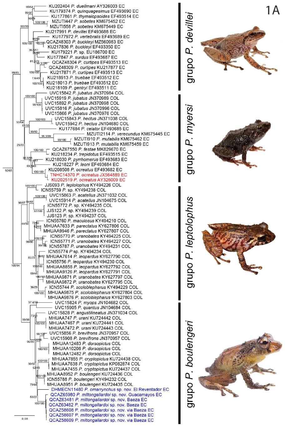
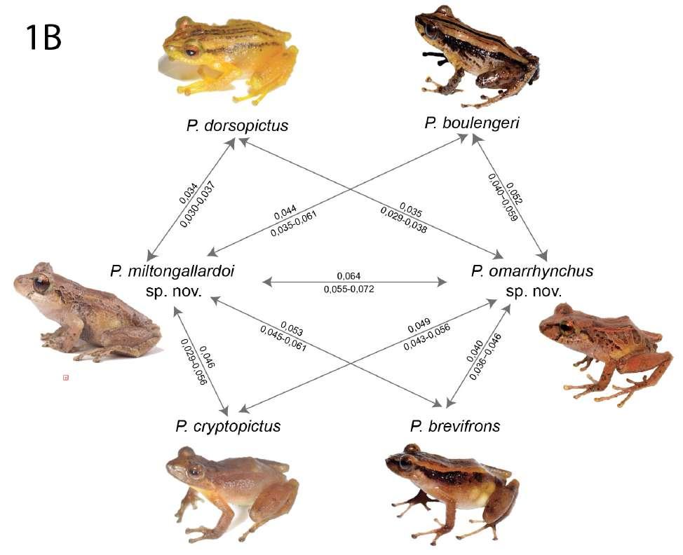
El análisis filogenético determinó que las nuevas especies se posicionan con un alto soporte (bootstrap = 99) en el clado conformado por el grupo de especies de Pristimantis boulengeri (sensu Gonzá lez Durán et al., 2017) (Fig. 1). Son parte de un clado que contiene a P. brevifrons + P. angustilineatus + P. urani + P. boulengeri + P. cryptopictus + P. dorsopictus con un alto soporte (bootstrap = 95; Fig. 1). Dentro del grupo de especies de P. boulengeri, el subclado formado por P. quantus + P. myops es hermano de un clado conformado por todas las demás especies. Las nuevas especies son hermanas entre sí con bajo soporte. Distancias genéticas de las nuevas especies con sus especies más cercanamente relacionadas son presentadas en la Figura 1B.
En nuestra filogenia, los especímenes corres pondientes a las secuencias AY326009 y JX564889, previamente reportados como “ P. myersi ” por Guayasamin et al., (2018), son genéticamente muy similares (distancia p no corregida para 16S = 0,1%) a los especímenes P. ocreatus KU 208508 (EF493682) y QCAZ 13664, además fueron colectados en la lo calidad tipo de P. ocreatus. Basados en esta evidencia concluimos que la identificación de “P. myersi” para las secuencias GenBank de Guayasamin et al., (2018) son incorrectas. Las mismas secuencias (AY326009 y JX564889), fueron identificadas incorrectamente como “P. thymelensis” por Zhang et al., (2013), Darst y Cannatella (2004) y trabajos subsecuentes (por ejemplo, Padial et al., 2014).
Posición filogenética Asignamos las nuevas especies al grupo de especies Pristimantis boulengeri (sensu González-Durán et al., 2017) con base a sus posiciones filogenéticas y a cinco características morfológicas propuestas como sinapomorfías para el grupo (González Durán et al 2017): (1) tubérculo subarticular distal doble en el dedo III de la mano, (2) tubérculo subarticular distal doble en el dedo IV de la mano, (3) peritoneo parietal cubierto con iridóforos; (4) saco vocal extendido; y (5) hocico con papila. Las distancias genéticas p no corregidas (16S) de las dos especies con respecto a las demás especies del grupo es > 3,3%; la distancia genética entre las dos nuevas especies es 6,4% (Fig. 1B).
Pristimantis omarrhynchus sp. nov. urn:lsid:zoobank.org:act:886B9E45-BCA0-4796-
P. Bejarano-Muñoz et al. - Dos nuevas especies del grupo Pristimantis boulengeri para Ecuador.
colección de anfibios del Instituto de Ciencias Na turales (ICN), Universidad Nacional de Colombia.
128
Cuad. herpetol. 36 (2):
129
125-154 (2022)
Figura 1. (B) Distancias genéticas p-no corregidas (16S) entre las nuevas especies y sus especies más cercanamente relacionadas (en base a 1A). El valor sobre la flecha corresponde al promedio de la distancia genética; bajo la flecha se muestra el rango. Fotografías: Patricia Bejarano, Santiago Ron y Mauricio Rivera-Correa, iNaturalist.
8AB9-C81AFF2FCD81
Pristimantis sp. 1 Bejarano-Muñoz et al., (2015) Holotipo (Fig. 2–4, 6, 7)
Hembra adulta DHMECN 11480 (tejido QCAZ 77345), colectada en la Reserva Cayambe-Coca, El Reventador (pueblo junto a la vía Quito-Nueva Loja); (0.086141 S; 77.599214 O, 1890 msnm), cantón Gonzalo Pizarro, parroquia El Reventador,
provincia de Sucumbíos, República del Ecuador, el 13 de diciembre de 2014 por Patricia BejaranoMuñoz, María Pérez, Mario H. Yánez-Muñoz, Jorge Brito M. y Glenda Pozo Z.
Paratipos
Hembra adulta DHMECN 11484, machos DH MECN 11476, 11482, 11485, 11487–8, 11491–2, con los mismos datos del holotipo. Hembra QCAZ
←Figura 1. (A) Relaciones filogenéticas del grupo de especies de Pristimantis boulengeri y clados cercanamente relacionados Árbol de máxima verosimilitud obtenido en base a una matriz de secuencias de ADN de un total de 3984 pares de bases (genes 12S, 16S, COI y RAG-1) y con un muestreo de 97 individuos, incluyendo especies de los grupos de P. leptolophus, P. devillei y P. myersi. Los valores de soporte se muestran como porcentajes junto a las ramas, SH-aLRT antes de la barra oblicua y bootstrap no paramétrico después. Los grupos externos no se muestran. El número del museo, código de acceso GenBank, nombre de la especie y país se presentan para cada muestra. Las abreviaturas son: COL Colombia y EC Ecuador. Los especímenes mostrados en rojo tienen identificaciones corregidas con respecto a identificaciones de estudios previos y el GenBank. El número de campo del espécimen KU 202519 fue corregido con respecto a la base de datos GenBank usando como referencia la base de datos de la colección de la Universidad de Kansas (disponible en https://collections.biodiversity.ku.edu/KUHerps/).
P. Bejarano-Muñoz et al. - Dos nuevas especies del grupo Pristimantis boulengeri para Ecuador.
130
10836, colectado el 5 de abril de 1996 por Andrew Gluesenkamp, en el Volcán Reventador (0.104389 S; 77.58833 O, 1700 msnm), cantón Gonzalo Pizarro, parroquia El Reventador, provincia de Sucumbíos, República del Ecuador.
Etimología
El epíteto específico es un patronímico que resulta de la combinación del nombre personal “Omar” y el término latín “rhyncus=nariz” y que hace alusión al sobrenombre bajo el cual los compañeros de aulas conocían al célebre herpetólogo ecuatoriano Omar Torres-Carvajal, curador de reptiles del Museo de Zoología QCAZ cuya notable y sobresaliente con tribución al estudio de los reptiles neotropicales es digna de reconocimiento.
Nombre común sugerido: Cutín de Omar Torres Nombre común en inglés sugerido: Omar´s Rain Frog
Diagnosis
Cuad. herpetol. 36 (2): 125-154 (2022)
Pristimantis omarrhynchus caracterizada por: (1) piel del dorso lisa con algunos tubérculos redondeados esparcidos en los flancos y pelvis con dos tubércu los subcónicos redondeados escapulares; vientre areolado; pliegues dorsolaterales continuos desde la parte posterior del párpado hasta la parte media del dorso en la cintura pélvica; pliegue discoidal presente; (2) anillo y membrana timpánica promi nente, redondeada 43–62% del diámetro del ojo, margen superior del anillo timpánico cubierto por el pliegue supratimpánico; con tubérculos postrictales subcónicos pequeños; (3) hocico subacuminado en vista dorsal y redondeado en vista lateral, con papila en la punta del hocico; (4) párpado superior con un tubérculo cónico prominente rodeado por algunos tubérculos redondeados pequeños; párpado más estrecho que la distancia interorbital; tubérculo inte rorbital pequeño; cresta craneal ausente; (5) procesos vomerinos presentes oblicuos de contorno con 2 a 4 dientes; (6) machos con hendiduras vocales, con saco vocal subgular y almohadillas nupciales pequeñas y oblicuas, no queratinizadas de color blanco; (7) dedo I de la mano más pequeño que el dedo II, discos li geramente expandidos en los dedos II-III-IV; dedos III y IV con tubérculo subarticular distal doble; (8) dedos sin rebordes cutáneos laterales; (9) tubérculos ulnares presentes; (10) talón con un tubérculo gran de, pequeños tubérculos en el borde externo de la pierna, rodilla y tarso; pliegue tarsal interno ausente; (11) tubérculo metatarsal interno oval, más de tres veces que el tubérculo metatarsal externo pequeño subcónico, pocos tubérculos supernumerarios bajos; (12) dedos del pie sin rebordes cutáneos laterales o indistintos; almohadillas ligeramente más grandes que el ancho de los dedos, sin membranas interdi gitales; dedo V más largo que el dedo III y alcanza al tubérculo subarticular del dedo IV; tubérculos hiperdistales presentes en todos los dedos; (13) co loración dorsal desde café claro verdoso hasta café oscuro rojizo, con una marca en forma )( café oscura; pliegues dorsolaterales crema; superficie de las extre midades, superficies ocultas de los muslos y flancos con barras café separadas por barras cremas; vientre y garganta cafés a crema amarillentos; superficies ocultas de las axilas e ingles cremas amarillentas a rojizas; superficies anteriores de los muslos y tibia cafés; iris plateado con retículos dorados verdoso y línea media café cobriza; banda cantal y timpánica café oscura a negra; superficies posteriores de los muslos con barras desde café oscuras a claro (ama
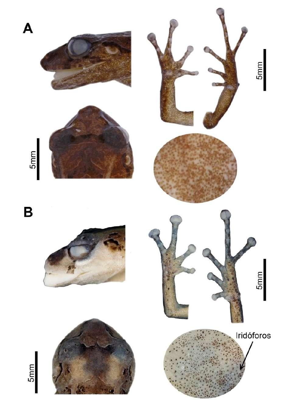
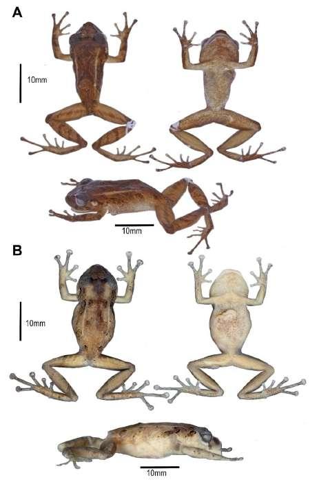 Figura 2. Aspecto dorsal, ventral y lateral en preservado (A) Pristimantis omarrhynchus sp. nov. Holotipo, hembra adulta (DHMECN 11480); (B) Pristimantis miltongallardoi sp. nov. Holotipo, hembra adulta (QCAZ 65980). Fotografías: M. H. Yánez-Muñoz y Santiago Ron.
131
rillentas en vida); (14) LRC en hembras 23,5–27,3 mm (promedio = 25,26 ± 1,9 n = 3); machos 12,1–20 mm (promedio = 16,7 ± 2,4 n = 15).
Comparación con especies similares Pristimantis omarrhynchus se diferencia de otros congéneres de Pristimantis en los Andes Norte de Sudamérica, entre la vertiente oriental de Ecuador y los Andes occidentales y centrales de Colombia, por la presencia de pliegues dorsolaterales bien de finidos, marcas dorsales generalmente en forma de )( café oscuro, tubérculos escapulares redondeados, hocico subacuminado en vista dorsal, vientre sin iridóforos, un tubérculo cónico en el párpado, papila en la punta del hocico, dígitos poco dilatados y pe queño tamaño corporal (hembras LRC < 28,2 mm). Su especie hermana (Fig 1), presenta iridóforos en el vientre y hocico en vista dorsal redondeado (sin iridóforos y hocico subacuminado en vista dorsal en P. omarrhynchus). A diferencia de otros miembros del grupo de especies de Pristimantis boulengeri (sensu González Durán et al., 2017), la nueva espe cie presenta pliegues dorsolaterales (ausentes en la mayoría de especies del grupo excepto P. baiotis y P. quantus), marcas en forma de )( café oscuro (marcas oscuras irregulares en P. boulengeri, P. brevifrons, P. cryptopictus, P. quantus y P. urani, diseño de rayas en P. angustilineatus y P. dorsopictus, manchas en forma de chevrones en P. quantus), tubérculos escapulares redondeados (ausente en las demás especies) un tubérculo cónico en el párpado (ausente en P. an gustilineatus, P. myops y P. urani; bajo, no cónico en P. boulengeri), tubérculos en el talón y tarso (ausente en P. angustilineatus, P. brevifrons, P. cryptopictus y P. urani; bajos, no cónicos, en P. boulengeri) Tabla 3. Otras diferencias con las especies del grupo son presentadas en el Apéndice III.
Descripción del holotipo
Hembra adulta, cabeza ligeramente más ancha que larga; hocico redondeado en vistas dorsal y lateral; distancia ojo narina 11.3% de la LRC, con un tu bérculo pequeño y aplanado en la punta del hocico; canto rostral recto con la región loreal ligeramente cóncava; narinas pequeñas dirigidas lateralmente; área interorbital plana con un tubérculo subcónico, más ancha que el párpado superior, el cual equivale al 82% la distancia interorbital; cresta craneal au sente; párpado superior con un tubérculo cónico rodeado de pequeñas verrugas elevadas; membrana timpánica diferenciada de la piel que la rodea, anillo
timpánico evidente y redondeado, con el margen superior cubierto 1/3 por el pliegue supratimpánico anterodorsal, tímpano visible dorsalmente, diáme tro del tímpano 51% del diámetro del ojo, pliegue supratimpánico cubierto por tubérculos postrictales subcónicos; coanas grandes y oblicuas de contorno, no cubiertas por el piso palatal del maxilar; procesos de los odontóforos vomerinos presentes, de contorno oblicuo con 2–3 dientes; lengua ligeramente más ancha que larga, de forma acorazonada, 3/4 adherida al piso de la boca.
Textura del dorso lisa, flancos y pelvis finamen te granular, vientre areolado, pliegues dorsolaterales que se extienden desde la parte posterior del ojo has ta la pelvis, pliegue discoidal conspicuo, se extiende desde la mitad de los flancos hasta la región ventral; cloaca rodeada por varias verrugas aplanadas. Bra zos esbeltos con tubérculos ulnares bajos; dedos sin rebordes cutáneos laterales, tres tubérculos palmares pequeños redondeados, tubérculo tenar ovalado; tubérculos subarticulares redondos, con tubérculos supernumerarios, dedos estrechos con almohadillas definidas por surcos circunmarginales, almohadilla del dedo I no expandida. Dedo III y IV de la mano con tubérculo subarticular distal doble.
Extremidades posteriores esbeltas, longitud de la tibia 53% de la LRC, tubérculos subcónicos en el borde externo de la rodilla y tarso; pliegue tarsal interno ausente; talón con un tubérculo cónico y varios subcónicos; dedos del pie estrechos, con débiles rebordes cutáneos laterales, sin membranas interdigitales; tubérculos subarticulares redondos y prominentes; tubérculo metatarsal interno ovalado, cuatro veces el tamaño del externo que es subcónico; tubérculos supernumerarios presentes, bajos; discos ligeramente expandidos en todos los dedos; dedo V mayor al III y alcanza la base del tubérculo subarti cular distal del dedo IV.
En vida, presenta el dorso café con marcas en forma de )( café oscuro a negro rodeada de café claro, cabeza con marcas café oscuro en fondo café claro, banda interorbital, párpados, banda cantal y supratimpánica café oscura a negra, superficies dorsales café con barras café oscuras; pliegues dorsolaterales café claro rojizo; manos y pies café rojizo con manchas café oscuras a negras, dígitos crema amarillentos; vientre, pecho y garganta café con pequeñas y tenues marcas negras, superficies ocultas de las extremidades color café cremoso; iris finamente reticulado dorado verdoso con una línea media café cobrizo (Fig. 4, 8, 12).
P. Bejarano-Muñoz et al. - Dos nuevas especies del grupo Pristimantis boulengeri para Ecuador.
132
Cuad. herpetol. 36 (2): 125-154 (2022)
Tabla 1. Variación morfométrica (en mm) de las series tipo de Pristimantis miltongallardoi sp. nov. y Pristimantis omarrhynchus sp. nov. Abreviaturas: ON = distancia órbita-narina; LC = longitud cefálica; DIO = distancia interorbital; EN = distancia internarinal; LRC = longitud rostro-cloacal; LT = longitud de la tibia; LP = longitud del pie; LM = longitud de la mano; DT = diámetro horizontal del tímpano.
Pristimantis omarrhynchus
Pristimantis miltongallardoi Hembras Machos Hembras Machos
Rango (media ± DE) n = 3
Rango (media ± DE) n = 15
Rango (media ± DE) n = 10
Rango (media ± DE) n = 4
LRC 23,5‒27,3 (25,2 ± 1,9) 12,1‒19,9 (16,7 ± 2,3) 22,3‒28,2 (25,2 ± 1,6) 17‒18,7 (17,7 ± 0,7)
LC 9,1‒9,4 (9,3 ± 0,2) 4,8‒7,1 (5,9 ± 0,7) 8,9‒10,04 (9,5 ± 0,5) 6,4‒7,3 (6,8 ± 0,3)
AC 9,2‒10,4 (9,6 ± 0,6) 3,9‒7,8 (6,3 ± 1,0) 9,1‒11 (9,9 ± 0,5) 6,6‒7,2 (6,8 ± 0,2)
DIO 2,8‒3,2 (3,1 ± 0,2) 1,3‒2,3 (1,9 ± 0,3) 2‒3,2 (2,8 ± 0,3) 2‒2,4 (2,2 ± 0,2)
ON 2,4‒2,9 (2,7 ± 0,3) 1,04‒6,1 (1,8 ± 1,2) 2,2‒3,5 (2,8 ± 0,4) 1,6‒2,2 (1,8 ± 0,3)
EN 2,4‒2,7 (2,5 ± 0,2) 1,1‒6,8 (2,3 ± 1,7) 1,09‒2,5 (2,2 ± 0,4) 1,1‒1,8 (1,5 ± 0,3)
DT 1,4‒1,7 (1,5 ± 0,1) 0,6‒5,9 (1,4 ± 1,3) 1,02‒1,7 (1,4 ± 0,2) 0,8‒1,1 (0,9 ± 0,1)
Tibia 12,4‒14,6 (13,3 ± 1,1) 6,7‒16,1 (9,7 ± 2,0) 12,4‒14 (13,1 ± 0,5) 9,1‒10,4 (9,7 ± 0,5)
Pie 11,7‒13 (12,2 ± 0,7) 5,04‒13,9 (8,3 ± 1,9) 10,7‒13,6 (12,6 ± 0,8) 7,9‒9,7 (8,8 ± 0,7)
Mano 7,08‒8,1 (7,5 ± 3,1) 3,1‒11,4 (5,5 ± 2,0) 7,03‒9,3 (8,2 ± 0,8) 5,1‒6,03 (5,5 ± 04)
En preservado, presenta coloración dorsal café oscura a café claro cremoso, con una marca en el dorso en forma de )( y manchas irregulares desde la cabeza hasta la cloaca café oscuro; banda supratimpánica café oscura desde la parte posterior del ojo hasta la inserción del brazo; pliegues dorsola terales café claros; flancos con barras café oscuras a negras con interespacios café claros a cremas, marcas blancas en la punta de los tubérculos; extremidades anteriores con marcas café oscuras a negras, dedos café claros con algunas marcas negras en los dedos III y IV; superficies dorsales de los muslos y extre midades posteriores con barras cafés oscuro sepa rado por café claro; ingles, axilas, vientre, garganta y superficies ocultas de las extremidades café claro a crema; superficies palmares y plantares café claro (Figs. 2, 3, 6, 7).
Medidas del holotipo (mm)
Longitud rostro cloaca LRC = 23,5; longitud de la tibia LT = 12,4; longitud del pie LP = 11,7; longitud de la cabeza LC = 9,1; ancho de la cabeza AC = 9,4; distancia interorbital DIO = 3,2; distancia internari nal EN = 2,4; distancia ojo narina ON = 2,8; diámetro horizontal del tímpano DT = 1,7.
Variación
La variación morfométrica más notable, es que las hembras en promedio son 1,5 veces más grandes que
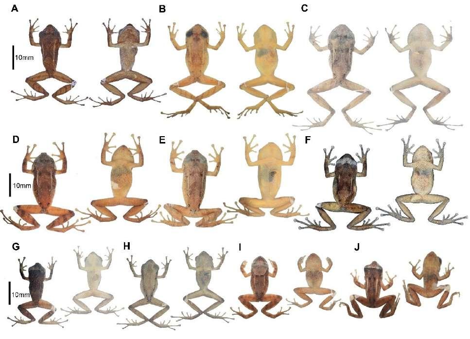
los machos (Tabla 1). La coloración en preservado de la serie tipo presenta dorso café oscuro, café claro o crema, pliegues dorsolaterales claros; región inte rorbital, banda cantal-supratimpánica café oscura o negra; barras de las extremidades y flancos café oscuro separado por barras cremas o poco definidas en algunos individuos, superficies anteriores de los muslos y piernas café claras; dígitos crema. Observa mos tres morfos de coloración en hembras, las mar cas café oscuro en el dorso son variables incluyendo formas de X, ^, y )( (Fig. 6). Los machos presentaron cinco morfos de coloración desde tonalidades café oscuro rojizo a café claro o crema grisáceo, con antebrazos y muslos amarillentos o blanquecinos, vientres y gargantas homogéneamente crema in maculado hasta café (Fig. 6); uno de los morfos es disruptivo de la serie tipo (DHMECN 11482), con la banda rostral-dorsolateral completa y sin diseños dorsales (Fig. 6H).
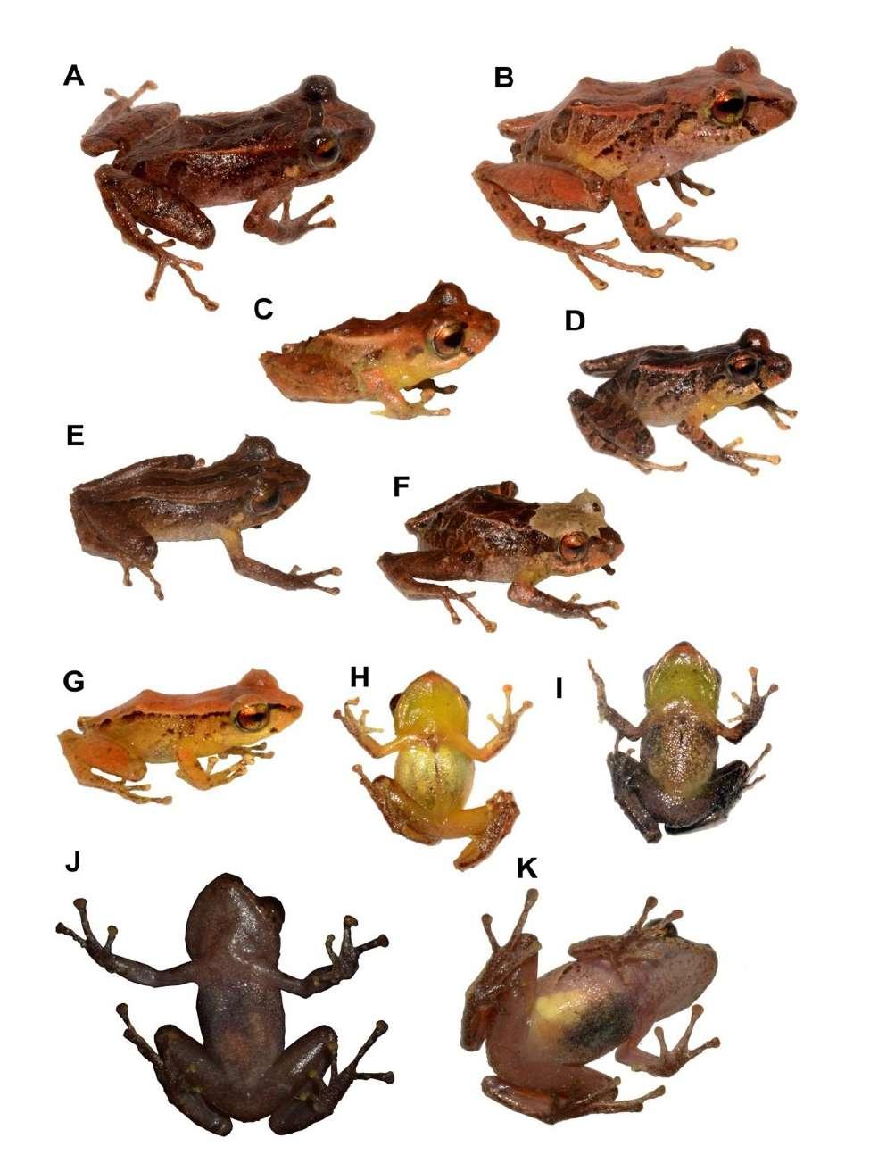
La coloración en vida de la serie tipo varía desde dorso café rojizo hasta naranja claro ama rillento sin diseño dorsal (DHMECN 11485) (Fig. 4G–H) o generalmente con una marca en forma )( café oscura. Otros individuos presentaron una barra mediodorsal café oscura (QCAZ 77383, Fig. 4E) o una mancha cefálica crema (QCAZ 77384, Fig. 4F); pliegues dorsolaterales naranja crema, superficie de las extremidades, superficies ocultas de los muslos y flancos con barras café oscuras separadas por barras
133
 P. Bejarano-Muñoz et al. - Dos nuevas especies del grupo Pristimantis boulengeri para Ecuador.
Figura 3. Detalle de la cabeza, patas y vientre en preservado (A) Pristimantis omarrhynchus sp. nov. Holotipo, hembra adulta (DHMECN 11480); (B) Pristimantis miltongallardoi sp. nov. Holotipo, hembra adulta (QCAZ 65980). Fotografías: M. H. YánezMuñoz y Santiago Ron.
134
Cuad. herpetol. 36 (2): 125-154 (2022)
Figura 4. Variación de coloración dorsal, lateral y ventral en vida de Pristimantis omarrhynchus sp. nov. (A y J) Holotipo hembra adulta (DHMECN 11480, LRC = 23,5 mm); (B y K) hembra adulta (DHMECN 11484 LRC = 27,3 mm); (C) macho (QCAZ 77386); (D) macho (QCAZ 77382); (E) macho adulto (QCAZ 77383); (F e I) macho (QCAZ 77384); (G y H) macho adulto (DHMECN 11485, LRC = 20 mm); Fotografías: M. H. Yánez-Muñoz. Fotografías sin escala.
135
cremas (Fig. 4); vientre y garganta cafés a crema amarillentos (Fig. 4H–K); superficies ocultas de las axilas e ingles crema amarillentas a rojizas en hem bras y machos; superficies anteriores de los muslos y piernas cafés; iris reticulado plateado verdoso con una línea media café cobrizo; banda interorbital, cantal y timpánica café oscuro a negra; superficies posteriores de los muslos con barras café oscuro a claro amarillento (Fig. 4).
Pristimantis omarrhynchus tiene plasticidad fenotípica, en vida, en la prominencia de sus tubér culos. En el caso del holotipo DHMECN 11480 y el paratipo DHMECN 11483, hubo una reducción en el tamaño de tubérculos a las 12 horas de haber sido colectados: el holotipo redujo el tamaño de sus tubérculos del párpado (Fig. 8G y 12A) y el paratipo redujo su papila en el hocico (Fig. 12B). Además, los tubérculos cónicos en párpados y talón, así como papila, fueron más desarrollados en machos voca lizadores registrados en septiembre de 2017 (Fig. 4 D–F) vs. machos sin actividad vocal registrados en diciembre de 2014 (Fig. 4C, G).
Descripción del canto
El canto de Pristimantis omarrhynchus (DHMECN 11483) es un clic que se emite en series de cinco o seis repeticiones (Fig. 9; Tabla 2). Su frecuencia dominante promedio es 4,04 kHz (DE = 0,06) y corresponde a la primera armónica; una segunda armónica tenue tiene 7,97 kHz (DE = 0,11). Los cantos tienen una sola nota; su duración promedio de 66 ms (DE = 11,9). El intervalo entre cantos es muy corto (promedio 9,2 ms, DE = 2,9) y las series de cantos se emiten a intervalos irregulares (rango 7,2–71,5 s). En la misma grabación hay un canto similar, pero de menor amplitud que probablemente corresponda a otro macho (no colectado) que can taba antífonamente. Esos cantos se diferencian por tener de 3 a 4 cantos por serie.
El canto de P omarrhynchus es similar a cantos de otras especies del grupo P. boulengeri. El canto de P. boulengeri también consiste en un clic emitido en series de 1 a 9 repeticiones (Ríos-Soto y Ospina 2018). Similarmente, el canto de P. dorsopictus y P. cryptopictus es un clic, con la diferencia de que no es emitido en series sino de modo individual (PatiñoOcampo et al., 2022). El canto de otras especies del grupo es desconocido.
Distribución e historia natural
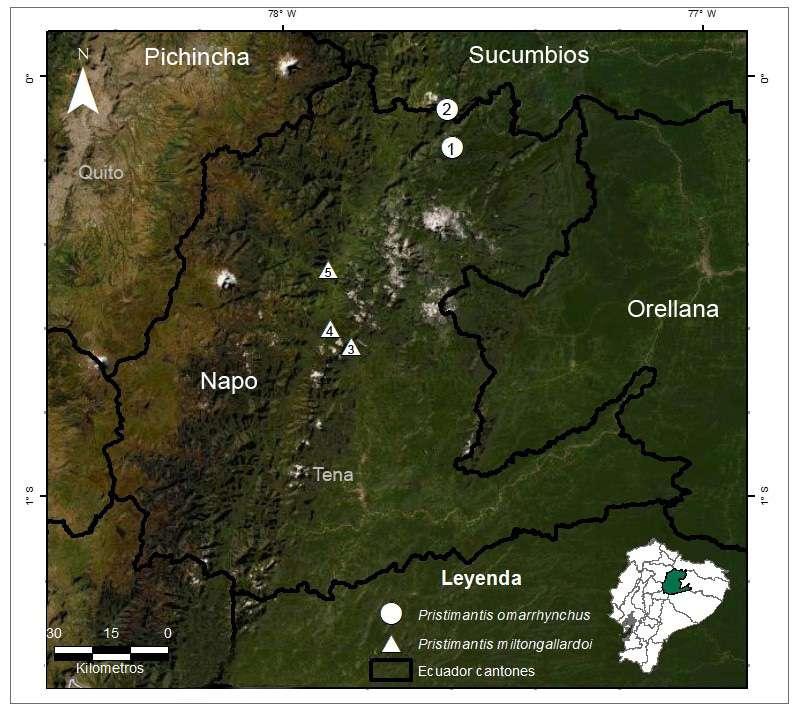
Pristimantis omarrhynchus se conoce para dos loca
lidades de las estribaciones orientales de los Andes de Ecuador en las provincias de Napo y Sucumbíos en los tributarios Quijos y Coca (Fig. 10; 11). Fue registrada en bosque maduro y secundario en las faldas del volcán Reventador entre los 1700 y 1800 msnm, en los bosques siempreverde montano y montano bajo de la cordillera oriental de los Andes norte de Ecuador (tipos de vegetación MAE 2013) y en Bosque Montano Oriental (Ron et al., 2022). Estás áreas corresponden a los límites del piso zoo geográfico Subtropical y piso Templado (Albuja et al., 2012). Los especímenes tipo fueron colectados en vegetación arbustiva hasta 1,5 m del suelo apro ximadamente, en su mayoría dentro del bosque maduro y algunos ejemplares en los claros, bordes de bosque y áreas intervenidas (Figs. 11-12). Las hembras grávidas (DHMECN 11480, 11484, QCAZ 10836) fueron colectadas en diciembre de 2014 y abril 1996, machos vocalizadores fueron registrados en septiembre de 2017, sugiriendo una reproducción continua a lo largo del año. La serie tipo colectada en los alrededores del volcán Reventador fue registrada durante actividad volcánica, antes y después de la caída de ceniza.
Pristimantis miltongallardoi sp. nov. urn:lsid:zoobank.org:act:3BEEB8E8-9970-4CEE938E-EF72C0158037
Pristimantis cf. petersi Guayasamin y Funk (2009) Pristimantis sp. 2 Funk et al., (2003)
Holotipo (Figs. 2, 3, 5, 7)
Hembra adulta QCAZ 65980, Reserva Ecológica Antisana, Cordillera de los Guacamayos, Sector la Virgen (sendero Jumandi); (0.64119 S; 77.8375 W, 1927 msnm), cantón Quijos, parroquia Cosanga, provincia de Napo, República del Ecuador, colec tado el 26 de noviembre de 2016 por Santiago Ron, Francisca Hervas, Gustavo Pazmiño, Javier Pinto, Jhael Ortega.
Paratipos
Todos los paratipos fueron colectados en la provin cia de Napo, República del Ecuador. QCAZ 65979, 65980–1, 65988 con los mismos datos del holotipo; Estación Científica Yanayacu (0.5988 S; 77.8895 W, 2100 msnm), cantón Quijos, parroquia Cosanga, hembra QCAZ 22377 colectada el 2 de junio de 1989 por Luis Coloma, QCAZ 23122 colectado el 20 de febrero de 2003 por Diego Almeida, QCAZ 33038
P. Bejarano-Muñoz et al. - Dos nuevas especies del grupo Pristimantis boulengeri para Ecuador.
136
Cuad. herpetol. 36 (2): 125-154
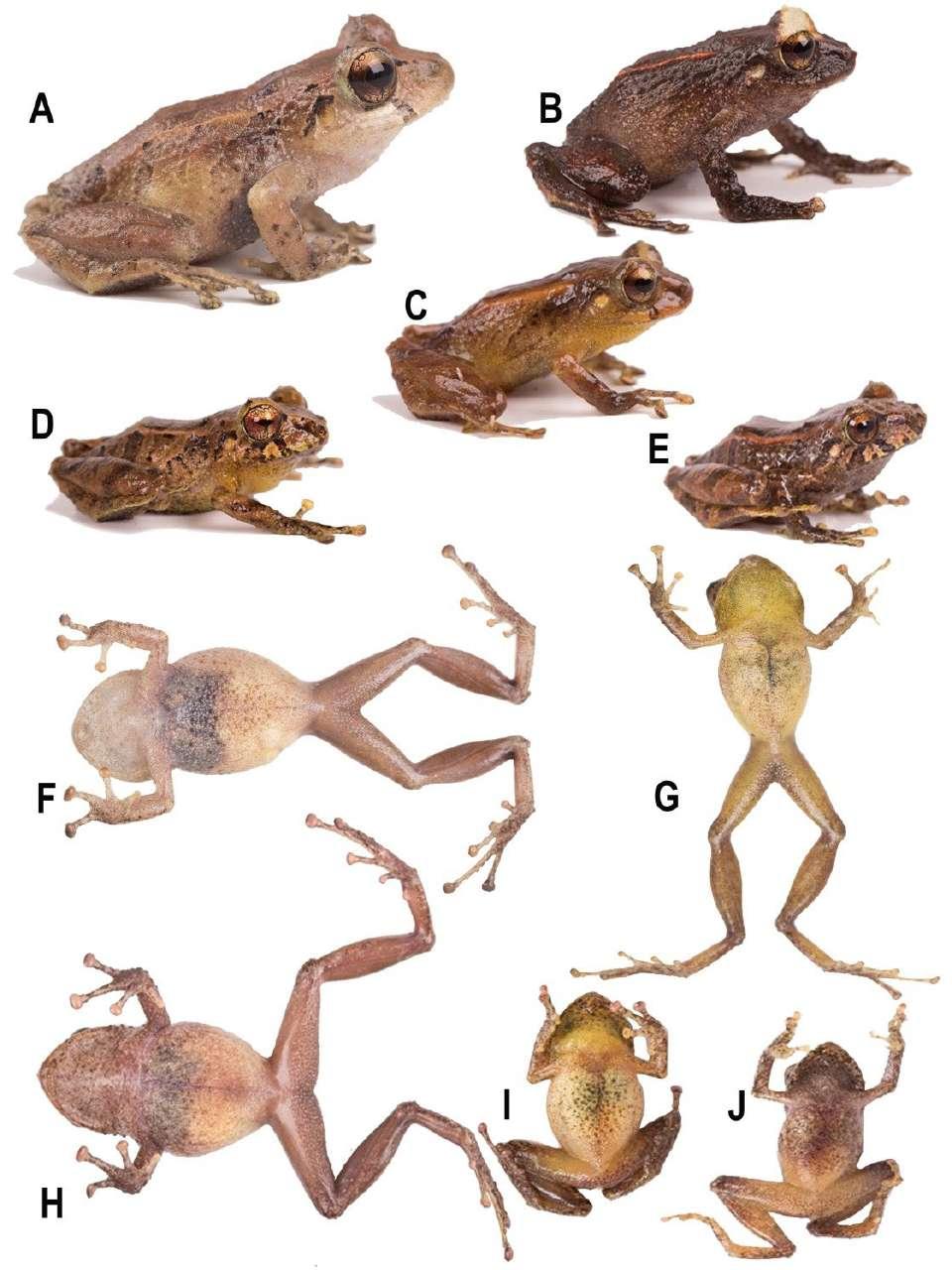
Figura 5. Variación de coloración lateral y ventral en vida de Pristimantis miltongallardoi sp. nov. (A y F) Holotipo hembra (QCAZ65980 LRC = 27mm); (B, H) hembra (QCAZ 65981, LRC = 25 mm); (C, G) macho (QCAZ 65979, LRC = 17 mm); (D, I) macho (QCAZ 63482); (E, J) juvenil (QCAZ 63481). Fotografías: Santiago Ron. Fotografías sin escala.
137
(2022)
Tabla 2. Estadísticas descriptivas de las variables acústicas del canto de Pristimantis omarrhynchus (macho DHMECN 11483).
Las abreviaciones son kHz = kilohertzios, ms = milisegundos, DE = desviación estándar.
P. omarrhynchus
Variable
sp. nov. Rango (media ± DE) n
Frecuencia Dominante (kHz) 3,90‒4,16 (4,05 ± 0,06) 25
Segunda Armónica (kHz) 7,90‒8,17 (7,97 ± 0,11) 5
Duración de la serie de cantos (ms) 341,1‒380,2 (362,6 ± 17,2) 5
Intervalos entre cantos (ms) 4,95‒14,4 (9,19 ± 2,88) 20
Duración de los cantos (ms) 55‒98 (66,15 ± 11,99) 25
Cantos/serie 5–6 5
colectado el 7 de julio de 2005 por Carolina Proaño; Cordillera de los Guacamayos, entre la Virgen y el Río Urcusiqui (0.6502 S; 77.8264 W 1800 msnm), cantón Quijos, parroquia Cosanga, hembras QCAZ 50075–076, machos QCAZ 50082–083, colectados el 31 de marzo de 1998 por Felipe Campos.
Etimología
El epíteto específico es un patronímico que resulta de la combinación del nombre y apellido de un admirado científico miembro de la Academia de Ciencias de América Latina, Milton H. Gallardo, Ph.D. en Biología y profesor de Genética y Evolución en la Facultad de Ciencias, Universidad Austral de Chile. Fue parte del programa Prometeo en Ecuador y desde esta posición impulsó la gestión del Museo Ecuatoriano de Ciencias Naturales y apoyó su tran sición al actual Instituto Nacional de Biodiversidad. Su obra “Evolución: el curso de la vida” es uno de sus legados más importantes de su carrera para las nuevas generaciones Latinoamericanas.
Nombre común sugerido: Cutín de Milton H. Gallardo
Nombre común en inglés sugerido: Milton´s Rain Frog
Diagnosis
Pristimantis miltongallardoi caracterizada por: (1) piel del dorso lisa con algunos tubérculos redon deados esparcidos en los flancos y pelvis con dos
tubérculos subcónicos redondeados escapulares; vientre areolado; pliegues dorsolaterales continuos desde la parte posterior del párpado hasta la parte media del dorso en la cintura pélvica; pliegue dis coidal presente; (2) anillo y membrana timpánica prominente, redondeada 38–52% del diámetro del ojo, margen superior del anillo timpánico cubierto por el pliegue supratimpánico; con tubérculos pos trictales subcónicos pequeños; (3) hocico redondea do en vistas dorsal y lateral, con papila en la punta del hocico; (4) párpado superior con un tubérculo cónico prominente rodeado por algunos tubérculos redondeados pequeños; párpado más estrecho que la distancia interorbital; tubérculo interorbital peque ño; cresta craneal ausente; (5) procesos vomerinos presentes oblicuos de contorno con 2 a 4 dientes; (6) machos con hendiduras vocales, con saco vocal sub gular y almohadillas nupciales pequeñas y oblicuas, no queratinizadas de color blanquecino; (7) dedo I de la mano más pequeño que el dedo II, discos li geramente expandidos en los dedos II-III-IV; dedos III y IV con tubérculo subarticular distal doble; (8) dedos sin rebordes cutáneos laterales; (9) tubérculos ulnares presentes; (10) talón con un tubérculo gran de, pequeños tubérculos en el borde externo de la pierna, rodilla y tarso; pliegue tarsal interno ausente; (11) tubérculo metatarsal interno oval, más de tres veces que el tubérculo metatarsal externo pequeño subcónico, pocos tubérculos supernumerarios bajos; (12) dedos del pie sin rebordes cutáneos laterales o indistintos; almohadillas ligeramente más grandes que los dedos, sin membranas interdigitales; dedo V más largo que el dedo III y alcanza al tubérculo subarticular del dedo IV; tubérculo hiperdistal presente en todos los dedos; (13) coloración dorsal desde café claro verdoso hasta café oscuro naranja o rojizo, con dos marcas en forma ^ café oscura; plie gues dorsolaterales desde crema hasta anaranjado; superficie de las extremidades, superficies ocultas de los muslos y flancos con barras café separadas por barras cremas; vientre y garganta cafés a crema amarillentos, peritoneo con iridóforos; superficies ocultas de las axilas e ingles cremas amarillentas a rojizas; superficies anteriores de los muslos y piernas cafés; iris con retículos dorado verdoso y con una línea media café cobriza; banda cantal y timpánica café oscura a negra; superficies posteriores de los muslos con barras desde café oscuras a claro (ama rillentas en vida); (14) LRC en hembras 22,3–28,2 mm (promedio = 25,2 ± 1,6 n = 10); machos 17–18,7 mm (promedio = 17,7 ± 0,7 n = 4).
P. Bejarano-Muñoz et al. - Dos nuevas especies del grupo Pristimantis boulengeri para Ecuador.
138
Cuad. herpetol. 36 (2): 125-154 (2022)
Figura 6. Variación de coloración en preservado del dorso y vientre. Pristimantis omarrhynchus sp. nov. (A) Holotipo, hembra adulta (DHMECN 11480); (B) hembra adulta (QCAZA 10836); (C) Paratipo hembra (DHMECN 11484); Pristimantis miltongallardoi sp. nov. (D) hembra (QCAZA 22377); (E) hembra (QCAZA 33038); (F) hembra (QCAZA 56981). Pristimantis omarrhynchus sp. nov. (G) Paratipo macho (DHMECN 11488). (H) Paratipo macho (DHMECN 11482); Pristimantis miltongallardoi sp. nov. (I) macho adulto (QCAZA 50082); (J) macho adulto (QCAZA 50083); Fotografías: M. H. Yánez-Muñoz y Santiago Ron.
Comparación con especies similares Pristimantis miltongallardoi se diferencia de sus congéneres del grupo P. boulengeri, por la presencia de pliegues dorsolaterales bien definidos, marcas dorsales en forma de ^ café oscuro, tubérculos es capulares redondeados, un tubérculo cónico en el párpado, hocico redondeado en vista dorsal, papila en la punta del hocico, vientre con iridóforos, dígitos dilatados y pequeño tamaño corporal (hembras LRC < 28,2 mm). Se diferencia de su especie hermana, Pristimantis omarrhynchus sp. nov. por la presencia de iridóforos en el peritoneo, hocico redondeado en vista dorsal y los dígitos dilatados (sin iridóforos, subacuminado y dedos estrechos en P. omarrhyn chus). Se diferencia de otros miembros del grupo Pristimantis boulengeri (sensu González Durán et al., 2017), por presentar pliegues dorsolaterales (ausen tes en la mayoría de las especies del grupo, excepto P. baiotis y P. quantus), marcas dorsales en forma
de ^ café oscuro (marcar oscuras irregulares en P. boulengeri, P. brevifrons, P. cryptopictus, P. quantus y P. urani, diseño de rayas en P. angustilineatus y P. dorsopictus, marcas en forma de chevrones en P. quantus), con tubérculos escapulares redondeados (ausente en los demás miembros a excepción de P. omarrhynchus), un tubérculo cónico en el párpado rodeado por algunos más pequeños (ausente en P. angustilineatus, P. myops y P. urani; bajo, no cónico en P. boulengeri), tubérculos en el talón y tarso (au sente en P. angustilineatus, P. brevifrons, P. crypto pictus y P. urani; bajos, no cónicos, en P. boulengeri) Tabla 3. Otras diferencias con las especies del grupo son presentadas en el Apéndice III.
Descripción del holotipo
Hembra adulta, cabeza ligeramente más ancha que larga; hocico redondeado en vistas dorsal y lateral; distancia ojo narina 11% de la LRC, con un tubér
139
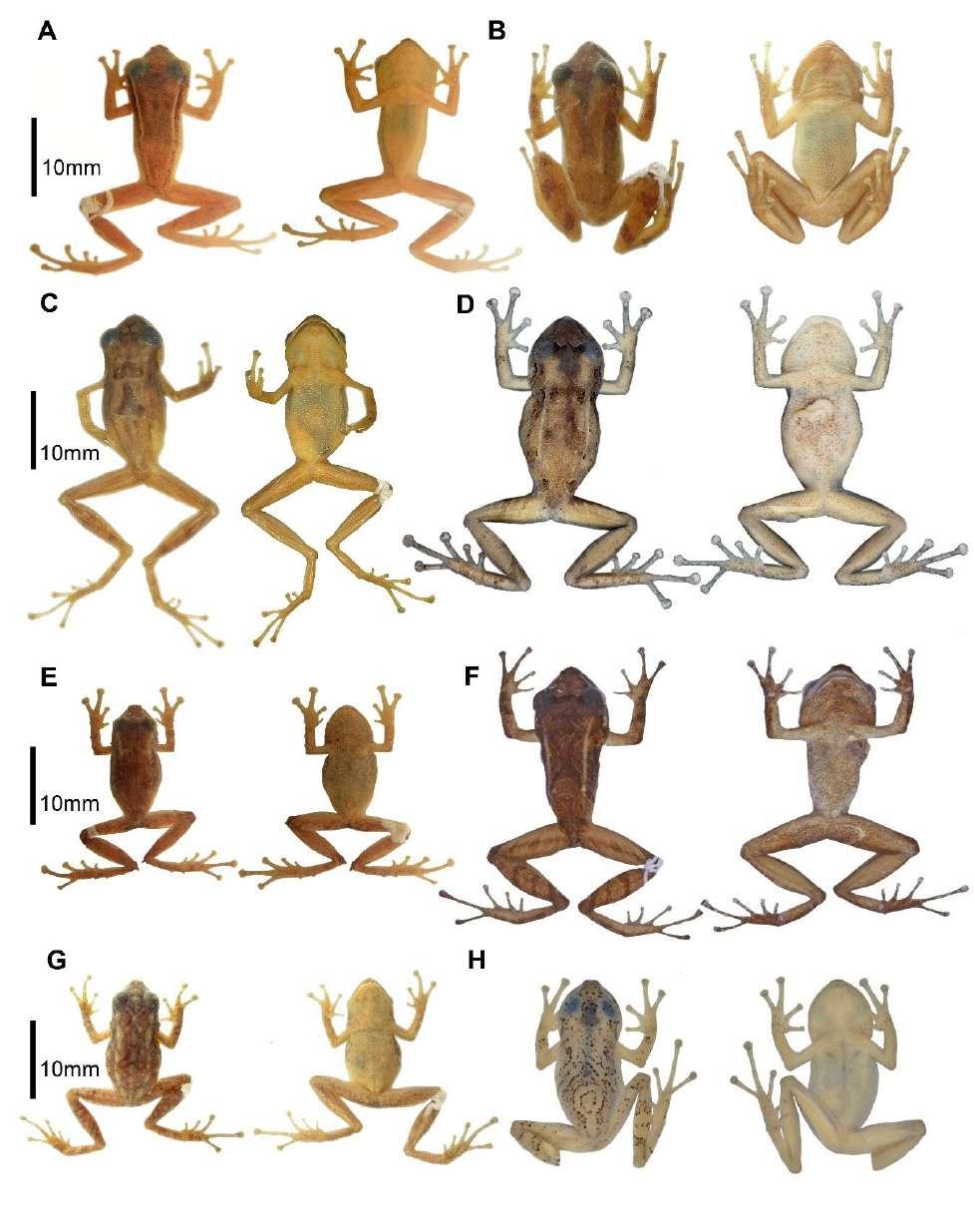
Figura 7. Vista dorsal y ventral de algunas especies del grupo Pristimantis boulengeri en preservado. (A) Pristimantis angustilineatus, Holotipo macho (ICN 39598, LRC = 19,8 mm); (B) Pristimantis boulengeri (ICN 34035, LRC=24,3 mm); (C) Pristimantis brevifrons, Holotipo (UMMZ 166572, LRC=22,3 mm); (D) Pristimantis miltongallardoi sp. nov., Holotipo (QCAZ 65980 LRC= 27 mm); (E) Pris timantis myops, Holotipo (ICN 39684, LRC= 15,6 mm); (F) Pristimantis omarrhynchus sp. nov., Holotipo (DHMECN 11480, LRC= 23,5); (G) Pristimantis quantus, Holotipo (ICN 29340, LRC= 15,9); (H) Pristimantis urani, Holotipo (MHUA-A 7471, LRC= 23,4). Fotografías: M. H. Yánez-Muñoz (A, B, E, F, G); The University of Michigan Museum of Zoology, Division of Reptiles and Amphibians (C); Santiago Ron. (D); Mauricio Rivera-Correa. (H).
 P. Bejarano-Muñoz et al. - Dos nuevas especies del grupo Pristimantis boulengeri para Ecuador.
140
culo pequeño y aplanado en la punta del hocico; canto rostral recto con la región loreal ligeramente cóncava; narinas pequeñas dirigidas lateralmente; área interorbital plana con un tubérculo subcónico, más ancha que el párpado superior, el cual equivale al 78% la distancia interorbital; cresta craneal au sente; párpado superior con un tubérculo cónico rodeado de pequeñas verrugas elevadas; membrana timpánica diferenciada de la piel que la rodea, anillo timpánico evidente y redondeado, con el margen superior cubierto 1/3 por el pliegue supratimpánico anterodorsalmente, tímpano visible dorsalmente, diámetro del tímpano 28% del diámetro del ojo, pliegue supratimpánico cubierto por tubérculos postrictales subcónicos; coanas grandes y oblicuas de contorno, no cubiertas por el piso palatal del maxilar; procesos de los odontóforos vomerinos presentes, de contorno oblicuo con 2–3 dientes; lengua ligeramente más ancha que larga, de forma acorazonada, ¾ adherida al piso de la boca.
Textura del dorso lisa, flancos y pelvis finamen te granular, vientre areolado, pliegues dorsolaterales que se extienden desde la parte posterior del ojo has ta la pelvis, pliegue discoidal conspicuo, se extiende desde la mitad de los flancos hasta la región ventral; cloaca rodeada por varias verrugas aplanadas. Bra zos esbeltos con tubérculos ulnares bajos; dedos sin rebordes cutáneos laterales, tres tubérculos palmares pequeños redondeados, tubérculo tenar ovalado; tubérculos subarticulares redondos, con tubérculos supernumerarios, dedos estrechos con almohadillas definidas por surcos circunmarginales, almohadilla del dedo I no expandida. Dedo III y IV de la mano con tubérculo subarticular distal doble.
Extremidades posteriores esbeltas, longitud de la tibia 46% de la LRC, tubérculos subcónicos en el borde externo de la rodilla y tarso; pliegue tarsal interno ausente; talón con un tubérculo cónico y varios subcónicos; dedos del pie estrechos, con débiles rebordes cutáneos laterales, sin membranas interdigitales; tubérculos subarticulares redondos y prominentes; tubérculo metatarsal interno ovalado, 4 veces el tamaño del externo que es subcónico; tu bérculos supernumerarios presentes, bajos; discos ligeramente expandidos en todos los dedos; dedo V mayor al III y alcanza la base del tubérculo subarti cular distal del dedo IV.
En vida, presenta el dorso café con marcas en forma de ^ café oscuro a negro rodeada de café claro, cabeza con marcas café oscuro en fondo café claro, banda interorbital, párpados, banda cantal
Cuad. herpetol. 36 (2): 125-154 (2022)
y supratimpánica café oscura a negra, superficies dorsales café con barras café oscuras; pliegues dorsolaterales café claro naranja; manos y pies café rojizo con manchas café oscuras a negras, dígitos crema amarillentos; vientre, pecho y garganta café con pequeñas y tenues marcas negras, peritoneo con iridóforos, superficies ocultas de las extremidades color café cremoso; iris finamente reticulado dorado verdoso con una línea media café cobrizo (Fig. 5, 8).
En preservado, presenta coloración dorsal café oscura a café claro cremoso, con una marca en el dorso en forma de ^ y manchas irregulares desde la cabeza hasta la cloaca café oscuro; banda supratim pánica café oscura desde la parte posterior del ojo hasta la inserción del brazo; pliegues dorsolaterales café claros; flancos con barras café oscuras a ne gras con interespacios café claros a cremas, marcas blancas en la punta de los tubérculos; extremidades anteriores con marcas café oscuras a negras, dedos café claros con algunas marcas negras en los dedos III y IV; superficies dorsales de los muslos y extre midades posteriores con barras cafés oscuro sepa rado por café claro; ingles, axilas, vientre, garganta y superficies ocultas de las extremidades café claro a crema; superficies palmares y plantares café claro (Figs. 2–3).
Medidas del holotipo (mm)
Longitud rostro cloaca LRC = 27; longitud de la ti bia LT = 12,39; longitud del pie LP = 12,8; longitud de la cabeza LC = 10,04; ancho de la cabeza AC = 10,25; distancia interorbital DIO = 2,56; distancia internarinal EN = 2,07; distancia ojo narina ON = 2,97; diámetro horizontal del tímpano DT = 1,02.
Variación
Las hembras tienen un tamaño promedio de 1,5 veces más grandes que los machos (Tabla 1). La coloración en preservado de la serie tipo presenta el dorso desde café oscuro, café claro o crema, plie gues dorsolaterales claros; región interorbital, banda cantal-supratimpánica café oscura o negra; barras de las extremidades y flancos café oscuro separado por barras cremas o poco definidas en algunos individuos, superficies anteriores de los muslos y piernas café claras; dígitos crema. Observamos tres morfos de coloración en hembras, desde café oscuro homogéneo a marcas variables café oscuro a claro en el dorso incluyendo formas de X y ^ y barra in ter orbital (Fig. 6 D, E, F). Los machos presentaron cuatro morfos de coloración desde tonalidades café
141
Patrón coloración dorsal
Tubérculos escapulares
Tubérculos en el borde externo del tarso
Tubérculos en el talón
Tubérculos en el párpado
Presentes, redondeados
Presentes, pequeños
Un tubérculo grande
Un tubérculo cónico prominente rodeado por algunos pequeños
Dorso con marcas en forma “)(“café oscuro
Pliegue dorso lateral
Presentes, redondeados
Presentes, pequeños
Un tubérculo grande
Un tubérculo cónico prominente rodeado por algunos tubérculos bajos
LRC
Especie
Presente
Ausente
Dorso sin diseño específico
Ausente
Dorso con marcas café y barra interor bital oscura
Ausente
Ausente
Dorso con o sin puntos o manchas irregulares en el cuerpo, sin franjas o líneas longitudinales
Ausente
Ausente
Ausentes
Presentes
Ausente
Presente
Presente
Subcónico
Tubérculos no cónicos
Ausente
17,7 (17,0–18,7; n = 5)
Dorso con marcas en forma “^” café oscuro P. omarrhynchus
P. miltongallardoi
25,2 (22,3–28,2; n = 10)
Presente
Sin tubérculos
Tubérculos cónicos
Bajos, no cónicos
16,7 (12,1–20,0; n = 15)
25,2 (23,5–27,3; n = 3)
Ÿ Tabla 3. Principales caracteres diagnósticos de las especies del grupo Pristimantis boulengeri.
Ausente
18,1 (15,8–20,4; n = 61)
Presente
Ÿ Ausente
Dorso con rayas finas dorsolaterales blancas delimitadas de negro por debajo P. baiotis
Ausente
Ausente
22,6 (20,8–24,8; n = 18)
Ž Bajos
P. angustilineatus
18,1–18,5
21,5
142 17,4 (15,1–19,7; n = 3)
Ÿ Dorso con marcas marrón oscuro
22,1 (18,6–25,6; n = 87)
30,0 (27,3–33,8; n = 17)
22,8 (21,2–25; n = 5)
Ÿ Un tubérculo redondea do rodeado por algunos pequeños
24,08 (20,6–27,2; n = 30)
Ž Ausente
Ž P. cryptopictus
P. boulengeri
P. brevifrons
P. Bejarano-Muñoz et al. - Dos nuevas especies del grupo Pristimantis boulengeri para Ecuador.
Ž
Ÿ
Ž
Ÿ
Ž
Ÿ
Dorso con bandas longitudinales o manchas en forma de V de color café oscuro
Ausente
Dorso con marcas dispersas oscuras
Ausente
Presentes, pequeños
Presentes, pequeños
Cuad. herpetol. 36 (2): 125-154 (2022)
Dorso con muchas manchas y marcas marrón oscuro dispersas
Ausente
Presentes, subcónicos
Presentes, pequeños
Granular
(21,31,6; n = 7)
Ausente
P. dorsopictus
Ausente
Ausentes
Ausentes
Ausente, pliegue inte rorbital y sacral
Ausente, interorbi tal presente, pliegue dérmico occipital en forma de W
Ÿ Tubérculo cónico
(19,0–22,0; n = 8)
Ž Pequeños subcónicos Pequeños subcónicos
11,9 (10,9–13,6; n = 34)
P. myops
15,5 (14,6–17,2; n = 38)
11,9 (11,6–14,5; n = 4)
25,3 (14,4–16,7; n = 9)
Ÿ Ausente
Ž Sin tubérculos
Dorso con verrugas y marcas oscuras verdosas en forma de “^” P. urani
P. quantus
18,9 (18,7–19,1; n = 2)
22,5 (21–23,4; n = 4)
143 Presente poco definidos
Ÿ
Ž
Ÿ
Ž
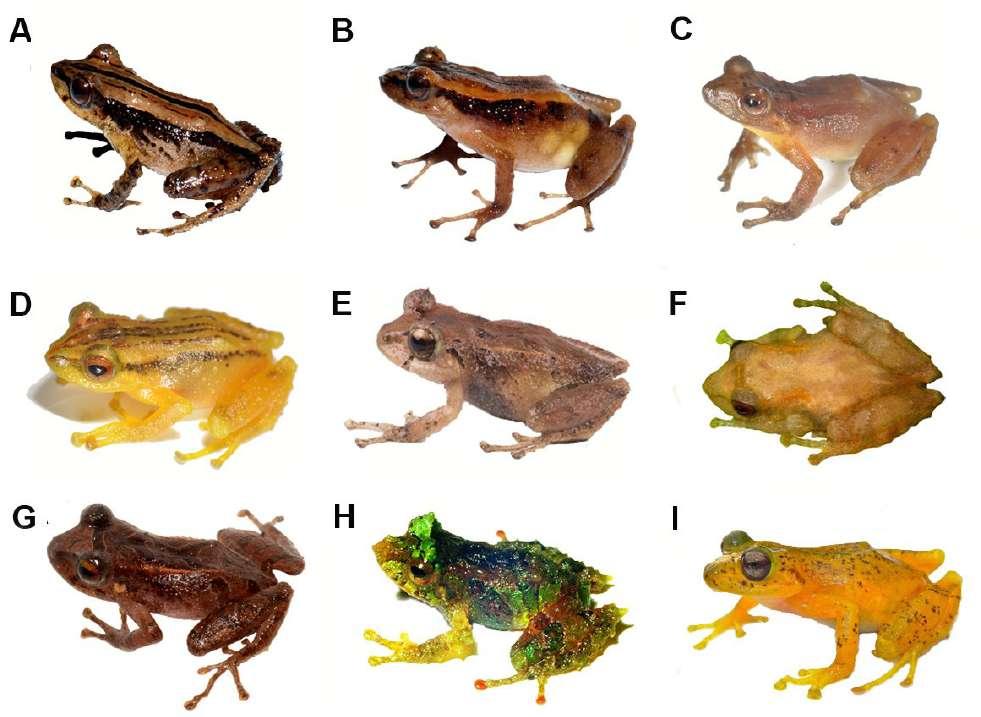
Figura 8. Variación de coloración en vida de algunas especies del grupo Pristimantis boulengeri. (A) Pristimantis boulengeri (MAR 2760); (B) Pristimantis brevifrons (MAR 2305); (C) Pristimantis cryptopictus (MHUA A12475); (D) Pristimantis dorsopictus (MHUA A12492); (E) Pristimantis miltongallardoi sp. nov., Holotipo (QCAZ 65980); (F) Pristimantis myops (iNaturalist); (G) Pristimantis omarrhynchus sp. nov., Holotipo (DHMECN 11480); (H) Pristimantis quantus (ICN 29315); (I) Pristimantis urani, Holotipo (MHUA-A 7471). Fotografías: M. Rada (A, B); Mauricio Rivera-Correa (C, D); S. Ron (E); J. J. Ospina-Sarria INaturalist (F); M. H. Yánez-Muñoz (G); J. D. Lynch (H); F. Duarte (I). Fotografías sin escala.
oscuro a crema grisáceo, con antebrazos y muslos amarillentos o blanquecinos, vientres y gargantas homogéneamente crema inmaculado hasta café; los machos QCAZ 50082 y 50083 presentan mancha cefálica o banda interorbital crema (Fig. 6I y 6J).
La coloración en vida de la serie tipo varía desde dorso café rojizo hasta naranja claro ama rillento sin diseño dorsal (QCAZ 65981) (Fig. 5B) con marcas en forma ^ en el dorso (Fig. 5A, C–E), pliegues dorsolaterales naranja crema, superficie de las extremidades, superficies ocultas de los muslos y flancos con barras café oscuras separadas por barras cremas (Fig. 5 A–G); vientre y garganta cafés a crema amarillentos (Fig. 5 F–J); superficies ocultas de las axilas e ingles crema amarillentas a rojizas en hem bras y machos; superficies anteriores de los muslos y piernas cafés; iris reticulado dorado verdoso con una línea media café cobrizo; banda interorbital,
cantal y timpánica café oscuro a negra; superficies posteriores de los muslos con barras café oscuro a claro amarillento (Fig. 5).
Distribución e historia natural Pristimantis miltongallardoi se conoce solo para la provincia de Napo en tres localidades de las estri baciones orientales de los Andes en los drenajes de la subcuenca del río Cosanga (Fig. 10; 11). Ha sido registrada en bosque secundario, claros, bordes de bosques y áreas intervenidas, algunos individuos fueron encontrados en la vía a Baeza a los 1840 msnm; en Cosanga, Estación Científica Yanayacu es una especie abundante que se encuentra en bosque primario en vegetación baja (Guayasamin y Funk, 2009), en un intervalo altitudinal entre 1800–2137 msnm y al sur de la cordillera de los Guacamayos a 1927 msnm. Habita los bosques siempreverde mon
P. Bejarano-Muñoz et al. - Dos nuevas especies del grupo Pristimantis boulengeri para Ecuador.
144
tano de la cordillera oriental de los Andes norte de Ecuador (MAE, 2013) y Bosque Montano Oriental acorde con Ron et al., 2022. Corresponde a los lí mites altitudinales de los pisos zoogeográficos Sub tropical y Templado (Albuja et al., 2012) (Fig. 10).
Discusión
Hasta el presente trabajo, el grupo de especies Pristimantis boulengeri era endémico de los Andes de Colombia (González-Durán et al., 2017; PatiñoOcampo et al., 2022). Pristimantis omarrhynchus y P. miltongallardoi corresponden a las primeras especies del grupo para los Andes de Ecuador y para la cuenca amazónica. Las nuevas especies pre sentan las sinapomorfías del grupo propuestas por González-Durán et al., (2017): (1) dedo III con un tubérculo subarticular distal doble; (2) tubérculo
Cuad. herpetol. 36 (2): 125-154 (2022)
subarticular distal doble del dedo IV; (3) peritoneo parietal cubierto con iridóforos (desconocido en P. myops y P. quantus); (4) saco vocal extendido; y (5) hocico con papila pequeña. Con la adición de las dos nuevas especies, el grupo Pristimantis boulengeri ahora incluye 11 especies: P. angustilineatus (Lynch, 1998); P. baiotis (Lynch, 1998); P. boulengeri (Lynch, 1981); P. brevifrons (Lynch, 1981); P. cryptopictus Patiño-Ocampo et al., (2022); P. dorsopictus (Rivero y Serna, 1988); P. miltongallardoi (presente estudio); P. myops (Lynch, 1998); P. omarrhynchus (presente estudio); P. quantus (Lynch, 1998), P. urani RiveraCorrea y Daza 2016. Pristimantis angustilineatus, P. baiotis, P. myops, P. quantus y P. urani se encuentran distribuidas en la cordillera Occidental de Colombia, desde los 1500 m a 2500 m de elevación; P. dorso pictus y P. cryptopictus son de la cordillera Central entre los 1800 m a 3100 m. Pristimantis boulengeri
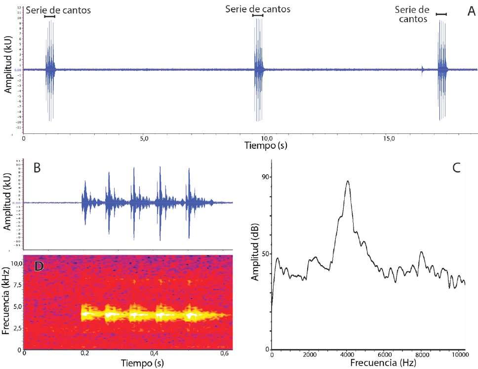 Figura 9. Canto de Pristimantis omarrhynchus sp. nov. A y B son oscilogramas, C es un espectro de poder y D es un espectrograma. (A) Tres series de cantos consecutivas; (B) y (D) serie de cinco cantos; (C) Espectro de poder de un canto. Macho DHMECN 11483, LRC = 17,6 mm, laderas del volcán Reventador, Provincia Sucumbíos, Ecuador.
145
y P. brevifrons se encuentran en ambas cordilleras entre los 1140 m a 3200 m (Lynch, 1981, 1998; Rivero y Serna, 1988; Rivera-Correa y Daza, 2016; PatiñoOcampo et al., 2022).
Recientes estudios (Ospina-Sarria y Duellman, 2019; Ron et al., 2020) han identificado que los tu bérculos hiperdistales también están presentes en otros grupos de Pristimantis (e.g., P. leptolophus y P. lacrimosus). Ese carácter al igual que la papila en la punta del hocico y saco vocal extendido, ha sido registrado en especies de Pristimantis del subgéne ro Huicundomantis o de los grupos de especies P. lacrimosus y P. unistrigatus (Lynch, 1979; Lynch y Duellman, 1980, 1997; Páez y Ron et al., 2022; Ron
et al., 2020). Por otro lado, el peritoneo con iridó foros ha sido observado por inspección directa en P. eremitus (grupo de especies de P. lacrimosus), P. calcarulatus (grupo de especies P. unistrigatus) y P. galdi (grupo de especies de P. lacrimosus) (Obs. Pers. MYM). Se requiere un análisis comprehensivo de la distribución de estos caracteres para determinar si son homólogos u homoplásicos entre los clados de Pristimantis que los presentan.
En Pristimantis omarrhynchus sp. nov. hay plasticidad fenotípica en rasgos taxonómicamente importantes y que se asumen fijos a nivel de especie (Lynch y Duellman, 1997; Duellman y Lehr, 2009), por ejemplo, los tubérculos cónicos sobre el párpado,
P. Bejarano-Muñoz et al. - Dos nuevas especies del grupo Pristimantis boulengeri para Ecuador.
Figura 10. Mapa de distribución de Pristimantis omarrhynchus sp. nov. (1) Localidad tipo, laderas del volcán Reventador; Río Azuela, Reserva Ecológica Cayambe-Coca; (2) Sector embalse compensador, Hidroeléctrica Coca Codo Sinclair. Pristimantis miltongallardoi sp. nov. (3) Localidad tipo, Cordillera de los Guacamayos; (4) Estación Científica Yanayacu; (5) Sector San Isidro antigua Vía Baeza-Cosanga.
146
talón o papila, variaron en pocas horas en el mismo individuo. La plasticidad fenotípica de la tubercula ción de la piel ha sido poco documentada y solo se conoce en P. mutabilis y P. sobetes (Guayasamin et al., 2015). Por lo tanto, es importante tenerla en cuenta para fotografiar y documentar los especímenes an tes y después de la manipulación, con ello evaluar si la plasticidad es más frecuente de lo hasta ahora reportado en el género Pristimantis
En la filogenia identificamos secuencias con determinación taxonómica incorrecta para los especímenes KU 202519 y TNHC-GDC14370, siendo inicialmente identificadas como P. thyme lensis (Hedges et al., 2008), y posteriormente como P. myersi (Guayasamin et al., 2018). No obstante, dichas secuencias son casi idénticas a P. ocreatus (KU 208508 y QCAZ 13664), ambos especímenes de la localidad tipo de esta última especie, lo que indica un error en la determinación.
En nuestro análisis y en filogenias previas (e.g., González-Durán et al., 2017; Rivera-Correa et al., 2017, Rivera-Correa y Daza, 2016; Jetz y Pyron, 2018), los grupos de especies P. boulengeri, P. lepto
Cuad. herpetol. 36 (2): 125-154 (2022)
lophus, P. myersi y P. devillei forman un clado con alto soporte (Fig. 1A). Este clado podría tener una equivalencia taxonómica de subgénero para el cuál estaría disponible el nombre Trachyphrynus (Goin y Cochran, 1963).
Agradecimientos
Agradecemos a la Fundación EcoCiencia financiada por el EcoFondo que, a través de las becas de inves tigación, apoyaron la realización y el desarrollo del proyecto “Priorización de áreas de conservación en el corredor Tropi-Andino OCP, a través del enfoque macroecológico de ranas endémicas Pristimantis (Anura: Craugastoridae). Del cual obtuvimos los registros ecológicos de la especie además de la colec ción de especímenes voucher para la descripción de Pristimantis omarrhynchus. Gracias a Rosita Alule ma de la Hostería el Reventador por su hospitalidad con todo el equipo durante toda la salida de campo.
A Marco Rada, Mauricio Rivera-Correa, Felipe Duarte, John D. Lynch, John J. Ospina y The Uni versity of Michigan Museum of Zoology, Division of
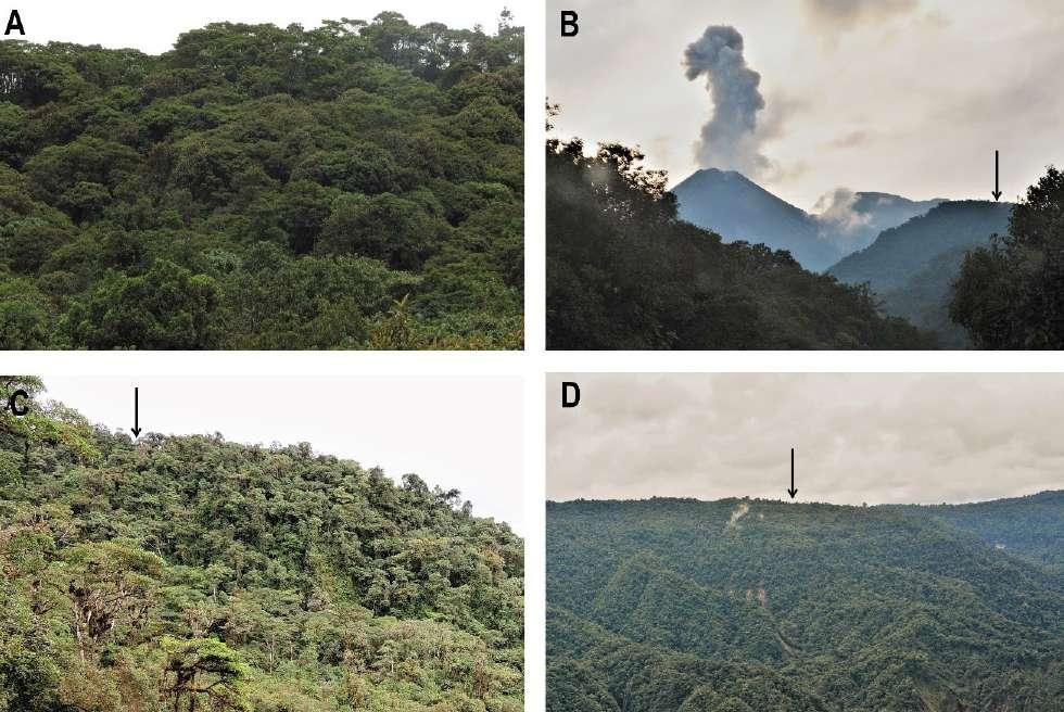
Figura 11. Paisaje general de las áreas de colección de Pristimantis omarrhynchus sp. nov. y Pristimantis miltongallardoi sp. nov. A) Estación Científica Yanayacu, B) Ladera del Volcán Reventador; C) Reserva Ecológica Cayambe Coca; D) Sector embalse compensador Hidroeléctrica Coca Codo Sinclair. Flechas indican áreas de colección. Fotografías: M.H. Yánez-Muñoz.
147
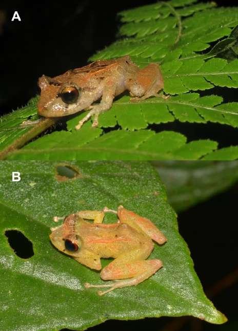
Figura 12. Pristimantis omarrhynchus sp. nov. in situ. (A) Holotipo, hembra adulta (DHMECN 11480, LRC = 23,5 mm) registrada a 0.80 cm del suelo en hoja de helecho; (B) Paratipo, macho adulto (DHMECN11483, LRC = 17,6 mm) registrado a 1 m. del suelo en el ápice de una hoja cantando. Fotografías: Patricia Bejarano-Muñoz (A); M.H. Yánez-Muñoz (B).
Reptiles and Amphibians por su contribución con el material fotográfico Colombiano. Al Ministerio del Ambiente por el permiso de investigación N° 010-14 IC-FAU_DNB/MA y permiso para acceso a recursos genéticos MAE-DNB-CM-2016-0045 and MAEDNB-CM-2019-0120, provistos por el Ministerio del Ambiente del Ecuador. A María Beatriz Pérez, Glenda Pozo y Jorge Brito M. por su colaboración en la obtención de datos en el campo y sus valiosos aportes al manuscrito. El trabajo de laboratorio y parte del trabajo de campo fue financiado por la Secretaría Nacional de Educación Superior, Ciencia, Tecnología e Innovación del Ecuador SENESCYT (iniciativa Arca de Noé; investigadores principales de SRR y Omar Torres) y proyectos de la Pontifi cia Universidad Católica del Ecuador, Dirección General Académica. John D. Lynch y Raúl Sedano brindaron las mejores condiciones durante la visita de MYM a las colecciones del Instituto de Ciencias Naturales (ICN) de la Universidad de Colombia y la Colección Herpetológica de la Universidad del valle
del Cauca (UVC). La visita a las colecciones en Co lombia de MYM fue posible gracias a la Fundación EcoMinga y El Jardín Botánico de Basell, a través de Javier Robayo, Lou Jost, Juan P. Reyes-Puig y Heinz Schneider. El trabajo de MYM forma parte del proyecto “Diversidad de Pequeños Vertebrados del Ecuador”, auspiciados por Diego Inclán y Francisco Prieto de INABIO. Patricia Bejarano Muñoz quiere dar un especial agradecimiento a sus padres y a toda la familia Muñoz-Ortiz en Medellín Colombia, por todo el amor brindado e inculcado por los animales. A todas nuestras familias por su apoyo incondicional y paciencia a lo largo de la investigación.
Literatura citada
Albuja, L.; Almendáriz, A.; Barriga, R.; Montalvo, L.D.; Cáceres, F. & Román, J.L. 2012. Fauna de Vertebrados del Ecuador. Instituto de Ciencias Biológicas. Escuela Politécnica Nacional. Quito, Ecuador.
Arteaga-Navarro, A.F. & Guayasamin, J.M. 2011. A new frog of the genus Pristimantis (Amphibia: Strabomantidae) from the high Andes of Southeastern Ecuador, discovered using morphological and molecular data. Zootaxa 2876: 17-29.
Arteaga-Navarro, A.F.; Bustamante L. & Guayasamin, J.M. 2013. The amphibians and reptiles of Mindo: life in the Cloud forest. Quito: Universidad Tecnológica Indoamérica.
Arteaga-Navarro, A.; Pyron, R.A.; Peñafiel, N.; Romero-Barreto, P.; Culebras, J.; Bustamante, L. & Guayasamin, J.M. 2016. Comparative phylogeography reveals cryptic diversity and repeated patterns of cladogenesis for amphibians and reptiles in northwestern Ecuador. Plos One 11(4): e0151746.
Barrio-Amorós, C.L.; Heinicke, M.P. & Hedges, S.B. 2013. A new tuberculated Pristimantis (Anura, Terrarana, Strabomantidae) from the Venezuelan Andes, redescription of Pristimantis pleurostriatus , and variation within Pristimantis vanadisae. Zootaxa 3647: 43-62.Bioacoustics Research Program. 2014. Raven Pro: interactive sound analysis software (version 1.5). Ithaca (NY): The Cornell Lab of Ornithology. Available from: http:// www.birds. cornell.edu/raven.
Chávez, G. & Catenazzi, A. 2016. A new species of frog of the genus Pristimantis from Tingo María National Park, Huánuco Department, central Perú (Anura, Craugastoridae). ZooKeys 610: 113-130.
Crawford, A.J.; Cruz, C.; Griffith, E.; Ross, H.; Ibáñez, R.; Lips, K.R. & Crump, P. 2013. DNA barcoding applied to ex situ tropical amphibian conservation programme reveals cryptic diversity in captive populations. Molecular Ecology Resources 13: 1005-1018.
Darst, C.R. & Cannatella, D.C. 2004. Novel relationships among hyloid frogs inferred from 12S and 16S mitochondrial DNA sequences. Molecular Phylogenetics and Evolution 31: 462-475.Pristimantis from eastern Brazilian Amazonia (Anura, Craugastoridae). ZooKeys 687: 101-129. Lehr, E. 2009. Terrestrial-breeding frog (Strabomantidae) in Perú. Naturund Tier-Verlag. Naturwissenschft. Munster. Germany.
Elmer, K.R.; Dávila, J.A. & Lougheed, S.C. 2007. Cryptic diversity and deep divergence in an upper Amazonian leaflitter frog,
P. Bejarano-Muñoz et al. - Dos nuevas especies del grupo Pristimantis boulengeri para Ecuador.
148
Eleutherodactylus ockendeni BMC Evolutionary Biology 7: 247.
Esselstyn, J.A.; García, H.J.D.; Saulog, M.G. & Heaney, L.R. 2008. A new species of Dermalopex (Pteropodidae) from the Philippines, with a phylogenetic analysis of the Pteropodini Journal of Mammalogy 89: 815-825.
Faivovich, J.; Haddad, C.F.B.; Garcia, P.C.A.; Frost, D.R.; Campbell, J.A. & Wheeler, W.C. 2005. Systematic Review of the Frog Family Hylidae, With Special Reference to Hylinae: Phylogenetic Analysis and Taxonomic Revision. Bulletin of the American Museum of Natural History 294: 240.
Flores, G. & Vigle, G.O. 1994 A new species of Eleutherodactylus (Anura: Leptodactylidae) from the lowland rainforests of Amazonian Ecuador, with notes on the Eleutherodactylus frater assembly. Journal of Herpetology 28: 416-424.
Fouquet, A.; Noonan, B.P.; Rodrigues, M. T.; Pech, N.; Gilles, A. & Gemmell, N.J. 2012. Multiple Quaternary refugia in the Eastern Guiana Shield revealed by comparative phylogeography of 12 frog species. Systematic Biology 61: 461-489.
Funk, W.C.; Almeida-Reinoso, D.; Nogales-Sornosa, F. & Bustamante, M.R. 2003. Monitoring population trends of Eleutherodactylus frogs. Journal of Herpetology 37: 245-256.
García-R, J.C.; Crawford, A.J.; Mendoza, Á.M.; Ospina, O.; Cárdenas, H. & Castro, F. 2012. Comparative Phylogeography of Direct-Developing Frogs (Anura: Craugastoridae: Pristimantis) in the Southern Andes of Colombia. PloS ONE 7: 1-9.
Goin, C.J. & Cochran, D.M. 1963. Two new genera of leptodactylid frogs from Colombia. Proceedings of the California Academy of Sciences Series 4, 31: 499-505.
González-Durán, G. A.; Targino, M.; Rada, M. & Grant, T. 2017. Phylogenetic relationships and morphology of the Pristimantis leptolophus species group (Amphibia: Anura: Brachycephaloidea), with the recognition of a new species group in Pristimantis Jiménez de la Espada, 1870. Zootaxa 4243: 42-74.uador, with a comparison of vertical microhabitat use among Pristimantis species and the description of a new species of the Pristimantis myersi group. Zootaxa 2220: 41-66.
Guayasamin, J.M.; Krynak, T.; Krynak, K.; Culebras, J. & Hutter, C.R. 2015. Phenotypic plasticity raises questions for taxonomically important traits: A remarkable new Andean rainfrog (Pristimantis) with the ability to change skin texture. Zoological Journal of the Linnean Society 173: 913-928.
Guayasamin, J.M.; Hutter, C.R.; Tapia, E.E.; Culebras, J.; Peñafiel, N.; Pyron, R.A. & Arteaga-Navarro, A. 2017. Diversification of the rainfrog Pristimantis ornatissimus in the lowlands and Andean foothills of Ecuador. PloS ONE 12: 1-21.A new (singleton) rainfrog of the Pristimantis myersi Group (Amphibia: Craugastoridae) from the northern Andes of Ecuador. Zootaxa 4527: 323-334.
Guindon, S.; Dufayard, J.F.; Lefort, V.; Anisimova, M.; Hordijk, W. & Gascuel, O. 2010. New algorithms and methods to estimate maximum-likelihood phylogenies: assessing the performance of PhyML 3.0. Systematic Biology 59: 307-321.
Hedges, S.B.; Duellman, W.E. & Heinicke, M.P. 2008. New World direct-developing frogs (Anura:Terrarana): Molecular phylogeny, classification, biogeography, and conservation. Zootaxa 1737: 1-182.
Heinicke, M.P.; Duellman, W.E. & Hedges, S.B. 2007. Major
Cuad. herpetol. 36 (2): 125-154 (2022)
Caribbean and Central American frog faunas originated by ancient oceanic dispersal. Proceedings of the National Academy of Sciences of the United States of America 104: 10092-10097.
Heinicke, M.P.; Duellman, W.E.; Trueb, L.; Means, B.D.; Macculloch, R.D. & Hedges, S.B. 2009. A new frog family (Anura: Terrarana) from South America and an expanded direct-developing clade revealed by molecular phylogeny. Zootaxa 2211: 1-35.
Heinicke, M.P.; Barrio-Amorós, C.L. & Hedges, S.B. 2015. Molecular and morphological data support recognition of a new genus of New World direct-developing frog (Anura: Terrarana) from an under-sampled region of South America. Zootaxa 3986: 151-172.
Heinicke, M.P.; Lemmon, A.R.; Lemmon, E.M.; McGrath, K. & Hedges, S.B. 2017. Phylogenomic support for evolutionary relationships of New World direct-developing frogs (Anura: Terraranae). Molecular Phylogenetics and Evolution 118: 145-155.
Hutter, C.R. & Guayasamin, J.M. 2015. Cryptic diversity concealed in the Andean cloud forests: two new species of rainfrogs (Pristimantis) uncovered by molecular and bioacoustic data. Neotropical Biodiversity 1: 36-59.
Jablonski, D.; Gruľa, D.; Barrio-Amorós, C.L. & Kok, P.J.R. 2017. Correspondence Molecular phylogenetic relationships among Pristimantis summit populations in the eastern Tepui chain: insights from P. aureoventris (Anura: Craugastoridae). Salamandra 53: 473-478.
Jetz, W. & Pyron, R.A. 2018 The interplay of past diversification and evolutionary isolation with present imperilment across the amphibian tree of life. Nature Ecology & Evolution 2: 850-858.
Köhler, J.; Jansen, M.; Rodríguez, A.; Kok, P.J.R.; Toledo, L.F.; Emmrich, M.; Glaw, F.; Haddad, C.F.B.; Rödel, M. & Vences, M. 2017. The use of bioacoustics in anuran taxonomy: theory, terminology, methods and recommendations for best practice. Zootaxa 4251: 1-124.
Kok, P.J.R.; MacCulloch, R.D.; Means, D.B.; Roelants, K.; Van Bocxlaer, I. & Bossuyt, F. 2012. Low genetic diversity in Tepui summit vertebrates. Current Biology 22: R589-R590. Kok, P.J.R.; Dezfoulian, R.; Means, D.B.; Fouquet, A. & BarrioAmorós, C.L. 2018. Amended diagnosis and redescription of Pristimantis marmoratus (Boulenger, 1900) (Amphibia: Craugastoridae), with a description of its advertisement call and notes on its breeding ecology and phylogenetic relationships. European Journal of Taxonomy 397: 1-30. Molecular Biology and Evolution 29: 1695-1701.
Lehr, E.; Moravec, J. & Cusi, J.C. 2012. Two new species of Phrynopus (Anura, Strabomantidae) from high elevations in the Yanachaga-Chemillén National Park in Perú (Departamento de Pasco). ZooKeys 235: 51-71.
Lehr, E. & Von May, R. 2017. A new species of terrestrialbreeding frog (Amphibia, Craugastoridae, Pristimantis) from high elevations of the Pui Pui protected forest in Central Perú. ZooKeys 660: 17-42.
Lehr, E.; Moravec, J.; Cusi, J.C. & Gvoždík, V. 2017. A new minute species of Pristimantis (Amphibia: Anura: Craugastoridae) with a large head from the Yanachaga-Chemillén National Park in central Perú, with comments on the phylogenetic diversity of Pristimantis occurring in the Cordillera Yanachaga. European Journal of Taxonomy 325: 1-22.
Lynch, J.D. & Duellman, W.E. 1973. A review of the
149
Centrolenidae frogs of Ecuador, with descriptions of new species. Occasional papers of the Museum of Natural History Lawrence, University of Kansas 66: 64-66.
Lynch, J.D. 1979. Leptodactylid frogs of the genus Eleutherodactylus from the Andes of Southern Ecuador. The University of Kansas, Museum of Natural History, Miscellaneous Publications 66: 1-62.
Lynch, J.D. & Duellman, W.E. 1980. The Eleutherodactylus of the Amazonian slopes of the Ecuadorian Andes (Anura: Leptodactylidae). Miscellaneous Publications of the Museum of Natural History Lawrence, University of Kansas 69: 1-86.
Lynch, J.D. 1981. Two new species of Eleutherodactylus from western Colombia (Amphibia: Anura: Leptodactylidae). Occasional papers of the Museum of Zoology, University of Michigan 697: 1-12.
Lynch, J.D. & Duellman, W.E. 1997. Frogs of the genus Eleutherodactylus in Western Ecuador: systematics, ecology, and biogeography. The University of Kansas, Natural History Museum, Special Publication 23: 1-236.
Lynch, J.D. 1998. New species of Eleutherodactylus from the cordillera occidental of western Colombia with a synopsis of the distribution of species in western Colombia. Revista de la Academia Colombiana de Ciencias Exactas, Físicas y Naturales 22: 118-148.
Maddison, W.P. & Maddison, D.R. 2011. Mesquite: a modular system for evolutionary analysis. Version 3.10. Available from: http://mesquiteproject.org
MAE (Ministerio del Ambiente del Ecuador). 2013. Sistema de clasificación de los ecosistemas del Ecuador continental Quito: Subsecretaría de Patrimonio Natural.
Morales-Mite, M. & Yánez-Muñoz, M.H. 2013. Anfibios y Reptiles. En: COCASINCLAIR. 2013. Flora y Fauna representativa de los bosques piemontanos y montanos bajo del proyecto hidroeléctrico coca codo Sinclair. Publicación Técnico-Divulgativa de la Empresa Pública Estratégica Hidroeléctrica Coca Codo Sinclair. Imprenta Murgraphic. Quito-Ecuador.
Morales-Mite, M.; Yánez-Muñoz, M.H.; Meza-Ramos, P. & Reyes-Puig, M. 2013. Herpetofauna en las reservas de la Fundación Jocotoco: Reserva Biológica Canandé. En: MECN (Eds.) Herpetofauna en Áreas Prioritarias para la Conservación: el sistema de reservas Jocotoco y Ecominga. Monografía 6 (pp. 1–408). Serie de Publicaciones del Museo Ecuatoriano de Ciencias Naturales (MECN). Quito: Fundación para la Conservación Jocotoco, Fundación Ecominga.
Nguyen, L.T.; Schmidt, H.A.; von Haeseler, A. & Minh, B.Q. 2015. IQ-TREE: A fast and effective stochastic algorithm for estimating maximum likelihood phylogenies. Molecular Biology and Evolution 32: 268-274.
Ortega-Andrade, H. M. & Venegas, P.J. 2014. A new synonym for Pristimantis luscombei (Duellman and Mendelson 1995) and the description of a new species of Pristimantis from the upper Amazon basin (Amphibia: Craugastoridae). Zootaxa 3895: 31-57.
Ospina-Sarria J.J, & Duellman W.E. 2019. Two new species of Pristimantis (Amphibia: Anura: Strabomantidae) from southwestern Colombia. Herpetologica 75: 85-95.
Padial, J.M.; Grant, T. & Frost, D.R. 2014. Molecular systematic of terraranas (Anura: Brachycephaloidea) with an assessment of the effects of alignment and optimality criteria. Zootaxa 3825: 1-132.
Páez, N.B. & Ron, S.R. 2019. Systematics of Huicundomantis, a new subgenus of Pristimantis (Anura, Strabomantidae) with extraordinary cryptic diversity and eleven new species. Zookeys 868: 1-112.
Patiño-Ocampo, E.; Duarte-Marín, S. & Rivera-Correa, M. 2022. Genética, bioacústica y morfología revelan una nueva especie oculta en Pristimantis dorsopictus (Anura: Strabomantidae). Revista Latinoamericana de Herpetología 5: 60-90.
Pinto-Sánchez, N.R.; Ibáñez, R.; Madriñán, S.; Sanjur, O.I.; Bermingham, E. & Crawford, A.J. 2012. The great American biotic interchange in frogs: multiple and early colonization of Central America by the South American genus Pristimantis (Anura: Craugastoridae). Molecular Phylogenetics and Evolution 62: 954-972.
Ríos-Soto, J.A. & Ospina-L, A.M. 2018. The advertisement call of Pristimantis boulengeri (Lynch, 1981) from a population in the Central Andes of Colombia (Anura: Craugastoridae). Herpetology Notes 11: 719-723.
Rivera-Correa, M. & Daza, J.M. 2016. Molecular phylogenetics of the Pristimantis lacrimosus species group (Anura: Craugastoridae) with the description of a new species from Colombia. Acta Herpetologica 10: 129-134.
Rivera-Correa, M.; Jimenez, C. & Daza, J.M. 2017. Phylogenetic analysis of the Neotropical Pristimantis leptolophus species group (Anura: Craugastoridae): molecular approach and description of a new polymorphic species. Zootaxa 4242: 313-343.
Rivera-Prieto, D.A.; Rivera-Correa, M. & Daza, J.M. 2014. A new colorful species of Pristimantis (Anura: Craugastoridae) from the eastern flank of the Cordillera Central in Colombia. Zootaxa 3900: 223-242.
Robert, E.C. 2004. MUSCLE: multiple sequence alignment with high accuracy and high throughput. Nucleic Acids Research 32: 1792-97.
Ron, S.R.; Carrión, J.; Caminer, M.A.; Sagredo, Y.; Navarrete, M.J.; Ortega, J.A.; Varela, A.; Maldonado, G.A. & Terán, C. 2020. Three new species of frogs of the genus Pristimantis (Anura, Strabomantidae) with a redefinition of the P. lacrimosus species group. ZooKeys 993: 121-155.
Ron, S.R.; Merino-Viteri, A. & Ortiz, D.A. 2022. Anfibios del Ecuador. Versión 2022.0. Museo de Zoología, Pontificia Universidad Católica del Ecuador. https://bioweb.bio/ faunaweb/amphibiaweb, fecha de acceso 14 de septiembre, 2022.
Shepack, A.; Von May, R.; Ttito, A. & Catenazzi, A. 2016. A new species of Pristimantis (Amphibia, Anura, Craugastoridae) from the foothills of the Andes in Manu National Park, Southeastern Perú. ZooKeys 594: 143-164.
Székely, P.; Cogălniceanu, D.; Székely, D.; Páez, N. & Ron, S.R. 2016. A new species of Pristimantis from southern Ecuador (Anura, Craugastoridae). ZooKeys 606: 77-97.
Vasconcelos, T.S.; da Silva, F.R.; dos Santos, T.G.; Prado, V.H.M. & Provete, D.B. 2019 Biogeographic Patterns of South American Anurans. Springer Nature Switzerland, Cham, 149 pp.
Von May, R.; Catenazzi, A.; Corl, A.; Santa-Cruz, R.; Carnaval, A.C. & Moritz, C. 2017. Divergence of thermal physiological traits in terrestrial breeding frogs along a tropical elevational gradient. Ecology and Evolution 7: 3257-3267.
Zhang, P.; Liang, D.; Mao, R.L.; Hillis, D.M.; Wake, D.B. & Cannatella, D.C. 2013. Efficient sequencing of anuran
P. Bejarano-Muñoz et al. - Dos nuevas especies del grupo Pristimantis boulengeri para Ecuador.
150
mtDNAs and a mitogenomic exploration of the phylogeny and evolution of frogs. Molecular Biology and Evolution 30: 1899-1915.
Cuad. herpetol. 36 (2): 125-154 (2022)
Zimmerman, B.L. & Simberloff, D. 1996. An historical interpretation of habitat use by frogs in a Central Amazonian forest. Journal of Biogeography 23: 27-46.
Apéndice I. Números de acceso para las secuencias de ADN generadas para el análisis filogenético. Todas las localidades están en Ecuador. Las secuencias corresponden al gen 16S a no ser que se especifique algo distinto.
Taxon Voucher Localidad Coordenadas Altitud (m) No. accesión GenBank
Pristimantis omarrhynchus sp. nov. DHMECN 11480 / QCAZ77345 El Reventador, Provin cia Sucumbíos 0.086141 S 77.599214 O 1890 OM339541
Pristimantis miltongallardoi sp. nov. QCAZ 58607
San Isidro, ~12 km SE Cuyuja, Provincia Napo 0.449579 S 77.95183 O 1994 MW504199
Pristimantis miltongallardoi sp. nov. QCAZ 58608 San Isidro, ~12 km SE Cuyuja, Provincia Napo 0.449579 S 77.95183 O 1994 MW504201
Pristimantis miltongallardoi sp. nov. QCAZ 58609 Reserva Yanayacu, Provincia Napo 0.5899 S 77.8755 O 2058 MW504200
Pristimantis miltongallardoi sp. nov. QCAZ 63481 Baeza antigua sendero al Río Papallacta, Provincia Napo 0.459500 S 77.89239 O 1843 MW504202
Pristimantis miltongallardoi sp. nov. QCAZ 63482 Baeza antigua sendero al Río Papallacta, Provincia Napo 0.459500 S77.89239 O 1843 MW504203
Apéndice II. Material examinado y referido.
Pristimantis omarrhynchus sp. nov. (Ecuador): Provincia Sucumbíos: El Reventador, Río Azuela: QCAZ 77382–391: 1727–2000 msnm.
Pristimantis miltongallardoi sp. nov. (Ecuador): Provincia Napo: Reserva Ecológica Antisana, Cordillera de los Guacamayos, Sector la Virgen (sendero Jumandi): QCAZ 65982, 65984–987, 65990–992, 65995: 1927 msnm. Provincia Napo: Cosanga, Estación Científica Yanayacu: QCAZ 18936–977, 18992, 19506–508, 19009–016, 19058–062, 19498–505, 19509, 22372–376, 39822–825: 2100 msnm. Provincia Napo: Cuyuja, antigua vía Napo Baeza, sendero que baja de la carretera Baeza-Papallacta al río Papallacta: QCAZ 63481–487: 1843 msnm. Provincia Napo; sector San Isidro aproximadamente a 12 km SE de Cuyuja: QCAZ 58607–609: Pristimantis omarrhynchus sp. nov. (Ecuador): Provincia Sucumbíos: El Reventador, Río Azuela: QCAZ 77382–391: 1727–2000 msnm.
Pristimantis miltongallardoi sp. nov. (Ecuador): Provincia Napo: Reserva Ecológica Antisana, Cordillera de los Guacamayos, Sector la Virgen (sendero Jumandi): QCAZ 65982, 65984–987, 65990–992, 65995: 1927 msnm. Provincia Napo: Cosanga, Estación Científica Yanayacu: QCAZ 18936–977, 18992, 19506–508, 19009–016, 19058–062, 19498–505, 19509, 22372–376, 39822–825: 2100 msnm. Provincia
Napo: Cuyuja, antigua vía Napo Baeza, sendero que baja de la carretera Baeza-Papallacta al río Papallacta: QCAZ 63481–487: 1843 msnm. Provincia Napo; sector San Isidro aproximadamente a 12 km SE de Cuyuja: QCAZ 58607–609: 1994 msnm.
Pristimantis angustilineatus (Colombia): Departamento Valle del Cauca, municipio El Cairo, vereda Las Amarillas, sitio El Boquerón, 19.85 km del cementerio de El Cairo: ICN 39598, Holotipo: 2140–2150 msnm.
Pristimantis boulengeri (Colombia): Departamento de Quindío: ICN 34035.
Pristimantis myops (Colombia): Departamento Valle del Cauca, municipio El Cairo, vereda Las Amarillas, El Boquerón (límite con Depto. Choco), 19.6 km del cementerio de El Cairo: ICN 39684, Holotipo: a 2130 msnm.
Pristimantis quantus (Colombia): Departamento de Valle del Cauca, Municipio El Cairo, vereda Las Amarillas, El Boquerón (límite con Depto. Choco): ICN 29340, Holotipo: 2100–2250 msnm.
Pristimantis baiotis (Colombia): Departamento de Antioquia, municipio de Urrao, Parque Natural Nacional Las Orquídeas, vereda río Calles, Quebrada Las Canoas: ICN 19170, Holotipo: 1780-1870 msnm.
151
P. Bejarano-Muñoz et al. - Dos nuevas especies del grupo Pristimantis boulengeri para Ecuador.
Apéndice III. Comparación de caracteres utilizados entre especies del grupo de especies de Pristimantis boulengeri.
Especie
LRC
P. miltongallardoi Ÿ 17,7(17,0–18,7; n = 5)
Pliegue dorsolateral Tímpano Hocico en vista Dorsal Hocico en vista lateral
Ž 25,2(22,3–28,2; n = 10) Presente Presente Redondeado Redondeado
P. omarrhynchus sp nov. Ÿ 16,7(12,1–20,0; n = 15)
Ž 25,2(23,5–27,3; n = 3) Presente Presente Subacuminado con papila en la punta Redondeado
P. angustilineatus Ÿ 18,1(15,8–20,4; n = 61) Ž 22,6(20,8–24,8; n = 18)
Ausente Presente Subacuminado con papila en la punta Agudamente redondeado
P. baiotis Ÿ 18,1–18,5 Ž 21,5 Presente Presente Acuminado Protuberante
P. boulengeri Ÿ 22,1(18,6–25,6; n = 87) Ž 30,0(27,3–33,8; n = 17) Ausente Presente Subacuminado con papila en la punta Agudamente redondeado
P. brevifrons
Ÿ 17,4(15,1–19,7; n = 3)
Ž 22,8(21,2–25; n = 5)
Ausente Presente Subacuminado con papila en la punta Protuberante
P. crytopictus Ÿ 24,08(20,6–27,2; n = 30) Ausente Presente Acuminado con papila en la punta
P. dorsopictus Ÿ (21,31,6; n = 7) Ž (19,0–22,0; n = 8) Ausente Presente Redondeado con protuberancia en la punta Redondeado, corto
P. myops Ÿ 11,9(10,9–13,6; n = 34) Ž 15,5(14,6–17,2; n = 38) Ausente Presente Ovoide Redondeado, corto
P. quantus Ÿ 11,9(11,6–14,5; n =4) Ž 25,3(14,4–16,7; n =9) Presente poco definidos Presente
P. urani Ÿ 18,9(18,7–19,1; n = 2)
Subacuminado con papila en la punta Agudamente redondeado
Ž 22,5(21–23,4; n =4) Ausente Presente Redondeado Truncado
Apéndice III. Continuación
Especie
Tubérculos en el párpado Vomerinos Hendiduras Almohadillas nupciales Reborde cutáneos mano
P. miltongallardoi sp nov. Un tubérculo cónico prominente rodeado por algunos pequeños Oblicuos, bajos 2 a 4 Presente Presentes Ausentes
P. omarrhynchus sp nov.
Un tubérculo cónico prominente rodeado por algunos tubérculos bajos Oblicuos, bajos 2 a 4 Presente Presentes Ausentes
152
P. angustilineatus
P. baiotis
Cuad. herpetol. 36 (2): 125-154 (2022)
Sin tubérculos Medianos, bajos de 2 a 4 Presentes Presente Ausente
Tubérculos cónicos Oblicuos, bajos Presentes Presente Presente
P. bounlengeri Bajos, no cónicos Oblicuos de 2 a 5 Presentes Presentes Ausente
P. brevifrons Bajos Ausentes o pequeños Presentes Ausentes Presente
P. crypopictus
P. dorsopictus
P. myops
Un tubérculo redon deado rodeado por algunos pequeños Pequeños, oblicuos de 3 a 5 Presentes Presente Ausente
Granular Pequeños Presentes Ausentes Ausente
Ausente, pliegue interorbital y sacral Ausentes Ausentes Ausentes Presente
P. quantus Tubérculo cónico Ausentes Presentes Ausentes Presentes
P. urani
Sin tubérculos Ausentes Presentes Presente Presentes
Apéndice III. Continuación
Especie
Tubérculo metatarsal externo Tubérculos supernumerarios Reborde cutáneos del pie Coloración
P. miltongallardoi sp. nov. Pequeño subcónico Presentes, bajos Ausente
Dorso café claro verdoso hasta café oscuro naranja o rojo, marcas en forma “^” café oscuro, flancos y superficies ocultas de muslos y ex tremidades con barras café separado por barras cremas, vientre y garganta café a crema amarillenta.
P. omarrhynchus sp. nov. Pequeño subcónico Presentes, bajos Ausente
Dorso café claro verdoso hasta café oscuro rojizo, marcas en forma “) (“ café oscuro, flancos y superficies ocultas de muslos y extremidades con barras café separado por barras cremas, vientre y garganta café a crema amarillento.
P. angustilineatus Redondeado Indistintos Estrechos
Dorso amarillo bronceado con rayas finas dorsolaterales blancas delimita das de negro por debajo.
P. baiotis
Oval Presentes Presentes
Dorso café, pliegues dorsolaterales crema, ingles incoloras, vientre crema con puntos negros.
153
P. Bejarano-Muñoz et al. - Dos nuevas especies del grupo Pristimantis boulengeri para Ecuador.
P. boulengeri Oval Numerosos Estrechos
Dorso café pálido con marcas café, barra interorbital oscura, vientre crema, superficies posteriores de los muslos café pálido
P. brevifrons Subcónico Presentes Presentes
Dorso café pálido amarillento con marcas marrón oscuro, superficies ocultas de las ingles café, vientre inmaculado.
P. cryptopictus Cónico Presentes, bajos Ausente
Dorso amarillo claro, naranja o café oscuro, con o sin puntos o manchas irregulares en el cuerpo, sin franjas o líneas longitudinales, vientre crema.
P. dorsopictus Alargado Presentes, bajos Ausente
Dorso amarillo verdoso con pintas grises, flancos rojizos, región in guinal a veces con puntos blancos, vientre inmaculado.
P. myops Redondeado Presente, indis tintos Presente
Dorso café con marcas dispersas os curas, ingles crema pigmentadas de amarillo, muslos salmón o naranjas, vientre fuertemente moteado de café o negro.
P. quantus Cónico Pequeños y bajos Ausente
Dorso café con marcas oscuras verdosas, garganta amarilla, puntos en los flancos amarillentos o rojizos, vientre crema reticulado café.
P. urani Redondeado Pequeños y bajos Ausente
Dorso verde amarillento con marcas marrón oscuro, vientre crema.
© 2022 por los autores, licencia otorgada a la Asociación Herpetológica Argentina. Este artículo es de acceso abierto y distribuido bajo los términos y condiciones de una licencia Atribución-No Comercial 4.0 Internacional de Creative Commons. Para ver una copia de esta licencia, visite http://creativecommons.org/licenses/by-nc/4.0/
154
Trabajo
Cuad. herpetol. 36 (2): 155-167 (2022)
Parasitic helminths in Boana pulchella (Duméril & Bibron, 1841) (Anura: Hylidae) and their relation with host diet, body size, and habitat
Emily Costa Silveira1, Carolina Silveira Mascarenhas1, Sônia Huckembeck2, Gertrud Müller1, Daniel Loebmann3
1 Universidade Federal de Pelotas/UFPel, Instituto de Biologia, Laboratório de Parasitologia de Animais Silvestres (LAPASIL), Campus universitário s/n, CEP 96160-000, Capão do Leão, Rio Grande do Sul, Brazil.
2 Universidad de la República/UdelaR, Facultad de Ciencias, Laboratorio de Sistemática e Historia Natural de Vertebrados, Iguá 4225, 11400 Montevideo, Uruguay.
3 Universidade Federal do Rio Grande/FURG, Instituto de Ciências Biológicas, Laboratório de Vertebrados, Campus universitário Carreiros, Rio Grande, CEP 96203-900, Av. Itália, Km 8, Rio Grande, Rio Grande do Sul, Brazil.
Recibido: 05 Marzo 2021
Revisado: 18 Julio 2021
Aceptado: 08 Septiembre 2022
Editor Asociado: C. Borteiro
doi: 10.31017/CdH.2022.(2021-024)
ABSTRACT
We analyzed the diet and helminthological fauna of the frog Boana pulchella from the extreme south of Brazil. A total of 100 males were collected from two wetland areas in the state of Rio Grande do Sul: Ilha dos Marinheiros (n = 50) in the Rio Grande municipality, and the UFPel/ Embrapa (n = 50) in the Capão do Leão municipality. Boana pulchella food items and helminths found in different organs were identified and quantified. We analyzed the relationship between helminth assemblage and host diet, body size, and sampling sites. Boana pulchella presented a generalist diet, composed mainly of terrestrial insects, with diet richness higher at Ilha dos Ma rinheiros. Helminth fauna was composed of Nematoda, Cestoda, Digenea, and Acanthocephala, with no difference in helminth richness and abundance between sampling sites. However, the abundance of helminths presented a significant correlation with the volume of items found in the gastrointestinal contents of the anurans from UFPel/Embrapa-CL. Although for some helminth taxa there were significant differences in prevalence and mean intensity of infection among host size classes, the GLM (generalized linear model) between helminth abundance and anuran SVL (snout-vent length) was not significant. Oxyascaris oxyascaris, Cosmocercinae gen. spp., Ochoterenella sp., Diplostomidae gen. spp., Pseudoacanthocephalus sp., and Centrorhynchus sp. were the main taxa constituting the helminth assemblage associated to B. pulchella males at the sampling sites. The occurrence of helminths at larval and adult stages suggests that B. pulchella may occupy different trophic levels in the biological cycles of those helminths. This helminth parasitic fauna associated with B. pulchella is mainly composed of taxa with heteroxenous cycles involving several intermediate and paratenic hosts, which agrees with the observations of a typical generalist anuran diet in this species.
Key words: Anurans; Diet; Helminths; Nematoda; Cestoda; Digenea; Acanthocephala; Swamps; Brazil.
Introduction
Parasitic infections can be influenced by several fac tors related to the host including diet, body size or mass, age, behavior, hormones, as well as immunity and genetic diversity (Wegner et al., 2003; Blanchet et al., 2009). The complex life cycle of several helminth species may involve prey-predator interactions, and
Author for correspondence: phrybio@hotmail.com
consequently, the analysis of host diet composition may provide hints of which helminth groups it can harbors (Brooks & Hoberg, 2000). Some studies on amphibian parasites suggest that they may become infected by gastrointestinal helminths through the ingestion of arthropods acting as intermediate or
155
E.C. Silveira et al. - Parasitic helminths in Boana pulchella
paratenic hosts, which are the main items of the diet in most species (Duré et al., 2004; Van et al., 2006; Akani et al., 2011; Klaion, 2011). Anuran body size is a relevant feature for parasitological studies, as it was observed that larger hosts usually present less nematode load, maybe due to enhanced resistance and/or more efficient physical protection mecha nisms (Santos et al., 2013).
Parasitological studies can provide indirect information about the environment since parasites can be viewed as indicators of environmental im pacts (Sures, 2004). According to Mackenzie (2007), landscape alterations caused by anthropic actions can induce changes in parasite transmission, incre asing or decreasing parasitism depending on the magnitude of the impact as well as the life history of the parasites and hosts. For example, eutrophica tion of aquatic environments can result in increased parasite populations (Spalding et al., 1993; Coyner et al., 2002).
Amphibians are important environmental regulators, contributing to the decomposition of organic matter and nutrient cycling, in addition to serving as bioindicators of pollution (Hocking & Babbit, 2014). Host and helminth community responses to environmental impacts may vary de pending on the type and intensity of stressors, the life cycle of the parasite, and time of exposure to the stressors (Marcogliese, 2004). For amphibians, a global decline of many species and/or populations has been recorded in the past decades (Kelehear et al., 2017; Guerrero & Yanez-Muñoz, 2018), which enhances the need for basic biological baseline in formation to be gathered.
Brazil harbors the highest anuran diversity worldwide with 1,155 known species distributed across 20 families (Frost, 2022). Boana pulche lla (Duméril & Bibron, 1841) (Hylidae) is a frog commonly found in southern Brazil, inhabiting vegetation near water bodies in open areas, and is also present in Argentina, Uruguay, and Para guay (Maneyro et al., 2017; Frost, 2022). Its diet is composed mainly of insects such as dipterans and coleopterans, as well as spiders (Maneyro & Rosa, 2004; Solé & Pelz, 2007; Rosa et al., 2011; Antoniazzi et al., 2013). Studies on the interactions between endo/ectoparasites and B. pulchella are limited, with records of Monogenea (Vaucher, 1987), Cestoda larvae (Borteiro et al., 2015) and intradermal mite larvae (Silveira et al., 2019). Additionally, Draghi et al. (2020) studied the helmintofauna of this species
from two agroecosystem areas in Argentina. The goals of our study were to identify the parasitic hel minth assemblage of B. pulchella in southern Brazil, and analyze its relationships with host diet, size, and sampling site.
Materials and methods
2.1 Study area
Fieldwork was carried out in two areas in the extreme south of Brazil, the municipalities of Capão do Leão (CL) (31º48'23” S and 52º25'07” W) and Rio Grande (RG) (32°00'00” S and 52°09'00” W). Both are in the coastal plains of the state of Rio Grande do Sul (Fig. 1). The regional climate is classified as humid subtropical with average temperatures ranging from 14.6°C in winter to 22°C in summer. Annual rainfall varies from 1,150 to 1,450 mm, and precipitation occurs all year round (Seeliger et al., 1998).
Sampling in the CL was carried out in an area of Universidade Federal de Pelotas, and Em presa Brasileira de Pesquisa Agropecuária (UFPel/ Embrapa-CL) (Fig. 1A). This area is characterized by temporary ponds with a predominance of her baceous vegetation as well as aquatic macrophyte species (e.g. Eichornia crassipes and Salvina herzogii). The vegetation cover is mostly of native grasslands and there are patches of Eucalyptus spp. trees, as those bordering the studied pond. Sampling in the RG was carried out in Ilha dos Marinheiros (IMRG) (Fig. 1B), a continental island located in the estuarine region of Lagoa dos Patos. The study site is characterized by the presence of ephemeral and permanent ponds which are formed on sandy te rrain, with partially exposed dunes where grass and shrub vegetation are predominant. Macrophytes like those in CL can be found near the water bodies in this area.
2.2 Collection and morphometric characterization of anurans
Field trips were carried out from August to Octo ber 2016, and from March to July 2017, since no individuals were found during summer. Sampling at each site was made every thirty days, during the first hours after sunset. We searched for B. pulchella for up to 3 hours or until we had captured 10 in dividuals, detected mostly by their advertisement call. We collected a total of 100 male specimens (n = 50 at CL, and n = 50 at RG), that once captured were stored in plastic containers and euthanized in
156
agreement with Resolution No. 1000 of the Federal Council of Veterinary Medicine (CFMV, 2012). The frogs were weighed, measured (snout-vent length), and refrigerated or frozen before necropsy. The study was licensed and approved by the Instituto Chico Mendes for Biodiversity Conservation (ICMBio no 43658-1), and approved by the Animal Ethics and Experimentation Commission (CEEA/UFPel no 6387 - 2016).
2.3 Qualitative and quantitative analysis of the diet
Host diet was investigated by analyzing the stomach and intestinal contents of 82 specimens (n = 50 from IM-RG, n = 32 from UFPel/Embrapa-CL).
Food items were identified to the lowest taxonomic level using a stereomicroscope, based on Needham & Needham (1978) and Mugnai et al. (2010). The volume of each item, presented in ml, was estimated by measuring length, width, and height (Huckem beck et al., 2014).
Frequency of occurrence (FO) and volume percentage (V) of food items were calculated respec tively, as the percentage of digestive tracts in which the food item was found, and the relative volume (%) of a given item to the total volume of all food
items found in the digestive tract. The alimentary index (AI) of each item was calculated as: AI = FO x V/∑(FO x V) x 100, where ∑(FO x V) is the sum of the products of all food items (Kawakami & Va zzoler, 1980).
Feeding strategy and the importance of items in the diet of B. pulchella males were analyzed using a graphic method by Costello (1990). This method allowed us to identify the food ecology of predators through the relationship between the volume per centage (V) of a specific prey and its frequency of occurrence (FO). Food items were classified into broad groups, that is, Gastropoda, Arachnida, Crus tacea, Entognatha, Insecta, and vegetable remains.
To compare the richness of the diet between the sampling sites, we used the rarefaction curve through incidence-type (Chao & Jost, 2012). This analysis was performed using the Inext package, in the R program (Hsieh et al., 2016).
2.4 Collection, preparation, identification of hel minths and infection parameters
All frogs were dissected searching for helminths, with particular attention to the oral cavity, esopha gus, stomach, intestines, bladder, kidneys, testicles,
 A
B
Figure 1. Collection sites of Boana pulchella (Duméril & Bribon, 1841) (Hylidae) in Universidade Federal de Pelotas, and Empresa Brasileira de Pesquisa Agropecuária (UFPel/Embrapa-CL), municipality of Capão do Leão (A) and Ilha dos Marinheiros (IM-RG), municipality of Rio Grande (B), Rio Grande do Sul State, southern of Brazil.
157 Cuad. herpetol. 36 (2): 155-167 (2022)
cloaca, lungs, heart, liver, and celomic cavity.
Helminths were fixed in AFA (70º ethanol, 37% formaldehyde, and acetic acid) for 24 hours and subsequently preserved in 70º ethanol. Some specimens of Digenea, Cestoda, and Acanthocephala were stained with Langeron’s carmine or Delafield’s hematoxylin and mounted in Canada balsam. Ne matodes were clarified in Amann's lactophenol on semi-permanent slides for taxonomic identification (Amato & Amato, 2010).
Initial parasite identification was done accor ding to Kiewiadomska (2002), Jones (2005) and Tkach (2008) for Digenea; Petrochenko (1971) for Acantocephala; Schmidt (1986) and Khalil et al. (1994) for Cestoda; and Chabaud (2009) and An derson & Bain (2009) for Nematoda. Specific iden tification followed Bacher & Vaucher (1985), Freitas (1958) and Travassos (1920) for Nematoda, and Tra vassos et al. (1969) for Digenea. Voucher specimens were deposited in the Coleção de Helmintos do Laboratório de Parasitologia de Animais Silvestres (714 – 757 CHLAPASIL/UFPel), Rio Grande do Sul, Brazil, and Coleção Helmintológica do Instituto Oswaldo Cruz (39321, 39322, 39746a, 39746b, 39746c, 39747 CHIOC), Rio de Janeiro, Brazil. Pre valence (P), mean intensity of infection (MII), and mean abundance (MA) were estimated according to Bush et al. (1997).
2.5 Analysis of helminth assemblage relative to host diet, size, and sampling sites
Spearman correlation tests were performed to assess whether diet composition and volume are related to helminth abundance, using the Past software (Ham mer et al., 2001) and STATA v. 15.
Boana pulchella individuals were grouped into two size classes based on snout-vent length (SVL). Class I was composed of 46 individuals with SVL = 27-37 mm (33.02 ± 2.70), and Class II was compo sed of 54 individuals with SVL = 38-49 mm (41.55 ± 2.36). The relationships of helminth assemblage composition with host size classes and sampling sites were assessed using P and MII values tested through chi-square (X2) and t-test (p≤0.05), respectively. The analyses were performed in the Quantitative Parasitology 3.0 software (Reiczigel et al., 2019). The chi-square and t-test were done for cases in which helminths prevalence was ≥10% (Bush et al., 1990).
A generalized linear model (GLM) analysis with a Poisson distribution and log link function was used to assess whether the abundance of parasites
varied according to host body size and sampling sites, using the Stata software (StataCorp, 2007). Finally, a rarefaction curve was employed to compare helminth richness between sites (Chao & Jost, 2012; Hsieh et al., 2016).
Results
3.1 Diet
Substantial gastrointestinal content was found in se venty anurans (85.4%). Animal and plant items, and also some of anthropic origin were identified (Table 1). The sampled males of B. pulchella displayed a generalist feeding strategy, mainly preying upon Insecta (Fig. 2). Coleoptera and Lepidoptera stood out among the insects with the highest alimentary indices (AI) being 20.9% and 5.1%. Diptera was present in 14.6% of anurans with an AI of 1.8%, and unidentified insects showed an AI of 26.4%. Araneae was the most frequent group among the Arachnida food items (14.6%), displaying the hig hest alimentary index (10.1%). Plant remains were recorded with a frequency of 35.4% and an AI of 28% (Table 1).
Through the rarefaction curve it was observed that the diet richness was higher in the IM-RG po pulation because their confidence intervals did not overlap (Fig. 3). Coleoptera was the group with the highest FO (32%) and AI (28.2%) among prey items. Diptera and Isopoda were also significant food items. Araneae was also important, with a FO of 18% and AI of 8.6%. Plant remains were found in 30% of the anurans, with an AI of 24.1% (Table 1).
Figure 2. Graphic representation of Costello (1990) of the frequency of occurrence (FO) and percentage of volume (V) of the groups of food items found in the diet of Boana pulchella (Anura: Hylidae) males in southern Brazil.
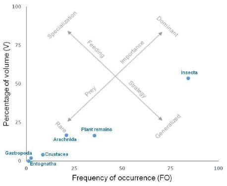 E.C. Silveira et al. - Parasitic helminths in Boana pulchella
158
ITEMS
GASTROPODA
ARACHNIDA
Acari
Araneae
Cuad. herpetol. 36 (2): 155-167 (2022)
SAMPLING SITES
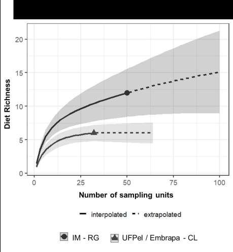
TOTAL SAMPLE (n=82) IM-RG (n=50) UFPel/Embrapa-CL (n=32)
FO V AI
2.4 1.9 0.2 2 2.6 0.2 3.1 0.6 0.1
4.9 0.1 0.02 8 0.1 0.04
14.6 14.4 10.1 18 10.9 8.6 9.4 20 9.8
Opiliones 1.2 2.6 0.2 2 4.3 0.4
CRUSTACEA
Isopoda 8.5 4.2 1.7 14 6.8 4.2
ENTOGNATHA
Collembola 1.2 0.03 <0.00 2 0.04 <0.00
INSECTA
Hymenoptera
2.4 0.1 0.02 4 0.2 0.04
Hemiptera 4.9 3.1 0.7 2 0.4 0.03 9.4 7.4 3.6 Coleoptera 23.2 18.2 20.9 32 20.1 28.2 18.8 15.2 14.8
Diptera 14.6 2.6 1.8 24 4.2 4.4 Lepidoptera (larvae) 9.8 10.8 5.1 8 7.5 2.6 12.5 16.2 10.5 Unidentified insects 29.3 18.8 26.4 32 16.9 23.7 25 21.9 28.5 Unidentified arthropods 9.8 4.3 2 12 6.3 3.3 6.3 1.2 0.4
Plant remains 35.4 16.5 28 30 18.3 24.1 43.6 13.7 31.1
Anthropic material (synthetic)
1.2 0.2 0.01 2 0.4 0.03
Unidentified items 3.7 2.1 0.4 4 1.0 0.2 6.3 3.9 1.3
The diet of UFPel/Embrapa-CL anurans inclu ded plant remains with the highest FO (43.6%) and AI (31.1%). Coleoptera at this site (FO of 18.8% and AI of 14.8%) and Lepidoptera (FO of 12.5% and AI of 10.5%) were the most important items of animal origin. Araneae was found with low FO (9.4%) and AI (9.8%), however it displayed a relatively high V (20%) (Table 1).
3.2 Helminth assemblage
Eighty-seven anurans (87%) presented helminths in the following sites of infection: lungs, liver, kidneys, stomach, intestines, and free or encysted in the celomatic cavity. Helminths belonging to Nematoda, Cestoda, Digenea, and Acanthocephala were identified, represented by an adult and larval forms (Table 2).
Figure 3. Rarefaction curve of the diet richness of Boana pul chella (Anura: Hylidae) males in two sampling sites (IM-RG, and UFPel/Embrapa-CL) in southern Brazil
Nematoda was the taxonomic group with the highest richness in the parasitic assemblage of B. pulchella. Cosmocercinae gen. spp. had the highest prevalence (P) and mean intensity of infection (MII)
Table 1. Frequency of occurrence (FO), volume (V) and alimentary index (AI) for the items found in the gastrointestinal contents of Boana pulchella (Anura: Hylidae) males from two sampling sites in southern Brazil. The values are presented as percentage.
159
FO V AI FO V AI
HELMINTHS
NEMATODA
SITES OF INFESTATION
P MII MA R
Oxyascaris oxyascaris Intestine 18 1.50 0.27 1-3
Falcaustra sp. Intestine 1 1.00 0.01 1
Cosmocercinae gen. spp. Intestine 33 5.88 1.94 1-31
Rhabdias sp. Lung 2 1.00 0.02 1
Ochoterenella sp. Intestine and coelomic cavity 10 3.80 0.38 1-12
Physaloptera sp. (larva) Stomach 1 1.00 0.01 1
Acuariinae gen. sp. (larvae) Liver, stomach, intestine and coelomic cavity 8 1.38 0.11 1-4
Unidentified larva Intestine 1 1.00 0.01 1
CESTODA
Cyclophyllidea gen. sp. Intestine 1 1.00 0.01 1
Unidentified larva Coelomic cavity 1 1.00 0.01 1
DIGENEA
Haematoloechus ozorioi Lung 1 1.00 0.01 1
Catadiscus sp. Intestine 5 1.60 0.08 1-2
Diplostomidae gen. spp. (metacercariae) Kidney 32 4.19 1.34 1-21
Crassiphialinae gen. sp. (metacercaria) Coelomic cavity 1 1.00 0.01 1
ACANTHOCEPHALA
Pseudoacanthocephalus sp. Intestine 27 2.22 0.60 1-7
Centrorhynchus sp. (cystacanths) Stomach, intestine and coelomic cavity 59 6.68 3.94 1-41
3.3 Boana pulchella helminth assemblage: aspects related to diet and host size, and sampling sites
The abundance of helminths presented a significant correlation only with the volume of items found in the gastrointestinal contents of the anurans from UFPel/Embrapa-CL (r² = 0.50, p <0.01).
Table 2. Helminths of Boana pulchella (Anura: Hylidae) males in southern Brazil, and their respective sites of infestation and parasi tological parameters (prevalence – P, as %, mean intensity of infection - MII, mean abundance – MA, and range - R). (33%; 5.88 helminths/host), followed by Oxyacaris oxyascaris Travassos, 1920 (18%; 1.50 helminths/ host) and Ochoterenella sp. (10%; 3.80 helminths/ host). Rhabdias sp. and Falcaustra sp. were found with P of 2% and 1%, respectively. Acuariinae stood out among the larval forms with a prevalence of 8% and an MII of 1.38 larvae/host, contrasting with the other taxa, which displayed an MII of 1 larva/host (Table 2).
Regarding Digenea, metacercariae of Diplos tomidae parasitized the kidney of 32% of anurans with MII of 4.19 helminth/host, followed by Cata discus sp. whose prevalence was 5% and MII of 1.60 helminth/host, while metacercaria of Crassiphiali nae gen. sp. and Haematoloechus ozorioi Freitas & Lent, 1939 were found in 1% of anurans with MII of 1 helminth/host (Table 2). Acanthocephala and Cestoda were represented by two taxa each (Table 2). Centrorhynchus sp. (cystacanths) infected 59% of anurans with MII of 6.68 helminths/host, while Pseu doacanthocephalus sp. was found with a prevalence of 27% and MII of 2.22 helminths/host. Cestoda was represented by a specimen of Cyclophyllidea gen. sp. and an unidentified larva (Table 2).
Analyses on helminth assemblage and host size revealed that O. oxyascaris and Cosmocercinae were significantly more prevalent in the anurans belonging to Class I, with P of 26.1% (X2, p = 0.026) and 39.1% (X2, p = 0.022) respectively. Acuarinae larvae and Pseudoacanthocephalus sp. had higher prevalences in Class II hosts, of 13% (X2, p = 0.047) and 35.2% (X2, p = 0.046) respectively (Table 3). Only the Cosmocercinae showed significant differences in the mean intensity of infection between the two size classes with greater MII for Class I (9.50 helminths/ host) (t-test, p = 0.0275) (Table 3). No significant differences were found in P and MII between host size classes infected by Digeneans belonging to Diplostomidae and Centrorhynchus sp. cystacanths (Table 3). Infections by the other helminths could not be compared as they were not detected in both classes. It is interesting to highlight that Ochoterene
E.C. Silveira et al. - Parasitic helminths in Boana pulchella
160
Cuad. herpetol. 36 (2): 155-167 (2022)
Table 3. Prevalence (P, as %), mean intensity of infestation (MII), mean abundance (MA) and range (R) of parasitic helminths of Boana pulchella (Anura: Hylidae) males in relation to the size classes (snout-vent length, SVL) in southern of Brazil.
HELMINTHS
NEMATODA
Oxyascaris oxyascaris
SVL
Class I (27- 37mm) (n=46) Class II (38 - 49mm) (n=54) P MII MA R P MII MA R
26.1a 1.50 0.41 1-3 9.3a 1.60 0.19 1-4
Falcaustra sp. 2.2 1.00 0.02 1 Cosmocercinae gen. spp. 39.1b 9.50bb 3.83 1-31 18.5b 1.80bb 0.33 1-5
Rhabdias sp. 0 3.7 1.00 0.04 1
Ochoterenella sp. 0 18.5 3.80 0.70 1-12 Physaloptera sp. (larva) 0 1.9 1.00 0.02 1 Acuariinae gen. sp. (larvae) 2.2c 1.00 0.02 1 13c 1.43 0.19 1-4
Unidentified larva 2.2 1.00 0.02 1 0
CESTODA
Ciclophyllidea gen. sp. 2.2 1.00 0.02 1 0 Unidentified larva 0 1.9 1.00 0.02 1
DIGENEA
Haematoloechus ozorioi 2.2 1.00 0.02 1 0 Catadiscus sp. 10.9 1.60 0.17 1-2 0
Diplostomidae gen. spp. (metacercariae) 37 5.12 1.89 1-14 27.8 3.20 0.89 1-21
Crassiphialinae gen. sp. (metacercaria) 0 1.9 1.00 0.02 1
ACANTHOCEPHALA
Pseudoacanthocephalus sp. 17.4d 1.63 0.28 1-2 35.2d 2.47 0.87 1-7 Centrorhynchus sp. (cystacanths) 56.5 6.73 3.80 1-41 61.1 6.64 4.06 1-39 Centrorhynchus sp. (cystacanths) 56.5 6.73 3.80 1-41 61.1 6.64 4.06 1-39
a - X2, p = 0.026; b - X2, p = 0.022; c - X2, p = 0.047; d - X2, p = 0.046; bb - t-test, p = 0.0275
lla sp. was found only in Class II anurans, with a P of 18.5% and an MII of 3.8 helminths/host. Besides, Catadiscus sp. was present in only 10.9% of Class I hosts, with an MII of 1.60 (Table 3). Although there were significant differences in the prevalence and mean intensity of infection for taxa among host size classes, the GLM between helminth abundance and anuran SVL was not significant (z = -1.55, p = 0.120).
The richness of helminths was not different between the sampling sites (Fig. 4). However, there was a significant difference in the prevalence of O. oxyascaris, Cosmocercinae gen. spp. and Ochotere nella sp. between the two sampled sites. A higher prevalence of the first two taxa was observed in anu rans from IM-RG (28%; X2, p = 0.009, and 52%; X2, p < 0.000, respectively), while Ochoterenella sp. was found with a higher prevalence (18%; X2, p < 0.008) in anurans from UFPel/Embrapa-CL. The mean in tensity of Cosmocercinae gen. spp. was significantly
higher in anurans from IM-RG (7.08 helminth/host; t-test, p = 0.0205) (Table 4). Catadiscus sp. occurred only in IM-RG parasitizing 10% of the hosts with an MII of 1.6 helminths/host. In general, although there is a variation in the prevalence of some taxa between sampled sites, the abundance of helminths did not vary significantly between the two localities (z = 1.76, p = 0.079).
Discussion
The study of the helminth fauna of B. pulchella in southern Brazil showed a greater number of taxa than a previous study in Buenos Aires province, Argentina, by Draghi et al. (2020). The prevalence of parasitism found by these authors was also lower as they recorded 35% of 150 hosts parasitized by at least one helminth species. Those differences may be associated with the characteristics of the sampling
161
sites, since Draghi et al. (2020) sampled cultivated and pasture areas.
Most parasitic helminths of B. pulchella have heteroxenous cycles that involve animals of several groups as intermediate and paratenic hosts, inclu ding anurans (Table 5). The generalist diet of B. pulchella observed in this study (insects, crustaceans, mollusks, and vegetation) is similar to previous ones (Maneyro & Rosa, 2004; Solé & Pelz, 2007; Rosa et al., 2011; Antoniazzi et al., 2013), and may provide the link between free-living forms of helminths and anurans. In this sense, studies on the diet and helminth fauna of anurans provide complementary information to improve our understanding of hostparasite relationships.
Some of the observed parasitic infections may be considered occasional since the prevalen ce of most helminths was less than 10%, and the intensity of infection of 11 taxa ranged from 1 to 7 helminths. The low rates of O. oxyascaris, Falcaustra sp., Physaloptera sp., Acuariinae gen. sp., H. ozorioi, and Cestoda may be related to the low consumption of intermediate and paratenic hosts that probably do not constitute prey commonly ingested by B. pulchella due to availability or preference bias for certain items. On the other hand, the infection rates of acanthocephalans may be related to the ingestion of arthropods, which were well represented in the diet of this species at both sampling sites. Accor ding to the literature, other taxa that compose the
helminth parasitic assemblage of B. pulchella (Cos mocercinae gen. spp., Rhabdias sp., Catadiscus sp. and Diplostomidae gen. spp.) can infect the anuran through the ingestion of immature forms during the tadpole phase or through active penetration of the skin (Table 5).
The infections by metacercariae of Diplosto midae and by cystacanths of Centrorhynchus sp. suggest that B. pulchella acts as an intermediate host for digeneans and as a paratenic host for the acanthocephalans since the above mentioned larval forms were found with expressive P% and MII va lues.This anuran act as a trophic bridge between the hosts at the lower level (invertebrates) and the hosts at the top of the trophic chain (birds and mammals, for example). The latter may acquire infections by ingesting anurans, thus ensuring the continuity of the parasitic cycle (Table 5). Centrorhynchus species, for example, were reported in natural predators of anurans, such as Rupornis magnirostris (Gmelin, 1788) (Accipitriformes) (Machado, 1940; Moura et al., 2017), and Guira guira (Gmelin, 1788) (Cucu liformes) (Lunaschi & Drago, 2010). On the other hand, a study carried out in agroecosystems in Argentina reported neither cystacanths nor meta cercariae associated with B. pulchella (Draghi et al., 2020). The different land use in the sampling sites of Brazil (present study) and Argentina (Draghi et al., 2020) may have influenced the parasitic infections in B. pulchella, since in the firstwe had no crops or livestock, as in Argentina. The sampling sites of the present study consist of temporary ponds with similar vegetation, to which anurans were closely associated, which may have contributed to the si milarity in the richness and abundance of parasitic helminths.
It is important to highlight that parasite-host relationships can be influenced by several other factors. Intrinsic characteristics related to hosts (e.g. maturity, gender, reproductive behaviour, size, diet) (Poulin, 1996; Von Zuben, 1997; Wilson et al., 2002; Klein, 2004) and parasites (e.g. size, quantity, fecundity rate, reproductive modes, dispersal ability) (Crofton, 1971; Von Zuben, 1997; Wilson et al, 2002; Khokhlova et al, 2010), as well as extrinsic factors (e.g. temperature, humidity, anthropogenic changes) (Marcogliese, 2005; Hudson et al. 2006; Koprivnikar et al., 2012), can act on the complex network of parasite-host-environment interactions.
Similarly to the present study, Draghi et al. (2020) found no relationship between host body
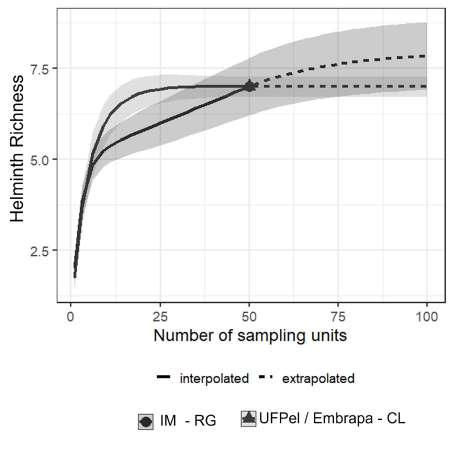 E.C. Silveira et al. - Parasitic helminths in Boana pulchella
Figure 4. Rarefaction curve of the parasitic helminth richness of Boana pulchella (Anura: Hylidae) males in two sampling sites (IM-RG, and UFPel/Embrapa-CL) in southern Brazil.
162
Cuad. herpetol. 36 (2): 155-167 (2022)
Table 4. Prevalence (P, as %), mean intensity of infestation (MII), mean abundance (MA) and range (R) of parasitic helminths of Boana pulchella (Anura: Hylidae) males in two sampling sites from southern Brazil.
HELMINTHS
NEMATODA
SAMPLING SITES
UFPel/Embrapa-CL (n=50) P MII MA R P MII MA R
IM-RG (n=50)
Oxyascaris oxyascaris 28a 1.43 0.40 1-3 8a 1.75 0.14 1-3
Falcaustra sp. 2 1.00 0.02 1 0 Cosmocercinae gen. spp. 52b 7.08bb 3.68 1-31 14b 1.43bb 0.20 1-3 Rhabdias sp. 4 1.00 0.04 1 0
Ochoterenella sp. 2c 1.00 0.04 1 18c 4.11 0.74 1-12
Physaloptera sp. (larva) 2 1.00 0.02 1 0 Acuariinae gen. sp. (larvae) 2 1.00 0.02 1 14 1.43 0.20 1-4 Unidentified larva 2 1.00 0.02 1 0
CESTODA
Ciclophyllidea gen. sp. 2 1.00 0.02 1 0 Unidentified larva 0 2 1.00 0.02 1
DIGENEA
Haematoloechus ozorioi 2 1.00 0.02 1 0 Catadiscus sp. 10 1.60 0.16 1-2 0 Diplostomidae gen. spp. (metacercariae) 36 5.17 1.86 1-14 28 3.00 0.82 1-21 Crassiphialinae gen. sp. (metacercaria) 0 2 1 0.02 1
ACANTHOCEPHALA
Pseudoacanthocephalus sp. 22 2.09 0.46 1-5 32 2.31 0.74 1-7
Centrorhynchus sp. (cystacanths) 58 6.69 3.88 1-41 60 6.67 4.00 1-39 a - X2, p = 0.009; b - X2, p = < 0.000; c - X2, p = 0.008; bb - t-test, p = 0.0205
Table 5. Helminth parasites of Boana pulchella (Anura: Hylidae) males in southern Brazil and their respective hosts and possible means of infestation, considering the information available for life cycles of similar species or species of the same family.
Helminths Definitive Host Intermediate host (IH)/ paratenic host (PH)
Nematoda
Oxyascaris oxyascaris
Amphibians Direct cycle – Invertebra tes (PH)
Possible means of infestation in anurans References
Ingestion of PH Anderson (2000)
Falcaustra sp. Amphibians, and reptiles Direct cycle – Freshwater mollusk (PH) Ingestion of PH Anderson (2000)
Cosmocercinae Amphibians Direct cycle
Rhabdias sp. Amphibians, and reptiles Direct cycle
Ingestion of larvae by tadpoles or skin penetration Anderson (2000)
Penetration of the larvae across the skin Anderson (2000)
Ochoterenella sp. Anurans Mosquito (IH) Ingestion of IH Anderson (2000)
163
Physaloptera sp. Birds, mammals, rep tiles, fish and amphi bians
Acuariinae Birds, and mammals
Insects (crickets, cockroa ches) (IH)
Insects (Coleoptera, Orthoptera) (IH)/Amphi bians and Reptiles (PH)
Ingestion of IH Anderson (2000)
Ingestion of IH Anderson (2000)
Cestoda Vertebrates
Digenea
Haematoloechus ozorioi Anurans
Vertebrates and Inverte brates (IH)
Ingestion of IH Olsen (1974)
Freshwater mollusk (1st IH), and Odonata (2nd IH) Ingestion of 2nd IH Olsen (1974)
Catadiscus sp. Anurans Mollusk Planorbidae (IH)
Tadpole ingests me taceraria present in aquatic vegetation Kehr & Hamann (2003)
Diplostomatidae Birds, and mammals
Acanthocephala
Pseudoacanthocephalus sp. Amphibians and reptiles
Freshwater mollusk (1st IH), fish, amphibians and occasionally mammals (2nd IH)
Aquatic arthropod (IH)
Penetration of the cercaria in the tadpole
Travassos et al. (1969), Hamann & Gonzáles (2009)
Ingestion of IH Barger & Nickol (1998)
Centrorhynchus sp. Birds, and mammals
Crustaceans or Insects (IH), Amphibians and rep tiles (PH)
1st IH=first intermediate host, 2nd IH=second intermediate host
size and helminth abundance. Host body size has also been considered a good indicator of parasite species richness since larger hosts may provide more space and other resources, possibly widening niche diversity for parasites (Campião et al., 2016). In addition, larger hosts live longer, establishing in less ephemeral habitats than smaller species and may have enhanced exposure to parasites (Poulin, 1997). According to our findings is possible to con clude that Boana pulchella males present a generalist feeding strategy with a diet composed of a wide variety of arthropods, mainly terrestrial insects. Their diverse helminth fauna was composed of lar val and adult forms, indicating that at least males of this anuran may act as definitive, intermediate, as well as paratenic hosts. The generalist diet and the association of the species with the vegetation close to aquatic environments are important factors for the persistence and the life cycle of its parasitic
Ingestion of IH
Travassos (1926), Petrochenko (1971), Schmidt (1985), Amato et al. (2003)
helminths. Oxyascaris oxyascaris, Cosmocercinae gen. spp., Ochoterenella sp., Diplostomidae gen. spp., Pseudoacanthocephalus sp., and Centrorhynchus sp. are the main taxa of this assemblage.
Acknowledgements
Special thanks to Bruna M. Chaviel, Ana Beatriz D. Henzel, and Julia V. Pereira for their assistance; Helena Silveira Schuch for helping us with statistical analyses; to Instituto Chico Mendes de Conservação da Biodiversidade (ICMBio No. 43658-1) for permis sions for collection of hosts; and to Coordenação de Aperfeiçoamento do Pessoal de Nível Superior (CA PES) for financial support (process No. 32/2010). DL acknowledges funding from CNPq (Productivity grant 310859/2020-4).
Literature cited
Aho, J.M. 1990. Helminth communities of amphibians and
E.C. Silveira et al. - Parasitic helminths in Boana pulchella
164
reptiles: comparative approaches to understanding patterns and processes, p.157-195. In: Esch GW, Bush 276 AO, Aho JM (eds.) Parasite communities: patterns and processes. Chapman and Hall, London.
Akani, G.C.; Luiselli, L.; Amuzie, C.C. & Wokem, G.N. 2011. Helminth community structure and diet of three Afrotropical anuran species: a test of the interactive-versusisolationist parasite communities hypothesis. Web Ecology 11: 11–19.
Amato, J.F.R.; Amato, S.B.; Araujo, P.B. & Quadros, A.F. 2003. First report of pigmentation dystrophy in terrestrial isopods, Atlantoscia floridana (van Name) (Isopoda, Oniscidea), induced by larval acanthocephalans. Revista Brasileira de Zoologia 20: 711–716.
Amato, J.F.R. & Amato, S.B. 2010. Técnicas gerais para coleta e preparação de helmintos endoparasitos de aves: 367–393. In: Von Matter S., F. C. Straube, I. Accordi, V. Piacentini, J. F. Cândido-Jr (ed.), Ornitologia e Conservação: Ciência Aplicada, Técnicas de Pesquisa e Levantamento. Technical Books Editora, Rio de Janeiro.
Anderson, R. C. 2000. Nematode parasites of vertebrates: their development and transmission. 2ª Ed. CABI International.
Anderson, R.C. & Bain, O. 2009. Capítulo 3: Rhaditida. In: Key to the Nematode Parasites of Vertebrates. CAB International, Massachusetts.
Antoniazzi, C.E.; López, J.A.; Duré, M. & Falico, D.A. 2013. Alimentación de dos especies de anfibios (Anura: Hylidae) en la estación de bajas temperaturas y su relación con la acumulación de energía en Santa Fe, Argentina. Revista de Biología Tropical 61: 875–886.
Bacher, M.R. & Vaucher, C. 1985. Parasitic helminths from Paraguay VII: systematic position of Oxyascaris Travassos, 1920 (Nematoda: Cosmocercoidea). Revue Suisse de Zoologie 92: 303–310.
Barger, M.A. & Nicko, B.B. 1998. Structure of Leptorhynchoides thecatus and Pomphorhynchus bulbocolli (Acanthocephala) eggs in habitat partitioning and transmission. Journal of Parasitology 84: 534-537.
Blanchet, S.; Rey, O.; Berthier, P.; Lek, S. & Loot, G. 2009. Evidence of parasite-mediated disruptive selection on genetic diversity in a wild fish population. Molecular Ecology 18: 1112–1123.
Borteiro, C., Castro, O., Sabalsagaray, M.J., Kolenc, F.; Debat, C.M. & Ubilla, M. 2015. Spargana in the Neotropical frog Hypsiboas pulchellus (Hylidae) from Uruguay. NorthWestern Journal of Zoology 11: 171–173.
Brooks, D.R. & Hoberg, E.P. 2000. Triage for the biosphere: the need and rationale for taxonomic inventories and phylogenetic studies of parasites. Comparative Parasitology 67: 1–21.
Bush, A.O.; Aho, J.M. & Kennedy, C.R. 1990. Ecological versus phylogenetic determinants of helminth parasite community richness. Evolutionary Ecology 4: 1–20.
Bush, A.O.; Lafferty, K.D., Lotz, J.M. & Shostar, A.W. 1997. Parasitology meets ecology on its own terms: Margolis et al. Revisited. Journal of Parasitology 83: 575–583.
Campião, K.M.; Dias, O.T.; Silva, R.J.; Ferreira, V.L. & Tavares, L.E.R. 2016. Living apart and having similar trouble: frog helminth parasites determined by the host or by the habitat? Canadian Journal of Zoology 94: 761–765.
Chabaud, A.G. 2009. Capítulo 9: Ascaridida. In: Key to the Nematode Parasites of Vertebrates. CAB International,
Cuad. herpetol. 36 (2): 155-167 (2022)
Massachusetts
Chao, A. & Jost, L. 2012. Coverage‐based rarefaction and extrapolation: standardizing samples by completeness rather than size. Ecology, 93: 2533-2547.
Costello, M.J. 1990. Predator feeding strategy and prey importance: a new graphical analysis. Journal of Fish Biology 36: 261-263.
Coyner, D.F.; Spalding, M.G. & Forrester, D. J. 2002. Epizootiology of Eustrongylides ignotus in Florida: distribution, density, and natural infections in intermediate hosts. Journal of Wildlife Diseases 38: 483-499.
CRMV. 2012. Conselho Federal de Medicina Veterinária. Available at: http://portal.cfmv.gov.br/ Accessed in: 25 September 2015.
Crofton, H.D. 1971. A quantitative approach to parasitism. Parasitology 62: 179-194.
Draghi, R.; Drago, F.B.; Saibene, P.E. & Agostini, M.G. 2020. Helminth communities from amphibians inhabiting agroecosystems in the Pampean Region (Argentina) Revue suisse de Zoologie 127: 261-274.
Duré, M.I.; Schaefer, E.F; Hamann, M.I. & Kehr, A.I. 2004. Consideraciones ecológicas sobre la dieta, la reprodución y el parasitismo de Psedopaludicola boliviana (Anura, Leptodactylidae) de Corrientes, Argentina. Phylomedusa 3: 121-131.
Freitas, J.F.T. 1958. Estudos sobre “Oxyascarididae” (Travassos, 1920) (Nematoda, Subuluroidea). Memórias do Instituto Oswaldo Cruz 56: 489–515.
Frost, D.R. 2022. Amphibian Species of the World: an Online Reference. Version 6.1 (Accessed June 20, 2022). Electronic Database accessible at https://amphibiansoftheworld.amnh. org/index.php. American Museum of Natural History, New York, USA.
Guerrero, R. & Yánez-Muñoz, M.H. 2018. Acarine biodiversity in Ecuador: two new species of endoparasitic chiggers (Acarina: Trombiculidae) from terrestrial andean anurans. MANTER: Journal of Parasite Biodiversity 9: 18.
Hamann, M.I. & González, C.E. 2009. Larval digenetic trematodes in tadpoles of six amphibian species from northeastern Argentina. Journal of Parasitology 95: 623-628.
Hammer Ø.;, Harper, D.A.T. & Ryan, P.D. 2001 PAST: Paleontological Statistics Software Package for Education and Data Analysis. Paleontological Statistics, PAST, 2.16. Available at: https://palaeo-electronica.org/2001_1/past/ issue1_01.htm Accessed 10 March 2020.
Hocking, D.J. & Babbitt, K.J. 2014. Amphibian contributions to ecosystem services. Herpetological Conservation and Biology 9: 1-17.
Hsieh, T. C., Ma, K. H. & Chao, A. 2016. iNEXT: an R package for rarefaction and extrapolation of species diversity (H ill numbers). Methods in Ecology and Evolution 7: 1451-1456.
Huckembeck, S.; Loebmann, D.; Albertoni, E.F.; Hefler, S.M.; Oliveira, M.C.L.M. & Garcia, A. 2014. Feeding ecology and basal food sources that sustain the Paradoxal frog Pseudis minuta: Multiple approaches combining stomach content, prey availability, and stable isotopes. Hydrobiologia 740: 253-264.
Hudson, P.J.; Dobson, A.P. & Lafferty, K.D. 2006. Is a healthy ecosystem one that is rich in parasites? Trends in Ecology and Evolution 21: 38-385.
165 Jones, A. 2005. Capítulo 25: Diplodiscidae Cohn, 1904. In: Key to the Trematoda. Volume 2. CABI Publishing, Massachusetts.
Kawakami, E. & Vazzoler, C. 1980. Método gráfico e estimative de índice alimentar aplicado no estudo de alimentação de peixes. Boletim do Instituto Oceanográfico 29: 205-207.
Kehr, AI. & Hamann, M.I. 2003. Ecological aspects of parasitism in the tadpole of Pseudis paradoxa from Argentina. Herpetological Review 34: 336-341
Kelehear, C.; Hudson, C.M.; Mertins, J.W. & Shine, R. 2017. First report of exotic ticks ( Amblyomma rotundatum ) parasitizing invasive cane toads (Rhinella marina) on the Island of Hawai’i. Ticks and tick-borne diseases 8: 330-333.
Khalil, L.F.; Jones, A. & Bray, R.A. 1994. Order Cyclophyllidea (diagnosis and key to families). In: Keys to the cestode parasites of vertebrates. CAB International, Wallingfors, U.K.
Khokhlova, I.S.; Serobyan, V.; Degen, A.A.; Krasnov, B.R. 2010. Host gender e offspring quality in a flea parasitic on a rodent. The Journal of Experimental Biology 213: 3299-3304.
Kiewiadomska, K. 2002. Família Diplostomidae. In: Gibson, D.I.; Jones, A. & Bray, R.A. Keys to the Trematoda – Volume I. CABI International e The Natural History Museum, Londres, UK.
Klaion, T. 2011. Diet and nematode infection in Proceratoprhys boiei (Anura: Cycloramphidae) from two Atlantic rainforest remnants in Southeastern Brazil. Anais da Academia Brasileira de Ciências 83: 1303-1312.
Klein, S.L. 2004. Hormonal e immunological mechanisms mediating sex differences in parasitic infection. Parasite Immunology 26: 247–264.
Koprivnikar, J.; Marcogliese, D.J.; Rohr, J.R.; Orlofske, S.A.; Raffel, T.R. & Johnson P.T.J. 2012. Macroparasite infections of amphibians: what can they tell us? EcoHealth 9: 342-360.
Lunaschi, L.I. & Drago, F.B. 2010. A new species of Centrorhynchus (Acanthocephala, Centrorhynchidae) endoparasite of Guira guira (Aves, Cuculidae) from Argentina. Helminthologia 41: 38-47.
Machado, D.A.F. 1940. Pesquisas helmintológicas realizadas no estado de Mato Grosso – Acanthocephala. Memórias do Instituto Oswaldo Cruz 35: 593-601.
Mackenzie, J.V. 2007. Human land use and patterns of parasitism in tropical anphibian host. Biological Conservation 137: 102-116.
Maneyro, R. & Rosa, I. 2004. Temporal and spatial changes in the diet of Hyla pulchella (Anura, Hylidae) in southern Uruguay. Phyllomedusa 3: 101-113.
Maneyro, R.; Loebmann, D.; Tozetti, A. & Fonte, L.F.M. 2017. Anfíbios das planícies costeiras do extremo sul do Brasil e Uruguai. Anolis Books Editora, 176 pp.
Marcogliese, D.J. 2004. Parasites: Small Players with Crucial Roles in the Ecological Theater. EcoHealth 1: 151-164.
Marcogliese D.J. 2005. Parasites of the superorganism: are they indicators of ecosystem health? International Journal for Parasitology 35: 705-716.
Moura, M.O.F.L.; Bregue, S.B. & Valente, A.L.S. 2017. Levantamento de helmintos de aves de rapina de Pelotas, Rio Grande do Sul. XXVI Congresso de Iniciação Científica, Pelotas, 4 pp.
Mugnai, R.; Nessimian, J.L & Baptista, D.F. 2010. Manual de identificação de macroinvertebrados aquáticos do estado do Rio de Janeiro. Technical Books Editora, 174 pp.
Needham, J.G. & Needham, P.R. 1978. Guía para el estudio de los seres vivos de las aguas dulces. Editora Reverté, S.A., 131 pp.
Olsen, O.W. 1974. Animal parasites: their life cycles and ecology.
Third Edition.
Petrochenko, V.I. 1971. Acanthocephala of domestic and wild animals. Israel Program for Scientific Translations, 465 pp.
Poulin, R. 1996. Sexual inequalities in helminth infections: a cost of being male? The American Naturalist 147: 287-295.
Poulin, R. 1997. Species richness of parasite assemblages: evolution and patterns. Annual Review of Ecology and Systematics 28: 341-358.
Reiczigel, J.; Marozzi, M.; Fábián, I. & Rózsa, L. 2019. Biostatistics for parasitologists – a primer to Quantitative Parasitology. Trends in Parasitology 35: 277-281.
Rosa, A.C.; Maneyro, R. & Camargo, A. 2011. Trophic niche variation and individual specialization in Hypsiboas pulchellus (Duméril and Bibron, 1841) (Anura, Hylidae) from Uruguay. South American Journal of Herpetology 6: 98-106.
Santos, V.G.T.; Amato, S.B. & Borges, M.M. 2013. Community structure of helminth parasites of the “Cururu” toad, Rhinella icterica (Anura: Bufonidae) from southern Brazil. Parasitology Research 112: 1097-1103.
Seeliger, U.; Odebrecht, C. & Castello, J.P. 1998. Fluxo de energia e habitats no estuário da Lagoa dos Patos: 326. In: Seeliger et al. Os ecossistemas costeiro e marinho do extremo sul do Brasil. Rio Grande.
Schmidt, G.D. 1986. Handbook of tapeworm identification Florida Constitution Revision Commission, 675 pp.
Silveira, E.C.; Mascarenhas, C.S.; Antunes, G.M.; Loebmann, D. 2019. Occurrence of Hannemania sp. (Acariformes: Leeuwenhoekiidae) larvae in males of Boana pulchella (Anura: Hylidae) from southern Brazil. Revista Mexicana de Biodiversidad 90: 1-5.
Solé, M. & Pelz, B. 2007. Do male tree frogs feed during the breeding season? Stomach flushing of five syntopic hylid species in Rio Grande do Sul, Brazil. Journal of Natural History 41: 2757-2763.
Spalding, M.G.; Bancroft, G.T. & Forrester, D.J. 1993. The epizootiology of eustrongylidosis in wading birds (Ciconiiformes) in Florida. Journal of Wildlife Diseases 29: 237-249.
StataCorp, L.P. 2007. Stata data analysis and statistical software. Special Edition Release (10.1 ed.), Stata, College Station. 733pp.
Sures, B. 2004. Environmental parasitology: relevancy of parasites in monitoring environmental pollution. Trends in Parasitology 20: 170-177.
Tkach, V.V. 2008. Family Haematoloechidae Freitas & Lent, 1939: 361-365. In: Keys to the Trematoda. v. 3. – CABI International, London.
Travassos, L. 1920 Contribuições para o conhecimento da fauna helmintológica brasileira. Memórias do Instituto Oswaldo Cruz 67:1–886. Archivos da Escola Superior de Agronomia e Medicina Veterinária 4: 17-20.
Travassos, L. 1926. Contribuição para o conhecimento da fauna helmintológica brasileira. XX. Revisão dos acantocéfalos brazileiros. Parte II. Fam. Echinorhynchidae. Sf. Centrarchinae Travassos, 1919. Memórias do Instituto Oswaldo Cruz 19: 31-125.
Travassos, L.; Freita, J.F.T. & Kohn, A. 1969. Trematódeos do Brasil. Memórias do Instituto Oswaldo Cruz 67: 1-886.
Van, S.M.; Schittini, G.M.; Marra, R.V.; Azevedo, A.R.M.; Vicente, J.J. & Vrcibradic, D. 2006. Body size, diet and endoparasites of the microhylid frog Chiasmocleis capixaba
E.C. Silveira et al. - Parasitic helminths in Boana pulchella
166
in an Atlantic Forest areaof southern Bahiastate, Brazil. Brazilian Journal of Biology 66: 167-173.
Vaucher, C. 1987. Polystomes d'Equateur, avec description de deux nouvelles especes. Bulletin de la Societe Neuchfteloise des Sciences Naturelles 110: 45-56.
Von Zuben, C.J. 1997. Implicações da agregação espacial de parasitas para a dinâmica populacional na interação hospedeiro-parasita. Revista de Saúde Pública 31: 523-530.
Wegner, K.M.; Kalbe, M.; Kurtz, J.; Reusch, T.B.H. & Milinski, M. 2003. Parasite selection for immunogenetic optimality.
Cuad. herpetol. 36 (2): 155-167 (2022)
Science 301: 1343. Wilson, K.; Bjørnstad; O.; Dobson, N.A.P.; Merler, S.; Poglayen, G.; Randolph, S.E.; Read, A.F & Skorping, A. 2002. Heterogeneities in macroparasite infections: patterns e processes, p 6-44. In: Hudson, P. J.; Rizzoli, A.; Grenfell, B. T.; Heesterbeek, H.; Dobson, A. P. (eds) The Ecology of Wildlife Diseases, United Kingdom, Oxford Press.
© 2022 por los autores, licencia otorgada a la Asociación Herpetológica Argentina. Este artículo es de acceso abierto y distribuido bajo los términos y condiciones de una licencia Atribución-No Comercial 4.0 Internacional de Creative Commons. Para ver una copia de esta licencia, visite http://creativecommons.org/licenses/by-nc/4.0/
167
Trabajo
Cuad. herpetol. 36 (2): 169-183 (2022)
DNA barcoding in Neotropical tadpoles: evaluation of 16S rRNA gene for the identification of anuran larvae from northeastern Brazil
Marcos J. Matias Dubeux1,2,3, Filipe A. Cavalcanti do Nascimento 2,3, Larissa L. Correia3, Tamí Mott2,3
1 Centro de Biociências, Universidade Federal de Pernambuco, Av. Prof. Moraes Rego, 1235, Cida de Universitária, CEP 50670-901, Recife, Pernambuco, Brazil
2 Instituto de Ciências Biológicas e da Saúde, Universidade Federal de Alagoas, Campus A.C. Simão, Av. Lourival Melo Mota, s/n, Tabuleiro do Martins, CEP 57072-970, Maceió, Alagoas, Brazil
3 Museu de História Natural, Universidade Federal de Alagoas, Av. Amazonas, s/n, Prado, CEP 57010-020, Maceió, Alagoas, Brazil
Recibido: 30 Abril 2021
Revisado: 07 Febrero 2022
Aceptado: 02 Agosto 2022
Editor Asociado: D. Baldo
doi: 10.31017/CdH.2022.(2021-030)
ABSTRACT
The challenge in studying Neotropical tadpoles is identifying species using only their external morphology. However, the DNA barcode protocol is often implemented to help elucidate ta xonomic issues. In fact, the identification of frogs through their unknown tadpoles has already been achieved accurately using this protocol. Despite the successful application of this tool, the efficiency of the 16S rRNA gene as a DNA barcode for Neotropical tadpoles has not been fully assessed. Herein we evaluate the efficacy of the 16S rRNA gene for identifying tadpoles from northeastern Brazil. Samples of 100 tadpole specimens from 12 locations were analyzed. The DNA sequences were individually submitted to a BLAST search and were then aligned with a matrix containing available sequences in the GenBank based on the anurans known to occur in the study area. The 16S rRNA fragment successfully identified the analyzed anuran species. Based on DNA barcoding, 8% of the tadpoles morphologically identified at the species level were incorrect. When an incongruence between morphological and molecular identifications was detected, the morphology of the target morphotype was reexamined, and previously ne glected morphological characteristics were identified. DNA barcoding using the 16S rRNA gene facilitated the assessment of tadpole richness in northeastern Brazil. This DNA protocol can be used as a starting point for detecting high levels of genetic divergence, highlighting potential taxa that should be studied from phylogenetic and taxonomic perspectives.
Key words: Amphibia; Mitochondrial Gene; Genetic Divergence; Species Diversity.
Introduction
Anuran amphibians generally have an aquatic larval phase, representing an important trophic com ponent of aquatic environments (Ranvestel et al., 2004; Rossa-Feres et al., 2004; Jordani et al., 2019). During certain periods, these tadpoles are the only evidence of anuran occurrence in some environ ments. Tadpoles are relatively abundant where they occur, and they are also easy to collect. However, for a long time, tadpoles were neglected by natu ralists and researchers, especially in megadiverse
Author for correspondence: marcosdubeux.bio@gmail.com
assemblages such as tropical regions (Provete et al., 2012; Rossa-Feres et al., 2015). Tadpoles have only recently started to be included more frequently in faunistic inventories and ecological, systematic, and taxonomic studies (Haas, 2003; Larson, 2005; Silva, 2010; Magalhães et al., 2013; Dubeux et al., 2020a). In fact, the tadpoles of a large portion of anuran species are unknown (Altig and McDiarmid, 1999; e.g., Provete et al., 2012; Schulze et al., 2015; Altig et al., 2021). This is an alarming scenario, considering
169
M. J. M. Dubeux et al. - DNA barcode in Neotropical tadpoles
that amphibians are the most threatened vertebrate group with known declining or extinct populations (IUCN, 2022). Additionally, many species will likely become extinct without formal descriptions or iden tification of their tadpoles (Crawford et al., 2010). In Brazil, the country with the highest worldwide anuran diversity (1,144 species; Segalla et al., 2021), the tadpoles of about half of identified species are unknown (Provete et al., 2012).
One significant challenge when studying tadpoles is species identification. When using only external morphology, species identification can be hampered by 1) the lack of knowledge of larval diagnostic characteristics (very similar morpholo gically or non-described tadpoles), 2) the absence of standardization in the nomenclature used for tadpole descriptions, or even 3) the scarcity of identification keys (Provete et al., 2012; Dubeux et al., 2020b). Until recently, the accurate identification of tadpoles was only possible in captivity, where tadpoles were held until they completed their metamorphosis, thereby allowing for the identification of species through their juveniles. However, this introduces the logistical challenge of reproducing environmental characteristics that mirror the natural environment where tadpoles are collected.
Currently, the integration of a molecular approach using the DNA barcode as a protocol has been promising in terms of facilitating tadpole identification (e.g., Vences et al., 2005b; Grosjean et al., 2015; Schulze et al., 2015). Although the original DNA barcoding protocol was used for identifying the information contained in a fragment of the mi tochondrial cytochrome c oxidase subunit 1 (COI) gene, in order to accelerate taxonomic descriptions for amphibians in general (Hebert et al., 2003, 2005), the barcode procedure using the mitochondrial 16S rRNA gene has been used in studies worldwide, due to its ease of use and high amplification success rate in different laboratory conditions (Vieites et al., 2009; Vences et al., 2005a, b; Lyra et al., 2017; Koroiva et al., 2020). The information in the 16S rRNA frag ment and the analysis of genetic similarity has been confirmed as an accurate method for identifying frog species, including their tadpoles. This approach has already been employed globally and has accurately identified tadpoles from Madagascar, Spain, Bolivia, and Southeast Asia (Thomas et al., 2005; Vences et al., 2005b, b; Grosjean et al., 2015; Schulze et al., 2015).
Despite the efficient application of the DNA
barcode tool in refining and accelerating the process of anuran identification, few studies have investiga ted the efficiency of the 16S rRNA gene as a molecu lar identification tool in Neotropical tadpoles (e.g., Schulze et al., 2015). Herein we evaluated the effecti veness of the 16S rRNA gene for the identification of specific and generic anuran taxa from northeastern Brazil, using their tadpoles. We assessed the rate of precise morphological identifications and discussed the challenges of the identification of tadpoles in me gadiverse assemblages such as Brazilian ecoregions. Additionally, we included some comments on cryp tic diversity in the local anurofauna based on genetic divergences and recent phylogenetic hypotheses.
Materials and methods
Sampling and DNA extraction
We used caudal musculature samples from 100 tadpoles obtained from 12 locations in the Caatinga and Atlantic Forest ecoregions in the state of Ala goas, northeastern Brazil (see Appendix I for more information about the locations). All the tadpoles were collected from 2013 to 2018 (licenses: SISBio/ ICMBIO 32920, 33507; CEUA 36/2015; SISGEN A6E0CAC). Firstly, we morphologically separated the tadpoles into 51 different morphotypes. Eighteen morphotypes were only identified at the generic level and two at the family level. All tadpoles were mor phologically allocated to eight families. Between one and three representatives of each morphotype were preserved in 92% alcohol, and the remaining speci mens were preserved in 10% formalin. All specimens were incorporated into the Coleção Herpetológica do Museu de História Natural of Universidade Federal de Alagoas (MHN-UFAL).
The total genomic DNA was extracted from a fragment of tadpole caudal musculature from each morphotype using the Phenol-Chloroform method (Sambrook et al., 1989) or by performing DNA extraction with salts (DNA Precipitation NaCl; Bruford et al., 1992), and stored in 15 μl of autoclaved distilled water.
Polymerase chain reaction and sequencing
A fragment of 550 base pairs (bp) of the 16S rRNA mitochondrial gene from each sample was amplified using the forward primer 16Sar: CGC CTG TTT ATC AAA AAC AT and reverse primer 16Sbr: CCG GTC TGA ACT CAG ATC ACG T (Palumbi et al., 2002) through the polymerase chain reaction
170
(PCR). Each PCR reaction had a final volume of 25 μl, comprised of 21.4 μl of MixMaster PCR, 0.8 μl of each primer, and 2 μl of DNA (20–100 ng/μl). The thermal cycling conditions followed an initial denaturation of 94° C (1–3 minutes), and 35 cycles of denaturation at 94° C for 45 seconds, pairing at 55° C for 45 seconds and extension at 72° C for one and half minutes. The PCR reactions were stained with syber safe, and electrophoresis in 1% agarose gel was performed to check for the presence of amplicons. The reactions were then visualized on the translumminator with ultraviolet light. The PCR products were purified with isopropanol and sequenced unidirectionally using the Sanger method after the Big Dye® terminator reaction.
Data analyses
The quality of the obtained sequences was checked and edited if necessary, using the software BioEdit Version 7.2.5 (Hall, 2011). Initially, in order to flag potential errors in tissue taxonomic labeling or DNA contamination, we performed Basic Local Align ment Search Tool (BLAST) analyzes on GenBank online platform. This analysis finds regions of local similarity between our samples and the sequences available in the repository database and calculates the statistical significance of the matches. We used the taxonomic identification of the most similar se quences in the database as a starting point to validate the identification of our samples. Each sample in our dataset was analyzed independently. The GenBank sequence with the greatest total similarity and the percentage of similarity is shown in Table 1. When an incongruence between morphological and mole cular identifications was detected, the morphology of the target morphotype was reexamined.
We then calculated genetic divergence between our samples and a selection of sequences obtained from the GenBank (selection criteria are presented below) using evolutionary models. This analysis aimed to compare our samples with: (1) conspecific samples of adult specimens collected in the study area or in geographically close areas, in order to associate tadpoles with their adult counterparts; (2) conspecific topotypical samples or samples from areas that are geographically close to the type loca lity of the nominal taxon, to obtain a preliminary assessment of the taxonomic status of populations in the study area (considering that nominal species with highly divergent populations may present an undescribed diversity) and, (3) sequences with grea
Cuad. herpetol. 36 (2): 169-183 (2022)
ter total similarity in the BLAST analysis, to validate the taxonomic identification obtained through this analysis.
Our sequences were aligned using the software MAFFT Version 7.310, implementing the iterative refinement method (automatic strategy selection) and default parameters (Katoh and Standley, 2013). They were then combined with a matrix containing sequences available in GenBank (see below for more details; Appendix II) of representatives of anuran species previously registered in the study area (fo llowing the anuran list available in Almeida et al., 2016). In the matrix, up to three DNA sequences from different localities for each anuran species were included, prioritizing regions close to the study area and the type locality of the species. When sequences were not available for a certain species (last consul tation in July 2020), a closely related representative was included (same genus and/or species group, following the current phylogenetic proposals [e.g., Carcerelli and Caramaschi, 1993; Lourenço et al., 2015; Lyra et al., 2020; specifically, Crossodactylus dantei (we used C. caramaschii), Physalaemus caete (we used P. signifier) and Boana exastis (we used B. pardalis)]. Additionally, we added the sequences with the highest total similarity in the BLAST analy sis, even if they had not been previously recorded in the study area (considering the possibility of new records and/or error in the labeling of the GenBank sequences). Following alignment, the tails of se quences imported from GenBank were manually trimmed using the software AliView Version 1.27 (Larsson, 2014) in order to eliminate gene fragments that were not of interest. The FFT-NS-i alignment strategy was selected by MAFFT and the final alig nment resulted in a 636 base pair matrix.
To estimate genetic divergences, distance esti mates were made from the sequence matrix using the Compute Pairwise Distances function implemented with the Kimura-2-parameters evolutionary model (K2P; Kimura, 1980) using the software MEGA X (Kumar et al., 2018). For a graphical visualization of the groupings, a dendrogram was generated using the Neighbor-Joining method (NJ) implemented with the K2P evolutionary model. The groupings obtained were validated using the bootstrap method (Felsenstein, 1985) with 1,000 pseudoreplicates. Bootstrap values above 97% were considered high and are indicated in the dendrogram. The sequences generated in this study were deposited in GenBank (OP022028 – OP022123; Table 1).
171
M. J. M. Dubeux et al. - DNA barcode in Neotropical tadpoles
Table 1. Molecular identification of tadpole specimens analyzed. Locations, associated vouchers and GenBank accession numbers of our samples. Genetic divergences [GD; in percentage (%)] calculated using the Compute Pairwise Distances function implemented with the Kimura-2-parameters evolutionary model including our samples and sequences from the GenBank. Sequences with the highest similarity, and their respective percentages, in BLAST analysis.
Species genetic identification Locality (municipality) Voucher (MHNUFAL)
Aromobatidae
GenBank access
GD closer to study area
GD closer to type locality BLAST (GenBank access)
Allobates olfersioides Teotônio Vilela 12465-1 OP022079 0 12.18 99.4 (KU495121) Bufonidae
Rhinella diptycha Traipu 13874 OP022111 0 99.7(MH004313)
Rhinella diptycha Arapiraca 13883 OP022116 0 100 (GU178784)
Rhinella diptycha Arapiraca 13885 OP022117 0 99.7(MH004313)
Rhinella diptycha Traipu 13897 OP022121 0 99.7 (DQ415572)
Rhinella granulosa Junqueiro 12462 OP022077 0.51 1.02 99.6 (KP685207)
Rhinella granulosa Arapiraca 13877 OP022112 0.51 1.02 99.8 (KP685207)
Rhinella granulosa Batalha 13882 OP022115 0.51 1.02 100 (KP685206)
Rhinella granulosa Batalha 13887 OP022118 0.51 1.02 99.8 (KP685205)
Rhinella hoogmoedi Murici 12502 OP022108 1.02 1.54 99.4(KU495521)■ Hylidae
Boana albomarginata
Limoeiro de Anadia 13901 OP022122 0 2.57 99.3 (AY549316)
Boana albomarginata Maceió 12259 OP022070 0 2.57 99.8 (AY549316)
Boana albomarginata Limoeiro de Anadia 12482 OP022094 0 2.57 99.8 (AY549316)
Boana albomarginata Coruripe 12474 OP022088 0 2.57 99.8 (AY549316) Boana albomarginata Barra de Santo Antônio 12497 OP022104 0 2.57 99.8 (AY549316)
Boana aff. atlantica ◊ Maceió 12079-1 OP022051 0.51 2.57 99.8 (MK348503)
Boana aff. atlantica ◊ Maceió 12079-2 OP022052 0.51 2.57 100 (MK348503)
Boana faber Limoeiro de Anadia 12478 OP022092 3.62* 3.62* 97.1 (KY002913)
Boana raniceps Coruripe 12132 OP022055 0 0.51 100 (KU495288)
Boana semilineata Maceió 11392-1 OP022028 0* 1.54 99.2 (MH004300)
Boana semilineata Maceió 11392-2 OP022029 0* 1.54 99.2 (MH004300)
Boana semilineata Maceió 12080-1 OP022053 0* 1.54 99.2 (MH004300)
Boana semilineata Maceió 12080-3 OP022054 0* 1.54 99.2 (MH004300)
Boana semilineata Maceió 12249-1 OP022056 0* 1.54 99.2 (MH004300)
Boana semilineata Maceió 12249-2 OP022057 0* 1.54 99.2 (MH004300)
Corythomantis greeningi Traipu 13889 OP022119 0 0.51 100 (MW243395)
Dendropsophus branneri Coruripe 12470 OP022083 0.51 0.51 99.2 (MT503865)
Dendropsophus branneri Maceió 12252-1 OP022060 0.51 0.51 99.6 (MT503865)
Dendropsophus branneri Maceió 12252-2 OP022061 0 0 99.8 (MT503865)
Dendropsophus branneri Maceió 12252-3 OP022062 0 0 99.8 (MT503865)
Dendropsophus sp.1 (aff. tapacurensis)
Dendropsophus sp.2 (aff. decipiens)
Barra de Santo Antônio 12491 OP022100 3.11 3.11 94.8 (MW026642)
Satuba 11866-1 OP022044 2.05 2.05 97.7 (MT503952)
172
Dendropsophus sp.2 (aff. decipiens)
Cuad. herpetol. 36 (2): 169-183 (2022)
Satuba 11866-2 OP022045 2.05 2.05 97.9 (MT503952)
Dendropsophus sp.2 (aff. decipiens) Satuba 11866-3 OP022046 2.05 2.05 97.9 (MT503952)
Dendropsophus sp.2 (aff. decipiens)
Dendropsophus sp.2 (aff. decipiens)
Dendropsophus sp.2 (aff. decipiens)
Satuba 11866-4 OP022047 2.05 2.05 97.9 (MT503952)
Satuba 11866-5 OP022048 2.05 2.05 97.9 (MT503952)
Satuba 12499 OP022106 2.05 2.05 97.7 (MT503952)
Dendropsophus aff. minutus ◊ Limoeiro de Anadia 13879 OP022114 0 3.09 100 (MK266721)
Dendropsophus aff. minutus ◊ Limoeiro de Anadia 13896 OP022120 0 3.09 100 (MK266721)
Dendropsophus aff. minutus ◊ Limoeiro de Anadia 13906 OP022123 0 3.09 100 (MK266721)
Dendropsophus aff. minutus ◊ Limoeiro de Anadia 12476 OP022090 0 3.09 100 (MK266721)
Dendropsophus aff. minutus ◊ Limoeiro de Anadia 12477 OP022091 0 3.09 100 (MK266721)
Dendropsophus aff. minutus ◊ Limoeiro de Anadia 12479 OP022093 0 3.09 100 (KJ833161)
Dendropsophus soaresi
Maceió 12251-1 OP022058 0 0 100 (MT503922) Dendropsophus soaresi Maceió 12251-2 OP022059 0 0 100 (MT503922)
Scinax similis ▲ Maceió 12256-1 OP022068 1.02 2.06 99.4 (MH206282)
Scinax similis ▲ Maceió 12256-2 OP022069 1.02 2.06 99.4 (MH206282)
Scinax similis ▲ Maceió 12254 OP022067 0.51 1.53 99.6 (MW114955)
Scinax similis ▲ Maceió 12492 OP022101 1.54 2.59 99.4 (MH206282)
Scinax auratus Maceió 11472-1 OP022034 5.83 5.83 95.2 (MH004316)
Scinax nebulosus Maceió 12493 OP022102 0 5.3 100 (EU201095)
Scinax nebulosus Maceió 11426-3 OP022031 0 5.3 100 (EU201095)
Scinax nebulosus Maceió 11426-2 OP022030 0.51 5.28 99.7 (EU201095)
Scinax nebulosus Maceió 11472-2 OP022035 0 5.3 99.7 (EU201095)
Scinax nebulosus Maceió 11473-1 OP022036 0 5.3 100 (EU201095)
Scinax nebulosus Maceió 11473-2 OP022037 0 5.3 100 (EU201095)
Scinax nebulosus Limoeiro de Anadia 13900 OP022109 0 5.3 100 (EU201095)
Scinax x-signatus
Coruripe 12469 OP022082 0 0 100 (MW114963)
Scinax x-signatus Junqueiro 12458 OP022074 0.51 0.51 99.7 (MW114964)
Scinax x-signatus
Maceió 12253-1 OP022063 1.02 1.02 99.0 (MW114963)
Scinax x-signatus Maceió 12253-2 OP022064 0 0 99.0 (MW114963)
Scinax x-signatus
Maceió 12253-3 OP022065 1.02 1.02 99.0 (MW114963)
Scinax x-signatus Maceió 12253-4 OP022066 1.02 1.02 99.0 (MW114963)
Leptodactylidae
Leptodactylus fuscus
Junqueiro 12456 OP022073 0 99.6 (AY911275)
Leptodactylus fuscus Junqueiro 12460 OP022076 0 99.6 (AY911275)
Leptodactylus fuscus Igaci 12486 OP022097 0 99.6 (AY911275)
Leptodactylus fuscus Teotônio Vilela 12467-2 OP022081 0 99.6 (AY911275)
173
M. J. M. Dubeux et al. - DNA barcode in Neotropical tadpoles
Leptodactylus fuscus Igaci 12487 OP022098 0 99.6 (AY911275)
Leptodactylus fuscus Maceió 12490 OP022099 0 99.6 (AY911275)
Leptodactylus macrosternum Arapiraca 13873 OP022110 1.02 1.02 99.8 (MH004302)
Leptodactylus macrosternum Coruripe 12475 OP022089 0 0 100 (MH206305)
Leptodactylus aff. mystaceus ◊ Maceió 12500 OP022107 0 5.89 99.8 (MT117857)
Leptodactylus natalensis Teotônio Vilela 12463 OP022078 0 1.02 99.8 (MH004304)
Leptodactylus troglodytes Junqueiro 12459 OP022075 0 0 99.3 (KM091620)
Leptodactylus troglodytes Coruripe 12473 OP022087 0 0 99.6 (MT117853)
Leptodactylus vastus Maceió 11433 OP022033 0 0 100 (KU495368)
Leptodactylus vastus Maceió 11867-1 OP022049 0.51 0.51 99.6 (KU495368)
Leptodactylus vastus Maceió 11867-2 OP022050 0.51 0.51 99.6 (KU495368)
Physalaemus cuvieri Coruripe 12467 OP022080 0 99.7 (KP146012)
Physalaemus cuvieri Coruripe 12471 OP022084 0 99.5 (KP146012)
Physalaemus cuvieri Barra de Santo Antônio 12494 OP022103 0 99.7 (KP146012)
Physalaemus cuvieri Maceió 12260 OP022071 0 100 (KP146012)
Pseudopaludicola aff. mystacalis ◊ Maceió 11429 OP022032 0 3.13 100 (KJ147044)
Microhylidae
Dermatonotus muelleri Arapiraca 13878 OP022113 0 2.57 99.8 (MH004297)
Elachistocleis cesarii Coruripe 12472-2 OP022086 1.02* 1.02 98.2 (KM509129)
Elachistocleis cesarii Coruripe 12472-1 OP022085 1.02* 1.02 98.2 (KM509129)
Odontophrynidae
Macrogenioglottus alipioi Maceió 11865-1 OP022041 0 0.51 99.6 (FJ685684)
Macrogenioglottus alipioi Maceió 11865-2 OP022042 0 0.51 99.6 (FJ685684)
Macrogenioglottus alipioi Maceió 11865-3 OP022043 0 0.51 99.6 (FJ685684)
Macrogenioglottus alipioi Maceió 12498 OP022105 0 0.51 99.2 (FJ685684)
Phyllomedusidae
Hylomantis granulosa Maceió 11864-1 OP022038 0 0.51 100 (GQ366224)
Hylomantis granulosa Maceió 11864-2 OP022039 0 0.51 100 (GQ366224)
Hylomantis granulosa Maceió 11864-3 OP022040 0 0.51 100 (GQ366224)
Pithecopus gonzagai Junqueiro 12455 OP022072 0 0 100 (KM387490)
Pithecopus gonzagai Igaci 12483 OP022095 0 0 100 (MW158678)
Pipidae
Pipa carvalhoi Igaci 12485 OP022096 0 0 100 (MT261652)
Legend of symbols: - Type location not known or not very specific; * Samples that are more than 1000 km away from the locality of interest (study area or taxon type locality); ◊ Lineage known to be distinct from that from the type locality of the taxon, which was assigned the “aff.” (see Results and discussion section for more details); ▲ Species never recorded for the study area; ■ Identified as Rhinella margaritifera (probably a misidentification).
Results and Discussion
The average length of the obtained sequences was 537 base pairs (440–594bp). The 51 analyzed mor photypes were molecularly designated to 33 taxa, representing 15 genera and eight families (Fig. 1). This represents 48% of anuran species with a regis tered tadpole phase in the study area (Dubeux et
al., 2019). In total, 32% (n = 32) of the specimens were only morphologically identified at a generic level, and only 6% (n = 6) at the family level (Fig. 2). Although incomplete, all these supra-specific identifications have been molecularly corroborated.
174 For the specimens that were morphologically identified at the specific level, misidentifications were made both at specific (2%, n = 2) and generic
(6%, n = 6) levels. Additionally, the tadpoles MHNUFAL 11472-1 and MHN-UFAL 11472-2, both collected on the same day and in the same pond and morphologically identified as having the same mor photype [Scinax nebulosus (Spix, 1824)], are actually two distinct species [S. nebulosus and S. auratus (Wied, 1821)]. This misconception can be explained by the fact that both species are representatives of
Cuad. herpetol. 36 (2): 169-183 (2022)
the S. rostratus clade, which are morphologically very similar, presenting the “labial arm” as a larval synapomorphy of the group (Gomes et al., 2014). After revisiting the morphology of these specimens, we could easily differentiate them by the number of labial teeth present in this structure (less than 10 in S. nebulosus, more than 15 in S. auratus; Dubeux et al., 2020b).
 Figure 1. Dendrogram of anurans (tadpoles and adult specimens) that occur in Alagoas state, Brazil. The diagram was built based on 636-bp of DNA sequences of 16S rRNA gene, using the Neighbor-Joining method implemented with the Kimura-2-parameters evolutionary model. Bold species names were sampled in this study. The terminals with acronym MUFAL represent our sequences (except MUFAL 2482, 14375, 14379). *Bootstrap values > 97%.
175
Figure 2. Percentage of morphological identification of tadpoles by family taxonomic level. CS = correct identification at specific level; IS = incorrect identification at specific level, but correct at generic level; IGS = incorrect generic and specific identifications; OG = identification only at generic level; OF = identification only at family level. Abbreviations below bars: AROMO. = Aromobatidae, BUFO. = Bufonidae, HYLI. = Hylidae, LEPTO. = Leptodactylidae, MICRO. = Microhylidae, ODON. = Odontophrynidae, PHYLLO. = Phyllomedusidae, PIPI. = Pipidae.
Additionally, some morphotypes identified as belon ging to species recorded in the study area were gene tically more similar to closely related taxa that have not been previously recorded in this region. Specifi cally, MHN-UFAL 1249, morphologically identified as Dendropsophus oliveirai (Bokermann, 1963), was genetically more similar to D. tapacurensis Olivei ra, Magalhães, Teixeira, Moura, Porto, Guimarães, Giaretta & Tinôco, 2021 (GD = 3.11%), which was recently described for the state of Pernambuco (municipality of São Lourenço da Mata, ~150 km away; Oliveira et al., 2021). There is no description for the tadpole of this species, which makes mor phological comparisons impossible. The specimens MHN-UFAL 11866(1-5) and MHN-UFAL 12499, morphologically identified as D. haddadi (Bastos & Pombal, 1996), were genetically more similar to D. decipiens (Lutz, 1925) (GD = 2.05%). Finally, spe cimens MHN-UFAL 12256(1-2) and MHN-UFAL 12254, morphologically identified as Scinax eurydice (Bokermann, 1968), were genetically more similar to S. similis (Cochran, 1952) (GD = 1.53 – 2.06%), a species whose type locality is in the state of Rio de Janeiro and whose distribution is only known for the Southeast and Central-west regions of Brazil
and Paraguay (Frost, 2022). The tadpoles of these two species present great morphological similarity to each other, as well as to the other representatives of the S. ruber clade, and no clear or unambiguous larval diagnostic characters were identified between these species (Rossa-Feres and Nomura, 2006; Dubeux et al., 2020b).
Considering the intraspecific divergence bet ween our sequences and those closest to the study area available in GenBank, the variation between conspecific pairs collected in the state of Alagoas (N = 13 species) ranged from zero (identical haploty pes) to 1.02%. Even when we added pairs of species that do not have adult sequences available for the study area, but that are available for locations up to 500km north of the state of Alagoas (covering the states of Pernambuco and Paraíba; N = 21 species), this range of variation does not change. However, when adding specimens collected up to 500km south of the state of Alagoas to the calculation (covering the states of Sergipe and Bahia; N = 29 species), the variation between conspecific pairs reached 5.83%. These high divergence values can be explained by the fact that the São Francisco River, which forms a southern boundary with the state of Alagoas, is
M. J. M. Dubeux et al. - DNA barcode in Neotropical tadpoles
176
known to be an important geographic barrier for the dispersion of anuran amphibians (e.g., Lima et al., 2019; Andrade et al., 2020). Additionally, it delimits important biogeographic regions of the At lantic Forest (Ribeiro et al., 2009) and creates zones of endemism (Asfora and Pontes, 2009; Dubeux et al., 2020b; França et al., 2020). It is expected that intraspecific genetic divergence would be greater in regions south of Alagoas compared to regions north of the state, either because they present lower gene flow or because they already represent independent evolutionary lineages.
All pairs of species had a smaller genetic di vergence in relation to the area closer to the study area than to their type locality. However, some populations from the study area were significantly different from those from the type locality of the no minal taxon. Some of these populations are known to represent independent evolutionary lineages or candidates for new species, according to recent phylogenetic studies, for example Leptodactylus aff. mystaceus (GD = 3.13%; see Silva et al., 2020), Den dropsophus aff. minutus (GD = 3.09%; see Gehara et al., 2014), Boana aff. atlantica (GD = 2.57%; see Lima et al., 2019), and Pseudopaludicola aff. mystacalis (GD = 3.13%; see Fávero et al., 2011).
However, there are no recent phylogenetic studies for other species and, based on the high genetic divergence identified here, in addition to other sources of evidence available in the literature, they should be considered as potential taxa for future phylogenetic studies (see below). For example, po pulations of Allobates olfersioides (Lutz, 1925) from Alagoas showed a genetic divergence of 12.18% with samples from localities close to their type locality in the state of Rio de Janeiro. The population of Ala goas, described as A. alagoanus (Bokermann, 1967), was synonymized to A. olfersioides by Verdade and Rodrigues (2007) based on the morphological simi larity of adult individuals. However, recent studies have highlighted the presence of marked differences in the larval morphology (Dubeux et al., 2020a, b) and acoustic repertoire (Forti et al., 2017) of these two populations.
Another example involves the representatives of the Dendropsophus decipiens species group, regis tered in the state of Alagoas. Although previously associated with D. oliveirai (hereafter referred to as Dendropsophus sp.1) and D. haddadi (hereafter referred to as Dendropsophus sp.2), the greater gene tic similarity with D. tapacurensis and D. decipiens,
Cuad. herpetol. 36 (2): 169-183 (2022)
respectively, raises interesting questions about the taxonomic status of these populations. Although genetically more similar (when compared to the sequences available in GenBank) and originating from locations less than 150 km away, the genetic divergence between Dendropsophus sp.1 and D. tapacurensis was 3.11%, which was higher than the divergence found between formally recognized lineages within this group (Oliveira et al., 2021). A similar situation was observed between Dendropso phus sp.2 and D. decipiens, where the identified di vergence was 2.05%. Multiple evolutionary lineages have already been identified in this species complex, including one lineage comprising populations from the state of Alagoas (D. decipiens VII; Oliveira et al., 2021). Phylogenetic studies with adequate sampling are required in order to assess the taxonomic status of these taxa. The type localities of some species were not included in the matrix due to the lack of geographic specificity in the designation of their locations, specifically: Rhinella diptycha (Cope, 1862), Leptodactylus fuscus (Schneider, 1799), and Physalaemus cuvieri Fitzinger, 1826.
One of the main bottlenecks in the develo pment of research focusing on larval life stages results as a consequence of the difficulty in iden tifying Neotropical anuran larvae, especially since assemblages often exceed 40 species (e.g., Brandão et al., 2004; Vancine et al., 2018) (Rossa-Feres et al., 2015; Dubeux et al., 2020b). The scarcity of this basic knowledge often becomes a barrier to the advance ment of other lines of research involving neotropical tadpoles. In the northern most portion of the At lantic Forest (north of the São Francisco River), for example, 78 anuran species have been recorded, of which 72 have a larval phase; however, 14% still lack a tadpole description [see Dubeux et al. (2020b) for a complete list]. Furthermore, until recently, there was no available identification key for this region (Dubeux et al ., 2020b). In this study, following sample collection, the tadpoles were identified by different undergraduate students, all with limited experience in this area of research, and the absence of identification keys for tadpoles in the study area or nearby regions (penalties available in 2020), made accurate identification even more difficult. This lack of experience and the scarcity of accessible tools, such as taxonomic keys, may explain, in part, the high rate of incomplete (38%) and incorrect (8%) morphological identification in our data.
With the popularization and low cost of single
177
M. J. M. Dubeux et al. - DNA barcode in Neotropical tadpoles
locus genetic analyzes, the use of molecular techni ques for more accurate assessments of biodiversity has become increasingly more common (Fouquet et al., 2007; Vieites et al., 2009; Perl et al., 2014; Grosjean et al., 2015; Schulze et al., 2015; Lyra et al., 2017; Koroiva et al., 2020; Vacher et al., 2020).
Several studies describing anuran larvae have used DNA barcoding to confirm the identity of species (e.g., Malkmus and Kosuch, 2000; Moravec et al., 2014; Dubeux et al., 2020c). However, even with the increasing number of sequenced species in recent decades, the under-sampling of species, mainly in re gions such as northeastern Brazil, still persists. In the state of Alagoas, for example, 74 anuran species are currently registered (Almeida et al., 2016; Roberto et al., 2017; Lisboa et al., 2019), of which six have no available DNA sequences (Frostius pernambucensis, Crossodactylus dantei, Scinax cretatus, S. muriciensis, S. skuki, and Physalaemus caete). This can hinder accurate assessments of local diversity, with some species remaining genetically unknown.
Despite the lack of available DNA sequences for some species, the 16S rRNA mitochondrial gene was found to be effective in associating tadpole spe cies with their adult counterparts and aided in asses sing anuran diversity in the state of Alagoas. Some advantages of using this marker in DNA barcode studies are 1) universal primers for vertebrates and 2) ease of use and high amplification success rate in different laboratory conditions (Vences et al., 2005b). Additionally, for anuran amphibians, the historical use of this gene has resulted in the construction of a database with approximately 75,000 available se quences (GenBank, accessed on April 27th, 2022). Furthermore, anuran amphibian lineages are rela tively old entities, and the mitochondrial gene has sufficient variation to unequivocally identify most species (Vences et al., 2005b; Thomas et al., 2005).
The cryptic diversity revealed through DNA barcoding has helped researchers in taxonomic re visions and estimations of local diversity (see below). In some countries anuran diversities have almost doubled when using the DNA barcode method (e.g., Fouquet et al., 2007 for French Guiana and Vieites et al., 2009 and Perl et al., 2014 for Madagascar). In Bra zil, recent large-scale studies have revealed diversities that have been neglected for decades (Lyra et al., 2017; Vacher et al., 2020; Koroiva et al., 2020). Con sidering the genetic lineages documented in previous phylogenetic studies (e.g., Gehara et al., 2014; Lima et al., 2019; Silva et al., 2020; Oliveira et al., 2021)
and the potential taxa for taxonomic studies based on the genetic diversity identified here, we suggest that new species should be described/revalidated in the coming years for the study area. Although the methods used here are suitable for associating tadpoles with their adult counterparts, they are not suitable for resolving phylogenetic relationships. These results should be considered as a starting point for the development of integrative studies that aim to investigate and solve taxonomic issues.
Acknowledgments
The authors thank the Museu de História Natural da Universidade Federal de Alagoas for allowing us to access the material; researchers involved for collecting the material and laboratory procedures, especially to Luana Lima, Isabela Nogueira, Adel Tenório, João Almeida e Ubiratan Gonçalves; to Ms. Rebecca Umeed for English editing; to project “Tadpoles from Brazil” (Edital SISBIOTA, Process Conselho Nacional de Desenvolvimento Científico e Tecnológico - CNPq 563075/2010-4 and Fundação de Amparo à Pesquisa do Estado de São PauloFAPESP 2010/52321-7); to Instituto Chico Mendes de Conservação da Biodiversidade (ICMBio) for collecting permits (license numbers: SISBIO 329201 and 33507-1) and to Comitê de Ética no Uso de Animais (CEUA) for permits (CEUA 36/2015); MJMD thanks Fundação de Amparo à Ciência e Tecnologia do Estado de Pernambuco - FACEPE (IBPG-1117-2.04/19) and TM thanks CNPq (309904/2015-3 and 312291/2018-3).
Literature cited
Almeida, J.P.F.; Nascimento, F.A.C.; Torquato, S.; Lisboa, B.S.; Tiburcio, I.C.S.; Palmeira, C.N.S.; Lima, M.G. & Mott, T. 2016. Amphibians of Alagoas State, northeastern Brazil. Herpetology Notes 9: 123-140.
Altig, R. & McDiarmid, R.W. 1999. Diversity: familial and generic characterizations. In: Tadpoles: the biology of anuran larvae. Edited by R.W. Mcdiarmid, R. Altig. University of Chicago Press, Chicago. pp. 295-337.
Altig, R.; McDiarmid, R.W. & Dias, P.H.S. 2021. Bibliography for the identification, morphology and development of amphibian gametes and larvae. https://www.researchgate. net/publication/359195811_Bibliography_for_the_ identification_morphology_and_development_of_ amphibian_gametes_and_larvae. Accessed on 07 February 2022.
Andrade, F.S.; Haga, I.A.; Ferreira, J.S.; Recco-Pimentel, S.M.; Toledo, L.F. & Bruschi, D.P. 2020. A new cryptic species of Pithecopus (Anura, Phyllomedusidae) in north-eastern Brazil. European Journal of Taxonomy 723, 108-134.
Asfora, P.H. & Pontes, A.R.M. 2009. The small mammals of the highly impacted North-eastern Atlantic Forest of Brazil,
178
Pernambuco Endemism Center. Biota Neotropica 9(1): 31-35.
Brandão, D.; Bastos, R.; De Souza, M.; Vieira, C.; Bini, L.; Oliveira, L. & Diniz-Filho, J.A. 2004. Spatial patterns in species richness and priority areas for conservation of anurans in the Cerrado region, Central Brazil. AmphibiaReptilia 25: 63-75.
Bruford, M.W.; Hanotte, O.; Brookfield, J.F.Y. & Burke, T. 1992. Single-locus and multilocus DNA fingerprint. In: Hoelzel AR (Ed.) Molecular Genetic Analysis of Populations: A Practical Approach. IRL Press, Oxford: pp. 225-270.
Carcerelli, L.C. & Caramaschi, U. 1993. Ocorrência do gênero Crossodactylus Duméril & Bibron, 1841 no nordeste brasileiro, com descrição de duas espécies novas (Amphibia, Anura, Leptodactylidae). Revista Brasileira de Biologia 52: 415-422.
Crawford, A.J.; Lips, K.R. & Bermingham, E. 2010. Epidemic disease decimates amphibian abundance, species diversity, and evolutionary history in the highlands of central Panama. Proceedings of the National Academy of Sciences 107(31): 13777-13782.
Dubeux, M.J.M.; Gonçalves, U.; Nascimento, F.A.C. & Mott, T. 2020a. Anuran amphibians of a protected area in the northern Atlantic Forest with comments on topotypic and endangered populations. Herpetology Notes 13: 61-74.
Dubeux, M.J.M.; Nascimento, F.A.C.; Lima, L.R.; Magalhães, F.D.M.; Silva, I.R.S.; Gonçalves, U.; Almeida, J.P.F.; Correia, L.L.; Garda, A.A.; Mesquita, D.O.; Rossa-Feres, D.D.C. & Mott, T. 2020b. Morphological characterization and taxonomic key of tadpoles (Amphibia: Anura) from the northern region of the Atlantic Forest. Biota Neotropica 20: 1-24.
Dubeux, M.J.M.; Silva, T.; Mott, T. & Nascimento, F.A.C. 2020c. Redescription of the tadpole of Leptodactylus natalensis Lutz (Anura: Leptodactylidae), an inhabitant of the Brazilian Atlantic Forest. Zootaxa 4732: 346-350.
Dubeux, M.J.M.; Silva, G.R.S.; Nascimento, F.A.C.; Gonçalves, U. & Mott, T. 2019. Síntese histórica e avanços no conhecimento de girinos (Amphibia: Anura) no estado de Alagoas, nordeste do Brasil. Revista Nordestina de Zoologia 12: 18-52.
Fávero, E.R.; Veiga-Menoncello, A.C.; Rossa-Feres, D.C.; Strüssmann, C.; Giaretta, A.A.; Andrade, G.D.; Colombo, P. & Recco-Pimentel, S.M. 2011. Intrageneric karyotypic variation in Pseudopaludicola (Anura: Leiuperidae) and its taxonomic relatedness. Zoological Studies 50(6): 826-836.
Felsenstein, J. 1985. Confidence limits on phylogenies: an approach using the bootstrap. Evolution 39: 783-791.
Forti, L.R.; Silva, T.R.Á. & Toledo, L.F. 2017. The acoustic repertoire of the Atlantic Forest Rocket Frog and its consequences for taxonomy and conservation (Allobates, Aromobatidae). ZooKeys (692): 141-153.
Fouquet, A.; Gilles, A.; Vences, M.; Marty, C.; Blanc, M. & Gemmell, N.J. 2007. Underestimation of species richness in Neotropical frogs revealed by mtDNA analyses. PLoS one 2: e1109.
França, R.C.; Morais, M.; França, F.G.; Rödder, D. & Solé, M. 2020. Snakes of the Pernambuco Endemism Center, Brazil: diversity, natural history and conservation. ZooKeys 1002: 115-158.
Frost, D.R. 2022. Amphibian Species of the World: Online Reference. Version 6.0. http://research.amnh.org/
Cuad. herpetol. 36 (2): 169-183 (2022)
herpetology/amphibia/index.php. Accessed on 07 February 2022.
Gehara, M.; Crawford, A.J.; Orrico, V.D.; Rodríguez, A.; Lötters, S.; Fouquet, A.; Barrientos, L.S.; Brusquetti, F.; de la Riva, I.; Ernst, R.; Gagliardi-Urrutia, G.; Glaw, F.; Guayasamin, J.M.; Hölting, M.; Jansen, M.; Kok, P.J.R.; Kwet, A.; Lingnau, R.; Lyra, M.; Moravec, J.; Pombal Jr., J.P.; Rojas-Runjaic, F.J.M.; Schulze, A.; Señaris, J.C.; Solé, M.; Rodrigues, M.T.; Twomey, E.; Haddad, C.F.B.; Vences, M. & Köhler, J. 2014. High levels of diversity uncovered in a widespread nominal taxon: continental phylogeography of the Neotropical tree frog Dendropsophus minutus PloS one 9: e103958.
Gomes, M.D.R.; Alves, A.C.R. & Peixoto, O.L. 2014. O girino de Scinax nebulosus (Amphibia, Anura, Hylidae). Iheringia. Série Zoologia 104(2): 184-188.
Grosjean, S.; Ohler, A.; Chuaynkern, Y.; Cruaud, C. & Hassanin, A. 2015. Improving biodiversity assessment of anuran amphibians using DNA barcoding of tadpoles. Case studies from Southeast Asia. Comptes Rendus Biologies 338(5): 351-361.
Haas, A. 2003. Phylogeny of frogs as inferred from primarily larval characters (Amphibia: Anura). Cladistics 19: 23-89. Hall, T. 2011. BioEdit: an important software for molecular biology. GERF Bull Biosci 2: 60-61.
Hebert, P.D.N. & Gregory, T.R. 2005. The promise of DNA barcoding for taxonomy. Systematic Biology 54: 852-859.
Hebert, P.D.N.; Cywinska, A.; Ball, S.L. & Waard, J.R. 2003. Biological identification through DNA barcodes. Proceedings of the Royal Society B 270: 313-321.
IUCN. 2022. The IUCN Red List of Threatened Species: Online Reference. Version 2020-1. http://www.iucnredlist.org. Accessed on 07 February 2022.
Jordani, M.X.; Mouquet, N.; Casatti, L.; Menin, M.; Rossa‐Feres, D.C. & Albert, C.H. 2019. Intraspecific and interspecific trait variability in tadpole meta‐communities from the Brazilian Atlantic rainforest. Ecology and Evolution 9(7): 4025-4037.
Katoh, K. & Standley, D.M. 2013. MAFFT multiple sequence alignment software version 7: improvements in performance and usability. Molecular Biology and Evolution 30(4): 772780.
Kimura, M. 1980. A simple method for estimating evolutionary rates of base substitutions through comparative studies of nucleotide sequences. Journal of Molecular Evolution 16: 111-120.
Koroiva, R.; Rodrigues, L.R.R. & Santana, D.J. 2020. DNA barcoding for identification of anuran species in the central region of South America. PeerJ 8: e10189.
Kumar, S.; Stecher, G.; Li, M.; Knyaz, C. & Tamura, K. 2018. MEGA X: molecular evolutionary genetics analysis across computing platforms. Molecular Biology and Evolution 35(6): 1547-1549.
Larson, P.M. 2005. Ontogeny, phylogeny, and morphology in anuran larvae: morphometric analysis of cranial development and evolution in Rana tadpoles (Anura: Ranidae). Journal of Morphology 264: 34-52.
Larsson, A. 2014. AliView: a fast and lightweight alignment viewer and editor for large datasets. Bioinformatics 30(22): 3276-3278.
Lima, L.R.; Dubeux, M.J.M.; Nascimento, F.A.C.; Bruschi, D.P. & Mott, T. 2019. Uncovering Neotropical treefrog diversity: integrative taxonomy reveal paraphyly in Boana atlantica (Amphibia, Anura, Hylidae). Amphibia-Reptilia 40: 511-521.
179
M. J. M. Dubeux et al. - DNA barcode in Neotropical tadpoles
Lisboa, B.; Santos, W.F.S.; Torquato, S.; Guarnieri, M.C. & Mott, T. 2019. A new state record of the glassfrog Vitreorana baliomma (Anura: Centrolenidae), with notes on its reproductive biology. Herpetology Notes 12: 957-960.
Lourenço, L.B.; Targueta, C.P.; Baldo, D.; Nascimento, J.; Garcia, P.C.; Andrade, G.V.; Haddad, C.F.B. & Recco-Pimentel, S.M. 2015. Phylogeny of frogs from the genus Physalaemus (Anura, Leptodactylidae) inferred from mitochondrial and nuclear gene sequences. Molecular Phylogenetics and Evolution 92: 204-216.
Lyra, M.L.; Haddad, C.F.B. & Azeredo‐Espin, A.M.L. 2017. Meeting the challenge of DNA barcoding Neotropical amphibians: polymerase chain reaction optimization and new COI primers. Molecular Ecology Resources 17(5): 966-980.
Lyra, M.L.; Lourenço, A.C.C.; Pinheiro, P.D.; Pezzuti, T.L.; Baêta, D.; Barlow, A.; Hofreiter, M.; Pombal, J.P.; Haddad, C.F.B. & Faivovich, J. 2020. High-throughput DNA sequencing of museum specimens sheds light on the long-missing species of the Bokermannohyla claresignata group (Anura: Hylidae: Cophomantini). Zoological Journal of the Linnean Society 190(4): 1235-1255.
Magalhães, F.M.; Dantas, A.K.B.P.; Brito, M.R.M.; Medeiros, P.H.S.; Oliveira, A.F.; Pereira, T.C.S.O.; Queiroz, M.H.C.; Santana, D.J.; Silva, W.P. & Garda, A.A. 2013. Anurans from an Atlantic Forest-Caatinga ecotone in Rio Grande do Norte State, Brazil. Herpetology Notes 6: 1-10.
Malkmus, R. & Kosuch, J. 2000. Beschreibung einer neuen Ansonia-Larve (Ansonia guibei) von Borneo. SalamandraBonn 36(2): 121-124.
Moravec, J.; Lehr, E.; Cusi, J.C.; Córdova, J.H. & Gvoždík, V. 2014. A new species of the Rhinella margaritifera species group (Anura, Bufonidae) from the montane forest of the Selva Central, Peru. ZooKeys 2014: 35-56.
Oliveira, R.F.D.; Magalhães, F.M.; Teixeira, B.F.D.V.; Moura, G.J.B.D.; Porto, C.R.; Guimarães, F.P.B.B.; Giaretta, A.A. & Tinôco, M.S. 2021. A new species of the Dendropsophus decipiens Group (Anura: Hylidae) from Northeastern Brazil. Plos one (7): e0248112.
Palumbi, S.; Martin, A.; Romano, S.; McMilan, W.O.; Stice, L. & Grabowski, G. 2002. The Simple Fool’s Guide to PCR. University of Hawaii.
Perl, R.G.B.; Nagy, Z.T.; Sonet, G.; Glaw, F.; Wollenberg, K.C. & Vences, M. 2014. DNA barcoding Madagascar’s amphibian fauna. Amphibia-Reptilia 35: 197-206.
Provete, D.B.; Garey, M.V.; Silva, F. & Jordani, M.X. 2012. Knowledge gaps and bibliographical revision about descriptions of free-swimming anuran larvae from Brazil. North-Western Journal of Zoology 8(2): 283-286.
Ranvestel, A.W.; Lips, K.R.; Pringle, C.M.; Whiles, M.R. & Bixby, R.J. 2004. Neotropical tadpoles influence stream benthos: evidence for the ecological consequences of decline in amphibian populations. Freshwater Biology 49: 274-285.
Ribeiro, M.C.; Metzger, J.P.; Martensen, A.C.; Ponzoni, F.J. & Hirota, M.M. 2009. The Brazilian Atlantic Forest: How much is left, and how is the remaining forest distributed? Implications for conservation. Biological Conservation 142: 1141-1153.
Roberto, I.J.; Araujo-Vieira, K.; Carvalho-e-Silva, S.P. & Ávila, R.W. 2017. A new species of Sphaenorhynchus (Anura: Hylidae) from northeastern Brazil. Herpetologica 73: 148161.
Rossa-Feres, D.C. & Nomura, F. 2006. Characterization and taxonomic key for tadpoles (Amphibia: Anura) from the northwestern region of São Paulo State, Brazil. Biota Neotropica 6: 1-26.
Rossa-Feres, D.D.C.; Jim, J. & Fonseca, M.G. 2004. Diets of tadpoles from a temporary pond in southeastern Brazil (Amphibia, Anura). Revista Brasileira de Zoologia 21: 745-754.
Rossa-Feres, D.D.C.; Venesky, M.D.; Nomura, F.; Eterovick, P.C.; Vera Candioti, M.F.; Menin, M.; Juncá, F.A.; Schiesari, L.C.; Haddad, C.F.B.; Garey, M.V.; Anjos, L.A. & Wassersug, R. 2015. Taking tadpole biology into the 21st century: a consensus paper from the First Tadpoles International Workshop. Herpetologia Brasileira 4(2): 48-59.
Sambrook, J.; Fritsch, E.F. & Maniatis, T. 1989. Molecular cloning: a laboratory manual (No. Ed. 2). Cold Spring Harbor Laboratory Press.
Schulze, A; Jansen, M & Köhler, G. 2015. Tadpole diversity of Bolivia's lowland anuran communities: molecular identification, morphological characterisation, and ecological assignment. Zootaxa 4016(1): 1-111.
Segalla, M.V.; Berneck, B.; Canedo, C.; Caramaschi, U.; Cruz, C.A.G.; Garcia, P.C.A.; Grant, T.; Haddad, C.F.B.; Lourenço, A.C.C.; Mângia, S.; Mott, T.; Nascimento, L.B.; Toledo, L.F.; Werneck, F.P. & Langone, J.A. 2021. List of Brazilian Amphibians. Herpetologia Brasileira 10(1): 121-216.
Silva, F.R. 2010. Evaluation of survey methods for sampling anuran species richness in the neotropics. South American Journal of Herpetology 5: 212-220
Silva, L.A.; Magalhaes, F.M.; Thomassen, H.; Leite, F.S.; Garda, A.A.; Brandao, R.A.; Haddad, C.F.B.; Giaretta A.A. & Carvalho, T.R. 2020. Unraveling the species diversity and relationships in the Leptodactylus mystaceus complex (Anura: Leptodactylidae), with the description of three new Brazilian species. Zootaxa 4779: 151-189.
Thomas, M.; Raharivololoniaina, L.; Glaw, F.; Vences, M. & Vieite, D.R. 2005. Montane tadpoles in Madagascar: molecular identification and description of the larval stages of Mantidactylus elegans, Mantidactylus madecassus, and Boophis laurenti from the Andringitra Massif. Copeia 2005: 174-183.
Vacher, J.P.; Chave, J.; Ficetola, F.G.; Sommeria‐Klein, G.; Tao, S.; Thébaud, C.; Blanc, M.; Camacho, A.; Cassimiro, J.; Colston, T.J.; Dewynter, M.; Ernst, R.; Gaucher, P.; Gomes, J.O.; Jairam, R.; Kok, P.J.R.; Lima, J.D.; Martinez, Q.; Marty, C.; Noonan, B.P.; Nunes, P.M.S.; Ouboter, P.; Recoder, R.; Rodrigues, M.T.; Snyder, A.; Marques-Souza, S. & Fouquet, A. 2020. Large‐scale DNA‐based survey of frogs in Amazonia suggests a vast underestimation of species richness and endemism. Journal of Biogeography 47: 1781-1791.
Vancine, M.H.; Duarte, K.D.S.; Souza, Y.S.; Giovanelli, J.G.R.; Martins‐Sobrinho, P.M.; López, A.; Bovo, R.P.; Maffei, F.; Lion, M.B.; Júnior, J.W.R.; Brassaloti, R.; Costa, C.O.R.; Sawakuchi, H.O.; Forti, L.R.; Cacciali, P.; Bertoluci, J.; Haddad, C.F.B. & Ribeiro, M.C. 2018. ATLANTIC AMPHIBIANS: a data set of amphibian communities from the Atlantic Forests of South America. Ecology 99: 1692-1692.
Vences, M.; Thomas, M.; Bonett, R.M. & Vieites, D.R. 2005a. Deciphering amphibian diversity through DNA barcoding: chances and challenges. Philosophical Transactions of the
180
Royal Society B: Biological Sciences 360: 1859-1868. Vences, M.; Thomas, M.; Van der Meijden, A.; Chiari, Y. & Vieites, D.R. 2005b. Comparative performance of the 16S rRNA gene in DNA barcoding of amphibians. Frontiers in Zoology 2: 1-12. Verdade, V.K. & Rodrigues, M.T. 2007. Taxonomic review of Allobates (Anura, Aromobatidae) from the Atlantic Forest,
Cuad. herpetol. 36 (2): 169-183 (2022)
Brazil. Journal of Herpetology 41(4): 566-580. Vieites, D.R.; Wollenberg, K.C.; Andreone, F.; Köhler, J.; Glaw, F. & Vences, M. 2009. Vast underestimation of Madagascar's biodiversity evidenced by an integrative amphibian inventory. Proceedings of the National Academy of Sciences USA 106: 8267-8272.
APPENDIX I. Locations with tadpoles sampled in this study, Alagoas state, Brazil.
Ecoregion Municipality
Atlantic Forest
Caatinga
Coordinates
Number of samples Number of species
Barra de Santo Antônio 9°23'27.6"S; 35°31'30.8"W 4 3
Coruripe 10°07'45.0"S; 36°11'09.5"W 11 8
Junqueiro 9°55'08.0"S; 36°28'46.6"W 7 5
Maceió 9°33'30.6"S; 35°47'57.5"W 44 16
Murici 9°12'59.3"S; 35°51'24.2"W 1 1
Satuba 9°34'51.4"S; 35°50'35.4"W 6 1
Teotônio Vilela 9°54'16.6"S; 36°22'21.9"W 3 3
Arapiraca 9°44'32.7"S; 36°37'57.2"W 5 4
Batalha 9°42'49.1"S; 37°06'02.4"W 2 1
Igaci 9°32'40.0"S; 36°37'53.0"W 4 3
Limoeiro de Anadia 9°44'16.5"S; 36°27'41.3"W 10 4
Traipu 9°57'39.8"S; 36°57'34.6"W 3 2
APPENDIX II. Additional 16S rRNA mitochondrial gene fragment sequences of anuran species used in the study.
Species Voucher Locality GenBank Aromobatidae
Allobates olfersioides
CFBHT01886 Alagoas: Passo de Camaragibe KU495122
Allobates olfersioides MNRJ79899 Rio de Janeiro: Maricá MF624179 Bufonidae
Rhinella crucifer TG094 Bahia: Salvador KU495499
Rhinella diptycha CFBH19523 Bahia: Maracás MW003646
Rhinella granulosa CFBH7341 Alagoas: Passo de Camaragibe KP685205
Rhinella granulosa CFBH21068 Bahia: Caetité KP685207
Rhinella hoogmoedi UFBA610 Bahia: Ilhéus MH538283 Centrolenidae
Vitreorana baliomma MCP14121 Bahia: Una MW366909 Ceratophryidae
Ceratophrys joazeirensis CFBH7411
Hylidae
Paraíba: Araruna KP295617
Aplastodiscus sibilatus MLL-2016 Alagoas: Murici KU184227
Aplastodiscus sibilatus CFBH32528 Bahia: Ibirapitanga KU184014
Boana albomarginata USNM284519 Pernambuco: Caruaru KF794116
Boana albomarginata UFBA380/7890 Bahia: Mata de São João MH004298
Boana atlantica MUFAL13067 Alagoas: Maceió MK348506
Boana atlantica CFBH16126 Bahia: Uruçuca MK348483
181
M. J. M. Dubeux et al. - DNA barcode in Neotropical tadpoles
Boana crepitans CFBHT07825
Alagoas: Campo Alegre KU495263
Boana crepitans CFBHT12841 Bahia: Camamu KU495262
Boana faber CFBHT04381 Rio de Janeiro: Petrópolis KU495265
Boana freicanecae MUFAL11072 Alagoas: Murici MT823773
Boana freicanecae ZUFRJ7941 Pernambuco: Jaqueira MT823774
Boana raniceps CFBHT07827
Alagoas: Campo Alegre MW197887
Boana raniceps MACN37795 Argentina: Santa Fé KF794140
Boana semilineata CFBH5424 Rio de Janeiro: Duque de Caxias AY843779
Corythomantis greeningi CFBH2968 Alagoas: Piranhas KF002247
Corythomantis greeningi CRS100 Bahia: Morro do Chapéu MW243375
Dendropsophus branneri CFBH20829 Pernambuco: Bonito MT503865
Dendropsophus dutrai MNRJ46746
Sergipe: Indiaroba MT503923
Dendropsophus elegans CFBH13294 Sergipe: Itabaiana MT503966
Dendropsophus elegans CFBH18699 Bahia: Prado KY348631
Dendropsophus haddadi FSFL1467 Bahia: Prado MT503942
Dendropsophus haddadi CFBH19472
Espírito Santo: Vitória MT503941
Dendropsophus minutus CFBH18567 Alagoas: Campo Alegre MK266721
Dendropsophus minutus CFBH24153 Rio de Janeiro: Resende MT503934
Dendropsophus nanus ZUEC:18018 Sergipe: Aracaju MN420278
Dendropsophus nanus MACN37785 Argentina: Entre Rios AY549346
Dendropsophus oliveirai AAGARDA12495 Pernambuco: São Lourenço da Mata MW026634
Dendropsophus oliveirai CFBH18799 Bahia: Maracás MT503956
Dendropsophus soaresi CFBH18575 Alagoas: Campo Alegre MT503922
Dendropsophus studerae URCAG767 Alagoas: Quebrangulo MT503894
Phyllodytes acuminatus CHP-UFRPE1598 Pernambuco: Buíque MN961791
Phyllodytes edelmoi MNRJ50118 Alagoas: Maceió MN961798
Phyllodytes gyrinaethes CFBH44634 Alagoas: Maceió MN961801
Scinax agilis UFBA562/11203 Bahia: Mata de São João MH004314
Scinax auratus UFBA618/11712 Bahia: Conde MH004316
Scinax eurydice UESC11005 Bahia OK161150
Scinax fuscomarginatus CFBH7349 Alagoas: Passo de Camaragibe KJ004153
Scinax fuscomarginatus
CFBHT1950 Minas Gerais: Santana do Riacho KJ004129
Scinax fuscovarius CFBHT03219 Minas Gerais: Araxá KU495554
Scinax melanodactylus CFBHT01137 Alagoas: Passo de Camaragibe KU495535
Scinax nebulosus AAG-UFU6251 Rio Grande do Norte: Nísia Floresta MK503366
Scinax nebulosus MPEG:31631 Pará: Belém MK503362
Scinax pachycrus UESC11027 Bahia OK161140
Scinax x-signatus CFBHT08860 Pernambuco: Fernando de Noronha KU495578
Scinax x-signatus CFBHT09136 Bahia: Caetité KU495576
Scinax x-signatus CFBHT09136 Bahia: Caetité KU495576
Sphaenorhynchus cammaeus MACNHe48851 Alagoas: Quebrangulo MK266727
Sphaenorhynchus prasinus MZUFBA6756 Bahia: Mata de São João MK266752
Trachycephalus mesophaeus CFBHT09297 Bahia: Aurelino Leal KU495600
Trachycephalus mesophaeus FRS1038 São Paulo: Apiaí MH206468
Leptodactylidae
Leptodactylus fuscus USNM284551 Pernambuco AY911279
182
Leptodactylus latrans MUFAL15265
Cuad. herpetol. 36 (2): 169-183 (2022)
Alagoas: Maceió MT495901
Leptodactylus macrosternum CFBHT01146 Alagoas: Passo de Camaragibe KU495341
Leptodactylus macrosternum FC18 Bahia: Terra Nova MT495860
Leptodactylus mystaceus AAGARDA12501 Pernambuco: São Lourenço da Mata MT117857
Leptodactylus mystaceus TG379 Amazonas: São Gabriel da Cachoeira MT117890
Leptodactylus natalensis MTR101P28
Alagoas: Maceió KU495347
Leptodactylus natalensis AAGARDA1980 Rio Grande do Norte: Parnamirim MW291330
Leptodactylus troglodytes USNM284553 Pernambuco KM091620
Leptodactylus vastus TG429 Paraíba: Guarabira KU495368
Physalaemus albifrons CFBH16137
Ceará: Viçosa do Ceará KP146010
Physalaemus cicada CFBH19395 Ceará: Novas Russas KP146064
Physalaemus cuvieri ZUEC17897 Pernambuco: Caruaru KP146012
Pleurodema diplolister TG427 Paraíba: Caiçara KU495455
Pleurodema diplolister CFBH16144 Ceará: Viçosa do Ceara JQ937185
Pseudopaludicola mystacalis ZUEC:13837 Maranhão: Barreirinhas KJ147006
Pseudopaludicola mystacalis ZUEC:14147 Mato Grosso: Cuiabá KJ146983
Microhylidae
Chiasmocleis alagoanus C2683 Alagoas: Maceió KC180030
Dermatonotus muelleri CFBHT07834 Alagoas: Campo Alegre KU495218
Dermatonotus muelleri T7 Paraguay KC179984
Elachistocleis cesarii ZUEC:DCC3301 Minas Gerais: Serra do Cipó JN604511
Elachistocleis cesarii CFBHT00912 São Paulo: Santa Fé do Sul KU495225
Stereocyclops incrassatus MUFAL2482 Alagoas: Marechal Deodoro KC180046 Stereocyclops incrassatus CFBHT02080 Espírito Santo Linhares KU495593
Odontophrynidae
Macrogenioglottus alipioi CFBHT04337 Bahia: Uruçuca KU495385
Odontophrynus carvalhoi JC1224 Bahia: Mucugê FJ685687
Proceratophrys cristiceps AAGARDA2739 Ceará: Crato KX855993
Proceratophrys renalis ZUFRJ8665 Pernambuco: Caruaru JN814584
Proceratophrys renalis UFBA187/6242 Bahia: Cachoeira MH004311
Phyllomedusidae
Hylomantis granulosa MNRJ50123
Alagoas: Murici GQ366225
Hylomantis granulosa CFBHT00392 Pernambuco: Jaqueira KU495255
Pithecopus gonzagai CFBH7330 Alagoas: Passo de Camaragibe GQ366330
Pithecopus gonzagai ZUEC19684 Pernambuco: Limoeiro MW158582
Pipidae
Pipa carvalhoi
MUFAL14375
Pipa carvalhoi MUFAL14379
Ranidae
Lithobates palmipes
CFBHT07835
Pernambuco: Buíque MT261672
Pernambuco: Buíque MT261653
Alagoas: Campo Alegre KU495377
Lithobates palmipes 01 Guiana AF467265
© 2022 por los autores, licencia otorgada a la Asociación Herpetológica Argentina. Este artículo es de acceso abierto y distribuido bajo los términos y condiciones de una licencia Atribución-No Comercial 4.0 Internacional de Creative Commons. Para ver una copia de esta licencia, visite http://creativecommons.org/licenses/by-nc/4.0/
183
Trabajo
Cuad. herpetol. 36 (2): 185-196 (2022)
Herpeto-commerce: A look at the illegal online trade of amphibians and reptiles in Brazil
Ibrahim Kamel Rodrigues Nehemy1, Thayllon Orzechowsky Gomes2, Fernanda Paiva2, Wesley Kauan Kubo2, João Emílio de Almeida Júnior1, Nathan Fernandes Neves2, Vinicius de Avelar São Pedro2
1Universidade Federal de Mato Grosso do Sul (UFMS), Instituto de Biociências, Laboratório Ma pinguari, Cidade Universitária, Av. Costa e Silva, s/nº, Bairro Universitário, 79.070900, Campo Grande, Mato Grosso do Sul, Brazil.
2Universidade Federal de São Carlos (UFSCar), Laboratório de Estudos Zoológicos do Alto Paranapanema (LEZPA), campus Lagoa do Sino, Rodovia Lauri Simões de Barros, Km 12 SP-189, Bairro Aracaçu, 18.290000, Buri, São Paulo, Brazil.
Recibido: 26 Abril 2022
Revisado: 28 Julio 2022
Aceptado: 26 Agosto 2022
Editor Asociado: G. Agostini
doi: 10.31017/CdH.2022.(2022-009)
ABSTRACT
The illegal sale of fauna and flora represents the third-largest illegal trade in the world. Social media has contributed considerably to the increase in this type of trade. We searched for posts announcing the sale of amphibians and reptiles in seven Facebook® groups (three public and four private groups) from 01 January 2019 to 31 July 2020. In total, we found 548 posts made by a total of 201 social network profiles announcing the sale of 1,049 animals. We found 58 herpetofauna species being traded in the network (15 amphibian and 43 reptile species). Most of the sale advertisements originated in Southeast Brazil, predominantly from the state of São Paulo. The most traded species were Pantherophis guttatus (N= 467), Eublepharis macularius (N= 152), and Boa constrictor (N=90). This study presents important data about the illegal herpetofauna trade through Facebook® in Brazil, proving this market is currently fully active. This trade has high growth potential, bringing possible risks to biodiversity and public health. In conclusion, we recommend the implementation of urgent, specific government measures for its regulation and effective inspection.
Key words: Herpetofauna; E-commerce; Animal Trafficking; Pet-trade; Facebook®
RESUMO
A venda ilegal de fauna e flora representa o terceiro maior comércio ilegal do mundo. As redes sociais têm contribuído consideravelmente para o aumento deste tipo de comércio. Buscamos postagens anunciando a venda de anfíbios e répteis em sete grupos do Facebook® (três grupos públicos e quatro privados) de 01 de janeiro de 2019 a 31 de julho de 2020. No total, encontramos 548 postagens feitas por um total de 201 perfis de redes sociais anunciando a venda de 1.049 animais. Encontramos 58 espécies de herpetofauna sendo comercializadas na rede (15 espécies de anfíbios e 43 espécies de répteis). A maior parte dos anúncios de venda teve origem no Sudeste do Brasil, predominantemente no estado de São Paulo. As espécies mais comercializadas foram Pantherophis guttatus (N= 467), Eublepharis macularius (N= 152) e Boa constrictor (N=90). Este estudo apresenta dados importantes sobre o comércio ilegal de herpetofauna através do Facebook® no Brasil, comprovando que este mercado está atualmente em plena atividade. Esse comércio tem alto potencial de crescimento, trazendo possíveis riscos à biodiversidade e à saúde pública. Em conclusão, recomendamos a implementação de medidas governamentais urgentes e específicas para sua regulamentação e fiscalização efetiva.
Palavras-chave: Herpetofauna; E-commerce; Tráfico de Animais; Comércio Pet; Facebook®
Introduction
Brazil is the most biodiverse country in the world, with approximately 117 thousand known animal species and around 50 thousand known plant
Author for correspondence: vasaopedro@gmail.com
species, showing a high rate of endemic species (Flora Brasileira, 2020; ICMBio, 2020; Charity and Ferreira, 2020). This large number of species makes
185
I. K. R. Nehemy et al. - Online trade of herpetofauna in Brazil
the country a target for intense smuggling of wild species, one of the primary causes of local extinction, along with deforestation, farming activities, and urbanization processes (Hernandez and Carvalho, 2006; Heliodoro, 2009; RENCTAS, 2016). The illegal wildlife trade is responsible for spreading diseases and introducing exotic species, jeopardizing structu red communities (Warchol, 2004; Carrete and Tella, 2008; Karesh et al., 2012). This trade is also related to the considerable increase in violence and corruption rates (Warchol, 2004).
In Brazil, the institution responsible for dealing with animal trade is IBAMA (Instituto Brasileiro do Meio Ambiente e dos Recursos Naturais Renováveis). This institute is also responsible for the supervision and enforcement of the legal purposes, while other agencies have scientific competence on the subject, such as RAN/ICMBio (Centro Nacional de Pesquisa e Conservação de Répteis e Anfíbios), responsible for the herpetofauna (RENCTAS, 2001). The legal definition of the act of illegal trade, described under article 29, section 1, III of Law nº. 9,605/98, which includes in all respects: " Those who sell, exposes for sale, exports or acquires, retains, keeps in captivity or storage, uses or transports eggs, larvae, wild or native species, or in migratory route, as well as products and objects originating from such species, from breeding sites that are not authorized, without proper permit, competent authority or authorization."
Despite the legislation, the wild fauna trade from irregular breedings sites or specimens caught in nature, is still widely practiced in the country (Charity and Ferreira, 2020). The lack of investiga tion efforts is one of the reasons why this trade still occurs. E-commerce has been neglected, and the trafficking structure seems to benefit from online spaces that are lawless. Animal trafficking seems increasingly interconnected to the online network, which results in higher successes in sales that use these new means and make online supervision difficult (Hernandez and Carvalho, 2006; Siriwat and Nijman, 2018).
Globally, the acquisition of wild animals as pets through the internet has grown over the last several years due to the emergence of websites and social network groups specifically focused on the subject (Jansen et al., 2018; Sy, 2018; Marshall et al., 2020; Strine and Hughes, 2020). In most cases, sale advertisements through social networks have ques tionable origins (Magalhães and São-Pedro, 2012). Araújo (2014) and Auliya et al. (2016a) highlight the
sale of amphibians and reptiles (herpetofauna) in this market. The authors also state that these animals are targeted due to the great variety of species, avai lability of individuals, and fewer care requirements when compared to mammals and birds.
Although the diversity of amphibians and rep tiles is high, few species are legally regulated to be traded. According to data published in 2016, out of 10,272 species of reptiles, less than 8% are regulated by the Convention on International Trade in Endan gered Species of Wild Fauna and Flora (CITES) and by the European Wildlife Trade Regulations (EWTR) (Auliya et al., 2016a). For amphibians, less than 3% of recognized species are listed in the three appendices of CITES (Auliya et al., 2016b).
In most cases, the trade of these species jeopardizes their conservation, putting them in endangered or vulnerable statuses. The IUCN Red List of Threatened Species presents more than two thousand species of reptiles as threatened under the category "Biological resource use". This is the third largest threat category for this group, where 769 species are intentionally targeted by collectors for hunting and capture (IUCN, 2022). Over 290 amphibian species from the IUCN Red List are tar geted for international pet trade and consumption purposes (Auliya et al., 2016b).
This study aims to shine a light on illegal herpe tofauna e-commerce due to the growing popularity of social networks in the last years and their increa sed use as a platform for worldwide illegal wildlife trading. Here, we collected quali-quantitative data from Brazilian public and private groups on the social network Facebook®, specifically created to sell or exchange herpetofauna individuals throughout the country.
Materials and methods
We searched for Brazilian groups on the social net work Facebook® applying the following keywords in Brazilian Portuguese: "anfíbios", "répteis", "ani mais exóticos", "compra e venda de exóticos", “pets exóticos”, “répteis e anfíbios” and “répteis e anfíbios venda” (English keywords: “amphibians”, “reptiles”, “exotic animals”, “exotic marketing”, “exotic pets”, “reptiles and amphibians”, and “reptiles and amphi bians for sale”). We selected the first 15 groups we found online and got access to seven (four private and three public) that became the object of our re search. We verified the number of members of each
186
group, the date they were founded, and number of advertisements (see ‘Information from analyzed Facebook® groups’ in Appendix S1, Supplementary information).
The research consisted of analyzing all posts between 01 January 2019 and 31 July 2020. We recorded the advertisements related to the sale of amphibian and reptile species, listing the total num ber of posts and traded animals. The names of the groups and their members will be kept confidential for legal reasons and to avoid higher visibility of the groups, following orientation from the Association of Internet Researchers Committee (Franzke et al., 2020).
We recognized the species mainly through images as well as scientific or popular names men tioned in the posts. We discarded posts that did not have pictures or any other means that would allow us to identify the advertised species correctly. We ba sed the identification of individuals (to species level when possible) on specialized literature. From posts that did not have pictures, we considered the scien tific name mentioned and current nomenclature. The nomenclature and taxonomic classification for species of reptiles were based on Uetz et al. (2022), and Frost (2021) for amphibians. We classified the species as native from the Brazilian fauna or exotic (non-native), following the lists of Brazilian reptiles (Costa and Bérnils, 2018) and amphibians (Segalla et al., 2021).
We investigated if the species found were included in the appendices of CITES (2021). The threat level for each species was verified in the Brazil Red Book of Threatened Species of Fauna (ICMBio, 2018) and the Red List of the International Union for Conservation of Nature (IUCN, 2022). We analyzed the frequency and location of each advertisement. Therefore, it was possible to understand which Brazi lian regions contribute the most to this animal trade.
Results
During the 19 months of sampling, we list a total of 548 posts advertising animals for sale made by 201 Facebook® profiles. In total, 1,049 individuals of herpetofauna were commercialized. We recorded the sale of 58 species (Table 1), belonging to five orders: Anura (37 individuals from 12 species), Caudata (ten individuals from three species), Crocodylia (six in dividuals from one species), Squamata (Snakes: 815 individuals from 23 species; Lizards: 115 individuals
Cuad. herpetol. 36 (2): 185-196 (2022)
from nine species) and Testudines (66 individuals from ten species) (Fig. 1). Between the groups we analyzed, two of them concentrated the majority of posts, with 218 and 186 on each one, with a total of 798 traded animals (see 'Information from analyzed Facebook® groups ' in Appendix S1, Supplementary information).
Thirteen individuals could not be identified to species level, belonging to the genera Ceratophrys sp., Chelonoidis sp., and Pantherophis sp. We added these species to Table 1; their conservation status was not specified, and their classification as native or exotic was not described for the first two species. The snake genus Pantherophis does not occur in Brazil, so we considered it an exotic species.
All analyzed posts refer to traded animals in Brazilian territory. Most animals were being traded in the state of São Paulo (n=506), followed by the state of Rio de Janeiro (n=82) and Distrito Federal (n=42) (Fig. 2). Most of the advertisements are concentrated in the southeast region. We could not verify the trading location of 358 announced indi viduals due to the information not being described in the posts.
Four announced species (Caiman latirostris, Acrantophis dumerili, Acrantophis madagascarien sis, and Python molurus) are listed in Appendix I of CITES (2021), which are species not allowed to be internationally traded due to being endangered. We found twenty-one species in Appendix II, that described species likely to become endangered in the future. In addition, a special license is required for their trade. Only one species (Crotalus durissus) is listed in Appendix III, included after a direct request from Honduras. This appendix lists species that need international control, so their exploitation is either restricted or prevented. Half of the species (n=29) do not appear on the CITES list (Table 1).
Concerning the species threat level, only 49 were assessed by IUCN Red List of Threatened Spe cies (2022), four species listed as Vulnerable (VU) (Podocnemis unifilis, Chelonoidis denticulatus, Corre lophus ciliatus, Python bivittatus), one listed as Cri tically Endangered (CR) (Ambystoma mexicanum), and two listed as Near Threatened (NT) (Python molurus and P. regius). Regarding the threat level in national scope, out of 28 native Brazilian species announced, only Ranitomeya ventrimaculata is not on the list. Twenty-four species are classified as Least Concern (LC), and three other species (Trachemys dorbigni, Podocnemis expansa, and Podocnemis uni
187
I. K. R. Nehemy et al. - Online trade of herpetofauna in Brazil
Table 1. List of reptile and amphibian species traded in Brazil through Facebook® groups from 01 January 2019 until 31 July 2020.
ORDER/FAMILY/SPECIES
Nº of Individuals CITES (2021)
ANURA
AMPHIBIA
Bombinatoridae
Conservation Status
Native or exotic IUCN Red List (2022) Red Book ICMBio (2018)
Bombina orientalis (Boulenger, 1890) 3 LC EX Brachycephalidae
Brachycephalus ephippium (Spix, 1824) 5 LC LC NA
Ceratophryidae
Ceratophrys aurita (Raddi, 1823) 1 LC LC NA
Ceratophrys sp. (Wied-Neuwied, 1824) 1
Dendrobatidae
Adelphobates galactonotus (Steindachner, 1864) 6 II LC LC NA
Dendrobates tinctorius (Cuvier, 1797) 2 II LC LC NA
Ranitomeya ventrimaculata (Shreve, 1935) 2 II LC EX
Hylidae
Dendropsophus minutus (Peters, 1872) 2 LC LC NA
Phyllomedusidae
Pithecopus azureus (Cope, 1862) 7 DD LC NA
Pithecopus nordestinus (Caramaschi, 2006) 1 DD LC NA
Pipidae
Xenopus laevis (Daudin, 1802) 5 LC EX Ranidae
Lithobates catesbeianus (Shaw, 1802) 2 LC EX/invasive species
CAUDATA
Ambystomatidae
Ambystoma mexicanum (Shaw & Nodder, 1798) 4 II CR EX
Salamandridae
Pleurodeles waltl Michahelles, 1830 4 NT EX
Triturus cristatus (Laurenti, 1768) 2 LC EX
REPTILIA
CROCODYLIA
Alligatoridae
Caiman latirostris (Daudin, 1802) 6 I LC LC NA
TESTUDINES
Chelidae
Hydromedusa tectifera Cope, 1870 1 LC NA
Mesoclemmys gibba (Schweigger, 1812) 2 LC NA Mesoclemmys tuberculata (Luederwaldt, 1926) 4 LC NA
Emydidae
Trachemys dorbigni (Duméril & Bibron, 1835) 20 NT NA
Geoemydidae
188
Cuad. herpetol. 36 (2): 185-196 (2022)
Rhinoclemmys punctularia (Daudin 1801) 1 LC NA Podocnemididae
Podocnemis expansa (Schweigger, 1812) 2 II CD NT NA Podocnemis unifilis Troschel, 1848 2 II VU NT NA Testudinidae
Chelonoidis carbonarius (Spix, 1824) 11 II LC NA
Chelonoidis denticulatus (Linnaeus, 1766) 12 II VU LC NA Chelonoidis sp. 11 SQUAMATA
Agamidae
Pogona vitticeps (Ahl, 1926) 33 LC EX Diplodactylidae
Correlophus ciliatus Guichenot, 1866 6 VU EX Rhacodactylus leachianus (Cuvier, 1829) 1 LC EX Eublepharidae
Eublepharis macularius (Blyth, 1854) 152 LC EX Iguanidae
Iguana iguana (Linnaeus, 1758) 49 II LC LC NA Polychrotidae
Polychrus acutirostris Spix, 1825 7 LC LC NA Polychrus marmoratus (Linnaeus, 1758) 1 LC LC NA Teiidae
Salvator merianae Duméril & Bibron, 1839 16 II LC LC NA Varanidae
Varanus exanthematicus (Bosc, 1792) 1 II LC EX Boidae
Acrantophis dumerili Jan, 1860 2 I LC EX Acrantophis madagascariensis (Duméril & Bi bron, 1844) 2 I LC EX
Boa constrictor Linnaeus, 1758 90 II LC LC NA
Boa imperator Daudin, 1803 1 II LC EX
Corallus hortulana (Linnaeus, 1758) 2 II LC LC NA
Epicrates assisi Machado, 1945 5 II LC LC NA Eryx colubrinus (Linnaeus, 1758) 12 II LC EX
Eunectes murinus (Linnaeus, 1758) 3 II LC LC NA Colubridae
Heterodon nasicus Baird & Girard, 1852 3 LC EX
Lampropeltis getula (Linnaeus, 1766) 19 LC EX
Lampropeltis californiae (Blainville, 1835) 7 LC EX
Lampropeltis polyzona Cope, 1860 6 LC EX
Pantherophis guttatus (Linnaeus, 1766) 467 LC EX
Pantherophis obsoletus (Say, 1823) 3 LC EX Pantherophis sp. 1
Pituophis catenifer (Blainville, 1835) 1 LC EX
Xenodon merremii (Wagler, 1824) 1 LC LC NA Pythonidae
189
I. K. R. Nehemy et al. - Online trade of herpetofauna in Brazil
Antaresia maculosa (Peters, 1873)
Morelia spilota (Lacépède, 1804)
2 II LC EX
2 II LC EX
Python bivittatus Kuhl, 1820 5 II VU EX
Python molurus (Linnaeus, 1758) 5 I NT EX
Python regius (Shaw, 1802) 23 II NT EX Viperidae
Crotalus durissus Linnaeus, 1758
2 III LC LC NA
Total 1049
filis) are considered Near Threatened (NT) (ICMBio, 2018). Nearly half of the traded species (n=27; 46%) are exotic, one of which (Lithobates catesbeianus) is considered an invasive species in Brazil (Both et al., 2011).
Discussion
Of the 19 sampling months in Facebook® groups, we recorded 548 posts, announcing a total of 1,049 individuals that belong to 58 herpetofauna species. Even though our data present a temporal overlap with those collected by Máximo et al. (2021), our stu dy was more comprehensive, including the analysis of more Facebook® groups and researching not only amphibians but also reptiles. Although our results still represents a small sample of the herpetofauna e-commerce in Brazil, they are enough to prove that this is an active and unregulated market in the country. Due to the lack of information in the posts,
it is not possible to confirm if the traded individuals come from illegal breeding sites, illegal imports, or if they were removed from the wild and introduced in the market, as observed by Máximo et al. (2021).
Figure 1. Representation of each herpetofauna taxonomic group, according to the number of species (A) and the number of indivi duals (B) announced for sale on Brazilian Facebook® groups, from 01 January 2019 until 31 July 2020. Species identified only to genus level were disregarded.
Figure 2. Number of herpetofauna individuals announced for sale on Facebook® from 01 January 2019 until 31 July 2020, in each Brazilian state.
190
However, the origin of the advertisements strongly suggests that they are mainly illegal trades. Even in advertisements with few potentially legal species, such as Boa constrictor, there is no mention of cer tificates that prove the origin of the animals.
The illegal herpetofauna trade is linked to se veral demands, from by-products exploitation (skin, meat, carapace, venom, etc.) to the pet market (Car penter et al., 2014; UNODC, 2020). Groups from the social network Facebook® we analyzed were specifi cally created to promote the trade of amphibians and reptiles as pets. The pet market is among the markets that benefit the most from resources offered by social networks for trading illegal or irregular products (Lavorgna, 2014). For this reason, it is fundamental to understand the consumers' motivations and the characteristics that might make certain animals attractive to this market. Therefore, strategies can be planned to inhibit or regulate such activities. Our data corroborate the demand for large-sized species (e.g. Ceratophrys sp., Lithobates catesbeianus, snakes of the families Boidae and Pythonidae) or bright-colored animals (e.g., anurans of the fami lies Dendrobatidae and Phyllomedusidae, snakes of the genera Lampropeltis sp. and Panterophis sp.), as pointed out by previous studies (Van Wilgen et al., 2009; Mohanty and Measey, 2019). Usually, the interest in keeping amphibians and reptiles as pets can be motivated by some specific issues, such as the opportunity to observe behaviors rarely seen in the wild (e.g., predation) and the relative ease of captivity maintenance (e.g., small space required, no bathing needed, infrequent feeding.) (Warwick, 2014; Mea sey et al., 2019). However, the false perception of the low captive herpetofauna maintenance results in the mortality of approximately 75% of acquired individuals after one year (Toland et al., 2012).
In our study, reptiles correspond to the majori ty of announced species (n=43; 71%), with a preva lence of snakes (n=22; 38%), showing the preference for these animals among the reptile breeders in Brazil (Alves et al., 2019). According to published data in the last report from World Wildlife Crime Report (UNODC, 2020), reptiles are considered the second most trafficked animal globally, behind mammals. Between 2007 and 2017, the main illegally traded living reptiles were tortoises and freshwater turtles (47.4%), followed by snakes (26.7%) and lizards (17.8%) (UNODC, 2020). Brazil has only contri buted with information to this report from 2015 to 2016, not providing any data since then. Unfortu
Cuad. herpetol. 36 (2): 185-196 (2022)
nately, this leaves a gap in the current knowledge regarding illegal wildlife trading.
One of the main environmental problems resulting from the pet market is the introduction of invasive species (Lockwood et al., 2019; Gippet and Bertelsmeier, 2021), which corresponds to one of the main current threats to biodiversity and ecosystems (Simberloff et al., 2013; Gallardo et al., 2015). Out of the 27 recorded exotic species, some have a high capacity to invade new environments (e.g., Lithobates catesbeianus, Xenopus laevis, Panterophis sp., Python sp.) (Kraus, 2009). Although the number of exotic species from the herpetofauna recorded in Brazil is growing (e.g., Eterovic and Duarte, 2002), their impacts on national ecosystems are still practically unknown, with few exceptions, such as the bullfrog (Silva et al., 2011; Both et al., 2014). However, the invasive herpetofauna might bring several negative consequences to the local biodiversity through predation, competition, hybridization, and disease transmission (Kraus, 2015). It can also bring risks to human health by spreading zoonoses (MendozaRoldan et al., 2021).
Although amphibian species are traded in a smaller proportion than reptiles, the possible impacts from the illegal trade of this group are not less significant. One of the main threats is related to the transmission of emerging diseases, such as chytridiomycosis, which represents the greatest loss of amphibian biodiversity ever caused by a disease (Scheele et al., 2019). Two of the species recorded in this study, the bullfrog (L. catesbeianus) and the African clawed frog (Xenopus laevis), are possibly the primary species responsible for the global dissemi nation of the fungus Batrachochytrium dendrobatidis (Bd) (Kilpatrick et al., 2010; O’Hanlon et al., 2018). These species are tolerant to chytrid infection and can act as a natural reservoir (James et al., 2015).
In a recent study, Máximo et al. (2021) tested the presence of Bd in illegally traded amphibians. The re searchers reported that none of the individuals were infected, which might indicate that fungus trans mission is low. However, this issue must be further investigated. Besides chytrid, the bullfrog can act as a vector of Ranavirus (Santos et al., 2020), an emer ging virus considered responsible for the mortality of ectothermic vertebrates worldwide (Duffus et al., 2015). There is another emerging disease, recently described, caused by the fungus Batrachochytrium salamandrivorans (Martel et al., 2013). Although it has only been recorded in European countries and
191
I. K. R. Nehemy et al. - Online trade of herpetofauna in Brazil
Asia, this disease may be spread to other countries through the global amphibian trade, resulting in another panzootic (Yap et al., 2017).
Regarding online trading in Brazil, we verified that the state of São Paulo is responsible for most advertisements and traded animals, followed by Rio de Janeiro and Distrito Federal. Alves et al. (2019) reported the same Federative units as the ones with the most owners of pet reptiles in Brazil. The researchers also indicated the probable existence of trafficking routes for these animals. According to Máximo et al., (2021), the highest concentration of sellers of pet amphibians in Brazil is located in the states of São Paulo and Rio de Janeiro. São Paulo state is considered a key state for understanding and combating this type of activity, due to being the country's primary destination of illegally traded fauna (Charity and Ferreira, 2020). Not accidentally, nearly half of the amphibians and reptiles announced on Facebook® , were for sale in São Paulo, proving the importance of this state in understanding the illegal herpetofauna e-commerce. This fact reinforces that animal trade through social networks strengthens the already existing wildlife trafficking and its distri bution networks (Nassaro, 2017; Siriwat and Nijman, 2018, Máximo et al., 2021).
As reported in World Wildlife Crime Report (UNODC, 2020), the trade in digital platforms such as Facebook® and other social networks is domi nant in the illegal wildlife trade, especially reptiles. Through e-commerce, anonymous traders with fake profiles are less subject to inspection and reach a much larger audience. We verified that most of the animal trading groups on Facebook® have restricted access, restraining the admittance of new members, consequently jeopardizing inspections. Other factors help explain the success of this clandestine online trade, such as the ease of purchase and lower pri ces when compared to legal trade. Legally traded animals can cost ten times more money than those illegally sold (Nassaro, 2017). Our data confirm this finding, as we found boa constrictor individuals (Boa constrictor) with prices between R$ 300.00 and R$ 1,500.00, while animals of the same species cost from R$ 3,500.00 to R$ 10,000.00 in a legal breeding website (T. Lima, personal communication, 2021). Owning pet reptiles in Brazil is found predominantly among people with medium to high purchasing power due to the high costs associated with pur chasing and maintaining these animals (Alves et al., 2019). That makes the low prices in e-commerce
an even more important factor in leveraging this market, making this product accessible to a much larger share of the population. This fact reinforces the global phenomenon of gradually replacing the clan destine sale of animals in markets, physical stores, and fairs in favor of illegal online trading (Nijman et al., 2019; Alves et al., 2019; UNODC, 2020).
It is fundamental to know and quantify the number of traded species, emphasizing those classi fied as threatened, to understand the impact caused by the wildlife trade (Marshall et al., 2020). Based on data from the IUCN Red List of Threatened Species (2022), out of the four species currently listed as vul nerable, only two have information on what causes their threats. Correlophus ciliatus is threatened by the categories "Biological resource use", "Natural system modifications" and "Invasive and other problematic species, genes & diseases" and its trade is related to the pet, display animals, and horticulture market. While Python bivitatus is mainly threatened by "Agriculture & aquaculture" and "Biological resource use" and its trade is associated with medicine, crafts, pet, clothing and food. Other studies show how the illegal trade of species for several purposes can lead to a considerable decrease in populations, as is the case of Astrochelys yniphora, a Madagascar endemic species of tortoise, considered at imminent risk of extinction in 2018, as a consequence of the illegal trade (Mandimbihasina et al., 2018).
The National Biodiversity Policy (Decree Nº 4.339 of 22 August 2002; Brasil, 2002) defines the importance of predicting, preventing, and acting against the origin of processes leading to the decrease or considerable loss of biodiversity. However, there are several flaws in the legislation, and the lack of government investment and attention to this acti vity in the country is sadly prevalent. The Brazilian legislation features the protection of the native fauna from the illegal trade of vertebrate animals. However, it lacks knowledge of key factors in wildlife trafficking. This interferes with the differentiation between animal traders and pet owners, as reported by Charity and Ferreira (2020) in their study on wildlife trafficking in Brazil.
This problem is also observed through Federal Law nº 9,605/1 998 (Brasil, 1998), where there is no definition for animal trafficking. Therefore, every act against wildlife (killing, chasing, catching, and using wild animals) is considered an "Environmental Crime", and is subject to a fine and penalty of six months to one year in prison. We highlight the Com
192
plementary Law nº 140 of 08 December 2011, which establishes that each Brazilian state is responsible for elaborating the assemblage of wild fauna species destined for breeding sites and scientific research (Brasil, 2011). However, to date, only Paraná state has provided this document (Paraná, 2015).
The lack of current documents defining the species likely to be traded possibly stimulates the internal wildlife market, aside from hindering inspections and differentiation of legal from illegal trade. This market is a worrying situation concerning the herpetofauna trade, especially in the Southeast and Midwest of Brazil. Additionally, the flexibility of legislation and mild penalties reinforce the neglect of protecting the wild fauna, and easing illegal activities in the country, especially through social networks, as demonstrated in this study. Since traffickers can ea sily migrate to other platforms (as observed in other countries) once their illegal activity in a determined social network is detected, governmental regulation of digital media is required (UNODC, 2020).
Aside from highlighting the primary negative aspects of social networks, it is essential to empha size that they can also be advantageous (Di Minin et al., 2015; Siriwat et al., 2020). Specifically, social networks have become an important place to obtain data that might help us understand the relation of users with issues involving biodiversity, which makes these networks a critical tool for the development of several policies and strategies for conservation (Di Minin et al., 2015; Roberge, 2014; Correia et al., 2021). Initiatives for environmental education and scientific dissemination created with support from social networks are increasingly common, expan ding the range of traditional activities, generating information, and stimulating the public interest in protecting biodiversity (Bik and Goldstein, 2013; Roberge, 2014; Irga et al., 2020).
Conclusion
Overall, our results show that herpetofauna e-com merce in Brazil happens with no legal obstacles. This market compromises the integrity of Brazilian reptile and amphibian species since native species proved to be the main target of the trade. Species with a high threat level can be the focus of "animal dealers" due to the profit acquired from them, bringing higher extinction risks to these species. This study presents the data from this form of trade, emphasizing how unrestrained animal trafficking is in Brazilian terri
Cuad. herpetol. 36 (2): 185-196 (2022)
tory. In conclusion, this study may be used as a tool to combat animal trafficking in Brazil, helping at the same time with the conservation of species that show some level of threat.
Acknowledgements
We thank Thiago Lima, from Jiboias Brasil, for pro viding the prices of legally traded snake individuals. We thank Luís Felipe Toledo and Sean KeuroghlianEaton for reviewing the manuscript and Julia Madrid Urbano for translating the article. We thank an anonymous reviewer for the valuable comments in the article. I. K. R. N. thanks CNPq (National Cou ncil for Scientific and Technological Development) for his scholarship granted (CNPq 133940/2020-9).
T. O. G. Thanks FPZSP (São Paulo Zoological Park Foundation) for funding his research. J. E. de A. J. thanks CAPES - Brazil (Coordination for the Impro vement of Higher Education Personnel) - Funding code 001. N. F. N. thanks CNPq for his scholarship granted (CNPq 128283/2020-3).
Literature cited
Alves, R.R.N.; Araújo, B.M.C. de.; Policarpo, I.S.; Pereira, H.M.; Borges, A.K.M.; Vieira, W.L.S. & Vasconcellos, A. 2019. Keeping reptiles as pets in Brazil: Ethnozoological and conservation aspects. Journal for Nature Conservation 49: 9-21. https://doi.org/10.1016/j.jnc.2019.02.002 Araújo, B.M.C. de. 2014. Utilização de Répteis Como Animais de Estimação: Implicações Conservacionistas. Unpublished results. Universidade Estadual da Paraíba Campus I, Campina Grande, Brazil.
Auliya, M.; Altherr, S.; Ariano-Sanchez, D.; Baard, E.H.; Brown, C.; Brown, R.M.; Cantu, J.-C.; Gentile, G.; Gildenhuys, P.; Henningheim, E.; Hintzmann, J.; Kanari, K.; Krvavac, M.; Lettink, M.; Lippert, J.; Luiselli, L.; Nilson, G.; Nguyen, T.Q.; Nijman, V.; Parham, J.F.; Pasachnik, S.A.; Pedrono, M.; Rauhaus, A.; Rueda Córdova, D.; Sanchez, M.E.; Schepp, U.; van Schingen, M.; Schneeweiss, N.; Segniagbeto, G.H.; Somaweera, R.; Sy E.Y.; Türkozan, O.; Vinke, S.; Vinke, T.; Vyas ,R.; Williamson, S. & Ziegler, T. 2016a. Trade in live reptiles, its impact on wild populations, and the role of the European market. Biological Conservation 204: 103-119. https://doi.org/10.1016/j.biocon.2016.05.017
Auliya, M.; García-Moreno, J.; Schmidt, B.R.; Schmeller, D.S.; Hoogmoed, M.S.; Fisher, M.C.; Pasmans, F.; Henle, K.; Bickford, D. & Martel, A. 2016b. The global amphibian trade flows through Europe: the need for enforcing and improving legislation. Biodiversity and Conservation 25: 2581-2595. https://doi.org/10.1007/s10531-016-1193-8
Bik, H. M. & Goldstein, M. C. 2013. An Introduction to Social Media for Scientists. PLOS Biology 11(4): 8. https://doi. org/10.1371/journal.pbio.1001535.
Both, C.; Madalozzo, B.; Lingnau, R. & Grant, T. 2014. Amphibian richness patterns in Atlantic Forest areas invaded by American Bullfrogs. Austral Ecology 39(7): 864874. https://doi.org/10.1111/aec.12155.
193
I. K. R. Nehemy et al. - Online trade of herpetofauna in Brazil
Both, C.; Lingnau, R.; Santos-Jr, A.; Madalozzo, B.; Lima, L.P. & Grant, T. 2011. Widespread Occurrence of the American Bullfrog, Lithobates catesbeianus (Shaw, 1802) (Anura: Ranidae), in Brazil. South American Journal of Herpetology 6(2): 127-134. https://doi.org/10.2994/057.006.0203
Brasil. Decreto nº 4.339, de 22 de agosto de 2002. Institui princípios e diretrizes para a implementação da Política Nacional da Biodiversidade. http://www.planalto.gov.br/ ccivil_03/decreto/2002/d4339.htm#:~:text=DECRETO%20 N%C2%BA%204.339%2C%20DE%2022%20DE%20 AGOSTO%20DE%202002&text=Institui% 20 princ%C3%ADpios%20e%20diretrizes%20para%20 a%20implementa%C3%A7%C3%A3o%20da%20 Pol%C3%ADtica%20Nacional%20da%20Biodiversidade (Last access: 12 february 2021).
Brasil. Lei Complementar nº 140, de 8 de dezembro de 2011. Fixa normas, nos termos dos incisos III, VI e VII do caput e do parágrafo único do art. 23 da Constituição Federal, para a cooperação entre a União, os Estados, o Distrito Federal e os Municípios nas ações administrativas decorrentes do exercício da competência comum relativas à proteção das paisagens naturais notáveis, à proteção do meio ambiente, ao combate à poluição em qualquer de suas formas e à preservação das florestas, da fauna e da flora; e altera a Lei no 6.938, de 31 de agosto de 1981. http://www.planalto.gov. br/ccivil_03/leis/lcp/lcp140.htm (Last access: 15 february 2021).
Brasil. Lei nº 9.605, de 12 de fevereiro de 1998. Dispõe sobre as sanções penais e administrativas derivadas de condutas e atividades lesivas ao meio ambiente, e dá outras providências. http://www.planalto.gov.br/ ccivil_03/leis/l9605.htm#:~:text=LEI%20N%C2%BA%20 9.605%2C%20DE% 2012%20DE%20FEVEREIRO%20 DE%201998.&text=Disp%C3%B5e%20sobre%20as%20 san% C3%A7%C3%B5es%20penais,ambiente%2C%20 e%20d%C3%A1%20outras%20provid%C3%AAncias (Last access: 15 february 2021).
Carrete, M. & Tella, J.L. 2008. Wild-bird trade and exotic invasions: A new link of conservation concern? Frontiers in Ecology and the Environment 6(4): 207-211.
Carpenter, A.I.; Andreone, F.; Moore, R.D. & Griffiths, R.A. 2014. A review of the international trade in amphibians: the types, levels and dynamics of trade in CITES-listed species. Oryx 48(4): 565-574. https://doi.org/10.1017/ S0030605312001627.
Charity, S. & Ferreira, J.M. 2020. Wildlife Trafficking in Brazil. TRAFFIC International, Cambridge, United Kingdom. https://www.traffic.org/site/assets/ files/13031/brazil_ wildlife_trafficking_assessment.pdf
CITES. 2021. Convention on International Trade in Endangered Species of Wild Fauna and Flora: Appendices I, II and III. https://cites.org/sites/ default/files/eng/app/2021/EAppendices-2021-02-14.pdf (Last access: 18 february 2021).
Correia, R.A.; Ladle, R.; Jarić, I.; Malhado, A.C.M.; Mittermeier, J.C.; Roll, U.; Soriano‐Redondo, A.; Veríssimo, D.; Fink, C.; Hausmann, A.; Guedes‐Santos, J.; Vardi, R. & Di Minin, E. 2021. Digital data sources and methods for conservation culturomics. Conservation Biology 35(2): 398-411. https:// doi.org/10.1111/cobi.13706
Costa, H.C. & Bérnils, R.S. 2018. Répteis do Brasil e suas Unidades Federativas: Lista de espécies. Herpetologia Brasileira 7(1): 11-57.
Di Minin, E.; Tenkanen, H. & Toivonen, T. 2015. Prospects and challenges for social media data in conservation science. Frontiers in Environmental Science 3(63): 6. https://doi. org/10.3389/fenvs.2015.00063.
Eterovic, A. & Duarte, M. R. 2002. Exotic snakes in São Paulo City, southeastern Brazil: why xenophobia? Biodiversity & Conservation 11(2): 327-339. https://doi. org/10.1023/A:1014509923673
Flora Brasileira. 2020. Projeto Flora do Brasil 2020 v. 393.341. Instituto de Pesquisas Jardim Botânico do Rio de Janeiro. http://floradobrasil.jbrj.gov.br/ (last access: 01 August 2022).
Franzke, A.S.; Bechmann, A.; Zimmer, M. & Ess, C.M. 2020. Internet research: Ethical guidelines 3.0. Association of Internet Researchers. https://aoir.org/reports/ethics3.pdf (last access: 24 August 2020).
Frost, D. R., 2021. Amphibian Species of the World: an Online Reference. Version 6.1. American Museum of Natural History, New York, USA. https://amphibiansoftheworld. amnh.org/index.php (Last access: 20 August 2021). http:// doi.org/10.5531/db.vz.0001
Gallardo, B.; Clavero, M.; Sánchez, M.I. & Vilà, M. 2015. Global ecological impacts of invasive species in aquatic ecosystems. Global Change Biology 22(1): 151-163. https:// doi.org/10.1111/gcb.13004
Gippet, J.M. & Bertelsmeier, C. 2021. Invasiveness is linked to greater commercial success in the global pet trade. Proceedings of the National Academy of Sciences 118(14): e2016337118. https://doi.org/10.1073/pnas.2016337118. Duffus, A.L.J.; Waltzek, T.B.; Stohr, A.C.; Allender, M.G.; Gotesman, M.; Whittington, J.; Hick, P.; Hines, M.K. & Marschang, R.E. 2015. Distribution and Host Range of Ranaviruses, in: Gray, M. J. & Chinchar, V. G. (Eds.). Ranaviruses: Lethal Pathogens of Ectothermic Vertebrates. Springer, New York: 9-57.
Heliodoro, L. 2009. Tráfico de animais silvestres deve aumentar muito no Brasil. Atualidades Ornitológicas 147: 24-25.
Hernandez, E.F.T. & Carvalho, M.S. de. 2006. O tráfico de animais silvestres no Estado do Paraná. Acta Scientiarum. Human and Social Sciences 2: 257-266. https:// doi. org/10.4025/actascihumansoc.v28i2.168.
ICMBio, Instituto Chico Mendes de Conservação da Biodiversidade. 2018: Livro Vermelho da Fauna Brasileira Ameaçada de Extinção. https://www.icmbio.gov.br/portal/ component/content/article/10187
ICMBio, Instituto Chico Mendes de Conservação da Biodiversidade. 2020. MINISTÉRIO DO MEIO AMBIENTE. Fauna brasileira. https://www.icmbio.gov.br/ portal_antigo/biodiversidade/fauna-brasileira.html (Last access: 11 august 2020).
Irga, P.J.; Dominici, L. & Torpy, F.R. 2020. The mycological social network a way forward for conservation of fungal biodiversity. Environmental Conservation 47(4): 243-250. https://doi.org/10.1017/s0376892920000363
IUCN, 2022. The IUCN Red List of Threatened Species. (Version 2022-1). http://www.iucnredlist.org (last access: 02 August 2022).
James, T.Y.; Toledo, L.F.; Rödder, D.; Silva Leite, D.; Belasen, A.M.; Betancourt‐Román, C.M.; Jenkinson, T.S.; Soto‐Azat, C.; Lambertini, C.; Longo, A.V.; Ruggeri, J.; Collins, J.P.; Burrowes, P.A.; Lips, K.R.; Zamudio, K.R. & Longcore, J.E. 2015. Disentangling host, pathogen, and environmental determinants of a recently emerged wildlife disease: lessons
194
from the first 15 years of amphibian chytridiomycosis research. Ecology and Evolution 5(18): 4079-4097. https:// doi.org/10.1002/ece3.1672.
Jensen, T.J.; Auliya, M.; Burgess, N.D.; Aust, P.W.; Pertoldi, C. & Strand, J. 2018. Exploring the international trade in African snakes not listed on CITES: highlighting the role of the internet and social media. Biodiversity and Conservation 28(1): 1-19. https://doi.org/10.1007/s10531-018-1632-9
Karesh, W.B.; Smith, K.M. & Asmussen, M.V. 2012. The unregulated and informal trade in wildlife: Implications for biodiversity and health. In: Karesh, W. & Machalaba, C. (Eds.). Compendium of the OIE global conference on wildlife, Paris, France: OIE (World Organisation for Animal Health): 51-57.
Kilpatrick, A.M; Briggs, C.J. & Daszak, P. 2010. The ecology and impact of chytridiomycosis: an emerging disease of amphibians. Trends in Ecology and Evolution 25(2): 109-118. https://doi.org/10.1016/j.tree.2009.07.011.
Kraus, F. 2009. Global trends in alien reptiles and amphibians. In: Genovesi, P. & Scalera, R. (Eds.). Aliens: The Invasive Species Bulletin 28: 13-18. http://citeseerx.ist.psu.edu/viewdoc/dow nload?doi=10.1.1.364.6246&rep=rep1&type=pdf#page=13
Kraus, F., 2015. Impacts from invasive reptiles and amphibians. Annu. Rev. Ecol. Evol. Syst. 46, 75–97. https://doi. org/10.1146/annurev-ecolsys-112414-054450
Lavorgna, A. 2014. Wildlife trafficking in the Internet age. Crime Science 3(5). https://doi.org/10.1186/s40163-014-0005-2.
Lockwood, J.L.; Welbourne, D.J.; Romagosa, C.M.; Cassey, P.; Mandrak, N.E.; Strecker, A.; Leung, B.; Stringham, O.C.; Udell, B.; Episcopio-Sturgeon, D.J.; Tlusty, M.F.; Sinclair, J.; Springborn, M.R.; Pienaar, E.F.; Rhyne, A.L. & Keller, R. 2019. When pets become pests: the role of the exotic pet trade in producing invasive vertebrate animals. Frontiers in Ecology and the Environment 17(6): 323-330. https://doi. org/10.1002/fee.2059
Magalhães, A. & São-Pedro, V. 2012. Illegal trade on non-native amphibians and reptiles in southeast Brazil: the status of e-commerce. Phyllomedusa: Journal of Herpetology 11(2): 155-160. https://doi.org/10.11606/issn.2316-9079. v11i2p155-160
Mandimbihasina, A.R.; Woolaver, L.G.; Concannon, L.E.; Milner-Gulland, E.J.; Lewis, R.E.; Terry, A.M.R.; Filazaha, N.; Rabetafika, L.L. & Young, R.P. 2018. The illegal pet trade is driving Madagascar’s Ploughshare tortoise to extinction. Oryx 54(2): 188-196. https://doi.org/10.1017/ s0030605317001880
Marshall, B.M.; Strine, C. & Hughes, A.C. 2020. Thousands of reptile species threatened by under-regulated global trade. Nature Communication 11(1): 29. https://doi.org/10.1038/ s41467-020-18523-4.
Martel, A.; Spitzen-van der Sluijs, A.; Blooi, M.; Bert, W.; Ducatelle, R.; Fisher, M.C.; Woeltjes, A.; Bosman, W.; Chiers, K.; Bossuyt, F. & Pasmans, F. 2013. Batrachochytrium salamandrivorans sp. nov. causes lethal chytridiomycosis in amphibians. Proceedings of the National Academy of Sciences 110(38): 15325-15329. https://doi.org/10.1073/ pnas.1307356110
Máximo, I.M.; Brandão, R.A.; Ruggeri, J. & Toledo, L.F. 2021. Amphibian Illegal Pet Trade and a Possible New Case of an Invasive Exotic Species in Brazil. Herpetological Conservation and Biology 16(2): 303-312.
Measey, J.; Basson, A.; Rebelo, A.D.; Nunes, A.L.; Vimercati, G.;
Cuad. herpetol. 36 (2): 185-196 (2022)
Louw, M. & Mohanty, N.P. 2019. Why Have a Pet Amphibian? Insights From YouTube. Frontiers in Ecology and Evolution 7: 52. https://doi.org/10.3389/fevo.2019.00052. Mendoza-Roldan, J.A.; Mendoza-Roldan, M.A. & Otranto, D. 2021. Reptile vector-borne diseases of zoonotic concern. International Journal for Parasitology: Parasites and Wildlife 22(15): 132-142. https://doi.org/10.1016/j. ijppaw.2021.04.00.
Mohanty, N.P. & Measey, J. 2019. The global pet trade in amphibians: species traits, taxonomic bias, and future directions. Biodiversity and Conservation 28(14): 3915-3923. https://doi.org/10.1007/s10531-019-01857-x
Nassaro, M.R.F. 2017. Wildlife trafficking in the state of Sao Paulo, Brazil. In: Rodríguez Goyes, D.; Mol, H.; Brisman, A. & South, N. (Eds.). Environmental Crime in Latin America. Palgrave Macmillan, London: 245-260. https:// doi.org/10.1057/978-1-137-55705-6_11
Nijman, V.; Morcatty, T.; Smith, J.H.; Atoussi, S.; Shepherd, C.R.; Siriwat, P.; Nekaris, A. & Bergin, D. 2019. Illegal wildlife trade-surveying open animal markets and online platforms to understand the poaching of wild cats. Biodiversity 20: 58-61. https://doi.org/10.1080/14888386.2019.1568915
O’Hanlon, S.J.; Rieux, A.; Farrer, R.A.; Rosa, G.M.; Waldman, B.; Bataille, A.; Kosch, T.A.; Murray, K.A.; Brankovics, B.; Fumagalli, M.; Martin, M.D.; Wales, N.; Alvarado-Rybak, M.; Bates, K.A.; Berger, L.; Böll, S.; Brookes, L.; Clare, F.; Courtois, E.A.; Cunningham, A.A.; Doherty-Bone, T.M.; Ghosh, P.; Gower, D.J.; Hintz, W.E.; Höglund, J.; Jenkinson, T.S.; Lin, C.F.; Laurila, A.; Loyau, A.; Martel, A.; Meurling, S.; Miaud, C.; Minting, P.; Pasmans, F.; Schmeller, D.S.; Schmidt, B.R.; Shelton, J.M.G.; Skerratt, L.F.; Smith, F.; SotoAzat, C.; Spagnoletti, M.; Tessa, G.; Toledo, L.F.; ValenzuelaSánchez, A.; Verster, R.; Vörös, J.; Webb, R.J.; Wierzbicki, C.; Wombwell, E.; Zamudio, K.R.; Aanensen, D.M.; James, T.Y.; Gilbert, M.T.P.; Weldon, C.; Bosch, J.; Balloux, F.; Garner, T.W.J. & Fisher, M. C. 2018. Recent Asian origin of chytrid fungi causing global amphibian declines. Science 360(6389): 621-627. https://doi.org/10.1126/science.aar1965. Paraná. 2015. Portaria IAP nº 246 de 17 de dezembro de 2015. Dispõe sobre o licenciamento ambiental, estabelece condições e procedimentos e dá outras providências, para empreendimentos que fazem uso e manejo de fauna nativa ou exótica no Estado do Paraná. http://celepar7.pr.gov.br/ sia/atosnormativos/form_cons_ ato1.asp?Codigo=3071 (Last access: 05 February 2021).
RENCTAS (Rede Nacional de Combate ao Tráfico de Animais Silvestres). 2016. I Relatório Nacional Sobre Gestão e Uso Sustentável da Fauna Silvestre. http://www.rebras.org.br/ rebras/userfiles/file/IREL_RENCTAS_2EDICAO_reduzido. pdf (Last access 17 December 2020).
RENCTAS (Rede Nacional de Combate ao Tráfico de Animais Silvestres). 2001. Relatório Nacional sobre o Tráfico de Fauna Silvestre. http://www.renctas.org.br/wp-content/ uploads/2014/02/REL_RENCTAS_pt_final.pdf (Last access: 11 August 2020).
Roberge, J.M. 2014. Using data from online social networks in conservation science: which species engage people the most on Twitter? Biodiversity and Conservation 23: 715-726. https://doi.org/10.1007/s10531-014-0629-2
Santos, R.S.; Bastiani, V.I.M.; Medina, D.; Ribeiro, L.P.; Pontes, M.R.; Leite, D.S.; Toledo, L.F.; Franco, G.M.S. & Lucas, E.M. 2020. High Prevalence and Low Intensity of Infection
195
I. K. R. Nehemy et al. - Online trade of herpetofauna in Brazil
by Batrachochytrium dendrobatidis in Rainforest Bullfrog Populations in Southern Brazil. Herpetological Conservation and Biology 15: 118-130.
Scheele, B.C.; Pasmans, F.; Skerratt, L.F.; Berger, L.; Martel, A.; Beukema, W.; Acevedo, A.A.; Burrowes, P.A.; Carvalho, T.; Catenazzi, A.; De la Riva, I.; Fisher, M.C.; Flechas, S.V.; Foster, C.N.; Frías-Álvarez, P.; Garner, T.W.J.; Gratwicke, B.; Guayasamin, J.M.; Hirschfeld, M.; Kolby, J.E.; Kosch, T.A.; La Marca, E.; Lindenmayer, D.B.; Lips, K.R.; Longo, A.V.; Maneyro, R.; McDonald, C.A.; Mendelson, J. 3rd.; PalaciosRodriguez, P.; Parra-Olea, G.; Richards-Zawacki, C.L.; Rödel, M.O.; Rovito, S.M.; Soto-Azat, C.; Toledo, L.F.; Voyles, J.; Weldon, C.; Whitfield, S.M., Wilkinson, M.; Zamudio, K.R. & Canessa, S. 2019. Amphibian fungal panzootic causes catastrophic and ongoing loss of biodiversity. Science 363(6434): 1459–1463. https://doi.org/10.1126/science. aav0379
Segalla, M.V.; Berneck, B.; Canedo, C.; Caramaschi, U.; Cruz, C.A.G.; Garcia, P.C.A.; Grant, T.; Haddad, C.F.B.; Lourenço, A.C.; Mângia, S.; Mott, T.; Nascimento, L.B.; Toledo, L.F.; Werneck, F.P. & Langone, J.A. 2021. List of Brazilian Amphibians. Herpetologia Brasileira 10(1): 121-217.
Silva, E.T.; Ribeiro Filho, O.P. & Feio, R.N. 2011. Predation of native anurans by invasive Bullfrogs in southeastern Brazil: spatial variation and effect of microhabitat use by prey. South America Journal of Herpetology 6: 1-10. https://doi. org/10.2994/057.006.0101.
Simberloff, D., Martin, J.L., Genovesi, P., Maris, V., Wardle, D.A., Aronson, J., Courchamp, F., Galil, B., García-Berthou, E., Pascal, M., Pyšek, P., Sousa, R., Tabacchi, E., Vilà, M., 2013. Impacts of biological invasions: what's what and the way forward. Trends in ecology & evolution, 28(1), 58–66. http://dx.doi.org/10.1016/j.tree.2012.07.013.
Siriwat, P. & Nijman, V. 2018. Illegal pet trade on social media as an emerging impediment to the conservation of Asian otters species. Journal of Asia-Pacific Biodiversity 11(4): 469-475.
S1
https://doi.org/10.1016/j.japb.2018.09.004
Siriwat, P.; Nekaris, K.A.I. & Nijman, V. 2020. Digital media and the modern-day pet trade: a test of the ‘Harry Potter effect’ and the owl trade in Thailand. Endangered Species Research 41: 7-16. https://doi.org/10.3354/esr01006
Sy, E.Y. 2018. Trading Faces: Utilisation of Facebook to Trade Live Reptiles in the Philippines. TRAFFIC, Petaling Jaya, Selangor, Malaysia.
Toland, E.; Warwick, C. & Arena, P.C. 2012. Pet hate: Exotic petkeeping is on the rise despite decades of initiatives aimed at reducing the trade of exotic and rare animals. Three experts argue that urgent action is needed to protect both animals and ecosystems. Biologist 59(3): 14-18.
Uetz, P.; Freed, P.; Aguilar, R. & Hošek, J. 2022. The reptile database. www.reptile-database.org (Last access: 02 August 2022).
UNODC (UNITED NATIONS OFFICE ON DRUGS AND CRIME). 2020. World wildlife crime report: Trafficking in protected species. United Nations Office on Drugs and Crime https://apo.org.au/node/65084 (Last access: 25 May 2021).
Van Wilgen, N.J.; Wilson, J.R.; Elith, J.; Wintle, B.A. & Richardson, D.M. 2009. Alien invaders and reptile traders: what drives the live animal trade in South Africa? Animal Conservation 13: 24-32. https://doi.org/10.1111/j.14691795.2009.00298.x.
Warchol, G.L. 2004. The Transnational Illegal Wildlife Trade. Criminal Justice Studies 17(1): 57-73. https://doi.org/10.10 80/08884310420001679334.
Warwick, C. 2014.The Morality of the Reptile "Pet" Trade. Journal of Animal Ethics 4(1): 74.
Yap, T.A.; Nguyen, N.T.; Serr, M.; Shepack, A. & Vredenburg, V.T. 2017. Batrachochytrium salamandrivorans and the Risk of a Second Amphibian Pandemic. EcoHealth 14(4) 851-864. https://doi.org/10.1007/s10393-017-1278-1
Information from analyzed Facebook® groups: Availability of groups to users of the social network (Public or private group); Number of participants (information collected on 19 August 2020, may have changed); Date of creation of groups; Number of reptiles and/or amphibians individuals being marketed in each group; Number of publications containing reptiles and/or amphibians being marketed in each group. The asterisk indicates that the group was deleted from the social network during the search period, making it impossible to collect some information.
Groups Public/Private Nº of Participants (19/08/2020)
Group 1
Creation Date Nº of individuals Nº of Publication
Private 1.831 01/02/2019 399 218
Group 2 Public 892 09/09/2013 106 66
Group 3* 100 60
Group 4 Public 329 29/04/2014 25 23
Group 5
Group 6
Group 7
Private 122 15/04/2020 10 7
Private 10.915 22/06/2015 399 168
Public 6.122 29/05/2013 10 6
© 2022 por los autores, licencia otorgada a la Asociación Herpetológica Argentina. Este artículo es de acceso abierto y distribuido bajo los términos y condiciones de una licencia Atribución-No Comercial 4.0 Internacional de Creative Commons. Para ver una copia de esta licencia, visite http://creativecommons.org/licenses/by-nc/4.0/
196
Appendix
Trabajo
Cuad. herpetol. 36 (2): 197-231 (2022)
Exploring the morphological diversity of Patagonian clades of Phymaturus (Iguania: Liolaemidae). Integrative study and the description of two new species
Fernando Lobo¹, Diego Andrés Barrasso²,³, Soledad Valdecantos¹, Alejandro R. Giraudo
, Diego Omar Di Pietro
, Néstor G. Basso²,³
¹ IBIGEO. Instituto de Bio y Geociencias del NOA (CONICET-UNSa), Salta, Argentina.
² Instituto de Diversidad y Evolución Austral (IDEAus-CONICET), Puerto Madryn, Chubut, Ar gentina.
3 Facultad de Ciencias Naturales y Ciencias de la Salud, Universidad Nacional de la Patagonia “San Juan Bosco” (UNPSJB), Puerto Madryn, Chubut, Argentina.
⁴ INALI (Instituto Nacional de Limnología, CONICET-UNL) Santa Fe, Argentina.
5 Sección Herpetología, División Zoología Vertebrados, Facultad de Ciencias. Naturales y Museo, Universidad Nacional de La Plata (UNLP), La Plata, Buenos Aires, Argentina.
Recibido: 03 Mayo 2022
Revisado: 30 Agosto 2022
Aceptado: 08 Septiembre 2022
Editor Asociado: S. Quinteros
doi: 10.31017/CdH.2022.(2022-015)
Phymaturus maquinchao: urn:lsid:zoobank.org:act:CF1712F1CE83-4308-8DAA-A82928BB6241
Phymaturus chenqueniyen: urn:lsid:zoobank.org:act:98154EF2E658-4AC2-878D-CA2534AB4599
ABSTRACT
In the present contribution, we revisited the taxonomy and phylogenetic relationships within the somuncurensis and spurcus clades of Phymaturus lizards. Based on 296 morphological characters and DNA sequences, we evaluated the taxonomic species status of each clade and of two populations sampled in a field trip. Based on this evidence, we describe two new taxa for the genus. We also studied the species recently described for Chubut province, we analyzed their phylogenetic relationships, and compared them with other Patagonian species. Here, we provide data of color in life, squamation, and measurements, and compare in detail the new taxa to other members of their respective clades. We found that the somuncurensis clade comprises nine species (plus two other candidate ones) distributed mostly peripherally to the Somuncurá plateau, and present on the margins of it in isolated creeks and small mountain chains. Recent molecular studies arrived to different conclusions about the taxonomic validity of closely re lated species of the spurcus clade of Phymaturus lizards. We decided to revisit this group and contribute with a more complete analysis for two reasons: none of these studies revisited care fully the overall morphology and type series, and the only article that revisited this complex of species studied only one color pattern character, without providing voucher information (matching color types-collection specimens-DNA samples-sites). We studied the type series of all species, revisited characters taken from squamation and measurements, revised the color pattern of all terminals, and performed statistical analysis. Our results discovered statistically significant characters, which provide enough morphological support to consider all species of the group as valid in congruence to the multilocus analysis that combined mitochondrial and nuclear data published recently. We also provide discrete color pattern characters that help to adequately differentiate these species.
Key words: Diversity; Lizards; Phylogenetics; Taxonomy; South America
RESUMEN
En la presente contribución, revisamos la taxonomía y las relaciones filogenéticas dentro de los clados somuncurensis y spurcus de las lagartijas del género Phymaturus. Con base en 296 caracteres morfológicos y secuencias de ADN, evaluamos el estado taxonómico de las especies de cada clado y de dos poblaciones muestreadas en un viaje de campo. Con base en este cuerpo de evidencia, describimos dos nuevos taxones. También estudiamos las especies recientemente descritas para la provincia de Chubut, analizamos sus relaciones filogenéticas y las comparamos con otras especies patagónicas. Aquí proporcionamos datos de color en vida, escamación y medidas, y comparamos en detalle los nuevos taxones con otros miembros de sus respectivos clados. El clado somuncurensis comprende nueve especies descriptas (más otras dos candidatas)
Author for correspondence: flobo@unsa.edu.ar
197
⁴
⁵
distribuidas principalmente en la periferia de la meseta de Somuncurá, presentes en quebradas aisladas y pequeñas cadenas montañosas. Estudios moleculares recientes llegaron a diferentes conclusiones sobre la validez taxonómica de especies estrechamente relacionadas dentro del clado spurcus de Phymaturus. Decidimos revisar este grupo y contribuir con un análisis más completo por dos razones: ninguno de estos estudios revisó cuidadosamente la morfología general y las series tipos, y el único artículo que revisó este complejo de especies estudió solo un carácter de patrón de color, sin pro porcionar información sobre vouchers (coincidencia de tipos de color-especímenes de recolección-muestras de ADN-lugares). Estudiamos la serie tipo de todas las especies, revisamos los caracteres tomados de la escamación y las medidas, revisamos el patrón de color de todos los terminales y realizamos análisis estadísticos. Nuestros resultados descubrieron caracteres estadísticamente significativos, que brindan suficiente apoyo morfológico para considerar válidas todas las especies en congruencia con el análisis multilocus que combinó datos mitocondriales y nucleares publicado hace poco tiem po. También proporcionamos caracteres de patrones de color discretos que ayudan a diferenciar adecuadamente estas especies.
Palabras claves: Diversidad; Reptiles; Filogenética; Taxonomia; America del Sur.
Introduction
The genus Phymaturus is known for its extremely endemic species, and often known only from their type locality, despite extensive sampling done over the years by different herpetologists. This distribu tion pattern is likely caused by the genus habitat, which consists of rocky outcrops with crevices that these animals use as refuge from predators. Unlike its morphologically diverse sister genus, Liolaemus, Phymaturus has a highly conserved body shape (González Marín et al., 2018), probably due to its restricted saxicolous life-style. Divergence time estimates of the sister genus Liolaemus support an Eocene origin, whereas the radiation of the current diversity of Phymaturus dates back to the Miocene (Hibbard et al., 2018; Esquerré et al., 2019a). Phy maturus lizards are also exclusively herbivorous and viviparous, with biennial reproduction (Boretto et al., 2007; Boretto and Ibargüengoytía, 2009). Due to this morphological conservatism, recognizing new species requires in-depth knowledge of these animal’s systematic and diagnostic traits. Fur thermore, given the extremely endemic nature of these species, their low population densities, and their biennial pattern of reproduction (Boretto and Ibargüengoytía, 2006; 2009), all Phymaturus were considered vulnerable in their latest categorization (Abdala et al., 2012). Therefore, recording the mor phological diversity and delineating species within this clade are primary goals for their conservation.
Etheridge (1995) divided the genus Phymaturus into two species groups, the patagonicus and the palluma groups, based on morphological characters. In his study, the author proposed apomorphies, but did not present a formal phylogenetic analysis. Ether idge’s division (1995) was corroborated later using phylogenetic methods with morphology (Lobo and Quinteros, 2005a; Lobo et al., 2012a) and molecular data (Morando et al , 2013). More recently, two total evidence studies were performed, one for each main species group of the genus (Lobo et al., 2016; Lobo et al., 2018). Knowledge about the diversity of this genus increased exponentially in the last 25 years, from Etheridge (1995), with the recognition of 10 species, to the currently 52 recognized species (Lobo and Barrasso, 2021), with several new species having been described in the last two decades (González Marín et al., 2016a; Scolaro et al., 2016; TroncosoPalacios et al., 2018; Lobo et al., 2018; Hibbard et al., 2019; Lobo et al., 2019; Lobo et al., 2021; Sco laro et al., 2021, among the most recent ones). New published information in the last years contributed to databases of DNA sequences and morphology for the genus Phymaturus (like Troncoso-Palacios et al., 2018; Quipildor et al., 2018a, 2018b; Lobo et al., 2019), providing a very interesting challenge for future research programs.
Within the patagonicus group, four clades were recovered in successive phylogenetic analyses (Lobo
F. Lobo et al. - Systematics of Phymaturus
198
et al., 2012a; Morando et al., 2013; Lobo et al., 2018): the indistinctus, payuniae, somuncurensis, and spur cus clades. In a recent contribution, the composition and phylogenetic relationships within the payuniae clade were analyzed and two new species were de scribed, Phymaturus niger and P. robustus (Lobo et al., 2021); however, several populations belonging to the patagonicus group remain unrevised.
In the present contribution, we study two clades: the somuncurensis and the spurcus clades. According to the last analysis (Lobo et al., 2018), the somuncurensis clade comprises eight species: P. calcogaster, P. camilae, P. ceii, P. etheridgei, P. siner voi, P. somuncurensis, P. tenebrosus, P. yachanana, and two candidate species: P. sp.22a and P. sp22b (Morando et al., 2013). Curiously, members of the somuncurensis clade are distributed along margins and surrounding the Meseta de Somuncurá, an extensive plateau in Argentinian Patagonia. Phyma turus yachanana, P. calcogaster, and P. camilae form a subclade , which is nested within the somuncurensis clade not forming an independent lineage (indicated as calcogaster group in Morando et al., 2013) . On the other hand, the spurcus clade is composed by P. spurcus, P. spectabilis, P. excelsus, and P. manuelae (Lobo et al., 2018). Lobo et al. (2012b) considered P. agilis Scolaro et al. (2008) as a junior synonym of P. spectabilis based on the lack of morphomet ric differences between these species for the same meristic characters studied by their authors. They also described and provided photographs of the dorsal color pattern of two neonates —one with the uniform dorsal pattern of P. agilis, the other with the bold pattern of P. spectabilis— born from a single female assignable to P. spectabilis. Corbalán et al. (2016) used the mitochondrial locus cytochrome c oxidase I to test if this molecular marker would reliably distinguish lizard species of the patagonicus group of Phymaturus (18 described species and two populations of unidentified species included in their study). They calculated intra- and inter-population genetic distances for all species and performed phylogenetic reconstructions. Based on the low genetic distances found, the authors concluded that P. agilis, P. excelsus, P. spectabilis, and P. spurcus are a single species with high polymorphism. Becker et al. (2018) arrived to a similar conclusion based on analyses of COI, fragments of Cytb, ND1, ND2 and eight transfer RNAs of P. agilis, P. excelsus, P. manu elae, P. spectabilis, and P. spurcus. Both Corbalán et al. (2016) and Becker et al. (2018) restricted their
Cuad. herpetol. 36 (2): 197-231 (2022)
morphological observations to only one character: the presence/ absence of brown morphs. More re cently, in a contribution that studied the evolution of body size and shape of the patagonicus group, González Marín et al. (2018) analyzed multiple loci simultaneously (mitochondrial and nuclear data). They found that their molecular analysis provided support for divergence among the species P. specta bilis, P. spurcus and P. excelsus and considered them valid species. Morando et al. (2020) considered that those species formed a single polymorphic species, following Corbalán et al. (2016) and Becker et al (2018).
Assignment to a clade of the patagonicus group is uncertain in two cases: P. curivilcun Scolaro et al. (2016) and P. katenke Scolaro et al. (2021); both species were described for Chubut province. Phyma turus curivilcun, for which no DNA information is available, has never been included in a phylogenetic analysis, whereas Phymaturus katenke was included in an analysis using only COI information (sp. 1 in Corbalán et al., 2016).
Taking into account the contradictory con clusions drawn from the last molecular analyses (especially in the case of the spurcus clade), we conducted a study to provide new morphological information, expand observations made in previous works and clarify certain interpretations. We carried out an exhaustive analysis of these species, studied the type series, performed statistical analyses, and showed the diagnostic characters that differentiate the species involved. We completed morphological data for Phymaturus curivilcun, P camilae, P. sinervoi, and P. katenke, and two unnamed populations, and added new DNA sequences. With this new evidence, we analyzed the phylogenetic relationships of all species mentioned and describe two new species for the genus Phymaturus, one belonging to the somuncurensis clade and the other to the spurcus clade. One of these two species was considered as candidate species (sp13) by Morando et al. (2013).
Materials and methods
We examined 356 specimens belonging to 19 species of Phymaturus of the patagonicus group, includ ing all members of the somuncurensis and spurcus clades, and two unnamed populations: 1) on RP N° 23, approx. 10 km N from Maquinchao town (41° 11.300' S, 68° 38.386' W, 876 m), Veinticinco de Mayo department, Río Negro province, Argentina, and 2)
199
F. Lobo et al. - Systematics of Phymaturus
on RN N° 1s40 (former RN° 40), approx. 7 km S of Las Bayas village (41° 29.238' S, 70° 41.557' W, 1139 m), climbing the plateau Meseta Chenqueniyén from Las Bayas, Ñorquinco department, Río Negro province, Argentina (see Appendix 1). We updated the morphological data matrix of Phymaturus, which includes 271 characters (see character lists in Lobo and Quinteros 2005a; Lobo, et al. 2012a, 2016, 2018, 2019, 2021). Part of this variation, 42% of those characters, involves informative features within the patagonicus group (114); these characters were revisited across the species included in the present study. Continuous characters were coded and scored following the method of Goloboff et al. (2006), as in previous studies. Fifty-three characters are continu ous, and 218 characters are discrete: 192 of external morphology (characters are of color pattern, scale counts, scale morphology and ornamentation, scale organs, integumentary glands, skin folding), 73 are anatomical (skeleton, muscles, hemipenis, viscera) and 6 are miscellaneous (chromosomes, fecundity, salt excretion). With respect to our contribution of Lobo et al. (2018), for this study, we improved our samples for the whole morphology of P. camilae, P. curivilcun, P. calcogaster, P. yachanana, P. somuncu rensis, P. ceii, P. sinervoi, and P. katenke, taking data from MLP (Museo de La Plata, Argentina), MCNUNSa (Museo de Ciencias Naturales, Universidad Nacional de Salta, Argentina) and IBIGEO (Reptile collection deposited at Instituto de Bio y Geocien cias del NOA, Salta, Argentina).The molecular dataset includes sequences of Cytb, COI, 12S, ND4, NTF3, PNN, PRLR, C-mos, and seven anonymous nuclear loci: Phy38, Phy41, Phy60, Phy64, Phy84, Phy87, Phy89, which were recorded by Morando et al. (2013), Corbalán et al. (2016), Lobo et al. (2018; 2021), and this study (see Appendix 2). To improve the molecular dataset, new DNA sequences of five markers (12S, COI, Cytb, ND4, C-mos) were ob tained for P. curivilcun, P. katenke, and for the un named population from Maquinchao area. For the unnamed population from Chenqueniyén plateau, only the ND4 fragment was amplified and combined with available markers labeled as Phymaturus sp. 13 uploaded by Morando et al. (2013). The genomic DNA was extracted from 96% ethanol-preserved muscle tissue samples using the phenol/chloroform method (Sambrook and Russell, 2001). Molecular markers were amplified following standard poly merase chain reaction (PCR) procedures, using the following primers: G73 (5´-GCGGT AAAGC AG
GTG AAGAAA-3´) and G78 (5´- AGRGT GATRW CAAAN GARTA RATGTC-3´) for the nuclear frag ment of C-mos (Saint et al. 1998); ND4 (5´- CACCT ATGAC TACCA AAAGC TCATG TAGAAGC-3´) and Leu (5´-CATTA CTTTT ACTTG GATTT GCACCA-3´) for the ND4 fragment (Arévalo et al. 1994); 12e (5´-GTRCG CTTAC CWTG TTACG ACT-3´) and tPhe (5´- AAAGC ACRGC ACTGA AGATGC-3´) for the 12S fragment (Wiens et al. 1999); and GLUDGL (5´-TGACT TGAAR AACCA YCGTTG-3´) and CB3–3´ (5´-GGCAA ATAGG AARTA TCATTC-3´) for Cytb (Palumbi, 1996), and T3-AnF1 (5´-AATAA CCCTC ACTAA AGACH AAYCA YAAAG AYATY GG-3´) and T7-AnR1 (5´-AATAC GACTC ACTAT AGCCR AARAA TCARA ADARR TGTTG-3´) for COI (Lyra et al., 2017). Sequencing reactions were run using Big Dye Terminators 3.1 in an ABI 3130 Genetic Analyzer (Applied Biosystems). All samples were sequenced in both directions and the contigs were made using DNA BASER 3 (HeracleBioSoft, Pitesti, Romania). Sequences were edited with BioEdit (Hall, 1999) and each gene was aligned with Clustal W (Thompson et al., 1994), and later concatenated using Sequence Matrix 1.7 (Vaidya et al., 2011). In total, 10,799 bp were obtained (Appendix 2).
To find the best-fitting model for each marker, we used Partition Finder v2 (Lanfear et al., 2017) ac cording to the Akaike Information Criterion (AIC) and using a greedy searching scheme (Lanfear et al., 2012). In each protein-coding gene, the codon positions were treated as a separate partition. We performed two independent analyses using default prior values in MrBayes v3.2 (Ronquist et al., 2012), each one with four independent Monte Carlo Mar kov chains of 20 000 000 generations, sampled every 2000 generations with a burn-in of 25% of trees. We examined the stationarity of parameters using TRACER 1.5 (Rambaut et al., 2018). Both Partition Finder and MrBayes were run on CIPRES Science Gateway website (Miller et al., 2010). Uncorrected p distance was estimated using the software MEGA 7 (Kumar et al., 2016).
We performed a total evidence analysis in cluding all the available morphological and DNA evidence (a data matrix used in a previous study for the patagonicus group, Lobo et al., 2018, adding new information and terminal taxa). Our popula tion sample from Meseta de Chenqueniyén has no morphological differences from samples of Museum of Vertebrate Zoology, California University (MVZ)
200
collected by R. Sage at Alto del Escorial, located at 3 km in straight line on the same plateau; therefore, we consider that they are the same species. We also consider that the sample mentioned as candidate species by Morando et al. (2013) (as P. sp13, 41°330' S, 70°400' W), about 6 km south of our finding is the same species. We analyzed our data matrix with TNT v. 1.5 applying strict parsimony (Goloboff et al., 2008). The search for the most parsimonious trees was performed applying equal weights (EW) to all characters. We made a “traditional search” ap plying tree bisection and reconnection (TBR) with 10.000 replications (saving 20 trees per replication). Including five species of Liolaemus (L. archeforus, L. lineomaculatus, L. buergeri, L. kingii, L. petrophilus), and three spp of the palluma group (P. palluma, P. vo ciferator, P. mallimaccii see Appendix 2) as outgroup taxa. Support for individual nodes was assessed with symmetric resampling (Goloboff et al., 2003) using 1000 replicates and a deletion value of 25%.
To explore the morphological diversity within each clade, we used continuous characters: 18 mor phometric measurements and 32 scale counts. The following measurements were taken: abdominal width (AW); fourth toe´s claw length (CL); eye length (measured between anterior and posterior commissural angles formed by ciliary scales) (EL); eye-auditory meatus distance (EM); foot length (FL); height head (HH); head length (HL); humerus length (HU); humerus width (Hu); head width (HW); internasal distance (measured between both medial borders of nasal openings (IN); interorbital distance (IO); auditory meatus height (MH); neck length (NL); snout-vent length (SVL); tibia length (Tb); trunk length (TL) and tail length (Tl). All measurements were taken using digital calipers at 0.02 mm of precision. Pictures of live specimens were taken in the field using a digital camera, and most character details were examined under a stereo microscope. The scale counts included were: scales along lateral neck fold till the antehumeral fold (AF); scales contacting interparietal (CI); scales contacting nasal (CN); scales contacting mental (CM); dorsal scales along the dorsal midline of trunk (counted along a head length distance) (DT); enlarged scales on the anterior margin of the auditory meatus (EM); subdigital plates of fourth finger (FF); scales between frontals and superciliaries (FS); subdigital plates of fourth toe (FT); gular scales taken in ventral view between both auditory openings (GS); number of scales counted along midline over dorsum of head
Cuad. herpetol. 36 (2): 197-231 (2022)
(Hellmich´s index) (He); infralabial scales (IS); in ternasal scales (Is); lorilabial scales in contact with subocular (LO); lorilabial scales (LS); number of scales counted around midbody (MS); neck scales counted along lateral neck fold (NS); postmental scales (PM); postocular scales (PO); number of pre cloacal pores in males (PP); postrostral scales (PR); scales between frontals and rostral scale (RF); super ciliaries scales (SC); the superciliary scale juxtaposed on both ends can be the fourth, fifth or sixth scale (Sc); average of scale organs counted on postrostral scales (SO); scales separating preocular from lorila bial row (SP); subocular row (SR); supralabial scales (SS); temporal scales (TS); temporal scales counted along a vertical line between labial commissure and the level of superciliaries (TV); upper ciliary scales (UC) and ventral scales (VS). Raw data of all the studied species of somuncurensis and spurcus clades are provided in S1 and S2 (Supplementaries files). With the only addition of the two measurements of humerus, all these continuous characters (scale counts and measurements) were included in the block of continuous characters in the phylogenetic matrix in previous works and also used for taxo nomic comparisons (Lobo et al., 2018, Lobo et al., 2021, among the most recent publications). Prin cipal component analyses (PCAs) were performed separately for scale counts and measurements, and for both the somuncurensis and the spurcus clades. The variables that most contributed to explain the variability were selected to perform the statistical test. The criterion used to choose the variables was that they contribute 80% or more to each principal component, considering the maximum contribution (100%) that of the character with the highest value. On the other hand, some variables were statistically tested but did not appear in the PCA; this is because some species did not have any data for this variable and to avoid eliminating the entire species from the PCA the variable was better removed. For more details see Supplementary file 3 (S3). The missing variable values were completed with the average obtained from the specimens of the species that did have that data. In the case of the measurements, the same procedure was followed but the sex of each spe cies was considered for the average. We performed Kruskal Wallis tests. All analyses were performed using the statistical package INFOSTAT (Di Rienzo et al., 2016). The procedure used to judge the sig nificance of multiple comparisons and postulated contrasts in Kruskal-Wallis analysis is that described
201
F. Lobo et al. - Systematics of Phymaturus
in Conover (1999). Since differences were found in SVL between the analyzed species, the residuals of the regression of each measurement character vs SVL were obtained to remove the possible effects of size across analyses.
Results
Relationships within the somuncurensis and spurcus clades and phylogenetic position of Phymaturus katenke and P. curivilcun.
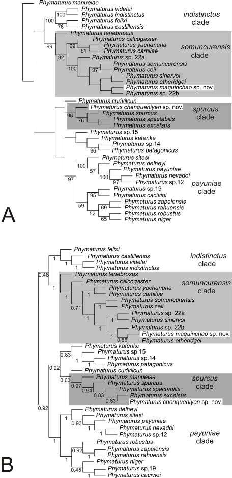
Phylogenetic analyses included the addition of DNA sequences (five markers) for the two new species, as well as for Phymaturus curivilcun and P. katenke, and new morphological information collected for the two clades. Figure 1A shows the tree obtained in a total evidence analysis that included morphology and molecules (parsimony) and Figure 1B depicts the Bayesian tree built based on all the molecular information available. The phylogenetic analyses found that each of the unnamed populations studied (Maquinchao and Chenqueniyén) belongs to two different clades. Phymaturus sp. nov. from Maqui nchao is found nested within the somuncurensis clade in the two analyses performed (total evidence and Bayesian). In the analysis of total evidence (Fig. 1A), Phymaturus tenebrosus is the sister taxon of all remaining species of the somuncurensis clade: then, towards the terminal branches, there are two sister subclades, the calcogaster subclade (formed by P. calcogaster, P. yachanana and P. camilae) and the ceii subclade (including the remaining species of the clade somuncurensis). Within the calcogaster sub clade, P. camilae is the sister taxon of P. yachanana, within the ceii subclade, P. ceii is the sister taxon of P. somuncurensis and P. sp. nov. from Maquinchao is more closely related to P. etheridgei and P. sinervoi.
In the Bayesian analysis (Fig. 1B), Phymaturus tenebrosus is the sister taxon of the remaining spe cies of the clade somuncurensis; then, towards the terminal branches, P. calcogaster is more basal (the calcogaster subclade is not recovered), P. yachanana is the sister taxon of P. camilae; within the ceii sub clade, P. ceii is a sister taxon to P. somuncurensis, P. sp. nov from Maquinchao is more closely related to P. etheridgei, and P. sinervoi is the sister taxon of P sp22a.
Phymaturus sp. nov. from Chenqueniyén is recovered into the spurcus clade. In the total evi dence analysis, P. curivilcun is the sister taxon of all
remaining species; then P. sp. nov. is sister taxon of all remaining species (96% Symmetric Resampling), then towards the terminal branches, P. spurcus is re lated to the pair of species formed by P. excelsus and P. spectabilis (76% SR). Phymaturus manuelae is not included in this clade. There is no strong statistical
Figure 1. Phylogenetic tree showing phylogenetic relationships within the patagonicus after the update of Lobo et al. (2018) study including more DNA sequences, morphological data and taxa (P. curivilcun, P. katenke, P. chenqueniyen sp. nov. and P. maquinchao sp. nov.). A- Total evidence analysis of parsi mony including all DNA data plus morphological information (running TNT). B- Bayesian tree obtained only for molecules (running MrBayes). The new species described in the present contribution and the somuncurensis and spurcus clades to which they belong are highlighted. Numbers under branch are support values (A: symmetric resampling, B: posterior probabilities).
202
support for the position of P. curivilcun.
In the Bayesian tree (Fig. 1B), P. curivilcun is weakly-supported as sister taxon of all other mem bers of the spurcus clade (0.63 pp); then P. manuelae is sister taxon of the remaining species (0.97 pp), P. spurcus is sister taxon of the other three species (0.94 pp), and P. spectabilis is sister taxon (0.83 pp) of the pair of species formed by P. sp. nov. (from Chenqueniyén) and P. excelsus (0.83 pp).
Because we extended the information available for P. katenke (only known for COI data in Corbalán et al., 2016) by sequencing Cytb, 12S, ND4, C-mos and revisiting all morphological characters (based on new samples), we checked its phylogenetic posi tion. In the two analyses, P. katenke was recovered related to P. sp15, P. sp14 and P. patagonicus. The Bayesian tree showed P. katenke as the sister taxon of the other three species (0.83 pp); in the total evidence analysis P. katenke was recovered as closely related to P. sp14-P. patagonicus with low support. In the case of P. curivilcun, we included DNA information of the species that was unknown until the present study, and we revisited its morphology based on the type series and a new sample. In the two analyses, P. curivilcun was recovered as sister of the wellsupported spurcus clade.
The somuncurensis clade: morphological compari sons among its members
Within the somuncurensis clade, several characters related to pattern and colors are quite informative for taxonomic purposes and carry phylogenetic information, as proved in previous articles (Lobo et al., 2012a; 2016; 2018). In those phylogenetic articles, 77 characters referred to pattern and colors were described for the entire genus. Here, we highlight only the main and more significant ones for this clade. Species of this clade can exhibit dorsal ocelli but less conspicuous than in the spurcus clade (see Lobo et al., 2018: Figure 5A and C), sometimes more often in females than in males. The number of ocelli in this group counted between shoulders and thighs is larger than in the spurcus clade (character 130). Phymaturus somuncurensis, P. tenebrosus, P. ceii and P. sinervoi share a black coloration along the flank (dark lateral band -character 120-, Lobo and Quinteros, 2005a), which is inconspicuous or absent in the remaining species. This character state also occurs in the species members of the payuniae clade (P. payuniae, P. nevadoi, P. sitesi, P. delheyi, P.
Cuad. herpetol. 36 (2): 197-231 (2022)
robustus, P. rahuensis, P. zapalensis, P. cacivioi, P. ni ger). Phymaturus camilae, P. sp. (from Maquinchao) and P. calcogaster show a pattern on dorsum of oc cipital region of head formed by black transversal bars (“head star pattern”) (character 296), which is shared with species of the indistinctus clade and with P. katenke. Dark pigmentation of infradigital lamel lae concentrated between central keels (remarked as a dark central line) (described in Lobo et al., 2010) is observed only within the clade in P. ceii, P. sp. (from Maquinchao) and P. tenebrosus. This character state is highly homoplastic, occurring in species of all the other clades within the patagonicus group. A mixed dorsal pattern consisting of small and medium-sized white spots (character 270) is present in P. sp. (from Maquinchao), P. etheridgei, the calcogaster subclade (P. calcogaster, P. yachanana and P. camilae), and within other clades in P. patagonicus, P. katenke and the northern subclade of the payuniae clade. A dorsal tail pattern of males (character 118) is also very informative within the somuncurensis clade; the calcogaster subclade exhibits ocellated/variegated tails, whereas the tail pattern of the remaining spe cies is markedly ringed or with almost inconspicuous ringing. Phymaturus tenebrosus, the sister species of all other members of the clade, lacks a dorsal tail pattern. Variation in the throat pattern was recorded. This pattern can consist of lines densely disposed but disrupted in P. sinervoi, P. etheridgei, P. yachanana and P. sp (from Maquinchao). This throat pattern may be scarce but formed by thick lines interrupted, as in P. camilae and P. calcogaster., as in P. camilae and P. calcogaster. In the latter species, the throat pattern can be absent in some individuals, as in P. tenebrosus, P. somuncurensis and P. ceii (see Figures in Lobo et al., 2020). Belly coloration of males is quite informative (character 127). It is yellow in P. calcogaster and P. ceii, orange in P. sinervoi, P. ether idgei, P. yachanana, and P. camilae, and mustard or red in P. tenebrosus. In P. sp. (from Maquinchao) most males have orange bellies, but an individual with yellow coloration was found. Other characters not referred to color pattern are also useful, i.e. opening of nares with a wide superficial platform inside (character 247); this character state is pres ent in almost all species, except for P. tenebrosus, P. somuncurensis and P. sp. (from Maquinchao). Those miscellaneous characters described above plus all anatomical characters were already included in character lists in Lobo et al. (2012a; 2016; 2018).
Several continuous characters exhibit signifi
203
F. Lobo et al. - Systematics of Phymaturus
cant variation among species. Table 1 shows char acters that exhibited significant variation among species of the two clades (in S1 we provide the raw results given by the analyses). Characters that exhibit significant variation among species were 17 of the 32 studied scale counts and 9 of 18 measurements taken. Table 2 shows characters that exhibited significant differences between all pairs of species within the somuncurensis clade after our statistical comparisons (scale counts and morphometric characters are in dicated below and above the diagonal, respectively). We did not find differences in scale counts between P. sinervoi and P. ceii. Phymaturus somuncurensis, P. ceii and P. sinervoi differed in only one measure ment character (interorbital distance –IO-). Body measurements did not show significant differences among P. somuncurensis, P. ceii, P. sinervoi, P. sp. (from Maquinchao), and P. camilae, but the latter two differ in scale count characters. Phymaturus ceii and P. somuncurensis are closely related (Fig. 1), which may explain the lack of greater morphological differentiation. Their morphological discrimination is based on a few characters, mostly of coloration patterns. Of the total pairwise comparisons (scale counts), characters that are discriminant in most comparisons were: scale organs counted on postros tral scales (SO= 15 times), scales along lateral neck fold till the antehumeral fold (AF= 15), neck scales counted along lateral neck fold (NS= 14), and ventral scales (VS= 13). Of the total pairwise comparisons (measurements), characters that were found as more discriminant in most comparisons were: abdominal width (AW= 13), internasal distance (IN= 10), inter orbital distance (IO= 11), and humerus length (HU= 9). The PCA allows us to explain the morphological variation in the somuncurensis clade based on four components. The S3 file shows the variables with the highest value (of the list of variables used in the analysis, the limit value selected to consider the characters that most contribute was the one calcu lated up to at least 80% of the maximum value found among the variables). All characters that exhibited significant differences among species are the same as those that exhibit the highest values in the PCA procedure.
Based on the significant amount of evidence provided by our morphological revision (color pattern, scale counts, and morphometry) and the genetic information available (Table 3), we are able to describe the population called P. sp. (from Maquinchao) so far in this article as a new species.
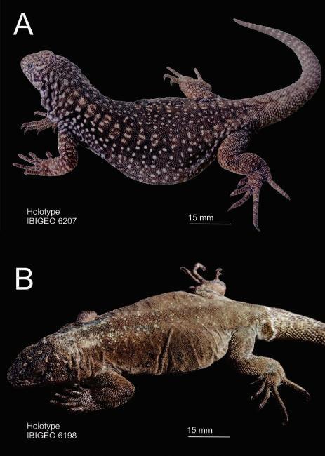
Figure 2. A- Holotype of Phymaturus maquinchao sp. nov. showing its typical color pattern. IBIGEO 6207 (male). Snoutvent length: 89.09 mm. B- Holotype of Phymaturus chenqueniyen sp. nov. IBIGEO 6198 (male). Snout-vent length: 90.00 mm.
Species description
Phymaturus maquinchao sp. nov. Holotype.- IBIGEO 6207. Male (Fig. 2A). Deposi ted at the Reptile collection of the Instituto de Bio y Geociencias del Noa (IBIGEO), Salta, Argentina. Provincial Route (PR) N° 23, approx. 10 km N from Maquinchao (41° 11.300' S, 68° 38.386' W; altitude: 876 m.) Veinticinco de Mayo department, Río Negro province, Argentina.
Paratypes.- IBIGEO 6203, 6205-06, 6208, 6210, 6213, 6215 (4 adult males, 3 juvenile males) and IBIGEO 6209, 6211-12, 6214, 6216 (4 adult females, 1 juvenile female). Deposited at the Reptile collection of the Instituto de Bio y Geociencias del Noa (IBIGEO), Salta, Argentina. Provincial Route (PR) N° 23, approx. 10 km N from Maquinchao (41° 11.300' S, 68° 38.386' W; altitude: 876 m.) Veinticinco de Mayo department, Río Negro province, Argenti na. DNA samples: IBIGEO 6211, 6214.
Diagnosis (Figs. 2, 3 & 4. Table 2, S1).- Phyma turus maquinchao sp. nov. belongs to the patagonicus group of Phymaturus because it exhibits flat and
204
imbricated superciliary scales, smooth tail scales, and a set of enlarged scales projected onto the audi tory meatus (Etheridge, 1995; Lobo and Quinteros, 1995). Within the patagonicus group, Phymaturus maquinchao sp. nov. belongs to the somuncurensis clade, supported by four morphological and 62 molecular sinapomorphies. The somuncurensis clade comprises nine species: P. calcogaster, P. camilae, P. ceii, P. etheridgei, P. maquinchao sp. nov., P. sinervoi, P. somuncurensis, P. tenebrosus and P. yachanana, and was found as a monophyletic group (Fig. 1; see also Lobo et al., 2018 Figure 1).
Phymaturus maquinchao sp. nov. is discrimi nated from the most closely related members of the somuncurensis clade (Fig. 1), P. ceii, P. somuncurensis, P. sinervoi and P. etheridgei, as follows: Phymatu rus ceii shows more conspicuous black coloration along its flank; males are ventrally yellow (most males of P. maquinchao sp. nov. are orange); there are completely melanic individuals (no melanism in P. maquinchao sp. nov.). The throat pattern in P. ceii is absent or inconspicuous (thin conspicuous pattern in males and females of P. maquinchao sp.
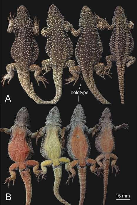
Cuad. herpetol. 36 (2): 197-231 (2022)
nov.). Phymaturus maquinchao sp. nov. also shows statistically significant differences (with overlapping ranges) from P. ceii in the following characters: it has fewer scales along lateral neck fold counted up to the antehumeral fold (AF), more enlarged scales in the anterior margin of the auditory meatus (EM) and shorter forelimbs (HU). Phymaturus somuncurensis exhibits a quite homogenous dorsal pattern; no ocelli are evident, as in P. maquinchao sp. nov Phymaturus somuncurensis shows black coloration along its flank more conspicuous than in P. maquinchao sp. nov. (inconspicuous or absent). Phymaturus somuncu rensis lacks throat reticulation, which is conspicu ous in P. maquinchao sp. nov. Males and females of P. somuncurensis show pink ventral coloration, but in males of P. maquinchao sp. nov. it is orange or yellow (Fig. 3). Phymaturus maquinchao sp nov. exhibits significant differences from P. somuncu rensis in two squamation characters: more scales contacting mental scale (CM) and enlarged scales on the anterior margin of the auditory meatus (EM), and in two morphometric characters: smaller eyes (EL) and shorter head (HL). No differences in scale counts were found between these species (Table 2). Phymaturus sinervoi lacks sexual dimorphism in its color pattern; it also lacks dorsal ocelli, which are present in all females of P. maquinchao sp. nov., and even in males, but are no so evident. Phymaturus sinervoi shows more conspicuous black coloration along its flank than P. maquinchao sp. nov., in which it is inconspicuous or absent. The anterior gular fold is almost conspicuous in P. sinervoi (absent in P. maquinchao sp. nov.). Enlarged scales on the anterior margin of auditory meatus are directed backwardly in P. sinervoi, but they are perpendicular to temporals in P. maquinchao sp. nov. There are two differences in scale counts: P. maquinchao sp. nov. exhibits more scales contacting mental scale (CM) and more enlarged scales on the anterior margin of the auditory meatus (EM); there are no differences in morphometry between these species (Table 2). Males of P. sinervoi and P. maquinchao sp. nov. can exhibit orange or yellow ventral color; in P. etheridgei, ven tral coloration is orange, and it can exhibit yellow mustard only in ventral surfaces of thighs. Contrary to P. maquinchao sp. nov., P. etheridgei has a dorsal black to dark brown coloration with very small white spots, contrasting the light brown coloration of tails; no dorsal ocelli are present in this species. Ventral coloration of P. etheridgei males is orange, turning to bright mustard/yellowish on ventral surfaces of
Figure 3. A- Male individuals of the type series of Phymaturus maquinchao sp. nov. Dorsal and ventral views.
205
F. Lobo et al. - Systematics of Phymaturus
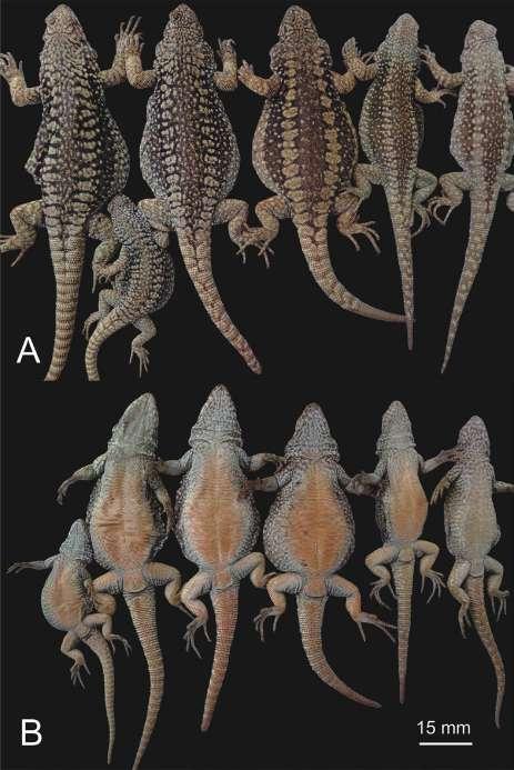
thighs and cloaca; in females, ventral coloration is pink, whereas in P. maquinchao sp. nov., both males and females exhibit orange ventral coloration (brighter in males), and some males have yellow coloration (Fig. 3). Phymaturus etheridgei presents the white dorsal spotting in two sizes: small to very small spots irregularly scattered and densely cover ing their backs; this condition of fore and hind limbs found in P. etheridgei is shared with P. yachanana P. maquinchao sp. nov. shows a similar condition of white spotting but scarcely distributed and not ex tended onto the limbs. Phymaturus maquinchao sp. nov. also differs from P. etheridgei in six scale count characters: it has more postrostral scales (PR), scales contacting nasal (CN), scales contacting mental scale (CM), scales in the subocular row (SR) and fewer scales along lateral neck fold up to the antehumeral fold (AF), and ventral scales (VS). They also differ in four morphometric characters: P maquinchao sp. nov. has smaller eyes (EL), shorter head (HL), lower internasal distance (IN), and greater abdominal width (AW).
Phymaturus maquinchao sp. nov. is discrimi nated from all other members of the somuncurensis clade as follows: Phymaturus maquinchao sp. nov. exhibits a quite different color pattern from that of P. tenebrosus; the latter species exhibits brown or red morphs, or even black individuals (Cerro Alto) with thin and homogeneously distributed white spotting. Brown morphs of P. tenebrosus can exhibit black coloration in flanks, as P. ceii, P. sinervoi and P. so muncurensis. Phymaturus tenebrosus never exhibits dorsal ocelli. Phymaturus maquinchao sp. nov. also differs from P. tenebrosus in having a higher number of: scales contacting nasal (CN), scales contacting mental (CM), dorsal scales along the dorsal midline of trunk (counted along a head length distance) (DT), enlarged scales on the anterior margin of the auditory meatus (EM), more scales counted along midline over dorsum of head (Hellmich’s index) (He), internasal scales (Is), lorilabial scales (LS), neck scales (NS), postrostral scales (PR), the superciliary scale juxtaposed on both ends tends to be the sixth rather than the fifth (Sc), and ventral scales (VS), and fewer scale organs on postrostral scales (SO). P. maquinchao shows a longer neck (NL), greater fore limb width (Hu) and abdominal width (AW). Dorsal white spots of P. calcogaster are large (formed by 9 to 16 scales, see fig. 2 in Lobo et al., 2018), contrary to P. maquinchao sp. nov. (no more than six or seven scales). The throat pattern consists of a dark thick
reticulation (especially in males) in P. calcogaster (thin reticulation in P. maquinchao sp. nov.), dorsal pattern of head reticulated in P. calcogaster, incon spicuous in P. maquinchao sp. nov. Males have yellow chests and abdomen in P. calcogaster (orange in 3 of 4 individuals in P. maquinchao sp. nov.). Dorsal ocelli are absent in P. calcogaster. In P. calcogaster, transversal rows of white spots are evident (absent in P. maquinchao sp. nov.). Phymaturus maquinchao sp. nov. also differs from P. calcogaster in four scale count characters: more postrostral scales (PR) and scales contacting nasal (CN), fewer scales organs in postrostrals (SO), and fewer scales along lateral neck fold up to the antehumeral fold (AF). No morpho metric characters exhibiting significant differences between these species were found. Dark brown to black dorsal pattern of trunk and limbs in P. camilae, differs from brown to light brown in P. maquinchao sp. nov. Dorsal pattern of head marked in P. camilae (light and almost inconspicuous in P. maquinchao sp. nov.), white scales on dorsum of tail contrasting from the brown background coloration in P. camilae
Figure 4. Female paratypes of Phymaturus maquinchao sp. nov. Dorsal and ventral views.
206
Cuad. herpetol. 36 (2): 197-231 (2022)
Table 1. Characters of squamation (scale counts) and measurements that exhibit significant differences among members of the somun curensis and spurcus clades after performing Kruskal Wallis tests. We provide the raw data for all species studied in Supplementary files1 and 2. Abbreviations of scale counts are as follow: Scales along lateral neck fold up to the antehumeral fold (AF); contacting nasal (CN); contacting mental (CM); dorsal scales along the dorsal midline of trunk (counted along a head length distance) (DT); enlarged scales at the anterior margin of the auditory meatus (EM); subdigital plates of fourth finger (FF); gular scales, between both auditory openings (GS); number of scales counted along midline over dorsum of head (Hellmich´s index) (He); infralabial scales (IS); internasal scales (Is); lorilabial scales in contact with subocular (LO); lorilabial scales (LS); number of scales counted around midbody (MS); neck scales counted along lateral neck fold (NS); postmental scales (PM); postocular scales (PO); postrostral scales (PR); scales between frontals and rostral scale (RF); superciliary scale juxtaposed to the others in both endings (Sc); average of scale organs counted on postrostral scales (SO); subocular row (SR); supralabial scales (SS); temporal scales counted in a vertical line between labial com missure and the superciliaries level (TV); upper ciliary scales (UC); ventral scales (VS). Measurements: abdominal width(AW); fourth toe´s claw length(CL); eye length (EL measured between anterior and posterior commissural angles formed by ciliary scales); height head (HH); head length (HL); humerus length (HU); humerus diameter (Hu); head width (HW); internasal distance (IN measured between both medial borders of nasal openings); interorbital distance (IO); auditory meatus height (MH); neck length (NL); trunk length (TL) and tail length (Tl).
somuncurensis clade spurcus clade
Character H P
F P
AF H=51.89 <0.0001 H=40.31 <0.0001
CM H=34.86 <0.0001
CN H=21.21 0.0027 H=13.07 0.0052
DT H=30.35 0.0002
EM H=22.98 0.0023
FF H=17.20 0.0208 H=16.81 0.0017
GS H=15.55 0.0036
He H=38.80 <0.0001 H=27.98 <0.0001
IS H=23.58 0.0006
Is H=23.74 <0.0001 H=10.75 0.0043
LO H=20.63 0.0001
LS H=34.21 <0.0001 H=13.22 0.0075
MS H=16.20 0.0027
NS H=46.86 <0.0001 H=29.01 <0.0001
PM H=17.32 0.0160 H=14.77 0.0015
PO H=15.46 0.0002
PR H=48.88 <0.0001 H=19.24 0.0001
RF H=21.43 0.0001
Sc H=11.52 0.0370
SO H=68.92 <0.0001 H=26.45 <0.0001
SR H=27.25 <0.0001
SS H=16.12 0.0006
TV H=14.51 0.0040
UC H=16.38 0.0018
VS H=49.03 <0.0001
AW H=44.74 <0.0001 H=15.87 0.0032
CL H=13.50 0.0091
EL H=22.41 0.0042 H=24.58 0.0001
HH H=18.50 0.0178
HL H=26.79 0.0008
HU H=31.98 0.0001
Hu H=24.41 0.0001
207
HW
IN H=39.92 <0.0001
IO H=25.21 0.0014
H=9.76 0.0448
MH H=14.32 0.0063
NL H=23.68 0.0006 H=11.41 0.0223
TL H=15.81 0.0451
Tl H=32.84 <0.0001
(inconspicuous in P. maquinchao sp. nov.). Phymatu rus maquinchao sp. nov. also differs from P. camilae in seven scale count characters: it has more scales contacting mental scale (CM), scales contacting na sal (CN), enlarged scales on the anterior margin of the auditory meatus (EM), postmental scales (PM) and postrostral scales (PR), fewer scales along lateral neck fold up to the antehumeral fold (AF) and scale organs on postrostral scales (SO). No morphometric characters exhibiting significant differences between these species were found. Phymaturus maquinchao sp. nov. has no reddish/clay coloration on dorsum, which is commonly found in P. calcogaster and P. yachanana. In P. yachanana dorsum of females has black transversal bars forming a longitudinal paired series, no ocelli are evident (they can be present in some females of P. maquinchao sp. nov. but as margins of light ocelli). Chest and abdomen of most individuals with light gray spotting in P. yachanana (immaculate in P. maquinchao sp. nov.). Phymaturus maquinchao sp. nov. also differs from P. yachanana in one scale count character: it has more neck scales (NS), ventral scales (VS) and the superciliary scale juxtaposed on both ends tends to be the sixth rather than the fifth (Sc). They differ in two morphometric characters: P. maquinchao sp. nov. smaller eye (EL) and greater head height (HH).
Description of holotype (Fig. 2A).- Male. SVL 89.09 mm. Head length: 15.42 mm. Head width: 14.98 mm. Head height (at parietal): 8.15 mm. Axilla-groin length: 47.93 mm (53.80 % of SVL). Tail length (complete, not regenerated): 97.88 mm.. Body moderately wide, trunk width: 37.74 mm (42.4 % of SVL). Twenty-two smooth dorsal head scales. Three scale organs in each of the four postrostrals. Nasal bordered by nine scales, not in contact with rostral. Canthal separated from nasal by two scales. Loreal region flat. Eight enlarged supralabial scales, none contacting subocular. Eight enlarged infralabials. Auditory meatus oval (height: 4.2 mm; width: 2.4 mm) with six enlarged, flat and smooth perpendicu
lar scales projecting on the anterior margin. Auricu lar scale absent. Ten convex, juxtaposed temporals. Auditory meatus - ciliary scales distance: 5.2 mm. Rostral undivided. Mental scale sub-pentagonal, in contact with six scales. Interparietal scale bordered by eight scales, larger than postparietals. Frontal region without an azygous scale. Supraorbital semi circles inconspicuous. No distinctly enlarged supra oculars. Nine juxtaposed superciliaries, 15 upper ciliaries and 11 lower ciliaries. Subocular unique (not fragmented). Eleven lorilabials, the 11th contacting subocular. Preocular smaller than canthal, these two scales separated by another one. Preocular separated from lorilabial row by two scales. Scales of throat round, small, and juxtaposed. Seventy-nine gulars between auditory meata. Lateral nuchal folds well developed, with granular scales on longitudinal fold. Antehumeral pocket well developed. Seventy-seven scales between auditory meatus and shoulder. Sixty scales between antehumeral fold and shoulder. In ventral view, anterior gular fold absent, posterior gular fold present with its anterior margins with two enlarged scales on their borders. Dorsal scales round, smooth and juxtaposed. Thirty-eight dor sal scales along midline of the trunk in a length equivalent to head length. Scales around midbody: 209. Ventral scales larger than dorsal scales. Ventral scales between mental and precloacal pores: 185. Seven precloacal pores in an undivided row without supernumerary pores. Four moderately enlarged postcloacal scales. Brachial and antebrachial scales smooth, with round posterior margins. Supracarpals laminar, round and smooth. Subdigital lamellae of fingers have three keels. Subdigital lamellae of finger (left manus) IV: 22. Supradigital lamellae convex, imbricate. Infracarpals and infratarsals have round margins and 2–3 keels. Supracarpals and supratarsals smooth, with rounded posterior margins. Subdigital lamellae of toe (left pes) IV: 27. Claws moderately long (fourth toe’s claw: 1.9 mm).
Coloration (in life).-The holotype exhibits a
F. Lobo et al. - Systematics of Phymaturus
208
Cuad. herpetol. 36 (2): 197-231 (2022)
Table 2. Continuous characters that exhibit significant variation between all pairs of species within the somuncurensis clade (17 of 32 scale counts studied and 9 of 18 measurements studied). Below the diagonal are scale counts characters, and above measurement characters. Same abbreviations of Table 1.
P. maquinchao
P.
calcogaster
P. camilae
P. ceii
P. etheridgei
CN
SO
AF CM
EM PM PR SO
ceii
etheridgei
sinervoi
somuncurensis
tenebrosus
yachanana
PM
EM EM IS PR SO Is PM PR SO
CM CN PR SR VS He IS SR PM VS
sinervoi CM EM EM SO SO
P. somuncurensis CM EM EM VS
P. tenebrosus
CM CN DT EM He Is LS NS PR Sc SO VS
AF CM DT EM He IS Is LS NS PR SO VS
HU
IN
AF DT He NS PM
SR VS
AW EL HL IN
SR
FF He SR
TL
HU
HU Is
NL TL
HU NL
TL
HH IN
HL IN
AF CM DT FF He Is LS NS PR SO VS
brown color as dorsal background, with a pair of longitudinal rows of ocelli (14 between shoulders and thighs). On the anterior ocelli (anterior half of trunk) transversal rows of white scales are conspicu ous. Upper half of flanks darker, almost black, with irregularly scattered white spots. The dorsal body pattern consists of white spots of two sizes is more evident on the vertebral band between the rows of ocelli (also commonly found in other species of the clade). The “star pattern” is conspicuous on the back of neck and nuchal region. Tail and limbs exhibit a light brown coloration. There is no evident pattern on the tail. In ventral view, throat, anterior half of chest and forearms are light gray. The throat exhibits thin reticulation. Posterior half of chest, abdomen, cloacal region, hind limbs and tail orange.
AF DT FF He Is LS NS PR SO SR VS
AF DT He Is LS NS PR SO VS
DT He Is IS LS NS PR SO VS
HH IN NL
P. surements only based on adult individuals. SVL 78.9–95.3 mm (mean = 88.09; SD = 5.8). Head length 16.3–18.2% (mean = 17.4%; SD = 0.1) of SVL. Tail length 1.04–1.26 (mean = 1.15; SD = 0.08) times SVL. Scales around midbody 204–231 (mean = 216.6; SD = 9.11). Dorsal head scales (Hellmich’s index) 19–26 (mean = 22.6; SD = 1.7). Ventral scales 161–188 (mean = 176.2; SD = 8.6). Scales surround ing interparietal 6–8 (mean = 7.2; SD = 0.7). Scales surrounding nasal 7–10 (mean = 8.9; SD = 0.8). Number of scale organs on postrostrals 2–5 (mean = 3.3; SD = 0.8). Superciliaries 7–11 (mean = 9.6; SD = 1.2). Subocular never fragmented (a single scale in all individuals). Mental scale in contact with six scales in all the samples. Number of chinshields 6–8 (mean = 6.7; SD = 0.7). Enlarged scales on the border of the posterior gular fold: 2–5 (mean = 3.3; SD = 1.0). Lorilabials 11–14 (mean = 11.8; SD = 1.1). Enlarged scales on the anterior border of the audi
209 Variation.- Squamation based on 13 specimens (8 males and 5 females), including four juvenile individuals (3 males and 1 female); mea
calcogaster
camilae
P. yachanana NS Sc VS AF He NS VS AF CN CM DT NS PM AF FF NS AF CM FF NS SR VS
AF CM NS PM SO VS
AF CM He NS PM VS
CM CN He Is LS PR SO
P.
P.
P.
P.
P.
P.
P.
maquinchao HU AW EL HL IN EL HL AW NL EL HH P.
AF
PR
IO NL AW EL HL IN IO IO HL HU AW HU HH
CN
EM
IO
AW EL
AW IO NL AW HH IO
AF
AW EL IO IO HU AW
TL HU IN TL
AF
PR
IN
EL
IO
AW HH HL IN IO
P.
SO
AW
IO
IO
FF
HH
AW
F. Lobo et al. - Systematics of Phymaturus
Table 3. Estimates of Evolutionary Divergence between Sequences. The number of base differences per site from between sequences are shown. The analysis involved 9 nucleotide sequences. All positions with less than 95% site coverage were eliminated. That is, fewer than 5% alignment gaps, missing data, and ambiguous bases were allowed at any position. There were a total of 838 positions in the final dataset. Evolutionary analyses were conducted in MEGA7. Above: Cytb pairwise comparisons; below: 12S.
somuncurensis clade P. calcogaster P. camilae P. ceii P. etheridgei P. maquinchao P. sinervoi P. somuncurensis P. tenebrosus P. yachanana
P. calcogaster 1.45 2.17 2.90 3.26 2.65 2.90 3.38 2.29
P. camilae 0.36 1.93 2.65 2.53 2.05 2.65 2.90 2.17
P. ceii 1.43 1.55 2.90 3.02 2.17 1.21 2.90 2.29
P. etheridgei 1.07 1.07 1.07 1.57 2.41 3.14 3.38 3.02
P. maquinchao 1.07 1.07 1.07 0.24 2.29 3.26 3.74 3.14
P. sinervoi 1.31 1.31 1.07 0.48 0.48 2.65 3.14 2.53
P. somuncurensis 1.31 1.31 0.36 0.72 0.72 0.72 3.14 2.53
P. tenebrosus 1.19 1.19 1.91 1.31 1.07 1.55 1.55 2.77
P. yachanana 0.95 1.07 1.91 1.55 1.55 1.79 1.79 1.67 spurcus clade P. chenqueni yen P. curivilcun P. excelsus P. manuelae P. spectabilis P. spurcus
P. chenqueniyen 2.32 1.22 2.32 1.46 1.46
P. curivilcun 1.56 1.83 2.20 1.83 1.83
P. excelsus 1.08 1.20 1.59 0.24 0.24
P. manuelae 0.96 1.32 0.84 1.83 1.83
P. spectabilis 0.96 1.08 0.36 0.72 0.24
P. spurcus 0.96 1.08 0.36 0.72 0.00
tory meatus 4–9 (mean = 6.8; SD = 1.7). Scales of neck along longitudinal fold from posterior border of auditory meatus to shoulder 74–88 (mean = 80.2; SD = 4.9). Gulars 68–89 (mean = 77.3; SD = 6.0). Scales between rostral and frontal 9–11 (mean = 9.7; SD = 0.8). Subdigital lamellae on fourth finger 21–26 (mean = 23.2; SD = 1.6). Subdigital lamellae on fourth toe 26–31 (mean = 28.1; SD = 1.6). Males with 7–9 precloacal pores (mean = 8.0; SD = 1.0). One fe male shows precloacal pores (3). Measurements: Eye length 3.4–3.7 (3.5; SD = 0.1). Head length 14,3–15,8 (mean =15.3; SD =0.5). Neck length 10.9–14.8 (mean = 13.4; SD = 1.3). Head width 12.8–15.5 (mean = 14.6; SD = 0.9). Head height 7.6–9.3 (mean = 8.5; SD = 0.6). Internares distance 2.4–2.8 (mean = 2.6; SD = 0.1). Interorbita distance 6.2–7.9 (mean = 7.1; SD = 0.5). Trunk length 37.5–50.6 (mean = 44.9;
SD = 4.2). Humerus length 12.5–15.5 (mean = 13.9; SD = 1.0). Humerus width 5.2–7.5 (mean = 6.5; SD = 0.7). Tibia length 14.9–17.5 (mean = 16.3; SD = 1.1). Foot length 22.8–24.8 (mean = 24.1; SD = 0.7). Tail length 93.9–114.0 (mean = 99.9; SD = 6.6). Eye -auditory meatus distance 4.4–5.3 (mean = 5.0; SD = 0.3). Auditory meatus height 3.3–4.2 (mean = 3.8; SD =0.3). Fourth toe´s claw length 1.7–2.1 (mean = 1.9; SD = 1.1). Abdominal width 31.0–43.6 (mean = 37.2; SD = 3.9).
210 Males exhibit a brown background coloration (Fig. 3); small to medium-sized white spots are widespread all over their backs, dorsum of neck, fore and hind limbs, but fading towards the tail. All males except one show a pair of longitudinal rows of dorsal ocelli that are different from the background coloration because of their lighter
coloration. Poorly conspicuous black transversal lines separate ocelli, and sometimes white small spots form a transversal line inside ocelli. Half of the males exhibit a conspicuous “star head pattern”, forming a reticulum in the posparietal area. There is no evident pattern on limbs, except for the white regular spotting. No pattern is evident on the tail, except for one specimen (ringed pattern). The throat is light gray, with slender black lines forming a re ticulum; one specimen has no pattern. Three males have homogeneously orange chest and bellies; one male has yellow ventral coloration. All males exhibit small orange precloacal pores. Poscloacal enlarged scales are more or less conspicuous. Orange or yellow coloration of their ventral surfaces is extended on the cloacal region, tail and thighs. Dorsal pattern of females is similar to that of males (Fig. 4), but ocelli rows are more conspicuous; the upper half of flanks is darker (lateral black band usually present in P somuncurensis, P. ceii, P. sinervoi and P. tenebrosus and in the payuniae clade), the white spotting is continuous on tails, and half of the females exhibit ringed tails. Ventral surfaces are of similar pattern and coloration to those of males.
Etymology.- Maquinchao refers to the locality where this species was found; it is an ancient native language word that means “site of wintering”. Distribution (Fig. 5).-This species inhabits the northwestern margin of Meseta de Somuncurá and is only known for the type locality. Morando et al. (2013) sampled individuals from two sites 35 km south to this place (candidate species P. sp22a and P. sp22b); further studies are needed to evaluate the identity of these populations. The Somuncurá plateau covers a vast territory of about 25,000 km² in Argentine Patagonia. This geological structure is located more than 1000 meters above sea level; it is a formation with several canyons generated by different water streams that drain in the lowlands. It is a basaltic plateau, with reliefs of volcanic cones, mountain ranges, hills that are almost 1900 meters above sea level, interspersed with temporary and clay lagoons. Figure 5 shows how this diversified clade of lizards (somuncurensis clade outlined in orange) inhabits mainly inside this plateau and all around its margins.
The spurcus clade: morphological comparisons among its members
Analysis of continuous characters taken from squa
Cuad. herpetol. 36 (2): 197-231 (2022)
mation and measurements. - Continuous characters exhibit significant variation among species. Table 1 shows characters that exhibited significant variation among species of both clades; scale counts characters are displayed below the diagonal and the morpho metric ones, above this diagonal (we provide the raw results of the analyses in S2). The characters that ex hibit significant variation among species were 18 of the 32 scale counts studied and 8 of the 18 measure ments taken. Table 4 shows characters that exhibited significant differences between all pairs of species within the spurcus clade. We did not find differences in measurements between P. sp (Chenqueniyén) and P. spectabilis (but they differ in 12 scale counts). Of all the pairwise comparisons (scale counts), the characters that are most discriminant and present in most comparisons were: scales between frontals and rostral scale (RF= 6 times), number of scales counted around midbody (MS= 6); scales along lateral neck fold up to the antehumeral fold (AF= 6); lorilabial scales in contact with subocular (LO= 4), and num ber of scale organs in postrostral scales (SO= 6). Of all the pairwise comparisons (measurements), the characters that were found as most discriminant and present in most comparisons were: tail length (Tl= 7), fourth toe´s claw length (CL= 6), abdomi nal width (AW= 5), auditory meatus height (MH= 5) and humerus width (Hu= 4). The PCA allows us to explain the morphological variation in the spur cus clade based on three components that account for 100%. The S3 file shows the variables with the highest value. All characters that showed significant differences among species are the same as those that exhibit higher values in the PCA. Figure 6 shows one of the PCA graphics obtained (squamation); in this case, PC1xPC2, PC2 discriminates P. spurcus, P. spectabilis and P. excelsus from each other, whereas PC1 discriminates P. manuelae and P. chenqueniyen from the other three. Tables with individual values of characters and for each component and accumulated percentages are included in S3.
Identification of brown morphs in the spurcus clade (Fig. 7).- Since coloration pattern was an es sential topic in a recent publication about this group of Phymaturus, we considered it important to revisit it and make the necessary remarks and observations to avoid confusion. In Lobo and Quinteros (2005a) we described P. excelsus and P. spectabilis from a restricted area of Río Negro province in Argentina. At that time, we considered certain brown morphs as a variation within P. spectabilis species. In the
211
Figure 5. Map of distribution of the somuncurensis (orange), spurcus (light blue), and P. katenke and related species (green) in Rio Negro and Chubut provinces of Argentina (Somuncura plateau is indicated, one of the most massive geomorphological formations in Patagonia). Numbers indicate: 1-P. somuncurensis, 2- P. yachanana, 3- P. calcogaster, 4- P. camilae, 5- P. katenke, 6- P. curivilcun, 7- P. etheridgei, 8- P. sinervoi, 9- P. ceii, 10- P. maquinchao sp. nov., 11- P. patagonicus, 12- P. tenebrosus, 13- P. manuelae, 14- P. sp14, 15- P. sp15, 16-P. chenqueniyen sp. nov., 17- P. spurcus, 18- P. spectabilis, 19- P. excelsus, 20- P. sp22a and P. sp22b. “P. sp.” are Morando et al. (2013) candidate species.
case of P. excelsus, we found only three individuals in Ojo de Agua (its type locality) exhibiting brown morphs; since these brown morphs in particular resemble P spurcus (Huanualuan), a species we resurrected in another article (Lobo and Quinteros, 2005b), we assumed that P spurcus could be syntopic to P excelsus. Later, after the description of P. agilis provided by Scolaro et al. (2008), we analyzed (Lobo et al., 2012b) this new species statistically for differ ent continuous characters and because we found a female giving birth both patterns “agilis” (brown morph) and ” spectabilis ” (ocellated morph), we concluded that these two species are synonymous. At that time, we also considered the brown morphs of P. excelsus as being part of that species, rejecting the idea of P spurcus being syntopic with P excelsus We discriminated brown morphs of P. spectabilis from P. excelsus (shown in Lobo et al., 2012b Fig. 2). At that time, we considered the brown morphs of the species to be different from each other and
very constant for each species. Figure 7A shows an individual of P. excelsus from the MCN collection, and an individual of “agilis” deposited at the MLP collection (Fig. 7B). As can be seen, the brown pat tern of P. spectabilis (= agilis) shows constancy in dorsal pattern, which is difficult to confuse with the P spurcus one; this “agilis pattern” is a brown dorsal pattern with darker coloration on both sides of the back, named in Lobo et al. (2018) as a kind of “elon gatus pattern” because it resembles the pattern found in members of the Liolaemus elongatus group. This color pattern is not present in any other Phymaturus species (P spurcus, P excelsus or P. sp. nov. from Chenqueniyén described in the present article). All specimens of the type series of P. agilis deposited at MLP, several other individuals deposited at the same museum, and all P. spectabilis specimens with brown pattern deposited at FML collection exhibit the same pattern. Pictures published in the description of P. agilis by Scolaro et al. (2008) are quite illustrative of
F. Lobo et al. - Systematics of Phymaturus
212
Cuad. herpetol. 36 (2): 197-231 (2022)
this pattern. Therefore, because this design of brown patterns is different among P. spurcus, P. excelsus and P. spectabilis, it is a mistake to call all of them simply “spurcus” morphotype based only on the color with out considering the design, as in Becker et al. (2018). The most similar brown morphs are the ones of P. excelsus with P. spurcus. For this reason, the brown excelsus found at that time was assigned to P. spurcus (Lobo and Quinteros, 2005a). But later, another case of intraspecific polymorphism was considered (Lobo et al., 2012b). All the examined individuals (MCNUNSa and FML collections) of brown morphs of P. excelsus exhibit a fading ocellated pattern in a lighter brown shade than the darker background color similar to that of individuals of P. sp. nov. from Chenqueniyén; in P. spurcus this fading ocellated pattern is absent or inconspicuous, but present in most newborns and small juveniles. This pattern of fading ocelli in brown morphs is completely absent in P. spectabilis brown morphs. Interparietal scale is white in both morphs of P. excelsus, in ocellated individuals of P. spectabilis and in a few individuals of P. sp. nov. from Chenqueniyén, but never in P. spurcus or in brown morphs of P. spectabilis. The throats do not exhibit a pattern in most individuals of P. excelsus or P. spurcus; when there is a pattern, in P. excelsus it is a vanished reticulation (light brown almost inconspicuous) but in P. spurcus it consists of small light brown dots. In addition, the abdominal region is yellow in P. spurcus males versus orange in those of P. excelsus and P. spectabilis.
How to distinguish the ocellated patterns of P. spectabilis and P. excelsus (Figs. 7C-D) .-The occurrence of specific brown morphs is not the only feature of color patterns that discriminates P. spectabilis from P. excelsus. Ocelli on dorsum of P. spectabilis and P. excelsus are constant in number. Lobo et al. (2012a, Fig. 6A) used this information for phylogenetic analysis (character 130) and de scribed the variation found within the patagonicus group. Species belonging to the somuncurensis clade, payuniae clade and P. manuelae (Lobo et al., 2018) showed more ocelli along their backs (9-11) counted between hips and shoulders, whereas P. spectabilis and P. excelsus had fewer ocelli, between 6-8 (Lobo et al ., 2012a). Fading pattern of ocelli (almost inconspicuous) on brown morphs of P. excelsus, P. spurcus and in P. sp. nov. (from Chenqueniyén) are 6-8, the same numbers of ocellated morphs of P. spectabilis and P. excelsus. The presence of this character in all species, except for P. manuelae (higher number of ocelli) or P. curivilcun (melanic), is an apomorphy of the subclade of the spurcus clade (with the addition of the occurrence of brown morphs, fixed in all population in P. spurcus and P. sp. (Chenqueniyén), and in part of the population in P. spectabilis and P. excelsus). Even though the number of ocelli on the backs of these two species is similar, they are different in size and coloration. In most P. spectabilis individuals, ocelli are larger, formed by more cream to white scales (Fig. 7D) than those of P. excelsus.There are more white scales irregularly
 Figure 6. PC1 versus PC2 of squamation characters. Phymaturus excelsus discriminated from P. spectabilis (PC2) and P. chenqueniyen sp. nov. from the three P. spurcus, P. spectabilis, and P. excelsus (PC1). Tables with individual values of characters and for each compo nent and accumulated percentages are shown in S3.
213
Figure 7. Typical color pattern of A- Phymaturus excelsus (MCN-UNSa 1385). B- Brown morph of Phymaturus spectabi lis (MLP 5346). C and D ocellated morphs of C- Phymaturus excelsus (MCN-UNSa 1336) and D- Phymaturus spectabilis (MCN-UNSa 1205). Typical banded pattern is present in all samples of brown morphs of P. spectabilis, absent in P. spurcus and P. excelsus. Ocelli are usually larger in P. spectabilis, limb pattern consisting of thicker lines, and small white spots more concentrated on the vertebral area.
scattered on the background black coloration of the back in P. excelsus (Fig. 7C) while in P. spectabilis these white scales are much fewer and concentrated along the vertebral midline. The rostral scale is cream or almost white in P. excelsus, but dark brown to black in P. spectabilis. Chinshields (posmental row) are white and differentiated in color from the rest of the throat in P. excelsus, but not differentiated in P spectabilis. Fore and hind limbs with a reticulated
pattern of black slender bands is typical of P. excel sus, being formed by thick bands and on a wider/ extended cream background in P. spectabilis. Based on the study of a population sampled from Meseta de Chenqueniyén and samples deposited at MVZ collection (considered in previous articles as P. spurcus), as well as on morphological (see Table 4) and genetic differences (Table 3) found in our com parisons, we conclude that these lizards represent an independent lineage (see phylogenetic tree in Figure 1) that deserves a formal description.
Species description
Phymaturus chenqueniyen sp. nov. (Figs. 2B and 8).
Phymaturus spurcus: Lobo and Quinteros (2005a), in part; Lobo et al. (2012a) in part; Lobo et al. (2018), in part.
Phymaturus sp. 13: Morando et al. (2013).
Holotype.- IBIGEO 6198. Male (Fig. 2B). Between Las Bayas village and Las Bayas hill (also named as Alto del Escorial), at the edge of the Chenqueni yén plateau (41° 29.238' S, 70° 41.557' W; 1139 m) Ñorquinco department, Río Negro province, Argen tina. Date 16 January 2020. Deposited at the Reptile collection of the Instituto de Bio y Geociencias del Noa (IBIGEO), Salta, Argentina.
Paratypes.- IBIGEO 6184, 6186, 6200; MVZ 188904-05, 247102-03, 247105 (6 adult males, 2 juvenile males); IBIGEO 6185, 6196-97, 6199; MVZ 247101, 247104, 247106, 188906-07(6 adult females, 3 juvenile females). Deposited at the Reptile collec tion of the Instituto de Bio y Geociencias del Noa (IBIGEO), Salta, Argentina, and Museum of Verte brate Zoology (Berkeley, California, USA). IBIGEO samples collected from Meseta Chenqueniyén, same site of Holotype between Las Bayas and Alto el Es corial or Cerro Las Bayas (41° 29.238' S, 70° 41.557' W; 1139 m). Ñorquinco department, Río Negro province, Argentina. Date 16 January 2020. MVZ samples: MVZ 188904-07. Ñorquinco department, along rimrock, 4 km S and 1 km E Alto del Escorial, elevation: 1100 m. Río Negro province, Argentina. 25 February 1982. MVZ 247101-07. Ñorquinco de partment, along rimrock, 4 km S and 1 km E Alto del Escorial, elevation: 1100 m. Río Negro province, Argentina. 23 November 1986. DNA samples: IBI GEO 6197, 6199.
F. Lobo et al. - Systematics of Phymaturus
214
Cuad. herpetol. 36 (2): 197-231 (2022)
Diagnosis (Fig. 2B and 8; Table 4; S2).- Phymaturus chenqueniyen sp. nov. belongs to the patagonicus group of Phymaturus because it exhibits flat and imbricated superciliary scales, smooth tail scales, and a set of enlarged scales projected onto the audi tory meatus (Etheridge, 1995; Lobo and Quinteros, 1995). Within the patagonicus group, Phymaturus chenqueniyen sp. nov. belongs to the spurcus clade because it shares the following morphological apo morphies (according to the total evidence analysis, excluding P. manuelae): females exhibit ocellated (not ringed) tails, presence of two longitudinal rows of ocelli in both sexes, and scale organs in rostral scale (six changes in DNA positions also support this clade). Since P. manuelae is included in the group (with high support value) in the Bayesian analysis, we also include this species in our comparisons Phymaturus chenqueniyen sp. nov. has dark brown coloration all over its head, body and limbs; half of the males studied exhibit irregularly distributed light brown markings (mostly on shoulders or neck); belly and chests of P. chenqueniyen sp. nov. are dark brown (light gray to white immaculate in all the other members of the spurcus group). It is the only species within the spurcus clade with melanic individuals, but at a low frequency (3/17, 17%). This melanism was reported in P. cacivioi, P. ceii, P. curivilcun, P. niger and P. tenebrosus (Lobo et al., 2021). Phymatu rus curivilcun is sister to the other taxa of the group but without support. Phymaturus chenqueniyen sp. nov. differs from P. manuelae because it lacks any kind of dorsal pattern, its coloration is dark brown with almost inconspicuous light brown ocelli (P. manuelae exhibits white small scattered spots, ir
regularly distributed or forming transversal rows, as well as conspicuous white dorsal ocelli along the paravertebral region between thighs and shoulder). The interparietal scale is white in P. chenqueniyen sp. nov. but brown and inconspicuous in P. manuelae Phymaturus chenqueniyen sp. nov. has spinier tail scales than P. manuelae (in this species, tail scales are not different from those of the somuncurensis or payuniae clades). Most of specimens of P. chen queniyen sp. nov. have enlarged (3-5) scales on the posterior gular fold margin (absent in P. manuelae), Phymaturus chenqueniyen sp. nov. has conspicu ous anterior gular fold, whereas it is absent in P. manuelae; P. chenqueniyen sp. nov. has more scales contacting nasal (CN), postocular scales (PO), scales between frontals and rostral scale (RF), and fewer scales counted around midbody (MS). Phymaturus chenqueniyen sp. nov. differs from P. manuelae in five body measurements: slender trunk (AW), lower auditory meatus height (MH), shorter fourth´s toe claw (CL), neck (NL), and tail (Tl). Phymaturus chenqueniyen sp. nov. differs from P. spurcus in that it exhibits a darker brown dorsal coloration; belly and chests are dark brown in P. chenqueniyen sp. nov. and light gray /white immaculate in P. spurcus The collected males or females of P. chenqueniyen sp. nov. did not exhibit any abdominal coloration typical of the patagonicus group (males and some females of P. spurcus have yellow abdomen). The interparietal scale is white in P. chenqueniyen sp. nov. but brown and inconspicuous in P. spurcus. Enlarged scales at the anterior border of the auditory meatus are perpendicular but in P. spurcus they extend backward, covering part of the auditory opening;
215
P. chenqueniyen P. excelsus P. manuelae P. spectabilis P. spurcus P. chenqueniyen CL EL Hu HW AW CL MH NL Tl AW Tl P. excelsus GS NS PM PR SO SS TV AW EL Hu HW MH NL Tl CL EL Hu HW MH AW CL EL Tl P. manuelae CN MS PO RF AF FF GS LO MS PM PR SO TV CL MH NL Tl CL Hu MH Tl P. spectabilis AF He Is LO LS NS PO PR RF SO SS UC AF FF GS He PM RF TV UC AF CN He Is Ls LO MS NS PR RF SO UC AW Tl P. spurcus FF MS PO PR SO SS AF FF GS MS NS PM TV RF SO AF He LO LS MS NS RF UC Table 4. Continuous characters that exhibit significant variation between all pairs of species within the spurcus clade (18 of 32 scale counts studied and 8 of 18 measurements studied). Below the diagonal are scale counts significant characters, and above measurement significant characters. Same abbreviations of Table 1.
most of P. chenqueniyen sp. nov. individuals have a dorsal fading tail pattern (absent in P. spurcus); 8/17 individuals exhibit a fading ocellated pattern in a lighter brown tone with respect to the darker color of background (absent or inconspicuous in adult P. spurcus, but present in most newborns and small juveniles); dorsal scales of tail more spiny and ending in dark brown spine (less spiny and with less developed light brown spine in P. spurcus); all ven
tral surface of fingers and toes dark brown to black (in P. spurcus a central longitudinal row is darker than the rest of finger or toes, see Lobo et al. (2010, Fig. 8D). Phymaturus chenqueniyen sp. nov. shows significant differences from P. spurcus in the follow ing continuous characters (eight characters): fewer scales counted around midbody (MS), supralabial scales (SS), postrostral scales (PR) and more scale organs over postrostral scales (SO), postocular scales
 F. Lobo et al. - Systematics of Phymaturus
Figure 8. Colors in life of Phymaturus chenqueniyen sp. nov. (individuals photographed on rocks of their typical environment). ADorsal view of a male (IBIGEO 6200); B- ventral view of the same individual; C- Dorsal view of a female (IBIGEO 6185); D- ventral view of the same individual; E- Dorsal view of a melanic female (IBIGEO 6196); F- ventral view of the same individual.
216
(PO), and subdigital plates of fourth finger (FF) and morphometric: slender trunk (AW) and longer tail (Tl). Phymaturus chenqueniyen sp. nov. differs from P. spectabilis in that all P. chenqueniyen sp. nov. are dark brown; they may exhibit some lighter color ation on the shoulders or neck and a fading (almost inconspicuous) pattern of ocelli, whereas the brown morphs of P. spectabilis show a brown dorsal pattern, speckled with lighter brown or some almost white scales scattered irregularly, sometimes concentrated on sides of dorsum of the trunk (Fig. 6B). Belly and chests are dark brown in P. chenqueniyen sp. nov., and light gray/white immaculate in P. spectabilis. Ocel lated morphs of P. spectabilis have black background coloration of the back, with large cream almost white ocelli. The interparietal scale is white in P. chenqueniyen sp. nov. but brown in brown morphs of P. spectabilis. Phymaturus chenqueniyen sp. nov. did not exhibit any abdominal coloration that is typical in the patagonicus group (males and females of P. spectabilis show abdominal orange color). Phyma turus chenqueniyen sp. nov. shows significant differ ences from P. spectabilis in the following continuous characters (12 characters): fewer scales on dorsum of head (He), postrostral scales (PR), supralabials (SS), upper ciliary scales (UC), scales between rostral and frontal (RF), lorilabial scales in contact with subocu lar (LO), lorilabial scales (LS) internasal scales (Is) more scales along lateral neck fold till the antehu meral fold (AF), neck scales (NS), postocular scales (PO) and scale organs counted on postrostral scales (SO). No morphometric differences between these two species were found. Phymaturus chenqueniyen sp. nov. differs from P. excelsus in the homogeneous dark brown coloration on the head, body and limbs, sometimes with lighter coloration on shoulders and neck, whereas P. excelsus brown morphs have lighter background coloration; the interparietal scale is white in P. chenqueniyen sp. nov. but brown and inconspicuous in brown morphs of P. excelsus. Belly and chests are dark brown in P. chenqueniyen sp. nov. and light gray/white immaculate in P. excelsus; the collected males and females of P. chenqueniyen sp. nov. did not exhibit any abdominal coloration (males and females of P. excelsus show abdominal orange color). Phymaturus chenqueniyen sp. nov. shows sig nificant differences from P. excelsus in the following continuous characters (11 characters): fewer neck scales (NS), postrostral scales (PR), supralabial scales (SS), postmental scales (PM), more scale organs in postrostrals (SO), gulars (GS), and temporal scales
Cuad. herpetol. 36 (2): 197-231 (2022)
counted in a vertical line between labial commissure and the superciliaries level (TV). Morphometric: shorter fourth´s toe claw (CL), smaller eyes (EL), wider head (HW), and wider forelimbs (Hu). Description of holotype (Fig. 2B).- Male. SVL 89.09 mm. Head length: 16.88 mm. Head width: 16.07 mm. Head height at parietal level 8.46 mm. Axilla-groin length 45.01 mm (44.4 % of SVL). Tail length (complete, not regenerated) 100.22 mm. Body moderately wide, trunk width 34.17 mm (38.3 % of SVL). Nineteen smooth dorsal head scales. Five, two and three scale organs in three postrostrals. Nasal bordered by seven scales, not in contact with rostral. Canthal separated from nasal by two scales. Loreal region flat. Nine enlarged supralabial scales, none contacting subocular. Eight enlarged infralabials. Auditory meatus oval (height 3.8 mm; width 2.7 mm) with six enlarged, flat and smooth perpen dicular projecting scales on the anterior margin. Auricular scale absent. Nine convex, juxtaposed temporals. Auditory meatus - ciliary scale distance: 5.5 mm. Rostral scale undivided. Mental scale subpentagonal, in contact with four scales. Interparietal scale bordered by eight scales, larger than postpari etals. Frontal region without an azygous scale. Su praorbital semicircles inconspicuous. No distinctly enlarged supraoculars. Six juxtaposed superciliaries, 14 upper ciliaries, and 12 lower ciliaries. Subocular fragmented in two scales. Ten lorilabials, ninth to tenth contacting subocular. Preocular of the same size as that of canthal, in contact. Preocular contact ing lorilabial row. Scales of throat round, small and juxtaposed. Eighty-three gulars between auditory meata. Lateral nuchal folds well developed, with granular scales on longitudinal fold. Antehumeral pocket well developed. Ninety-three scales between auditory meatus and shoulder. Seventy-four scales between antehumeral fold and shoulder. In ventral view, anterior gular fold absent, posterior gular fold present with its anterior margins with three en larged scales on their borders. Dorsal scales round, smooth and juxtaposed. Forty dorsal scales along midline of the trunk in a length equivalent to head length. Scales around midbody: 218. Ventral scales larger than dorsal scales. Ventral scales between mental and precloacal pores: 174. Nine precloacal pores in an undivided row without supernumerary pores. Two enlarged postcloacal scales. Brachial and antebrachial scales smooth, with round posterior margins. Supracarpals laminar, round and smooth. Subdigital lamellae of fingers have three keels.
217
F. Lobo et al. - Systematics of Phymaturus
Subdigital lamellae of finger (left manus) IV: 22. Supradigital lamellae convex, imbricate. Infracar pals and infratarsals have round margins and 2–3 keels. Supracarpals and supratarsals smooth, with rounded posterior margins. Subdigital lamellae of toe (left pes) IV: 28. Claws moderately long (fourth toe’s claw: 2.4 mm).
Coloration in life (Fig. 2B).- The holotype exhibits a homogeneous brown dorsal background, with light brown cream colored scales on dorsum of neck and shoulders. Toward the trunk, these cream scales become more scattered and irregularly distrib uted, and are almost absent towards the posterior half of trunk. Dorsum of tail of the same creamy coloration (no obvious ringed or variegated pattern). Head uniformly dark brown, with this coloration extended on the lateral neck folds. Interparietal scale white. Two large melanic spots are conspicuous on the right side, one anterior to the forearm and the other on the shoulder. Flank,fore and hind limbs brown, as the rest of body. Throat immaculate light gray with no variegation. Chest, abdomen, ventral surface of limbs, and tail light gray. Ventral surface of tail does not have any pattern.
Variation.- Squamation based on thirteen adult specimens (7 males and 6 females) and five juvenile individuals (2 males and 3 females); mea surements only based on adult individuals. SVL 75.1–99.4 mm (mean = 91.4; SD = 6.8). Head length 16.4–19.9% (mean = 17.7%; SD = 0.9) of SVL. Tail length 0.95–1.12 (mean = 1.05; SD = 0.05) times SVL. Scales around midbody 185–234 (mean = 207.4; SD = 11.9). Dorsal head scales (Hellmich’s index) 17–21 (mean = 19.1; SD = 1.2). Ventral scales 155–182 (mean = 167.2; SD = 6.5). Scales contact ing interparietal 7–9 (mean = 7.7; SD = 0.6). Scales surrounding nasal 7–11 (mean = 8.6; SD = 1.1). Number of scale organs on postrostrals 3–8 (mean = 4.8; SD = 1.2). Superciliaries 6–9 (mean = 7.6; SD = 0.8). Subocular fragmented in 1–4 scales (mean = 2.6; SD = 0.9). Mental scale in contact with 4–5 (mean = 4.1; SD = 0.3). Number of chinshields 4–7 (mean = 5.9; SD = 0.9). Twelve of 18 specimens ex hibit enlarged scales on the border of the posterior gular fold: 0–5 (mean = 2.4; SD = 1.7). Lorilabials 8–13 (mean = 10.1; SD = 1.4). Enlarged scales on the anterior border of the auditory meatus 4–8 (mean = 6.8; SD = 1.3). Scales of neck along longitudinal fold from posterior border of auditory meatus to shoul der 77–97 (mean = 87.4; SD = 5.8). Gulars 71–96 (mean = 78.7; SD = 6.3). Scales between rostral
and frontal 7–10 (mean = 8.5; SD = 0.9). Subdigital lamellae on fourth finger 19–27 (mean = 22.9; SD = 2.2). Subdigital lamellae on fourth toe 23–31 (mean = 27.4; SD = 1.9). Males with 8–11 precloacal pores (mean = 9.0; SD = 1.1). No females show precloacal pores. Eye length 3–4.1 (mean =3.4; SD = 0.4). Head length 13,4–17,3 (mean =15.9; SD =1.0). Neck length 10.7–16.4 (x = 13.3; SD = 1.2). Head width 13.2–16.9 (mean = 15.5; SD = 1.1). Head height 7.0–9.9 (mean = 8.5; SD = 0.8). Internares distance 2.3–3.3 (mean = 2.9; SD = 0.3). Interorbital distance 5.9–7.6 (mean = 7.1; SD = 0.5). Trunk length 35.6–54.5 (mean = 46.9; SD = 4.9). Humerus length 12.2–16.4 (mean = 13.8; SD = 1.0). Humerus width 4.7–6.7 (mean = 5.9; SD = 0.7). Tibia length 13.5–18.8 (mean = 16.9; SD = 1.4). Foot length 19.9–25.6 (mean = 24.2; SD = 1.5). Tail length 85.2–104.7 (mean = 96.8; SD = 5.8). Eye -auditory meatus distance 4.3–5.9 (mean = 5.3; SD = 0.4). Auditory meatus height 3.1–4.2 (mean = 3.8; SD =0.3). Fourth toe´s claw length 2.3–0.3 (mean = 2.3; SD = 0.3). Abdominal width 28.9–44.3 (mean = 35.7; SD = 4.3). Individuals of Phymaturus chenqueniyen sp. nov. (Fig. 8) exhibit a brown coloration all around their trunks. This color is extended on the tail and fore and hind limbs. Four males (of a total of nine) also exhibit an irregular spotting of a lighter coloration (cream) on neck and shoulders. Eight of 18 individuals (independent of sexes) show a vanishing pattern of lighter brown ocelli than the dark brown background coloration. In addition, three individuals are completely melanic (Fig. 8E and F). Only six specimens have a white interparietal. Five individuals show few scattered black scales on throats. Tails of half of the sample of individuals exhibit reticulated pattern (not very conspicuous), with irregular light brown cream spots scattered on the darker brown coloration. Females with dorsal background coloration brown all over head, trunk, tail and limbs. Interparietal brown, inconspicuous. Light brown coloration speckled on flank and on the mid-dorsal line. Most of the ventral surfaces immaculate, brown to light brown. Darker coloration on ventral surfaces of jaws than in the middle of throat. Tail light-brown creamy, no pattern is evident.
Etymology.-Its name refers to the Patagonian pla teau called Chenqueniyén.
Distribution (Fig. 5).- The species occurs in Me seta de Chenqueniyén, a flat elevated plateau area
218
of about 260 square kilometers. Known from three close sites along National Road 1s40 (ex 40), with less than 6-km distance between the two extreme sites.
Discussion
Relationships within the somuncurensis and spurcus clades
Relationships among clades of the patagonicus group remain uncertain, with none of the hypothesis ob tained in the last years having support to be accepted with confidence. Our inclusion of two new taxa, more sequences (five DNA markers of four species) and additional morphological data for several spe cies has not modified this situation. Our results about the somuncurensis clade agree with those obtained by Lobo et al. (2018). Phymaturus tenebrosus is the sister taxon of the other members of the somuncurensis clade and the subclade calcogaster (P. calcogaster, P. camilae, and P. yachanana) is sister to the remaining species. The relationships found among most terminal taxa differ from those reported by Lobo et al. (2018). Phyma turus etheridgei is sister taxon of P. sp. 22b and P. sinervoi of P. sp. 22a in Lobo et al. (2018) but it is sister taxon of P. sinervoi in the present total evidence analysis and of P. maquinchao sp. nov. in the bayes ian tree (Fig 1B). The inclusion of P. maquinchao sp. nov. with additional sequences and morphology changes this position (Fig. 1). Our Bayesian analysis breaks the monophyly of the calcogaster subclade recovered in Lobo et al. (2018); the remaining rela tionships are the same, i.e. P. maquinchao sp. nov. is sister taxon of P. sp. 22b. The inclusion of new data (taxa, morphology, and DNA sequences) can affect the results of the analyses and, consequently, the topologies; however, we consider that such effect is not very drastic. The completion of our knowledge of morphologies and DNA markers in candidate spe cies, and the more careful exploration of this clade will probably provide a more complete picture of the somuncurensis clade, its composition and evolution ary relationships.
The inclusion of Phymaturus curivilcun and P. chenqueniyen sp. nov. did not affect the main topol ogy; in the total evidence analysis, P. manuelae is not related to somuncurensis clade, and P. curivilcun –a species that was not previously evaluated– is recovered as sister of the remaining species of the spurcus clade but with weak statistical support. Then,
Cuad. herpetol. 36 (2): 197-231 (2022)
P. chenqueniyen sp. nov. (P. sp. 13 in Morando et al., 2013) is the sister taxon of the remaining species (in Lobo et al., 2018, sister taxon of P. excelsus), and P. spurcus is the sister taxon of the clade formed by P. excelsus and P. spectabilis. In comparison to Lobo et al. (2018), with the inclusion of P. curivilcun and P. chenqueniyen sp. nov, the Bayesian topology only changed in the position of P. spurcus, now placed as sister taxon of P. spectabilis plus the clade formed by P. excelsus and P. chenqueniyen sp. nov. In contrast to findings reported by González Marín et al. (2018), we do not consider P. calcogaster as an independent clade because 1) P. yachanana, P. camilae and P. cal cogaster (forming or not a monophyletic grouping) are nested within the somuncurensis clade, and 2) the authors include P. patagonicus within their calco gaster clade, and our previous phylogenetic analysis does not support this relationship (P. patagonicus is outside the somuncurensis clade and more related to P. katenke, and P. sp14 and P. sp15.) (Lobo et al., 2018; Morando et al., 2020). In addition, we consid ered P. tenebrosus as member of the somuncurensis clade (recovered in both the total evidence analysis and the Bayesian tree), González Marín et al. (2018) included this species in the spurcus clade.
More information on morphology is available for the study of Phymaturus
Morphological data can be used for many purposes, sometimes for studying the evolution and the effect of selection in certain systems of characters (e.g. Tulli et al., 2011; Reaney et al., 2018; Valdecantos et al., 2019; among others), for resolving taxonomic prob lems or the description of the diversity of a group (e.g. Avila et al., 2011; Scolaro et al., 2012; Avila et al., 2014; Scolaro et al., 2016; González Marín et al., 2016a; 2016b; among others), or for providing phy logenetic information (Lobo and Quinteros, 2005a; Lobo et al., 2012a; 2016; 2018). Morphology is not re stricted to the revision of a limited set of characters, as is done in certain publications, but involves much more diverse data. Therefore, authors’ conclusions in those examples often suggest the power of morphol ogy in this group as highly conservative and not very informative. Their conclusions sometimes are based on the study of a subset of characters or systems of characters, which should not be taken as a general morphological rule, but which were obviously use ful just for meeting the objectives that they set. In fact, in a recent study, González Marín et al. (2018)
219
F. Lobo et al. - Systematics of Phymaturus
analyzed the body shape morphology (based on 11 linear measurements and the geometric analysis of heads in species of the patagonicus group). “In this study we quantify levels of morphological divergence (size and shape) among the multiple species relative to interspecific molecular divergence, and show that most species have not diverged significantly in size and/or shape to permit unambiguous species diag nosis with morphological data alone”. It is a type of generalization that we do not agree with the authors’ conclusion indicates that morphological data are insufficient to make species diagnosis, since diagno ses are usually built based on squamation and color pattern characters. They studied a reduced dataset with respect to the 271 characters described in the literature (see character lists in Lobo and Quinteros 2005; Lobo et al., 2012a; 2016; 2018; 2019; 2021) for the genus Phymaturus; of those characters, 114 were found informative for the patagonicus group. The need for such claims is unclear, since, to our knowledge, there is no description of Phymaturus based solely on body size or body shape characters. Similarly, if we study only a couple of DNA markers and state that genomes of these species are invariant or constant, probably we may be arbitrary in our conclusions. After 15 years of morphological, taxo nomic and phylogenetic studies, we have incorpo rated new characters that we can be used to address new questions. Over this period, we have gathered a body of more than 300 characters that can be used (see character lists in Lobo et al., 2012a; 2019; 2021). Anyway, our results show that most morphometric (linear) characters have ranges of values that over lap among species and are not useful to elaborate a diagnosis of a species, but they are very informative to determine the taxonomic status of different popu lations (when added to other sources of evidence) see Tables 1-2 and 4. In the present contribution, we have added this information to qualitative characters derived from scalation, bones, hemipenis, precloacal glands, colors, and color patterns (see Lobo et al., 2021). Curiously, González Marín et al. (2018, Table 2) found significant differences in measurements among most of the species of the patagonicus group, even though these characters alone cannot be used to make a taxonomic diagnosis. How does evolution occur through the different character systems? Do integumentary traits, like scale numbers or orna mentation features, exhibit the same evolutionary degree of change as that of color and patterns, or morphometric traits?. In Lobo et al. (2021) we
measured those sets of characters separately for the entire patagonicus group. In the phylogenetic trees inferred for the patagonicus group, we estimated the evolutionary lability index (ELI) for the whole mor phological data set sensu Poe and Wake (2004), who proposed this method for ontogenetic changes in evolution. We calculated the index for each character as follows: (number of changes − number of stases) / (number of changes + number of stases). A stasis is a branch of the tree that does not exhibit a change in the character. For example: character 120 (presence/ absence of dark lateral band) changed three times on the tree, whereas the number of branches in stasis is 67, so the lability index is (3 − 67) / (3 + 67) = −0.914. Values range between -1 and 1. Continuous characters were divided into two subsets: morpho metric (20 characters) and scale counts (21). We found the first subset to be more conservative, given that the average ELI of all morphometric characters was −0.885 versus −0.764 for the scale counts. The discrete characters were divided into scalation (33 characters) versus pattern and color (43 characters). The scalation characters were slightly less conserva tive than pattern and color characters (scalation = −0.801; pattern and colors = −0.852).
According to these results, when reviewing the literature of species descriptions of Phymaturus and phylogenetic studies, we found that scale characters (scale counts and discrete characters), the most widely used were those that exhibited most of the changes during the evolution of the group (after calculating ELI), followed by the characters refer ring to pattern designs and colors. However, our analyses in Lobo et al. (2021) are preliminary. Have we addressed only a part of the general morphol ogy of these animals, and what would happen if we also analyzed the anatomical features, like skeletal, muscular, or genital characters? Regarding the tax onomy objectives of this article, we demonstrate that overall morphology is more informative than can be expected, even in clades and subclades of closely related species like in Phymaturus (see Tables1-2 and 4). This amount of information, along with other qualitative characters, like scale ornamentation and color patterns, is useful for analyzing the taxonomic status of the described species, even when mtDNA distances can be small in some complexes.
The truly integrative taxonomy
Corbalán et al. (2016) remarked the low genetic
220
distance among P. ceii , P. somuncurensis , and P. sinervoi, as for species of the spurcus clade (P. spur cus, P. excelsus, and P. spectabilis), but they made an asymmetric decision, since they proposed only the synonymy for all species of the latter clade. Contrary to DNA information, our morphological analyses of continuous characters including scale counts and morphometry (see Tables 1, 2 and 4) show more morphological divergence among species of the spurcus clade than that detected among the three species of the somuncurensis clade. In fact, within the somuncurensis clade we found: P. camilae P. calcogaster (scalation: 0 character; morphometry: 1); P. camilae P. sinervoi (1/0); P. sinervoi P. ceii (0/1), P. camilae P. somuncurensis (0/0); P. somuncurensis P. maquinchao sp. nov. (0/2); P. somuncurensis P. cal cogaster (0/4); P. sinervoi P. somuncurensis (1/0), P. ceii P. somuncurensis (2/1).
Within the spurcus clade we found: P. excelsus P. spectabilis (8/5), P. excelsus P. spurcus (6/3) and P. spectabilis P. spurcus (8/2). We consider here all taxa (of the two clades) as valid, taking into account that, beyond the comparisons we made on continuous characters, there are also characters of color and patterns that discriminate all these species.
Brown morphs of Phymaturus spectabilis, al ways show the striped type, unlike those of P. spurcus and P. excelsus, which exhibit the homogeneous brown, not striped type (Fig. 7). That was probably one of reasons why Scolaro et al. (2008) described Phymaturus agilis. In Becker’s (2018) contribution, the authors advocated, even in the title, for the integrative taxonomy as the preferred tool to dis criminate and/or delimit species, but in this pursue they just made some remarks on what they call the “spurcus” morph. They restricted their analysis of integrative taxonomy to a single morphological char acter (presence or absence of a “spurcus morphot ype”). Here we analyzed 49 continuous characters of squamation and measurements plus four color pattern characters, and revised the brown morphs of all species, the only morphological character studied by Becker et al. (2018). Confusion arose and led authors to wrong conclusions because they assigned the same character state to all members of the group, P. spurcus, P. excelsus and P. spectabilis, since all of them exhibited the same “ spurcus morphotype”. Their observations on the offspring recorded from P. agilis and P. spectabilis confirmed our observations (Lobo et al. 2012b). Their observations on brown morph giving birth to an excelsus in Ojo de Agua is
Cuad. herpetol. 36 (2): 197-231 (2022)
also not surprising, since we pointed out that it is the polymorphism of that species. Unfortunately, Becker et al. (2018) did not show pictures of patterns of the female and the newborn. Becker et al. (2018) were not able to discriminate that brown morph (spurcus) from the spectabilis one. What we never found until today is a single P. spurcus individual from its type locality (Estancia Huanuluan) showing the ocellated pattern. Noticeably, some brown morphs of other species of the patagonicus group are more similar to spurcus than to spectabilis or excelsus ones (see Fig. 5 in Lobo et al., 2018). Here, based on different collec tion samples, we demonstrate that each of the above mentioned species have its particular brown morph (see Fig. 7). In addition, we provide morphometric and squamation characters that show statistically significant differences among species (Tables 1, 2 and 4), and five characters of color pattern: abdominal life color (yellow in P. spurcus but orange in P. spec tabilis and P. excelsus), brown morphs, limb pattern, size and shape of dorsal ocelli, and dorsal white spots distribution (Fig. 7). Morphology can provide more information than that used so far; regarding this aspect, Becker et al. (2018) indicate in the dis cussion section: "The P. spurcus populations studied here might be useful in identifying similar processes of incipient speciation, given that their dorsal pattern polymorphisms are also associated with ventral color polymorphisms (Fernández JB, Boretto JM, Ibargüen goytía NR, and Sinervo B, unpublished observations)."
No shared haplotypes among species supported by morphology
In both analyses, Corbalán et al. (2016) and Becker et al. (2018), built their tree using COI, and recovered the monophyly of P. spurcus and P. excelsus, but not of P. spectabilis and/or P. agilis. Because this com plex of species does not match their BIN definition (barcoding index), they concluded that P. spurcus, P. spectabilis, P. agilis and P. excelsus are the same spe cies. Corbalán et al. (2016) after their comparisons considered P. cei P. somuncurensis P. sinervoi; P. indistinctus- P. videlai; and P. payuniae P. nevadoi as valid species despite having a very short genetic distance (such as the genetic distance found between P. spurcus P. excelsus P. spectabilis). Their interpreta tion was different for both clades only based on the fact that the species of the somuncurensis clade are geographically separated by greater distances. We consider that geographic/spatial isolation can occur
221
F. Lobo et al. - Systematics of Phymaturus
between populations and cause speciation at a much smaller geographic scale than the one exhibited by for example between P. ceii and P. somuncurensis In their final considerations, Corbalán et al. (2016) indicate that “species delimitation ideally requires data from many different sources such as morphol ogy, behavior, and multiple molecular markers (Funk and Omland, 2003; Hajibabaei et al., 2007)” and: “Therefore, DNA barcoding can fail to identify spe cies when introgression, incomplete lineage sorting, or complex species are involved (Vences et al., 2005a, b; Smith et al., 2008). In such cases, nuclear loci are necessary to reliably identify species (Hebert et al., 2003a; Murphy et al., 2013).” In their Discussion section, Becker et al. (2018) remark: "Despite their relatively narrow distribution, gene flow seems to be restricted among P. spurcus populations as revealed by genetic structure, except between the North and South Yuquiche Hill, whose haplotypes were found in common. Thus, P. spurcus appears to be a single highly structured species whose populations seem to be experiencing a process of divergence in morphometric and meristic characteristics and dorsal color patterns from a common, ancestral population. The star-shaped haplotype network agrees with this hypothesis, and the small genetic pairwise distances among haplotypes are in accordance with a recent diversification. "
Becker et al . (2018) found no shared hap lotypes among spurcus type locality, excelsus and spectabilis / agilis, but they only found shared hap lotypes between south and north of Yuquiche Hill (individuals identified as spectabilis and agilis), in agreement with Lobo et al. (2012b), who showed that P. spectabilis and P. agilis are synonyms, based on the lack of statistical differences in measurements or squamation, and on the evidence of a mother giving birth individuals with both patterns. Divergence in morphometric and meristic characteristics as well as color patterns suggests that these lizards may be distinguishable species; in such a case, we agree that it must be a recent process of isolation. Anyway in trogression and the existence of shared haplotypes have been detected among different closely related vertebrate species (i.e. Méndez Rodríguez et al., 2021 in bats, Chen et al., 2009 in frogs, etc.); therefore, this fact cannot be a sufficient argument to synonymize the described species. We agree that the genetic dis tance is quite low for mitochondrial DNA among P. spurcus, P. excelsus and P. spectabilis (Table 3).
González Marín et al. (2018) performed a com bined analysis of mitochondrial and nuclear mark
ers, and indicated that the limitation of these two studies (Corbalán et al., 2016; and Becker et al., 2018) to detect the independence of lineages in the case of the spurcus clade is that they are almost exclusively based on mtDNA. In fact, our morphological results agree with findings of González Marín et al. (2018, Fig.2), who provided molecular support for diver gence for the species P. spectabilis, P. spurcus, and P. excelsus. In a recent contribution to the taxonomy of a Liolaemus complex of species (leopardinus group) made by Esquerré et al. (2019b), they found strong conflicting signals between phylogenetic analyses of the nuclear and mtDNA data, but they discovered a consistent match between nuclear and morphologi cal data. The authors stressed the importance of us ing multiple lines of evidence to resolve evolutionary histories, and the potential misleading results from relying solely on mtDNA. Our results are similar to previous findings in other groups of vertebrates, such as those reported by Pedraza Marrón et al. (2019). The authors combined different molecular sources of evidence and discriminated two species of fishes that were previously lumped after only mtDNA analyses. The conclusions of Pedraza Marrón et al. (2019) are in agreement with the old morphology-based taxonomy of the group that discriminated these two species just based on two morphological traits.
Conclusion
Phymaturus katenke is related to P. patagonicus and other two candidate species (P. sp. 14 and P. sp. 15), one of them close to the locality of El Sombrero. Lobo and Quinteros (2005b) collected a sample of specimens from that locality and at that time as signed it to P. patagonicus, indicating some differ ences with the type locality population (Dolavon, Chubut province), such as the presence of two rows of dorsal ocelli. Further studies are needed to revise those two candidate species. This natural group might be recognized as the katenke clade (Fig. 1 and Fig. 5 green circles), but its relationships with other clades is uncertain and not well supported. Phy maturus curivilcun is recovered in both analyses as sister to all members of the spurcus clade, but without support; indeed, because of the extreme melanism of this species, it was not possible to record color characters and patterns that are so informative in the systematics of this group. Its phylogenetic posi tion continues to be a subject pending investigation. The somuncurensis clade still needs more studies. P.
222
sinervoi, P. ceii, and P. somuncurensis exhibit quite low morphological differentiation among them; DNA markers studied to date also show restricted to null distance among them (Corbalán et al., 2016). A similar situation is observed within the spurcus clade, but in the latter case more morphological differentiation is evident (see Tables 2 and 4). Phy maturus maquinchao sp. nov. is closely related to P. etheridgei and P. sp. 22b; the morphology of P. sp. 22a and P. sp. 22b is lacking and should be analyzed. It would also be very useful to add ND4 sequences because they were proved to be very informative for recovering phylogenetic relationships (see Lobo et al., 2018: Table 2). Although the title of Becker et al. (2018) claims "An integrative approach to elu cidate the taxonomic status...", the work analyzed a single morphological character (presence/absence of "spurcus morphotype. This "spurcus morphotype" was wrongly interpreted as being the same for all taxa, and, what is more controversial, the authors arrived to that conclusions without studying the type series of the involved species. Furthermore, they did not show vouchers of individuals that allow the scrutiny of other researchers about the identity of specimens, and the correspondence between phe notypes, haplotypes and localities. They indicate: "Species assignation was based on external morphol ogy. Most lizards (N = 130) were released at their exact site of capture within 48 h after tissue sampling from the tip of the tail, and only seven individuals were euthanized and tissue samples taken from liver." Their Appendix 2 shows the number of DNA extracts deposited at the MACN barcoding laboratory, but no vouchers of specimens are mentioned. The lack of vouchered samples does not allow other authors to corroborate their interpretations and the proper taxonomic identification. Because results shown in the present study (statistical analysis of continuous characters and the revision of color pattern varia tion) and results obtained by Becker et al. (2018) that did not find shared haplotypes among species (only within spectabilis-agilis samples), we consider that the taxonomic status of P. spurcus Barbour 1921, P. excelsus Lobo and Quinteros 2005, P. spectabilis Lobo and Quinteros 2005 should be maintained. Our results indicate several morphological traits showing significant differences among species/ populations within the spurcus clade. If these enti ties are not species, is it possible to have such levels of morphological differentiation? If these cannot be considered full species, then can we say that we are
Cuad. herpetol. 36 (2): 197-231 (2022)
able to discriminate populations within Phymaturus based on morphological characters? In terms of con servation, recent diversification processes should be considered, reported and classified. From the point of view of conservation, in the case of P. spurcus, P. excelsus and P. spectabilis, if we consider them only a single species distributed in a vast extension, this would require less concern and conservation priorities than if we valued them as they really are: independent entities that carry their own particular phenotypic diversity. There are no sufficient argu ments to change the taxonomy of the spurcus clade as it is known up to now (Lobo and Quinteros, 2005a; Lobo et al., 2012a; Morando et al., 2013; González Marín et al., 2018; Lobo et al., 2018).
Acknowledgements
Suggestions made by two anonymous reviewers improved the quality of this article. Thanks to J. Wil liams (Herpetology Department, Fac. Cs. Naturales y Museo, Universidad Nacional de La Plata) for his permanent support and for allowing us access to lab facilities. We are grateful to M. Olmos (Division de Herpetología, MACN Buenos Aires) for his invalu able help during our field trip. Financial support for this research was provided by CONICET (PIP 8071) to FL and ANCyT (PICT 4066) to FL and DB. We thank the following colleagues (and museums) for allowing FL to study specimens under their care recently and over the last decade: B. Espeche (Unidad de Herpetología - Facultad de Química, Bioquímica y Farmacia - Universidad Nacional de San Luis, curator of the Diagnostic Collection José Miguel Cei), R. Espinoza (CSUN Herpetological Collection), E. Pereyra (Instituto de Biología Animal, Universidad Nacional de Cuyo, Mendoza), E. Lavilla and S. Kretzschmar (Instituto de Herpetología, Fun dación Miguel Lillo, Tucumán), J. Faivovich and S. Nenda (Museo Argentino de Ciencias Naturales, Buenos Aires), J. Williams and L. Alcalde (Museo de La Plata), A. Scolaro (CENPAT, Puerto Madryn).
Literature cited
Abdala, C.S., J.L. Acosta, J.C. Acosta, … & S.M. Zalba. 2012. Categorización del estado de conservación de las lagartijas y anfisbenas de la República Argentina. Cuadernos de Herpetología 26: 215-248.
Avila, L.J., C.H.F. Pérez, D.R. Pérez, & M. Morando, 2011. Two new mountain lizard species of the Phymaturus genus (Squamata: Iguania) from northwestern Patagonia, Argentina. Zootaxa 2924: 1-21.
Avila, L.J., C.H.F. Pérez, I. Minoli, & M. Morando. 2014. A new lizard of the Phymaturus genus (Squamata:
223
F. Lobo et al. - Systematics of Phymaturus
Liolaemidae) from Sierra Grande, northeastern Patagonia, Argentina. Zootaxa 3793: 99-118. https://d oi.org/10.11646/ zootaxa.3793.1.4
Barbour, T. 1921. On a small collection of reptiles from Argentina. Proceedings of the Biological Society of Washington 34: 139-141.
Becker, L.A., J.M. Boretto, F. Cabezas-Cartes, S. Márquez, E. Kubisch, J.A. Scolaro, B. Sinervo, & N.R. Ibargüengoytía, 2019. An integrative approach to elucidate the taxonomic status of five species of Phymaturus Gravenhorst, 1837 (Squamata: Liolaemidae) from northwestern Patagonia, Argentina. Zoological Journal of the Linnean Society of London. 185: 268-282.
Boretto, J.M., & N.R. Ibargüengoytía. 2006. Asynchronous spermatogenesis and biennial female cycle of the viviparous lizard Phymaturus antofagastensis (Squamata: Liolaemidae): reproductive responses to high altitudes and temperate climate of Catamarca, Argentina. Amphibia-Reptilia 27: 25-36.
Boretto, J.M., & N.R. Ibargüengoytía. 2009. Phymaturus of Patagonia, Argentina: Reproductive biology of Phymaturus zapalensis (Liolaemidae) and a comparison of sexual dimorphism within the genus. Journal of Herpetology 43: 96-104.
Boretto, J.M., N.R. Ibargüengoytía, J.C. Acosta, G.M. Blanco, H.J. Villavicencio, & J.A. Marinero. 2007. Reproductive biology and sexual dimorphism of a high-altitude population of the viviparous lizard Phymaturus punae from the Andes in Argentina. Amphibia-Reptilia 28: 427-432.
Chen, W., Bi, K., & J.K. Fu. 2009. Frequent mitochondrial gene introgression among high elevation Tibetan megophryid frogs revealed by conflicting gene genealogies. Molecular Ecology 18: 2856-2876.
Conover, W.J. 1999. Practical Nonparametric Statistics. John Wiley & Sons, Inc., New York.
Corbalán, V., G. Debandi, J.A. Scolaro, & A. Ojeda, 2016. DNA barcoding of Phymaturus lizards reveals conflicts in species delimitation within the patagonicus clade. Journal of Herpetology 50: 654-666.
Di Rienzo, J.A., F. Casanoves, M.G. Balzarini, L. González, M. Tablada, & C.W. Robledo. 2016. InfoStat versión 2016. Grupo InfoStat, FCA, Universidad Nacional de Córdoba, Argentina. Disponible en: http://www. infostat.com.ar.
Esquerré, D., I.G. Brennan, R.A. Catullo, F. Torres-Pérez, & J.S. Keogh. 2019a. How mountains shape biodiversity: The role of the Andes in biogeography, diversification, and reproductive biology in South America’s most species-rich lizard radiation (Squamata: Liolaemidae). Evolution 73: 214-230.
Esquerré, D., D. Ramírez-Álvarez, C.J. Pavón-Vázquez, J. Troncoso-Palacios, C.F. Garín, J.S. Keogha, & A.D. Leaché. 2019b. Speciation across mountains: Phylogenomics, species delimitation and taxonomy of the Liolaemus leopardinus clade (Squamata, Liolaemidae). Molecular Phylogenetics and Evolution 139: 106524. https://doi.org/10.1016/j. ympev.2019.106524
Etheridge, R.E. 1995. Redescription of Ctenoblepharys adspersa Tschudi, 1845, and the taxonomy of Liolaeminae (Reptilia: Squamata: Tropiduridae). American Museum of Novitates 3142: 1-34.
Goloboff, P.A., C.I. Mattoni, & A.S. Quinteros. 2006. Continuous characters analyzed as such. Cladistics 22: 589-601. https://
doi.org/10.1111/j.1096-0031.2006.00122.x
Goloboff, P.A., J. Farris, & K. Nixon. 2008. TNT, a free program for phylogenetic analysis. Cladistics 24: 774-786. doi:10.1111/ j.1096-0031.2008.00217.x.
Goloboff, P.A., J.S. Farris, M. Källersjö, B. Oxelman, M.J. Ramírez, & C.A. Szumik. 2003. Improvements to resampling measures of group support. Cladistics 19: 324-332. https:// doi.org/10.1111/j.1096-0031.2003.tb00376.x
González Marín, A., C.H.F. Pérez, I. Minoli, M. Morando, & L.J. Avila. 2016a. A new lizard species of the Phymaturus patagonicus group (Squamata: Liolaemini) from northern Patagonia, Neuquén, Argentina. Zootaxa 4121: 412-430. https://doi.org/10.11646/zootaxa.4121.4.3
González Marín, A., M. Morando, & L.J. Avila. 2016b. Morfología lineal y geométrica en un grupo de lagartijas patagónicas del género Phymaturus (Squamata: Liolaemini). Revista Mexicana de Biodiversidad 87: 399-408. https://doi. org/10.1016/j.rmb.2016.04.009
González Marín, A., M. Olave, L.J. Avila, J. W. Sites Jr., & M. Morando. 2018. Evidence of body size and shape stasis driven by selection in Patagonian lizards of the Phymaturus patagonicus clade (Squamata: Liolaemini). Molecular Phylogenetics and Evolution 129: 226-241. https:// doi.org/10.1016/j.ympev.2018.08.019
Hall, T.A. 1999. BioEdit: a user–friendly biological sequence alignment editor and analysis program for Windows 95/98/ NT. Nucleic Acids Symposium Series 41: 95-98.
Hibbard, T.N., M.S. Andrade-Díaz, & J.M. Díaz-Gómez. 2018. But they move! Vicariance and dispersal in southern South America: Using two methods to reconstruct the biogeography of a clade of lizards endemic to South America. PLoS ONE 13: e0202339. https://doi.org/10.1371/ journal.pone.0202339https://doi.org/10.1371 / journal. pone.0202339
Hibbard T.N., S.J. Nenda & F. Lobo. 2019. A new species of Phymaturus (Squamata: Liolaemidae) from the Auca Mahuida Natural Protected Area, Neuquén, Argentina, based on morphological and DNA evidence. South American Journal of Herpetology 14: 123-135. https://doi.org/10.2994/ SAJH-D-17-00067.1
Kumar, S., G. Stecher, & K. Tamura. 2016. MEGA7: Molecular Evolutionary Genetics Analysis version 7.0 for bigger datasets. Molecular Biology and Evolution 33: 1870-1874. https://doi.org/10.1093/molbev/msw054
Lanfear, R., B. Calcott, S.Y.W. Ho, & S. Guindon. 2012. PartitionFinder: Combined selection of partitioning schemes and substitution models for phylogenetic analyses. Molecular Biology and Evolution 29: 1695-1701. https://doi. org/10.1093/molbev/mss020
Lanfear, R., P.B. Frandsen, A.M. Wright, T. Senfeld, & B. Calcott. 2017. Partitionfinder 2: New methods for selecting partitioned models of evolution for molecular and morphological phylogenetic analyses. Molecular Biology and Evolution 34: 772-773. https://doi.org/10.1093/molbev/ msw260
Lobo F., & S. Quinteros. 2005a. A morphological approach on the phylogenetic relationships within the genus Phymaturus (Iguania: Liolaemidae). The description of four new species from Argentina. Papeis Avulsos de Zoologia 45: 143-177. doi:10.1590/S003110492005001300001.
Lobo, F., & S. Quinteros. 2005b. Taxonomic studies of the genus Phymaturus (Iguania: Liolaemidae): Redescription of
224
Phymaturus patagonicus Koslowsky 1898, and revalidation and redescription of Phymaturus spurcus Barbour 1921. Journal of Herpetology 39: 533-540.
Lobo, F., Abdala, C.S., & S. Valdecantos. 2010. Taxonomic studies of the genus Phymaturus (Iguania: Liolaemidae): description of four new species. South American Journal of Herpetology 5: 102-126. doi:10.2994/057.005.0205.
Lobo, F., C.S. Abdala, & S. Valdecantos. 2012a. Morphological diversity and phylogenetic relationships within a SouthAmerican clade of iguanian lizards (Liolaemidae: Phymaturus ). Zootaxa 3315: 1-41. doi:10.11646/ zootaxa.3315.1.1.
Lobo, F., F.B. Cruz, & C. Abdala. 2012b. Multiple lines of evidence show that Phymaturus agilis Scolaro, Ibarguengoytía and Pincheira-Donoso, 2008 is a junior synonym of Phymaturus spectabilis Lobo and Quinteros, 2005. Cuadernos de Herpetología 26: 21-27.
Lobo, F., D.A. Barrasso, T. Hibbard, & N.G. Basso. 2016. On the evolution and diversification of an Andean clade of reptiles: Combining morphology and DNA sequences of the palluma group (Liolaemidae: Phymaturus). Zoological Journal of the Linnean Society 176: 648-673. https://doi. org/10.1111/zoj.12335
Lobo, F., D.A. Barrasso, M. Paz, & N.G. Basso. 2018. Phylogenetic relationships within a patagonian clade of reptiles (Liolaemidae: Phymaturus ) based on DNA sequences and morphology. Journal of Zoological Systematics and Evolutionary Research 2018: 1-21. doi:10.1111/jzs.12221.
Lobo, F., T. Hibbard, M. Quipildor, & S. Valdecantos. 2019. A new species of lizard endemic to Sierra de Fiambalá, Northwestern Argentina (Iguania: Liolaemidae: Phymaturus). Integrated Taxonomy Using Morphology and DNA Sequences: Reporting Variation Within the antofagastensis Lineage. Zoological Studies 58: 1-18 doi:10.6620/ZS.2019.58-20
Lobo, F., & D.A. Barrasso. 2021. Diversidad insospechada y explosión de estudios en las lagartijas saxícolas del género Phymaturus. In: Las lagartijas de la Familia Liolaemidae: Sistemática, distribución e historia natural de una de las familias de vertebrados más diversas del cono sur de Sudamérica (Abdala, C.S., A. Laspiur, G. Scrocchi, R. Semhan, F. Lobo, and P. Valladares eds.). Universidad de Tarapacá, Arica, Chile. In press.
Lobo, F., D.A. Barrasso, T. Hibbard, M. Quipildor, D. Slodki, S. Valdecantos, & N.G. Basso. 2021. Morphological and genetic divergence within the Phymaturus payuniae clade (Iguania: Liolaemidae): description of two new species. South American Journal of Herpetology 20: 42-66.
Lyra, M.L., C.F.B. Haddad, & A.M.L. de Azeredo-Espin. 2017. Meeting the challenge of DNA barcoding Neotropical amphibians: Polymerase chain reaction optimization and new COI primers. Molecular Ecology Resources 17: 966-980. https://doi.org/10.1111/1755-0998.12648
Méndez-Rodríguez, A., J. Juste , A. Centeno-Cuadros, F. Rodríguez-Gómez, A. Serrato-Díaz, J.L. García-Mudarra, L.M. Guevara-Chumacero, & R. López-Wilchis. 2021. Genetic Introgression and Morphological Variation in Naked-Back Bats (Chiroptera: Mormoopidae: Pteronotus Species) along Their Contact Zone in Central America. Diversity 13: 194. https://doi.org/10.3390/d13050194
Miller, M.A., W. Pfeiffer, & T. Schwartz. 2010. Creating the CIPRES Science Gateway for inference of large phylogenetic
Cuad. herpetol. 36 (2): 197-231 (2022)
trees. Proceedings of the Gateway Computing Environments Workshop (GCE), New Orleans, 1–8.
Morando, M., L.J. Avila, C.H. Pérez, M.A. Hawkins, & J.W. Sites Jr. 2013. A molecular phylogeny of the lizard genus Phymaturus (Squamata, Liolaemini): Implications for species diversity and historical biogeography of southern South America. Molecular Phylogenetics and Evolution 66: 694-714. doi:10.1016/j.ympev.2012.10.019.
Morando, M., C.D. Medina, I. Minoli, C.H.F. Pérez, J.W. Sites Jr., & L.J. Avila. 2020. Diversification and Evolutionary Histories of Patagonian Steppe Lizards: 217–254. En: M. Morando & L. Avila (eds.), Lizards of Patagonia. Diversity, Systematics, Biogeography and Biology of the Reptiles at the End of the World. Springer Nature Switzerland AG 2020. 432 pp. https://doi.org/10.1007/978-3-030-42752-8
Palumbi, S.R. 1996. Nucleic acids I: The polymerase chain reaction. En D.M. Hillis, C. Moritz, & B. K. Mable (eds.), Molecular systematics (2nd ed., pp. 205–247). Sunderland, MA: Sinauer Associates Inc.
Pedraza-Marrón C. del R. et al. 2019. Genomics overrules mitochondrial DNA, siding with morphology on acontroversial case of species delimitation. Proceedings of the Royal Society B 286: 20182924. http://dx.doi.org/10.1098/ rspb.2018.2924
Poe S., M.H. Wake 2004. Quantitative tests of general models for the evolution of development. American Naturalist 164: 415–422. https://www.journals.uchicago. edu/doi/10.1086/422658
Quipildor, M., A.S. Quinteros, & F. Lobo. 2018a. Structure, variation, and systematic implications of the hemipenes of liolaemid lizards (Reptilia: Liolaemidae). Canadian Journal of Zoology 96: 987-995. dx.doi.org/10.1139/cjz-2017-0245.
Quipildor, M., V. Abdala, R. Santa Cruz Farfán, & F. Lobo. 2018b. Evolution of the cloacal and genital musculature, and the genitalia morphology in liolemid lizards (Iguania: Liolaemidae) with remarks on their phylogenetic bearing. Amphibia-Reptilia 39: 63-78. https://doi. org/ 10.1163/15685381-00003139.
Rambaut, A., A.J. Drummond, D. Xie, G. Baele & M.A. Suchard 2018. Posterior summarisation in Bayesian phylogenetics using Tracer 1.7. Systematic Biology. syy032. doi:10.1093/ sysbio/syy032
Reaney, A., M. Saldarriaga-Córdoba, & D. Pincheira-Donoso. 2018. Macroevolutionary diversification with limited niche disparity in a species-rich lineage of cold-climate lizards. BMC Evolutionary Biology 18: 16
Ronquist, F., M. Teslenko, P. Van Der Mark, D.L. Ayres, A. Darling, S. Höhna, B. Larget, L. Liu, M.A. Suchard, & J.P. Huelsenbeck. 2012. MrBayes 3.2: Efficient bayesian phylogenetic inference and model choice across a large model space. Systematic Biology 61: 539-542. https://doi. org/10.1093/sysbio/sys029
Sambrook, J., & D.W. Russell. 2001. Molecular cloning: a laboratory manual, Vol. 1, 3rd edn. New York: Cold Spring Harbor Laboratory Press.
Scolaro, J.A., N.R. Ibargüengoytía & D. Pincheira-Donoso. 2008. When starvation challenges the tradition of niche conservatism: On a new species of the saxicolous genus Phymaturus from Patagonia Argentina with pseudoarboreal foraging behavior (Iguania, Liolaemidae). Zootaxa, 1786: 48-60.
Scolaro, J.A., F. Méndez de la Cruz, & N.R. Ibarguengoytía.
225
F. Lobo et al. - Systematics of Phymaturus
2012. A new species of Phymaturus of the patagonicus clade (Squamata, Liolaemidae) from isolated plateau of southwestern Rio Negro Province, Argentina. Zootaxa 3451: 17-30.
Scolaro, J.A., M. Jara, & D. Pincheira-Donoso. 2013. The sexual signals of speciation? A new sexually dimorphic Phymaturus species of the patagonicus clade from Patagonia Argentina. Zootaxa 3722: 317-332. https://doi.org/10.11646/ zootaxa.3722.3.2
Scolaro, J.A., V. Corbalán, F.O. Tappari, & L . Obregon Streitenberger. 2016. Lizards at the end of the world: A new melanic species of Phymaturus of the patagonicus clade from rocky outcrops in the northwestern steppe of Chubut province, Patagonia Argentina (Reptilia: Iguania: Liolaemidae). Boletín del Museo Nacional de Historia Natural. Santiago de Chile 65: 137-152.
Scolaro, J.A., V. Corbalán, L. Obregón Streitenberger, & O.F. Tappari. 2021. Description of Phymaturus katenke, a new species of lizard (Iguania: Liolaemidae) discovered through DNA barcoding. North-Western Journal of Zoology 2021: e201511.
Thompson, J.D., D.G. Higgins, & T.J. Gibson. 1994. CLUSTAL W: improving the sensitivity of progressive multiple sequence alignment through sequence weighting, position– specific gap penalties and weight matrix choice. Nucleic Acids Research 22: 4673-4680.
Troncoso-Palacios, J., F. Ferri-Yáñez, A. Laspiur, & C. Aguilar. 2018. An updated phylogeny and morphological study of the Phymaturus vociferator clade (Iguania: Liolaemidae). Zootaxa 4441: 447-466.
Tulli, M.J., V. Abdala, & F.B. Cruz. 2011. Relationships among morphology, clinging performance and habitat use in Liolaemini lizards. Journal of Evolutionary Biology 24: 843855. doi: 10.1111/j.1420-9101.2010.02218.x
Vaidya, G., D. Lohman, & R. Meier. 2011. SequenceMatrix: concatenation software for the fast assembly of multi–gene datasets with character set and codon information. Cladistics 27: 171-180.
Valdecantos, S., F. Lobo, M.G. Perotti, D.L. Moreno Azócar, & F.B. Cruz. 2019. Sexual size dimorphism, allometry and fecundity in a lineage of South American viviparous lizards (Liolaemidae: Phymaturus). Zoologischer Anzeiger 279: 152163 https://doi.org/10.1016/j.jcz.2019.02.003
Wiens, J.J., Reeder, T.W., & A.N. Montes de Oca. 1999. Molecular phylogenetics and evolution of sexual dichromatism among populations of the Yarrow’s Spiny lizard (Sceloporus jarrovii). Evolution 6: 1884-1897.
SUPLEMENTARY FILES
Supplementary materials cited in this article are available upon request from FL
Supplementary 1. All character information (average, standard deviation, and sample size) taken for comparisons among species of the somuncurensis clade and outgroup species.
Supplementary 2. All character information (average, standard deviation, and sample size) taken for comparisons among species of the spurcus clade and outgroup species.
Supplementary 3. PCA tables resuming information of analyses.
APPENDIX 1
Specimens included in the present study (type series data of Phymaturus chenqueniyen sp. nov. and Phymaturus
maquinchao sp. nov. are provided in their respective descriptions).
Phymaturus calcogaster (n = 16)
MACN 39990–91 (paratypes), JAS-DC 799, 803,1096–97: Laguna de las Vacas, Telsen Dept, Chubut Province, Argentina; JAS-DC 1154–55, Bajo Amarillo, Telsen Dept, Chubut Province, Argentina. MCN-UNSa 4295–98, 4301–04, Laguna de las Vacas, southwestern end of the lake (42°29054.60''S, 67°21007.03''O, 651 m), Telsen Dept, Chubut Province, Argentina.
Phymaturus camilae (n = 4)
MLP5786 (holotype), MLP 5787-89 (paratypes). In volcanic rocky outcrops (1100 m) of Sacanana stream bridge, adjacent to Provincial Road 4, (42º27'55.4”S, 68º43'33.3”W), Chubut Province, Argentina.
Phymaturus ceii (n = 21)
MCN-UNSa 910–18, RP No. 8, 17 km S of San Antonio del Cuy, 25 de Mayo Dept, Río Negro Province, Argentina. MACN 44738 (ex MCN-UNSa 3914), MACN 44739 (ex MCN-UNSa 3918), MACN 44740 (ex MCN-UNSa 3921), MACN 44741 (ex MCN-UNSa 3923), MACN 44742 (ex MCN-UNSa 3928), MACN 44743 (ex MCN-UNSa 3941), MCN-UNSa 3913, 3916, 3920, 3939–40, 3942, on RP No. 6 (40°20047.1''S, 68°58050.3''W, 1,194 m), El Cuy Dept, Río Negro Province, Argentina.
Phymaturus curivilcun (n = 8)
MLP 6339 (holotype). 6340-41, 6342 (three individuals), 6343. Paraje El Mirador (42º 27’ S; 70º 03’ W; 1100 m, datum = WGS84), Provincial road Nº 4, approximately 80 km NW of Gastre, Cushamen Department, Chubut Province, Argentina. IBIGEO 6180. Paraje El Mirador 42° 26.980' S 70° 02.154' W. 1178m. Provincial road Nº 4, approximately 80 km NW of Gastre, Cushamen Department, Chubut Province, Argentina.
Phymaturus etheridgei (n = 17)
FML 23495 (holotype) FML 23496–501 (paratypes), MCNUNSa 4305, 07–08, 10, on RP No. 76, between Ingeniero Jacobacci and Moligüe (41°34047.2''S, 69°23033.0''W, 818 m), 25 de Mayo Dept, Río Negro Province, Argentina. FML 8435, MCN-UNSa 3109–13, 43 km N of Moligüe (41°35.8800S, 69°22.6280W), 25 de Mayo Dept, Río Negro Province, Argentina.
Phymaturus excelsus (n = 13)
MCN-UNSa 1582 (holotype), MCN-UNSa 1582-89, RP No. 6, 1 km NW from Ojo de Agua (41°32030''S, 69°51033''W, 1,141 m), Ñorquinco Dept, Rio Negro Province, Argentina. MCN-UNSa 1590. RP No. 6, 1 km NW from Ojo de Agua (41°32030''S, 69°51033''W, 1,141 m), Ñorquinco Dept, Rio Negro Province, Argentina. MCN-UNSa 1386,88. from Ojo de Agua, Ñorquinco Dept, Rio Negro Province, Argentina. MCN-UNSa 1385,87. from Ojo de Agua, Ñorquinco Dept, Rio Negro Province, Argentina.
Phymaturus indistinctus (n = 24)
IBA 666-1, (Holotype), IBA 666-2–3, 2 km W Lago Munsters, Las Pulgas (700–800 m), Sarmiento Dept., Chubut Province, Argentina. MCN-UNSa 1274–77, Las Pulgas, Sarmiento Dept, Chubut Province, Argentina. MCN-UNSa 3943–55. RP No. 20, 19 km W to Los Manantiales (45°270S, 69°420W, 669 m).
Phymaturus katenke (n = 8) IBIGEO 6165-72. Los Adobes, Paso de Indios, Chubut Province. Argentina. (43°15'37.59"S; 68°53'16.83"W 839m).
226
Phymaturus manuelae (n = 7)
UNCo-PH 201–02 (paratypes), JAS-DC 1251, 26 km W Comallo, adjacent to RN No. 23, Pilcaniyeu Dept, Río Negro Province, Argentina. MCN-UNSa 3929–30, 3932–33, between Pilcaniyeu and Las Bayas on RN1S40 (ex- RN No. 40; 41°12011.1''S, 70°41030.9''W, 1,014 m), Pilcaniyeu Dept, Río Negro Province, Argentina.
Phymaturus patagonicus (n = 37)
MLP 778 (lectotype), MLP 777 (paralectotype), Chubut Province, Patagonia, Argentina. FML 10079–85, 1 km W from juction of RP 53 and RP 90, 2.2 km SW Meseta El Sombrero, Paso de Los Indios Dept, Chubut Province, Argentina. IADIZA 80, 40 km W Dolavon, 350 m, Gaiman Dept, Chubut Province, Argentina. IBA 783(4), IBA 785, 20 km W from Sombrero, Paso de Los Indios Dept, Chubut Province, Argentina. IBA 787, IBA 789 (7), MCN-UNSa 1284– 86, 40 km W Dolavon, Gaiman Dept, Chubut Province, Argentina. MCN-UNSa 1250–58, 1261, hills in front of El Sombrero, Paso de Los Indios Dept, Chubut Province, Argentina. SDSU 1980, 40 km WSW Dolavon, Gaiman Dept, Chubut Province, Argentina.
Phymaturus payuniae (n = 45)
IBA 769-2, 769-4–8, 769–10, 76912, 769–17, 769–20, 769–24, 769–26 (type series), Payún Plateau (2,000 m), 5 km from Volcán Payún Malargüe Dept, Mendoza Province, Argentina. IADIZA 87-8–9, 20 km SE Volcán Payún (1,800 m) Malargüe Dept, Mendoza Province, Argentina. MCZ 152079–81, basaltic rocks of the Payún Plateau, Malargüe Dept, Mendoza Province, Argentina. REE-SDSU 2330–32, 2339, SDSU 1981–84, 10 km SW base of Volcán Payún, Mendoza Province, Malargüe Dept, Argentina. MCN-UNSa 3648–51, 3665–79, on RP No. 183, 16 km S to Payún vulcano (36°40020.8''S, 69°16010.9''W, 1,737 m).
Phymaturus sinervoi (n=9)
MLP 5660 (holotype); MLP 5664; 5920-22; 5929 (paratypes). In rocky outcrops (1000 m) of Cari Laufquen basaltic Tableland in Abi-Saad farm (41°02'12"S, 70°24'30.6"W), adjacent to Provincial Road 6, 61 km north of Ingeniero Jacobacci town, Rio Negro Province, Argentina.
Phymaturus spectabilis (n = 38)
MCN-UNSa 1203 (holotype), MCN-UNSa 1204–11, 1214 (paratypes), on RP No. 6, 28 km S Ingeniero Jacobacci, 25 de Mayo Dept, Rio Negro Province, Argentina. MCN-UNSa (agilis) 1212-13, 1215. on RP No. 6, 28 km S Ingeniero Jacobacci, 25 de Mayo Dept, Rio Negro Province, Argentina. MLP (agilis) 5343 (Holotype), 5344-46, collected in rocky tableland (41º 25’ 40’’ S; 69º 45’ 07’’ W; 1030 m), close to Provincial road 6 south of Ingeniero Jacobacci, Rio Negro Province, Argentina. 10 March 2006. MLP (agilis) 5880-83. 41,4342 S; 69,7534 W. 25 de Mayo Dept, Rio Negro Province, Argentina. 5/2/2011. MLP 5877-79. 41,4342 S; 69,7534 W. 25 de Mayo Dept, Rio Negro Province, Argentina 5/2/2011. FML 23502–15. On provincial road 6, approximately 27 km S of intersection with provincial road 23, Río Negro, Argentina (41°25’43.25"S, 69°45’24"W; 924 m). FML (agilis) 23503–23505, 23508– 09. On Ruta Prov. 6, approximately 27 km S of intersection with Ruta Prov. 23, Río Negro, Argentina (41°25’43.25"S, 69°45’24"W; 924 m).
Phymaturus spurcus (n = 17)
MCZ 14791 (Holotype), MCZ 14915 (paratype) Huanuluan,
Cuad. herpetol. 36 (2): 197-231 (2022)
Pilcaniyeu Dept, Rio Negro Province, Argentina. MCNUNSa 1237–44, 1246– 49, hills opposite of Estancia Huanuluan, RN No. 23, 22 km W from Ingeniero Jacobacci, 25 de Mayo Dept, Rio Negro Province, Argentina. MVZ 6177. Huanuluan, Pilcaniyeu Dept, Rio Negro Province, Argentina.
Phymaturus somuncurensis (n = 29)
IBA 470, IBA 472 (type series), MACN 37436–40, MCZ 156909, 170443–44, Laguna Raimunda, Meseta de Somuncurá, 9 de Julio Dept, Rio Negro Province, Argentina. FML 1038, Laguna Raimunda, Meseta de Somuncurá (1400 m) 9 de Julio Dept., Rio Negro Province Argentina. IADIZA 212, Meseta de Somuncurá, Cerro Corona, 9 de Julio Dept., Rio Negro Province, Argentina. IBA 507, 4, Laguna Raimunda, Meseta de Somuncurá, Rio Negro Province, 9 de Julio Dept., Argentina. MACN 37431–35, 2 km N Casco Cecchi, Meseta de Somuncurá, 9 de Julio Dept, Rio Negro Province Argentina. REE-SDSU 2433–35, N from Laguna Raimunda, Meseta de Somuncurá. 9 de Julio Dept, Rio Negro Province, Argentina. SDSU 1780–83, 2 km N Laguna Raimunda, Meseta Somuncurá, 9 de Julio Dept, Rio Negro Province, Argentina. MCN-UNSa 4550 (SJ 25) (41°12013.95''S, 66°53031.94''W, 1060 m), Meseta Somuncurá. 9 de Julio Dept, Rio Negro Province, Argentina.
Phymaturus tenebrosus (n = 18) MCN-UNSa 1271 (Holotype), MCN-UNSa 1264–70, 1272–73 (paratypes), RN No. 40, 20 km S Cerro Alto; Pilcaniyeu Dept, Río Negro Province, Argentina. MCN-UNSa 1591–95, 1597–99, RN No. 23 between San Carlos de Bariloche and Pilcaniyeu, Pilcaniyeu Dept, Río Negro Province, Argentina.
Phymaturus yachanana (n = 14)
MLP 2636 (holotype). 1.74 km South of the Sierra Grande town, east of National Road 3 (41º37’S, 65º20’W, 270 m, datum = WGS 84), San Antonio department, Río Negro province, Argentina. MCN-UNSa 1334-35. Eight kilometer north from junction of RP No. 8 and RP No. 4, Sierra Colorada, Telsen Dept, Chubut Province, Argentina. MCN-UNSa 3281, MCN-UNSa 4314, 4319–20, 8 km north of junction between RP No. 8 and RP No. 4 (on RP No. 8–42°41040.9''S, 65°49017.7''W), Telsen Dept, Chubut Province, Argentina. MCN-UNSa 3272,3274, 3276-78, 3280. Provincial road 8, 80 km NW from intersection of provincial road 4. 42º 11' 20.8" S; 66º 23' 6.7"W. IBIGEO 6228. A 64 km de Pto Madryn cerca del cruce rutas 4 y 8, sobre ruta 8.
Outgroups (DNA sequences)
L. archeforus LJAMM–CNP 9240; L. buergeri LJAMM–CNP 2744; L. kingii LJAMM–CNP 326/LJAMM3040; L. lineomaculatus LJAMM–CNP 7471/SDSU4268; L. petrophilus LJAMM–CNP 11121/BYU47098. P. palluma MCN 3627. P. vociferator LJAMM–CNP 3432. P. mallimaccii LJAMM–CNP 2035/MCN1741.
Morphology data:
Liolaemus kingii: SDSU 1670-71, 3378. MCZ 150291. Golfo de San José, Pen. Valdéz, Chubut, Argentina. MCZ 11837, 3940. Patagonia. MCZ 18948-49. Ultra Cautín, Prov. Cautín Chile. MCN 1545-50. Rio Seco, Ruta Nac. 3 entre San Julián y Tres Cerros, S 48°31.817’; O 67°44.081’. MCN 1551-52. Tres Cerros, S48°07.160'; O67°38.384'. MCN 1324. Las
227
F. Lobo et al. - Systematics of Phymaturus
Pulgas (Cerro frente a Gruta de la virgen) Dpto. Sarmiento, Prov. Chubut, Argentina.
Phymaturus mallimaccii (N = 17): FML 21114. Camino entre Famatina y Mina la Mejicana, 3460 m a.s.l. 28°58'45,9"; S67°42'58,1"W. FML 21117. Camino entre Alto Carrizal y Cueva de Pérez, pasando Famatina 8 kms al sur. 3460 m a.s.l. 28°58'45,9"S; 67°42'58,1"W. FML 1721. (6 ejemplares) Nevado de Famatina, Cueva de Pérez. 3800-4000 m a.s.l. MCN-UNSa 920 (LA 2779) on the road to La Mejicana (28°54'43'S, 67°42'47'W, 3430 m a.s.l.), Sierra de Famatina, Famatina Department, La Rioja Province, Argentina. MCN-UNSa 1483-84 (LA 2035 2002). Camino a la Mejicana, 3430 m a.s.l. 28°54’43”S; 67°42’47”W. Dpto. Famatina. Prov. de La Rioja (CS). MCN-UNSa 1741, Cueva de Pérez, Famatina Department, La Rioja Province, Argentina. MCN-UNSa 3567 (LA 2196). Camino a Mina La Mejicana, 3420 m a.s.l. 28º54´43´´S; 67º42´47´´W. Famatina Department, Prov. La Rioja. MLP 5360. Quebrada de Ampallados, Cerro de la Cueva de Pérez. S 28 99798; W 67 73719. REE–CSUN 183, 489–491, Sierra de Famatina, Cueva de Pérez, Famatina Department, La Rioja Province, Argentina.
Phymaturus palluma (= Phymaturus gynechlomus ; N = 32). MCN 3130–3131, Portillo Argentino (Cordón del Portillo, 33°36'53.8''S, 69°29'16.7''W), Mendoza Province,
Argentina. MCN 3612–13, 3619–22, Portillo Argentino, Arroyo Guardia Vieja (33°36'53.8''S, 69°29'16.7''W), Mendoza Province, Argentina. MVZ 126991, Valle Hermoso (35°20'S, 70°15'W), Malargüe Departament, Mendoza Province, Argentina. MVZ 126992–126894, Lago de la Niña Encantada (33°18'S, 69°83'W, 2000 m a.s.l.), 6 km east of Molles, Mendoza Province, Argentina. MVZ 126995, at the north end of Valle Hermoso (35°11'S, 70°10'W), Malargüe Departament, Mendoza Province, Argentina. MVZ 126996–126999, 4 km NW from Cerro Chupasangral (33°21'S, 69°51'W, 2800 m a.s.l.), Quebrada de Chupasangral, Tupungato Department, Mendoza Province, Argentina. MVZ 127025–127027, 2 km east from Agua Botada (35°62'S, 69°95'W), Malargüe Departament, Mendoza Province, Argentina. MVZ 180771–180774, Quebrada Cruz de Piedra (34°26'S, 68°90'W), San Carlos Departament, Mendoza Province, Argentina. MCN 3627–30, 3635–43, 3645, Road to Laguna Diamante (34°14'33.6''S, 69°24'00.0''W), San Carlos Departament, Mendoza Province, Argentina.
Phymaturus vociferator (N= 7) (P. cf. palluma CH, P sp2., P. sp chi in Lobo and Quinteros, 2005; Lobo et al 2012; Lobo et al 2016). MVZ 199435-38 & 230992. Hotel Termas de Chillán. Región VIII (= Región del Bío Bío), Chile. MCZ 165456. Cordillera de Chillán. Chile. MCZ 169935. Chile.
228
Nota
Cuad. herpetol. 36 (2): 233-235 (2022)
First record of myiasis in Physalaemus cuvieri Fitzinger, 1826 (Anura: Leptodactylidae) by Diptera
Bryan da Cunha Martins1, Leandro Silva Barbosa3, Rafael Scherrer Mathielo2
1 Programa de pós-graduação em Zoologia, Universidade Federal do Amazonas, Instituto de Ciências Bioló gicas, 68067-005, Manaus, Brasil.
2 Herpeto Capixaba, Guarapari, Espírito Santo, 29206-090, Brasil.
3 Universidade Federal do Rio de Janeiro (UFRJ), Museu Nacional, Departamento de Entomologia, Laboratório de Diptera - Rio de Janeiro, RJ, Brasil.
Recibida: 21 Abril 2021
Revisada: 15 Junio 2021
Aceptada: 14 Febrero 2021
Editor Asociado: C. Borteiro
doi: 10.31017/CdH.2022.(2021-025)
ABSTRACT
Myiasis is a parasitic disease caused by some species of Diptera, whose larvae feed on their host’s tissues. Records of myiasis in Brazilian anurans are scarce in the literature with single records in 12 species divided into 5 families. We report herein the observation of the first case of myiasis in the frog Physalaemus cuvieri from an individual found dead.
Key Words: Ecology; Interaction; Natural History; Parasitism.
Myiasis is a parasitic disease caused by larvae of some species of Diptera. In anurans, the most represen tative families of these parasites are Calliphoridae, Chloropidae, Muscidae, and Sarcophagidae (Eizem berg et al., 2008). The larvae feed on tissues of their host, frequently causing death, but in unusual cases individuals can survive (Eaton et al. 2008; SouzaPinto et al. 2015).
Records of dipteran infestations are well do cumented in humans and domestic animals for the reason of their impacts in public health and econo mics (Hall and Wall, 1995). Despite several records of myiasis in wild animals have been made, studies on Neotropical amphibians are not so common (Travers and Townsend, 2010; Pinto et al., 2017). Among them, there are some records in anurans of the Brachycephalidae (Brachycephalus ephippium), Bufonidae (Rhinella margaritifera, Rhinella pombali), Leptodactylidae (Leptodactylus latrans, Physalaemus albonotaus), Hylidae (Aplastodiscus arildae, Boana beckeri, Boana atlantica, Scinax fuscovarius, Scinax ruber, Dryaderces inframaculata) and Ranidae (Litho bates catesbeianus) families (Schwartz and Sebben, 1992; Eizemberg et al. 2008; Souza Jr. et al. 1990;
Author for correspondence: bryancmartins@hotmail.com
Carvalho-Filho et al. 2010; Mello-Patiu and LunaDias 2010; Oliveira et al. 2012, Müller et al. 2015; Souza-Pinto et al., 2015; Pinto et al. 2017, Mulieri et al., 2018). In this work we report the first case of myiasis in Physalaemus cuvieri.
Physalaemus cuvieri is widely distributed throughout Brazil, parts of Argentina, Bolivia, Paraguay, and Venezuela (Frost, 2021). On April 3, 2021, at 11:30 h, during fieldwork in the muni cipality of Pinheiros (-18,4111233, -40,3013780, WGS 84; 131 m), Espírito Santo state, southeastern Brazil, we found an individual of P. cuvieri (Fig. 1A) parasitized by larvae Diptera apparently of the Sarcophagidae family (Fig. 1B). The larvae were not collected, and for this reason specific identification of the specimens was not possible; only adult indi viduals of this dipteran family can be confidently identified (Mello-Patiu and Luna-Dias, 2010). The individual of P. cuvieri was found dead in the leaf litter. When handled, two larvae left the host (one through the mouth and another one through a hole on the left side of its abdomen) (Fig. 1C). During the observation of the event, many other juvenile individuals of P. cuvieri were jumping on the litter,
233
which confidently allowed the identification of the host (Fig. 1D). The study locality is part of the At lantic Forest biome domains (Garbin et al., 2017), and corresponds to an understory area of dense ombrophylous forest with some invasive species such as Acacia. The climate is tropical humid with a monthly average of 95 mm (Governo-ES, 2021; Climatempo, 2021). There is also a small water body that flows into a permanent lake.
The frog P. cuvieri is the second species of the genus registered as parasitized by Diptera. Pre viously, Physalaemus albonotatus, was observed to be attacked by Lepidodexia (Notochaeta) adelina (Mulieri et al., 2018). In South America, Sarcoph agidae is the main cause of anuran myiasis (Kraus, 2007; Mello-Patiu and Luna-Dias, 2010). However, only three groups, one genus and two subgenera, were registered parasitizing amphibians: Sarcophaga, Peckia (Sarcodexia) and Lepidodexia (Notochaeta)
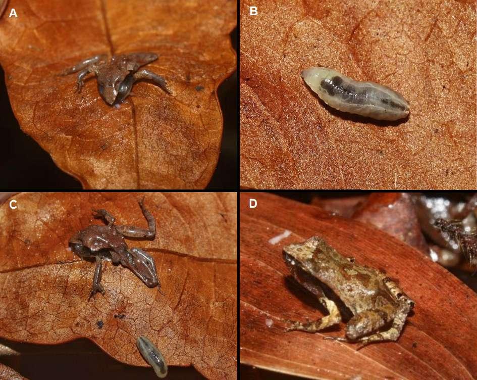
(Souza et al., 1989; Hagman et al., 2005).
Records generally point to the death of the anuran by the end of the parasitic stage, or after the larvae abandonment (Lopes 1942; Crump and Pounds 1985; Eizemberg et al., 2008). This fact is probably due to the voracity of the last larval instar, characteristic of dipterans, especially of parasitic and parasitoid species (Coupland and Barker, 2004). Mello-Patiu and Luna-Dias (2010) pointed to the rapid death of anurans, followed by the host abandonment of the larvae hours after capture. This occurrence, also observed in other works, suggests the possibility that parasites respond to the stress caused on the host (Muratori et al. 2010).
Acknowledgments
We are grateful for the consideration and guidance of Dr. Elidiomar Ribeiro, who made possible this paper, and we also thank the editors for their careful review.
B. da Cunha Martins et al. - First record of myiasis in Physalaemus cuvieri
Figure 1. A) Individual of Physalaemus cuvieri (juvenile) killed by the larvae of Diptera; B) Close view of parasitic larva; C) Larvae abandoning the anuran carcass from a hole on the left side of its abdomen; D) Live juvenile of P. cuvieri from the same locality.
234
Literature cited
Carvalho-Filho, F.D.S.; Gomes, J.O.; Maciel, A.O.; Sturaro, M.J. & Silva, K.R.A. 2010. Rhinella margaritifera (NCN). Parasites. Herpetological Review 41: 479-478.
Climatempo. 2021. Previsão do tempo: Dados históricos de chuva em Pinheiros. Available at: https://www.climatempo. com.br/previsao-do-tempo/cidade/2016/pinheiros-es. Accessed April 9, 2021.
Coupland, J. & Barker, G. 2004. Flies as Predators and Parasitoids of Terrestrial Gastropods, with Emphasis on Phoridae, Calliphoridae, Sarcophagidae, Muscidae and Fanniidae (Diptera, Brachycera, Cyclorrhapha): 85-158. In: Barker, G.M. (ed.) Natural enemies of terrestrial molluscs. CAB International, Wallingford.
Crump, M.L.& Pounds, J.A. 1985. Lethal parasitism of an aposematic anuran ( Atelopus varius ) by Notochaeta bufonivora (Diptera: Sarcophagidae). Journal of Parasitology 71: 588-591.
Eaton, B.R.; Moenting, A.E.; Paszkowski, C.A. & Shpeley, D. 2008. Myiasis by Lucilia silvarum (Calliphoridae) in amphibian species in boreal Alberta, Canada. Journal of Parasitology 94: 949-952.
Eizemberg, R.; Sabagh, L.T & Mello, R.S. 2008. First record of myiasis in Aplastodiscus arildae (Anura: Hylidae) by Notochaeta bufonivora (Diptera: Sarcophagidae) in the Neotropical area. Parasitology Research 102: 329-331.
Frost, D.R. 2021. Amphibian Species of the World: An Online Reference. Version 6.0. Available at: https:// amphibiansoftheworld.amnh.org. Accessed February 9, 2021.
Garbin, M.L.; Saiter, F.Z.; Carrijo, T.T. & Peixoto, A.L. 2017. Breve histórico e classificação da vegetação capixaba. Rodriguésia 68: 1883-1894.
Governo do Estado do Espírito Santo. 2021. Geográfia. Available in: https://www.es.gov.br/geografia. Accessed April 9, 2021.
Hagman, M.; Pape, T. & Schulte, R. 2005. Flesh fly myiasis (Diptera, Sarcophagidae) in Peruvian poison frogs genus Epipedobates (Anura, Dendrobatidae). Phyllomedusa 4: 69-73.
Hall, M. & Wall, R. 1995. Myiasis of humans and domestic animals. Advances in Parasitology 35: 257-334.
Kraus, F. 2007. Fly parasitism in Papuan frogs, with a discussion of ecological factors influencing evolution of life-history differences. Journal of Natural History 41: 1863-1874.
Lopes, H.S. 1981. Notes on American Sarcophagidae (Diptera). Revista Brasileira de Biologia 41: 149-152.
Cuad. herpetol. 36 (2): 233-235 (2022)
Mello-Patiu, C.A. & Luna-Dias, C. 2010. Myiasis in the Neotropical amphibian Hypsiboas beckeri (Anura: Hylidae) by a new species of Lepidodexia (Diptera: Sarcophagidae). Journal of Parasitology 96: 685-688.
Mulieri, P.R.; Schaefer, E.F.; Duré, M.I. & González, C.E. 2018. A new flesh fly species (Diptera: Sarcophagidae) parasitic on leptodactylid frogs. Parasitology Research, 117: 809-818. doi:10.1007/s00436-018-5755-4.
Müller, G.A.; Lehn, C.R.; Bemvenuti, A. & Marcondes, C.B. 2015. Primer registro de myiasis (Diptera: Sarcophagidae) en anuros de Leptodactylidae (Amphibia). Revista Colombiana de Ciencia Animal l7: 217-220.
Muratori, F.B.; Borlee, S. & Messing, R.H. 2010. Induced niche shift as na anti-predator response for an endoparasitoid. Proceedings of the Royal Society of London (B) 277: 14751480.
Pape, T. 1996. Catalogue of the Sarcophagidae of the World (Insecta: Diptera), Memoirs on Entomology, International, Vol. 8. Gainesville, Florida, 558 p.
Oliveira, R.M.; Mendes, C.V.M.; Ruas, D.S.; Solé, M.; Pinho, L.C. & Rebouças, R. 2012. Myiasis on Hypsiboas atlanticus (Caramaschi and Velosa, 1996) (Anura: Hylidae) from southern Bahia, Brazil. Herpetology Notes 5: 493-494.
Pinto, K.C.; Padilha, B.C.; Cruz, L.S.S.; Batista, G.A.; Rossi, M.D.P.; Martins, D.L.; Penhacek, M.; Vaz-Silva, W. & Neves, J.M. 2017. Myiasis caused by Sarcophagidae fly in Dryaderces inframaculata (Boulenger, 1882) (Anura: Hylidae) in the north of Mato Grosso, Brazil. Herpetology Notes 10: 147-149.
Schwartz, C.A. & Sebben, A. 1992. Predação de Brachycephalus ephippium (Amphibia, Anura, Brachycephalidae) por larvas de Notochaeta bufonivora (Diptera, Sarcophagidae). In: Proc. XII Congr Latino-Americano Zool. e XIX Congr. Bras. Zool. Belém, Brazil; p. 119.
Souza, F.L.S.; Souza C.W.O.; Hipolito, M.; Baldassi, L.; & Martins, M.L. 1989. Cases of buccal myiasis in the bullfrog (Rana catesbeiana Shaw, 1802), with larvae of Notochaeta sp. Aldrich, 1916 (Diptera: Sarcophagidae) in São Paulo, Brazil. Memórias do Instituto Oswaldo Cruz 84:517-518.
Souza-Pinto, F.C.; França, I.F. & Mello-Patiu, C.A. 2015. Brief description of myiasis cases in three amphibian species from Atlantic Forest located in the central region of the state of Minas Gerais, Brazil. Herpetology Notes 8: 287-290.
Travers, S.L. & Townsend, J.H. 2010. Myiasis on a Neotropical leaf frog Agalychnis saltator Taylor, 1955. Herpetology Notes 3: 355-357.
© 2022 por los autores, licencia otorgada a la Asociación Herpetológica Argentina. Este artículo es de acceso abierto y distribuido bajo los términos y condiciones de una licencia Atribución-No Comercial 4.0 Internacional de Creative Commons. Para ver una copia de esta licencia, visite http://creativecommons.org/licenses/by-nc/4.0/
235
Cuad. herpetol. 36 (2): 237-244 (2022)
New additions to the anuran fauna of the Cancão Municipal Natural Park, Serra do Navio, state of Amapá, Brazil
Carlos Eduardo Costa-Campos1, Patrick Ribeiro Sanches2, Fillipe Pedroso-Santos3, Vinicius A. M. B. de Figueiredo1, Rodrigo Tavares-Pinheiro1
1 Universidade Federal do Amapá, Departamento de Ciências Biológicas e da Saúde, Laboratório de Herpeto logia, Macapá, AP, Brazil, CEP: 68.903-419.
2 Instituto Nacional de Pesquisas da Amazônia, Programa de Pós-Graduação em Ecologia (Biologia), Manaus, Amazonas, Brasil.
3 Universidade Federal do Amapá, Programa de Pós-Graduação em Biodiversidade Tropical, Macapá, Amapá, Brasil.
Recibida: 03 Diciembre 2021
Revisada: 13 Diciembre 2022
Aceptada: 02 Agosto 2022
Editor Asociado: J. Goldberg
doi: 10.31017/CdH.2022.(2021-065)
ABSTRACT
Twelve species of anurans (Amazophrynella teko, Rhinella castaneotica, Hyalinobatrachium mondolfii, H. tricolor, Pristimantis gutturalis, P. inguinalis, Ranitomeya variabilis, Osteocephalus leprieurii, Scinax proboscideus, Leptodactylus petersii, Chiasmocleis hudsoni, Synapturanus miran daribeiroi), into the seven families (Bufonidae, Centrolenidae, Craugastoridae, Dendrobatidae, Hylidae, Leptodactylidae, Microhylidae) are reported for the first time from Cancão Municipal Natural Park, state of Amapá, North Brazil. The total number of species of anurans known from the park now stands at 61 species. Our results contribute to an increase in the knowledge of the anuran fauna of Eastern Amazonia and Guiana Shield.
Key Words: Amphibians; Natural History; Protected Areas; Amazonia.
The state of Amapá is located in the extreme northeast of the Brazilian Amazon and belonging to the Guiana Shield region. Amapá plays an important role in Brazil’s conservation with more than 95% of its original vegetation well-preserved and close to 70% of its extent lying within protected areas (Hilário et al., 2017). The state has 14 protected areas (except private reserves) and five indigenous reserves (Drummond et al., 2008). Despite this good conservation status, little is known about the diversity of anurans of this part of eastern Amazonia (Azevedo-Ramos and Galatti, 2002; Benício and Lima, 2017; Costa-Campos and Freire, 2019). Thus, inventories regarding the biodiversity are a conser vation priority, especially because several studies based on deforestation have detected changes in the habitats potentially contributing to declines of some species (Becker et al., 2016).
Cancão Municipal Natural Park is a Municipal Protection Conservation Unit that composes the
Author for correspondence: dududueducampos@gmail.com
Protected Areas Mosaic of the eastern Brazilian Amazonia, located in the municipality of Serra do Navio, in the northwest center portion of Amapá state, North Brazil. The Protected Areas that make up the Mosaic of the Eastern Amazon are: Tumucu maque Mountains National Park, Amapá National Forest, Iratapuru River Sustainable Development Reserve, Amapá State Forest, Cancão Municipal Natural Park, Extractive Reserve Beija-Flor Brilho de Fogo and Indigenous Land Wajãpi, Tumucumaque Mountains National Park e Rio Paru D’Este (Drum mond et al., 2008).
Of the Protected Areas that make up the Mosaic of the Eastern Amazon only four have am phibians’ inventories: Tumucumaque Mountains National Park (Lima, 2008); Amapá National Fo rest (Benício and Lima, 2017), Cancão Municipal Natural Park (Silva-e-Silva and Costa-Campos, 2018), and Extractive Reserve Beija-Flor Brilho de Fogo (Pedroso et al., 2019). For Iratapuru River
Nota 237
C. E. Costa-Campos et al. - Anuran species of Serra do Navio, Brazil
Sustainable Development Reserve only records and new amphibian’s species distributions have been registered (Costa-Campos et al., 2020; Figueiredo et al., 2020; Figueiredo et al., 2021; Tavares-Pinheiro et al., 2021).
Until recently the only literature report on anuran richness from municipality of Serra do Navio was a survey conducted by Silva-e-Silva and CostaCampos (2018), and although the sampling effort was limited, they recorded significant richness (n = 49 species) for four areas in the Cancão Municipal Natural Park. Here, we provide new additions to the anurans of the municipality of Serra do Navio, in Amapá state, Brazil, and with some comments on the natural history and taxonomic notes.
The sampled area was conducted in the Cancão Municipal Natural Park (0°54’8.82”N; 52°0’19.62”W). The park has an area of 370,26 ha of terra firme forest, belonging to the Amazonian forest domain. The climate is classified as Am (Equatorial, Köpper-Geiger classification), with average tempe rature of 26.1 ºC and rainfall annual of 2,450 mm (NHMET database, 2022).
The first fieldwork was conducted in the park during January to December 2013 (see Silva-e-Silva and Costa-Campos, 2018), whereas the second field work occurs during March 2018 to February 2019 and July 2022. We using “visual encounter surveys” during the day (6:00 – 9:00 hs) and about the first three hours after dark on three nights. Surveys were made in two trails (Fig. 1): i) trail at Cancão forest (0°54’9.72”N, 52°0’17.64”W); and ii) right margin of the River Amapari trail (0°54’2.88”N, 52°0’48.44”W).
Specimens were anesthetized with 5% lidocai ne, fixed with 10% formalin and preserved in 70% ethanol (Heyer et al., 1994). Voucher specimens were deposited in the Herpetological Collection of Universidade Federal do Amapá (CECC). The taxo nomic nomenclature applied herein follows Segalla et al. (2021) for amphibians with modifications made by Dubois (2017). The conservation status of spe cies was obtained from the Red List of endangered species (IUCN, 2022).
We recorded 12 species of anurans (Fig. 2) belong to families Bufonidae ( Amazophrynella teko, Rhinella castaneotica), Centrolenidae (Hyalin obatrachium mondolfii, H. tricolor), Craugastoridae (Pristimantis gutturalis, P. inguinalis), Dendrobatidae ( Ranitomeya variabilis ), Hylidae ( Osteocephalus leprieurii , Scinax proboscideus ), Leptodactylidae
(Leptodactylus petersii), and Microhylidae (Chias mocleis hudsoni , Synapturanus mirandaribeiroi ), undetected in previous inventories at the Cancão Municipal Natural Park (see Silva-e-Silva and CostaCampos, 2018).
Amazophrynella teko Rojas, Fouquet, Ron, Hernan dez-Ruz, Melo-Sampaio, Chaparro, Vogt, Carvalho, Pinheiro, Avila, Farias, Gordo & Hrbek, 2018. Is a small toad of the family Bufonidae (SVL 12.9–15.8 mm in males and 17.9–21.5 mm in females; Rojas et al., 2018). This species occurs in French Guiana, southern region of Suriname and Brazil (Amapá state), at elevations ranging from 70–350 m. The species is diurnal and crepuscular (Rojas et al., 2018). The conservation status of this species is Not Evalu ated under IUCN Red List of Threatened Species. Voucher number CECC 3168.
Rhinella castaneotica (Caldwell, 1991). A mediumsized species of the Rhinella margaritifera species group (SVL 18.4–23.6 mm in males and 18.9–26.3 mm in females; Caldwell, 1991). It is known from the Amazon Basin in Bolivia, Colombia, eastern Peru, Brazil (Amazonas, Amapá, Pará, and Rondônia), but likely occurs wider in the upper Amazon Basin. Rhinella castaneotica are nocturnal and terrestrial toads and natural habitats are tropical moist oldgrowth lowland forests. It is a forest floor species that breeds in Brazil nut capsules and temporary pools (Caldwell, 1993). There are no known signi ficant threats to this species. Rhinella castaneotica is listed as Least Concern under IUCN Red List of Threatened Species. Voucher number CECC 2145.
Hyalinobatrachium mondolfii Castroviejo-Fisher, Vilà, Ayarzagüena, Blanc & Ernst, 2011. Is a small glassfrog (SVL 20.7–23.0 mm in adult males; unk nown in females; Castroviejo-Fisher et al., 2011). The species inhabits primary tropical floodplain forest of the eastern Guiana Shield (15–200 m) and the western Amazon. It has exclusively been found in vegetation associated with streams. This spe cies has a broad distribution through the lowland Amazon rainforests, occurring in Bolivia, Brazil (Amapá state), Colombia, French Guiana, Guyana, Suriname and Venezuela (Castroviejo-Fisher et al., 2011; Figueiredo et al., 2020). Calling males were found perched on the underside of leaves at night, at a height of 3 m above ground. Hyalinobatrachium mondolfii is listed as Least Concern under IUCN
238
Red List of Threatened Species. Voucher number CECC 1813.
Hyalinobatrachium tricolor Castroviejo-Fisher, Vilà, Ayarzagüena, Blanc & Ernst, 2011. Is a small glassfrog (SVL 20.3–21.0 mm in adult males; unk nown in females; Castroviejo-Fisher et al., 2011).
Males call on vegetation 4–5 m above streams of
herpetol. 36
0.5–1.5 m deep. This species occurs in a riparian zone on the right bank of the Amapari River, which has some anthropic disturbance (Costa-Campos et al., 2021) in the Amapá state, and in Crique Wapou, Kaw, French Guiana (type locality) at low elevations to 100 m (Castroviejo-Fisher et al., 2011; Vacher et al., 2020). Males call from the underside of leaves
 Figure 1. Trails and habitats sampled at the Cancão Municipal Natural Park, municipality of Serra do Navio, Amapá state: A) trail at Cancão forest; B) right margin of the River Amapari trail; C) Treefall gap at Cancão forest; D) temporary pond at river Amapari trail; E) igapó forest; F) terra firme forest.
239 Cuad.
(2): 237-244 (2022)
during night and close to egg clutches. Hyalinoba trachium tricolor is listed as Least Concern under IUCN R ed List of Threatened Species. Voucher number CECC 2783.
Pristimantis gutturalis (Hoogmoed, Lynch & Lescure, 1977). A medium-sized species belongs to Crau gastoridae family (SVL 19.0–20.3 mm in males and
17.9–40.9 mm in females; Hoogmoed et al., 1977). Is a diurnal and terrestrial frog found in leaf litter in tropical moist lowland forests and is known from Northern Brazil (Amapá), southern French Guiana, and eastern Surinam (Ouboter and Jairam, 2012; Frost, 2022). Pristimantis gutturalis is listed as Least Concern under IUCN Red List of Threatened Spe cies. Voucher number CECC 3486.
 C. E. Costa-Campos et al. - Anuran species of Serra do Navio, Brazil
Figure 2. Anurans recorded at the Cancão Municipal Natural Park, municipality of Serra do Navio, Amapá state: A) Amazophrynella teko; B) Rhinella castaneotica; C) Hyalinobatrachium mondolfii; D) H. tricolor; E) Pristimantis gutturalis; F) P. inguinalis); G) Ranitomeya variabilis); H) Osteocephalus leprieurii; I) Scinax proboscideus; J) Leptodactylus petersii; K) Chiasmocleis hudsoni; L) Synapturanus mirandaribeiroi
240
Pristimantis inguinalis (Parker, 1940). Adult males measure on average 20.2 mm (Fouquet et al., 2013). Is a arboreal frog of the family Craugastoridae. It is found in Guyana, Suriname, French Guiana, and northern Brazil (Amapá state). Occurs in primary forests at elevations of 50–700 m. Males call from trees 4–6 m above the ground (Cole et al., 2013; Fouquet et al., 2013). It is a common species, and no significant threats to it are known. Its range overlaps with several protected areas (IUCN SSC Amphibian Specialist Group, 2018). Pristimantis inguinalis is listed as Least Concern under IUCN Red List of Threatened Species. Voucher number CECC 3498.
Ranitomeya variabilis (Zimmermann & Zimmer mann, 1988). Is a small (adult males has a mean SVL 17.4 mm and 18.0 in females; Brown et al., 2008), diurnal and semi-arboreal dendrobatid, with inhabits in primary and secondary rainforests in the understory, canopy, and sometimes in leaf litter at elevations of up to 900-1200 m above sea level (Brown et al., 2008). Its habitat is in the Amazon Rainforest. It most frequently uses bromeliads for breeding, approximately 2.5 m above the forest floor (Brown et al., 2011; Simões et al., 2019). Known dis tribution consists of widely separated populations, one in northeastern Amazonian Peru and extreme southeastern Colombia, and expected in the adjacent Brazil, Venezuela; extreme southern Guyana; eastern French Guiana; the mouth of the Amazon in Brazil (Frost, 2022; Muell et al., 2022). Ranitomeya variabi lis is listed as Data Deficient under IUCN Red List of Threatened Species. Voucher number CECC 2609.
Osteocephalus leprieurii (Duméril & Bibron, 1841). The species is a moderate-sized treefrog (SVL max size 63 mm, unknown sex; Lescure and Marty, 2000; Ouboter and Jairam, 2012) distributed throughout the Guiana Shield in French Guiana, Surinam, Guyana, Venezuela, Ecuador, Peru, Bolivia, and northern Brazil in Amazonas and Amapá (Santana et al., 2008; Barrios-Amorós et al., 2019; Figueiredo et al., 2021). This species is an explosive breeder associated to lowland rainforest, where it occurs both in terra firme and in seasonally flooded forest (Jungfer and Hödl, 2002). Osteocephalus leprieurii is listed as Least Concern under IUCN Red List of Threatened Species. Voucher number CECC 3843.
Scinax proboscideus (Brongersma, 1933). The species is a moderate-sized (SVL max size 46 mm, unknown
Cuad. herpetol. 36 (2): 237-244 (2022)
sex; Lescure and Marty, 2000; Ouboter and Jairam, 2012). Is a species of frog in the family Hylidae, currently known from French Guiana, Guyana, Su riname, and expected to be found in Brazil (Frost, 2022). Its natural habitats are subtropical or tropical moist lowland forests and intermittent freshwater marshes (Lescure and Marty, 2000). This species is categorized as Least Concern according to the IUCN Red List of Threatened Species. Voucher number CECC 2740.
Leptodactylus petersii (Steindachner, 1864). Is a small to moderate size (SVL 26.6–41.1 mm in males and 31.2–51.3 mm in female; De Sá et al., 2014). Is a species of frog in the family Leptodactylidae found widely in the Guianas and the Amazon Basin. It has found in tropical rainforest, forest edge, open areas, savanna enclaves in the tropical rainforest, and open cerrado formations below 600 m (De Sá et al., 2014). This nocturnal frog is usually found on the ground near water. Eggs are laid in a foam nest near water, to which the tadpoles will later move (Lima et al., 2012). Leptodactylus petersii is listed as Least Concern under IUCN Red List of Threatened Species. Voucher number CECC 2398.
Chiasmocleis hudsoni Parker, 1940. Is a small-sized (SVL 14.9–16.4 mm in males and 19.9 mm in the single female; Costa-Campos et al., 2019) common species living in tropical rainforests at elevations below 300 m, nocturnal, terrestrial and fossorial that inhabits streamside ponds (Rodrigues et al., 2008). Is found in French Guiana, Suriname, Guyana, Guianan Venezuela, Colombia (Amazonas), and in Brazil at southern Amazonas, as well as at several localities in central and center-east Pará and from Serra do Navio municipality, Amapá state (Lima et al., 2012; Peloso et al., 2014; Costa-Campos et al., 2019). Chiasmocleis hudsoni is listed as Least Con cern under IUCN Red List of Threatened Species. Voucher number CECC 2262.
Synapturanus mirandaribeiroi (Nelson & Lescure, 1975). Is a medium-sized (SVL 26.2–30.8 in males and 28.6–34.4 in females; Fouquet et al., 2021). The species is found in terra firme forests between 100–400 m above sea from the eastern Guiana Shield in Guyana, Suriname, south to Parque Estadual Rio Negro, Amazonas, Brazil; probably occurs in the northern part of the states of Amapá, Pará, and Roraima, in Brazil (Fouquet et al., 2021).
241
Synapturanus mirandaribeiroi is listed as Least Con cern under IUCN Red List of Threatened Species. Voucher number CECC 3847.
Previous research in the Cancão Municipal Natural Park represents the only officially published list of anurans present in the area (Silva-e-Silva and Costa-Campos, 2018). The authors recorded 49 anuran species of anurans belonging to 11 families: Allophrynidae (1); Aromobatidae (2), Bufonidae (5), Centrolenidae (1), Craugastoridae (5), Dendrobati dae (2), Eleutherodactylidae (1), Hylidae (18), Lep todactylidae (10), Phyllomedusidae (3) and Pipidae (1). In our study, we identify 12 species not registered in the previous study (i.e., Silva-e-Silva and CostaCampos, 2018) increasing a known richness of 61 anurans species for the area.
Although the Cancão Municipal Natural Park is located on the edge of the urban expansion area of the municipality of Serra do Navio (Drummond et al., 2008), the current number of 61 anuran species uncovered a representative sample of the anurofau na, when compared to other conservation units that are part of the Mosaic of the Eastern Amazon: 25 spe cies listed for Extractive Reserve Beija-Flor Brilho de Fogo (Pedroso-Santos et al., 2019); 53 species listed for Amapá National Forest (Benício and Lima, 2017), and 70 species listed for Tumucumaque Mountains National Park (Lima, 2008). The presence of new records in the study area (e.g., Hyalinobatrachium mondolfii, H. tricolor and Chiasmocleis hudsoni) according to previously studies (Costa-Campos et al., 2019; Figueiredo et al., 2020; Costa-Campos et al., 2021) evidence the incipience of knowledge in the context of anuran fauna regional and revealing the importance of the anurans inventories in areas not samples.
Our results contribute to an increase in the knowledge of the anuran fauna of Eastern Amazonia and Guiana Shield, in which a lowland tropical for est in the municipality of Serra do Navio is inserted. With the increase in deforestation and logging in Brazilian Amazonia (Fearnside, 2005), the protec tion of these areas is importance for the conservation of the local anurofauna, considering the descriptions from new species (Taucce et al., 2020; Carvalho et al., 2022) and the extension restricted and ende mism of anurans in the region (Costa-Campos et al., 2016; Pezzuti et al., 2022). We recommended that future sampling designs include these areas to better characterize the amphibian diversity of
Amazonian fauna.
Acknowledgments
We are grateful to all colleagues of “Laboratório de Herpetologia” for supporting during our field work, and in particular Wirley Almeida-Santos for their help and facilitation of this study. We would like to thank to the Instituto Chico Mendes de Conservação da Biodiversidade (ICMBio) for providing collection permits (SISBIO #48102-2) and Prefeitura Municipal de Serra do Navio for authorizing us to conduct the research in the Cancão Municipal Natural Park, and Christoph Jaster (ICMBio/Tumucumaque Moun tains National Park) for logistical support during the fieldwork.
Literature cited
Azevedo-Ramos, C. & Galatti, U. 2002. Patterns of amphibian diversity in Brazilian Amazonia: Conservation implications. Biological Conservation 103: 103-111.
Barrio-Amorós, C.L.; Rojas-Runjaic, F.J.M. & Señaris, J.C. 2019. Catalogue of the amphibians of Venezuela: Illustrated and annotated species list, distribution, and conservation. Amphibian & Reptile Conservation 13(1) [Special Section]: 1-198.
Becker, G.; Rodriguez, D.; Longo, A.V.; Toledo, L.F.; Lambertini, C.; Leite, D.S.; Haddad, C.F.B. & Zamudio, K.R. 2016. Deforestation, host community structure, and amphibian disease risk. Basic and Applied Ecology 17: 72-80.
Benício, R.A. & Lima, J.D. 2017. Anurans of Amapá National Forest, Eastern Amazonia, Brazil. Herpetology Notes 10: 627-633.
Brongersma, L.D. 1933. Ein neuer Laubfrosch aus Surinam. Zoologischer Anzeiger 103: 267-270.
Brown, J.L.; Twomey, E.; Morales, V. & Summers, K. 2008. Phytotelm size in relation to parental care and mating strategies in two species of Peruvian poison frogs. Behaviour 145: 1139-1165.
Brown, J.L.; Twomey, E.; Amézquita, A.; de Souza, M.B.; Caldwell, J.P.; Lötters, S.; von May, R.; Melo-Sampaio, P.R.; Mejía-Vargas, D.; Pérez-Peña, P.E.; Pepper, M.; Poelman, E.H.; Sanchez-Rodriguez, M. & Summers, K. 2011. A taxonomic revision of the Neotropical frog genus Ranitomeya (Amphibia: Dendrobatidae). Zootaxa 3083: 1-120.
Caldwell, J.P. 1991. A new species of toad in the genus Bufo from Para, Brazil, with an unusual breeding site. Papéis Avulsos de Zoologia 37: 389-400.
Caldwell, J.P. 1993. Brazil nut fruit capsules as phytotelmata: interactions among anuran and insect larvae. Canadian Journal of Zoology 71: 1193-1201.
Carvalho, T.R.; Fouquet, A.; Lyra, M.L.; Giaretta, A.A.; CostaCampos, C.E.; Rodrigues. M.T.; Haddad, C.F.B.; Ron, S.R. 2022. Species diversity and systematics of the Leptodactylus melanonotus group (Anura, Leptodactylidae): review of diagnostic traits and a new species from the Eastern Guiana Shield. Systematics and Biodiversity 20: 2089269.
C. E. Costa-Campos et al. - Anuran species of Serra do Navio, Brazil
242
Castroviejo-Fisher, S.; Vilà, C.; Ayarzagüena, J.; Blanc, M. & Ernst, R. 2011. Species diversity of Hyalinobatrachium glassfrogs (Amphibia: Centrolenidae) from the Guiana Shield, with the description of two new species. Zootaxa 3132(1): 1-55.
Cole, C.J.; Townsend, C.R.; Reynolds, R.P.; MacCulloch, R.D. & Lathrop, A. 2013. Amphibians and reptiles of Guyana, South America: illustrated keys, annotated species accounts, and a biogeographic synopsis. Proceedings of the Biological Society of Washington 125(4): 317-578.
Costa-Campos, C.E.; Bang, D.L.; Figueiredo, V.A.M.B.; Tavares-Pinheiro, R. & Fouquet, A. 2021. New records and distribution extensions of the glassfrogs Hyalinobatrachium taylori (Goin, 1968) and H. tricolor Castroviejo-Fisher, Vilà, Ayarzagüena, Blanc & Ernst, 2011 (Anura, Centrolenidae) in Amapá, Brazil. Check List 17(2): 637-642.
Costa-Campos, C.E.; Figueiredo, V.A.M.B.; Lima, J.R.F. & Lima, J.D. 2020. New record and distribution map of the glassfrog Vitreorana ritae (Lutz, 1952) (Anura: Centrolenidae) from Amapá state, Eastern Amazon. Herpetology Notes 13: 733737.
Costa-Campos, C.E.; Sousa, J.C. & Menin, M. 2019. Chiasmocleis hudsoni Parker, 1940 (Anura, Microhylidae): a new record for Amapá State, Brazil. Herpetology Notes 12: 405-408.
Costa-Campos, C.E. & Freire, E.M.X. 2019. Richness and composition of anuran assemblages from an Amazonian savanna. ZooKeys 843: 149-169.
Costa-Campos, C.E., Lima, A.P. & Amézquita, A. 2016. The advertisement call of Ameerega pulchripecta (Silverstone, 1976) (Anura, Dendrobatidae). Zootaxa 4136(2): 387-389.
De Sá, R.O.; Grant, T.; Camargo, A.; Heyer, W.R.; Ponssa, M.L. & Stanley, E. 2014. Systematics of the Neotropical Genus Leptodactylus Fitzinger, 1826 (Anura: Leptodactylidae): Phylogeny, the Relevance of Non-molecular Evidence, and Species Accounts. South American Journal Herpetology 9: 1-128.
Drummond, J.A.; Dias, T.C.A.C. & Brito, D.M.C. 2008. Atlas das Unidades de Conservação do Estado do Amapá. MMA/ IBAMA, GEA/SEMA. Macapá, Amapá.
Dubois, A. 2017. The nomenclatural status of Hysaplesia , Hylaplesia, Dendrobates and related nomina (Amphibia, Anura), with general comments on zoological nomenclature and its governance, as well as on taxonomic databases and websites. Bionomina 11: 1-48.
Duméril, A.M.C. & Bibron, G. 1841. Erpétologie Genérale ou Histoire Naturelle Complète des Reptiles. Volume 8. Librarie Enclyclopedique de Roret. Paris.
Fearnside, P.M. 2005. Deforestation in Brazilian Amazonia: History, Rates, and Consequences. Conservation Biology 19(3): 680-688.
Figueiredo, V.A.M.B.; Tavares-Pinheiro, R.; Freitas, A.P. & Costa-Campos, C.E. 2021. New geographic record for Osteocephalus leprieurii (Duméril & Bibron, 1841) (Anura, Hylidae) from Amapá State, northern Brazil. Herpetology Notes 14: 827-831.
Figueiredo, V.A.M.B.; Tavares-Pinheiro, R.; Freitas, A.P.; DiasSouza, M.R. & Costa-Campos, C.E. 2020. First records of the glass frogs Hyalinobatrachium cappellei (van Lidth de Jeude, 1904) and H. mondolfii Señaris & Ayarzagüena, 2001 (Anura, Centrolenidae) in the state of Amapá, Brazil. Check
Cuad. herpetol. 36 (2): 237-244 (2022)
List 16(5): 1369-1374.
Fouquet, A.; Leblanc, K.; Fabre, A.-C.; Rodrigues, M.T.; Menin, M.; Courtois, E.A.; Dewynter, M.; Hölting, M.; Ernst, R.; Peloso, P.L.V. & Kok, P.J.R. 2021. Comparative osteology of the fossorial frogs of the genus Synapturanus (Anura, Microhylidae) with the description of three new species from the Eastern Guiana Shield. Zoologischer Anzeiger 293: 46-73.
Fouquet, A.; Martinez, Q.; Courtois, E.A.; Dewynter, M.; Pineau, K.; Gaucher, P.; Blanc, M.; Marty, C. & Kok, P.J.R. 2013. A new species of the genus Pristimantis (Amphibia, Craugastoridae) associated with the moderately evelated massifs of French Guiana. Zootaxa 3750(5): 569-586.
Frost, D.R. 2022. Amphibian Species of the World: an Online Reference. Version 6.1. Available at http://research.amnh. org/herpetology/amphibia/index.html. American Museum of Natural History, New York, USA. Last access: 07 April 2022.
Heyer, W.R.; Donnelly, M.A.; McDiarmid, R.W.; Hayek, L.A.C. & Foster, M.S. 1994. Measuring and monitoring biological diversity: standard methods for amphibians. Smithsonian Institution Press. Washington.
Hilário, R.R., Toledo, J.J., Mustin, K., Castro, I.J., Costa-Neto, S.V., Kauano, E.E.; Eiler, V.; Vasconcelos, I.M.; MendesJúnior, R.N.; Funi, C.; Fearnside, P.M.; Silva, J.M.C.; Euler, A.M.C. & Carvalho, W.D. 2017. The Fate of an Amazonian Savanna: Government Land-Use Planning Endangers Sustainable Development in Amapá, the Most Protected Brazilian State. Tropical Conservation Science 10: 1-8.
Hoogmoed, M.S.; Lynch, J.D. & Lescure, J. 1977. A new species of Eleutherodactylus from Guiana (Leptodactylidae, Anura). Zoologische Mededelingen 51: 33-41.
IUCN. 2022. The IUCN Red List of Threatened Species. Version 2021.2. Available at http://www.iucnredlist. org. Last access: 08 April 2022.
IUCN SSC Amphibian Specialist Group. 2018. Pristimantis inguinalis. The IUCN Red List of Threatened Species 2018. Available at https://dx.doi.org/10.2305/IUCN.UK.2018-2. RLTS.T56671A61411006.en. Last access: 07 April 2022.
Jungfer, K.-H.; Hödl, W. 2002. A new species of Osteocephalus from Ecuador and a redescription of O. leprieurii (Duméril & Bibron, 1841) (Anura: Hylidae). Amphibia-Reptilia 23: 21-46.
Lescure, J. & Marty, C. 2000. Atlas des Amphibiens de Guyane. Collections Patrimoines Naturels 45: 1-388.
Lima, J.D. 2008. A herpetofauna do Parque Nacional do Montanhas do Tumucumaque, Amapá, Brasil, Expedições I a V: 38-50. In: Bernard, E. (ed.). Inventários Biológicos Rápidos no Parque Nacional Montanhas do Tumucumaque, Amapá, Brasil. RAP Bulletin of Biological Assessment. Arlington, VA, Conservation International.
Lima, A.P.; Magnusson, W.E.; Menin, M.; Erdtmann, L.K.; Rodrigues, D.J.; Keller, C. & Hödl, W. 2012. Guia de Sapos da Reserva Adolpho Ducke, Amazônia Central. Second Edition. Editora INPA. Manaus.
Muell, M.R., Chávez, G., Prates, I., Guillory, W.X., Kahn, T.R., Twomey, E.M., Rodrigues, M.T. & Brown, J.L. 2022. Phylogenomic analysis of evolutionary relationships in Ranitomeya poison frogs (Family Dendrobatidae) using ultraconserved elements. Molecular Phylogenetics and
243
C. E. Costa-Campos et al. - Anuran species of Serra do Navio, Brazil
Evolution 168: 107389.
Nelson, C.E. & Lescure, J. 1975. The taxonomy and distribution of Myersiella and Synapturanus (Anura: Microhylidae). Herpetologica 31: 389-397.
NHMET database. 2022. Banco de dados do Núcleo de Hidrometeorologia e energias renováveis do Instituto de pesquisas científicas e tecnológicas do estado do Amapá, NHMET/IEPA, Available at http://www.iepa.ap.gov.br/ meteorologia/. Last access: 05 March 2022.
Ouboter, P.E. & Jairam, R. 2012. Amphibians of Suriname. Brill. Leiden.
Parker, H.W. 1940. Undescribed anatomical structures and new species of reptiles and amphibians. Annals and Magazine of Natural History, Series 11 5: 257-274.
Pedroso-Santos, F.; Sanches, P.R. & Costa-Campos, C.E. 2019. Anurans and reptiles of the Reserva Extrativista Beija-Flor Brilho de Fogo, Amapá state, eastern Amazon. Herpetology Notes 12: 799-807.
Peloso, P.L.V.; Sturaro, M.J.; Forlani, M.C.; Gaucher, P.; Motta, A.P. & Wheeler, W.C. 2014. Phylogeny, taxonomic revision, and character evolution of the genera Chiasmocleis and Syncope (Anura, Microhylidae) in Amazonia, with descriptions of three new species. Bulletin of the American Museum of Natural History 2014: 1-112.
Pezzuti, T.L., Araújo, R.B., Sanches, P.R., Pedroso-Santos, F., DiasSouza, M.R. & Costa-Campos, C.E. 2022. The tadpole of the endemic poison frog Ameerega pulchripecta (Silverstone, 1976) with the description of its chondrocranium (Anura: Dendrobatidae: Colostethinae). Zootaxa 5115(2): 295-300.
Rodrigues, D.J., Menin, M., Lima, A.P. & Mokross, K.S. 2008. Tadpole and vocalizations of Chiasmocleis hudsoni (Anura, Microhylidae) in Central Amazonia, Brazil. Zootaxa 1680: 55-58.
Rojas, R.R.; Fouquet, A.; Ron, S.R.; Hernández-Ruz, E.J.; Melo-Sampaio, P.R.; Chaparro, J.C.; Vogt, R.C.; Carvalho, V.T.; Pinheiro, L.C.; Avila, R.W.; Farias, I.P.; Gordo, M. & Hrbek, T. 2018. A Pan-Amazonian species delimitation: high species diversity within the genus Amazophrynella (Anura: Bufonidae) PeerJ 6: e4941.
Santana, D.J.; São-Pedro, V.A.; Costa, H.C.; Feio, R.N. 2008. Amphibia, Anura, Hylidae, Osteocephalus leprieurii :
Distribution extension. Check List 4(4): 453-454.
Segalla, M.V.; Berneck, B.; Canedo, C.; Caramaschi, U.; Cruz, C.A.G.; Garcia, P.C.A.; Grant, T.; Haddad, C.F.B.; Lourenço, A.C.; Mângia, S.; Mott, T.; Nascimento, L.B.; Toledo, L.F.; Werneck, F.P. & Langone, J.A. 2021. Herpetologia Brasileira 10(1): 121-216.
Silva-e-Silva, Y.B. & Costa-Campos, C.E. 2018. Anuran species composition of Cancão Municipal Natural Park, Municipality of Serra do Navio, Amapá state, Brazil. ZooKeys 762: 131-148.
Simões, P.I., Rojas-Runjaic, F.J.M., Gagliardi-Urrutia, G. & Castroviejo-Fisher, S. 2019. Five new country records of Amazonian anurans for Brazil, with notes on morphology, advertisement calls, and natural history. Herpetology Notes 12: 211-219.
Steindachner, F. 1864. Batrachologische Mittheilungen. Verhandlungen des Zoologisch-Botanischen Vereins in Wien 14: 239-288.
Taucce, P.P.G., Costa-Campos, C.E., Haddad, C.F.B. & de Carvalho, T.R. 2020. A New Amazonian Species of the Diminutive Frog Genus Adelophryne (Anura: Brachycephaloidea: Eleutherodactylidae) from the State of Amapá, Northern Brazil. Copeia 108(4): 746-757.
Tavares-Pinheiro, R.; Figueiredo, V.A.M.B. & Costa-Campos, C.E. 2021. New state record for Ctenophryne geayi Mocquard, 1904 (Anura, Microhylidae), with an updated distribution map. Herpetology Notes 14: 883-886.
Vacher J.-P.; Chave, J.; Ficetola, F.; Sommeria-Klein, G.; Tao, S.; Thébaud, C.; Augustín, B.; Camacho, J.; Cassimiro, T.J.; Colston, M.D.; Ernst, R.; Gaucher, P.; Gomes, J.O.; Jairam, R.; Kok, P.J.R.; Lima, J.D.; Martinez, Q.; Christian, M.; Noonan, B.P.; Sales Nunes, P.M.; Ouboter, P.; Recorder, R.; Rodrigues, M.T.; Snyder, A., Marques-Souza, S. & Fouquet, A. 2020. Large-scale DNA-based survey of frogs in Amazonia suggests a vast underestimation of species richness and endemism. Journal of Biogeography 47(8): 1781-1791.
Zimmermann, H. & Zimmermann, E. 1988. Etho Taxonomie und zoogeographische Artengruppenbildung bei Pfeilgiftfroschen (Anura: Dendrobatidae). Salamandra 24: 125-160.
© 2022 por los autores, licencia otorgada a la Asociación Herpetológica Argentina. Este artículo es de acceso abierto y distribuido bajo los términos y condiciones de una licencia Atribución-No Comercial 4.0 Internacional de Creative Commons. Para ver una copia de esta licencia, visite http://creativecommons.org/licenses/by-nc/4.0/
244
Nota
Cuad. herpetol. 36 (2): 245-249 (2022)
Amelanism in Amphisbaena darwinii Duméril & Bibron, 1839 (Squamata: Amphisbaenidae)
Carolina L. Paiva1, Mateo Cocimano2, Ricardo Montero3, Henrique C. Costa1
1 Departamento de Zoologia, Universidade Federal de Juiz de Fora, 36036-900, Juiz de Fora, Minas Gerais, Brazil.
2 Almirante Brown 776, Quilmes, Buenos Aires, Argentina
3 Facultad de Ciencias Naturales, Universidad Nacional de Tucumán, Tucumán, Argentina.
Recibida: 30 Noviembre 2021
Revisada: 12 Febrero 2022
Aceptada: 22 Febrero 2022
Editor Asociado: C. Borteiro
doi: 10.31017/CdH.2022.(2021-069)
ABSTRACT
Color anomalies are rarely reported in Amphisbaenia. We present the first record of amelanism in this group based on a specimen of Amphisbaena darwinii from Argentina. The photos were uploaded to a citizen science platform, reinforcing the positive impact of citizen science to filling gaps in our knowledge about biodiversity.
Key Words: Amphisbaenia; Citizen Science; Color Anomaly; Hypopigmentation; iNaturalist
Conspicuous chromatic anomalies occur due to pigmentation production disturbances causing aber rant coloration of the skin (Rook et al., 1998). Such anomalies are not common in wild squamates, but have been frequently reported for snakes, especially cases of hypopigmentation (Borteiro et al., 2021). Traditionally, hypopigmentation anomalies were classified as albinism, leucism, and piebaldism. In albinism, there is a complete absence of pigmenta tion of the skin and eyes caused by hereditary dis position compromising melanocytes, responsible for melanin production (Griffiths et al., 2016). On the other hand, in leucism and piebaldism, which are also known as ‘partial albinism’, the eyes are pig mented, but there is an almost complete absence of pigmentation in skin (leucism) or there is a pattern of unpigmented patches along the body (piebaldism) (Prüst, 1984; Bechtel, 1991; Lamoreux et al., 2010; Abreu et al., 2013). Recently, Borteiro et al. (2021) reviewed color anomalies in Neotropical snakes and proposed a standardized terminology to be used in reptiles, particularly in cases of hypopigmentation: amelanism, albinism, hypomelanism, leucism, and piebaldism.
Hypopigmentation is rarely reported in worm lizards (Amphisbaenia), although it is suggested that
Author for correspondence: ccostah@gmail.com
such color anomalies would be of little adaptative harm to fossorial species (Sazima & Di-Bernardo, 1991; Kornilios, 2014; Perez & Alvares, 2020). There are records of hypopigmentation in Amphisbaena munoai (Perez & Alvares, 2020), A. darwinii (cited as A. d. trachura) (Chalkidis & Di-Bernardo, 2004), Blanus strauchi (Avcý et al., 2018; Kazilas et al., 2018), and B. vandellii (cited as B. cinereus) (Malk mus, 1997; Cabana & Vázquez, 2008). With the ex ception of an albino specimen of B. strauchi (Avcý et al., 2018), those reports refer to cases of piebaldism, sometimes cited as partial albinism (Malkmus, 1997; Chalkidis & Di-Bernardo, 2004; Cabana & Vázquez, 2008) or even complete albinism (Fig. 1 in Cabana & Vázquez 2008) (Table 1).
On 24 October 2021, at 6:16 p.m., in La Ca pilla, Buenos Aires Province, Argentina (34.8925° S, 58.2827° W), the father of MC was shoveling in the backyard, when he unearthed a specimen of Amphisbaena darwinii which was buried about 50 cm deep (Fig. 1). The specimen was photographed by MC and released. The photos were uploaded to the citizen science website iNaturalist (https://www. inaturalist.org/observations/99297581), where they caught the attention of the remaining authors.
Amphisbaena darwinii is known to occur from
245
 C. L. Paiva et al. - Amelanism in Amphisbaena darwinii
Figure 1. Amelanistic specimen of Amphisbaena darwinii unearthed at La Capilla, Buenos Aires Province, Argentina, while a backyard was being shoveled. A) dorsal view of the specimen; B) detail of the head and anterior portion of the body; C) detail of the posterior portion of the body and the tail (note that the tail is not tuberculate and exhibits a pale-yellow color).
246
Cuad. herpetol. 36 (2): 245-249 (2022)
Table 1. Published reports of color anomalies in Amphisbaenia.
Taxon Color anomaly Source
Amphisbaena darwinii “heterozonata”
Amphisbaena darwinii “trachura”
Amphisbaena munoai
Blanus strauchii
Blanus strauchii
Blanus vandellii
Amelanism
This study
Piebaldism (Chalkidis and Di-Bernardo, 2004)
Piebaldism (Perez and Alvares, 2020)
Albinism (Avcý et al., 2018)
Piebaldism (Kazilas et al., 2018)
Piebaldism (Malkmus, 1997; Cabana and Vázquez, 2008)
eastern Bolivia to central Argentina (Montero, 2016). Three subspecies were traditionally recognized: A. d. darwinii Duméril & Bibron, 1839, A. d. heterozonata Burmeister, 1861 and A. d. trachura Cope 1885 –although darwini is used by many authors (Gans, 1966; Vanzolini, 2002; Montero, 2016), the original spelling is darwinii (Duméril & Bibron, 1839). Some authors have considered these subspecies as valid species (Vanzolini, 2002; Gans, 2005; Perez et al., 2012), but Montero (2016) argued that differences observed are due to clinal variation in morphology, synonymizing A. heterozonata and A. trachura with A. darwinii, without recognition of subspecies. The taxonomy of these taxa was recently reviewed, but still not formally published (Perez, 2016).
Although the photographed specimen was not collected, we can confidently assign its identity to A. darwinii due to the following characters that fall within the known variation of the species (Mon tero, 1996; Vanzolini, 2002): snout rounded, annuli (body+lateral+(autotomy)caudal), 186+2+(8)17; dorsal segments, 13. The number of body annuli (186) falls slightly below the known range of 188–209 in populations from central-eastern Argentina (tra ditionally assigned to A. d. heterozonata; Montero, 1996), but is consistent with the low count observed in the southernmost populations; nevertheless, the counts may be inaccurate as they are based on a photo in dorsal view (Fig. 1A), and the position of the cloaca was estimated based on the posterior end of the lateral sulci. The specimen exhibits a smooth surface at the end of the tail, in congruence with
central-eastern Argentina populations (tuberculated in Brazilian populations, traditionally assigned to A. d. trachura). We can eliminate the other amphisbae nian species that may occur in the area (A. kingii and A. angustifrons angustifrons) (Montero, 1996), based on the rounded head shape (keeled in A. kingii) and the presence of a well-marked autotomy annulus (absent in A. a. angustifrons) (Gans & Diefenbach, 1972).
Typical specimens of A. darwinii are brown dorsally (with pigmentation more concentrated in the center of each dorsal segment/scale), darker in the head and tail (Gans, 1966). The photographed specimen reported here clearly lacks normal pig mentation, except for some pale-yellow segments on the posteriormost portion of the body, including the tail (Fig. 1C). Based on the terminology proposed by Borteiro et al. (2021), the present case cannot be considered albinism, as there is not a ‘total absence of pigments’, evidenced by the presence of yellowish stains on the body. The absence of melanin rules out that it may be a case of hypomelanism, leucism or piebaldism (Borteiro et al., 2021). Therefore, we can assume the individual here reported was amelanistic in the sense of Borteiro et al. (2021). To the best of our knowledge, this is the first published report of amelanism in Amphisbaenia (Table 1). In the past decades, RM examined hundreds of A. darwinii in collections (Montero, 2016) and never recorded naturally unpigmented specimens, suggesting hy popigmentation is a rare condition in this taxon.
Citizen science is gaining space in the last years
247
as an important tool to increase our understand ing of biodiversity (Suprayitno et al., 2017; Rowley 2020; Maritz & Maritz, 2020; Yves et al., 2021), and iNaturalist stands out as one of the main platforms for citizen scientists (Hochmair et al., 2020; Maritz & Maritz, 2020; Marshall et al., 2020). Our report reinforces the relevance and positive impact of citizen science in contributing to filling gaps in our knowledge about the natural world, including the secretive worm lizards.
Acknowledgments
We are grateful to an anonymous referee and the subject editor Claudio Borteiro for valuable com ments on a first version of this article; and to Ross D. MacCulloch for English review.
Literature cited
Abreu, M.S.L., Machado, R., Barbieri, F., Freitas, N.S. & Oliveira, L.R. 2013. Anomalous colour in Neotropical mammals: a review with new records for Didelphis sp. (Didelphidae, Didelphimorphia) and Arctocephalus australis (Otariidae, Carnivora). Brazilian Journal of Biology 73: 185-194.
Avcý, A., Üzüm, N., Bozkurt, E. & Olgun, K. 2018. The uncommon morphological feature in reptiles: Albinism in the Turkish worm lizard, Blanus strauchi (Bedriaga, 1884) (Amphisbaenia, Blanidae). Russian Journal of Herpetology 25: 154-156.
Bechtel, H.B. 1991. Inherited color defects: comparison between humans and snakes. International Journal of Dermatology 30: 243-246.
Borteiro, C., Abegg, A.D., Oda, F. H., Cardozo, D., Kolenc, F., Etchandy, I., Bisaiz, I., Prigioni, C. & Baldo, D. 2021. Aberrant colourations in wild snakes: case study in Neotropical taxa and a review of terminology. Salamandra 57: 124-138.
Cabana, M. & Vázquez, R. 2008. Albinismo parcial y total de Blanus cinereus (Vandelli, 1797) en la Península Ibérica. Boletín de la Asociación Herpetológica Española 19: 39-40.
Chalkidis, H.M. & Di-Bernardo, M. 2004. Amphisbaena darwinii trachura (Worm Lizard). Albinism. Herpetological Review 35: 165.
Duméril, A.M.C. & Bibron, G. 1839. Erpétologie Générale ou Histoire Naturelle Complète des Reptiles. Paris: Librairie Encyclopédique de Roret. 854 pp.
Gans, C. 1966. Studies on Amphisbaenids (Amphisbaenia, Reptilia) 3. The small species from southern South America commonly identified as Amphisbaena darwini. Bulletin of the American Museum of Natural History 134: 185-260.
Gans, C. 2005. Checklist and bibliography of the Amphisbaenia of the World. Bulletin of the American Museum of Natural History 289: 1-130.
Gans, C. & Diefenbach, C.O. 1972. Description and geographical variation of the South American Amphisbaena angustifrons: the southernmost amphisbaenian in the World (Reptilia, Amphisbaenia). American Museum Novitates 2494: 1-20.
Griffiths, A.J.F., Wessler, S.R., Carrol, S.B. & Doebley, J. 2016.
Introdução à Genética. Rio de Janeiro: Guanabara-Koogan. 178 pp.
Hochmair, H.H., Scheffrahn, R.H., Basille, M. & Boone, M. 2020. Evaluating the data quality of iNaturalist termite records. PLoS ONE 15: e0226534.
Kazilas, C., Kalaentzis, K. & Strachinis, I. 2018. A case of piebaldism in the Anatolian Worm Lizard, Blanus strauchi (Bedriaga, 1884), from Kastellorizo Island, Greece (Squamata: Blanidae). Herpetology Notes 11: 527-529.
Kornilios, P. 2014. First report of piebaldism in scolecophidians: a case of Typhlops vermicularis (Squamata: Typhlopidae). Herpetology Notes 7: 401-403.
Lamoreux, M.L., Delmas, V., Laure, L. & Bennett, D.C. 2010. The color of mice. A model genetic network. Bryan: WileyBlackwell. 59 pp.
Malkmus, R. 1997. Partieller Albinismus bei der Netzwühle, Blanus cinereus (Vandelli, 1797) in Portugal (Reptilia: Amphisbaenidae). Sauria 19: 45-46.
Maritz, R.A. & Maritz, B. 2020. Sharing for science: highresolution trophic interactions revealed rapidly by social media. PeerJ 8: e9485.
Marshall, B.M., Freed, P., Vitt, L.J., Bernardo, P., Vogel, G., Lotzkat, S., Franzen, M., Hallermann, J., Sage, R.D., Bush, B., Duarte, M.R., L. Avila, J., Jandzik, D., Klusmeyer, B., Maryan, B., Hosek, J. & Uetz, P. 2020. An inventory of online reptile images. Zootaxa 4896: 251-264.
Montero, R. 1996. Lista de localidades de Amphisbaenia de la República Argentina. Cuadernos de Herpetología 10: 25-45.
Montero, R. 2016. On the validity of several Argentinian species of Amphisbaena (Squamata, Amphisbaenidae). Journal of Herpetology 50: 642-653.
Perez, R. 2016. Revisão taxonômica e sistemática filogenética do complexo de espécies associadas à Amphisbaena darwinii (Amphisbaenia: Amphisbaenidae) a partir de dados morfológicos e moleculares. PhD Thesis. Porto Alegre: Universidade Federal do Rio Grande do Sul.
Perez, R. & Alvares, D.J. 2020. First record of piebaldism in the Munoa worm lizard (Amphisbaena munoai). Herpetological Bulletin 154: 35-36.
Perez, R., Ribeiro, S. & Borges-Martins, M. 2012. Reappraisal of the taxonomic status of Amphisbaena prunicolor (Cope 1885) and Amphisbaena albocingulata Boettger 1885 (Amphisbaenia: Amphisbaenidae). Zootaxa 3550: 1-25.
Prüst, E. 1984. Albinism in snakes. Litteratura Serpentium 4: 6-15.
Rook, A., Wilkinson, D.S., Ebling, F.J.B., Champion, R.H. & Burton, J.L. 1998. Textbook of Dermatology. Boston: Blackwell Science. 3683 pp.
Rowley, J.J.L. 2020. Citizen Science and Herpetology: joining forces to increase our impact. Herpetological Review 51: 412-413.
Sazima, I. & Di-Bernardo, M. 1991. Albinismo em serpentes neotropicais. Memórias do Instituto Butantan 53: 167-173.
Suprayitno, N., Narakusumo, R.P., von Rintelen, T., Hendrich, L. & Balke, M. 2017. Taxonomy and Biogeography without frontiers – WhatsApp, Facebook and smartphone digital photography let citizen scientists in more remote localities step out of the dark. Biodiversity Data Journal 5: e19938.
Vanzolini, P.E. 2002. An aid to the identification of the South American species of Amphisbaena (Squamata,
C. L. Paiva et al. - Amelanism in Amphisbaena darwinii
248
Amphisbaenidae). Papéis Avulsos de Zoologia 42: 351- 362. Yves, A., Rios, C.H.V., Lima, L.M.C., Araújo, S.M.C., Ferreira, J.G., Mendonça, S.H.S.T. & Costa, H.C. 2021. Predation
Cuad. herpetol. 36 (2): 245-249 (2022)
attempt of Ameivula cipoensis (Squamata: Teiidae) by Tropidurus montanus (Squamata: Tropiduridae): A citizen science case. Herpetologia Brasileira 10: 139-143.
© 2022 por los autores, licencia otorgada a la Asociación Herpetológica Argentina. Este artículo es de acceso abierto y distribuido bajo los términos y condiciones de una licencia Atribución-No Comercial 4.0 Internacional de Creative Commons. Para ver una copia de esta licencia, visite http://creativecommons.org/licenses/by-nc/4.0/
249
Nota
Cuad. herpetol. 36 (2): 251-257 (2022)
Alcéster Diego Coelho-Lima1, Oberdan Coutinho Nunes1,2,3, George Washington Neves Soares1 , Tarcísio Jesus Santana1, Ericarla Barbosa Santana1, Alexandre Magno Pais Araújo1, Elaine Larissa Cardoso Lima1, Vashtir Ramalho dos Santos Braga1, Cristiano Eduardo Amaral Silveira-Júnior1 , Arthur de Souza Magalhães1, Lyse Panelli de Castro Meira1, Maria José Pereira Fernandes4, Daniel Cunha Passos5
1 Bioconsultoria Ambiental LTDA., Caetité, Bahia, Brazil.
2 Brazilae Consultoria Ambiental - BRAZILAE, Lauro de Freitas, Bahia, Brazil.
3 Centro Universitário UNIFAS/UNIME, Lauro de Freitas, Bahia, Brazil.
4 AES Brasil, Guanambi, Bahia, Brazil.
5 Universidade Federal Rural do Semi-Árido, Centro de Ciências Biológicas e da Saúde, Departamento de Biociências, Programa de Pós-Graduação em Ecologia e Conservação, Laboratório de Ecologia e Comporta mento Animal, Mossoró, Rio Grande do Norte, Brazil.
Recibida: 04 Febrero 2022
Revisada: 12 Abril 2022
Aceptada: 03 Junio 2022
Editor Asociado: J. Goldberg
doi: 10.31017/CdH.2022.(2022-006)
ABSTRACT
During wildlife rescue and monitoring activities, we recorded 142 individuals of Phyllopezus lutzae in the municipalities of Tucano and Nova Soure, state of Bahia, Northeastern Brazil. These records are the first of this species in the Caatinga domain. Moreover, an adult individual of Tropidurus hispidus was recorded attempting to subdue an adult P. lutzae. Beyond to expand the known distribution range of the species, our records show that P. lutzae inhabits an ecological and climate domain different from Atlantic Forest where it was previously known, and that it is a potential prey of T. hispidus
Key Words: Caatinga; Distribution; Predator-prey interaction; Squamata.
The Caatinga domain (Queiroz et al., 2017) was re cognized as little diverse in Squamata reptiles, being represented by a set of species shared with other domains of the diagonal of open formations in South America (Vanzolini, 1974, 1988). In recent years, this comprehension has changed, with an increase in the number of known species and endemisms for Caatinga (Guedes et al., 2014; Mesquita et al., 2017). Currently, it is known that the heterogeneity of vegetation types and morphoclimatic conditions within the Brazilian semiarid region contribute to a representative diversity, one of the most important among semiarid areas around the world (Silva et al., 2017a). However, there are still deep scientific gaps
Author for correspondence: alcester.bio@gmail.com
about the composition and geographic distribution of species, the Wallacean Shortfall (Lomolino, 2004), including regarding reptiles.
Phyllodactylidae comprises 160 species be longing to 10 genera (Dubeux et al., 2022; Uetz et al., 2022), of which 14 species are recorded in Brazil (Costa et al., 2022 “2021”; Dubeux et al., 2022).
Phyllopezus comprises eight species distributed throughout South America (Dubeux et al., 2022; Gamble et al., 2012; Uetz et al., 2022), six of which occur in the Brazilian territory (Costa et al., 2022 “2021”; Dubeux et al., 2022). Phyllopezus lutzae (Loveridge, 1941) (Fig. 1A) is endemic to Brazil and has its distribution restricted to the Northeastern
Hidden among bromeliads in the Brazilian semiarid: first records of Phyllopezus lutzae for the Caatinga domain and its predation by Tropidurus hispidus
251
Atlantic Forest, occurring from the state of Paraíba to Southern Bahia (Albuquerque et al., 2019) (Table 1, Fig. 2). This species inhabits rainforests and restinga habitats (vegetation types with marine influence and associated with coastal sand deposits; Costa et al., 2018), intimately associated with bromeliads (Al buquerque et al., 2019; Loveridge, 1941). Here, we report the first records of this species in the Brazilian semiarid and its attempted predation by the lizard Tropidurus hispidus (Spix, 1825).
During wildlife rescue and monitoring ac tivities for the installation of wind power plants (Complexo Eólico Tucano) between February and November 2021, 141 individuals of P. lutzae were registered in the municipality of Tucano (11º11’ 58.80’’ S, 38º46’57.02’ ‘ W, 405 m elevation) and one in the municipality of Nova Soure (11º17’04.67’’ S, 38º28’07.14” W, 239 m elevation), in the Raso da Catarina ecoregion (Veloso et al., 2002; Silva et al., 2017b), Bahia, Brazil. The region is located in the part of Brazil most affected by drought, known as the “Polygon of Droughts” (Ab’Sáber, 2003). The
climate of both municipalities is classified as DdA´a´ according Thornthwaite (1948), with average annual rainfall of 561.2 mm in Tucano and 891 mm in Nova Soure (SEI, 1999). During the wildlife rescue, the in dividuals were captured manually during vegetation suppression activities, and later they were released in areas close to the activity sites. For wildlife mo nitoring activity, individuals were recorded during nocturnal searches. The specimens were always found associated to ground bromeliads, on the lea ves, inside rosettes or in the vicinities of them when felled during vegetation removal.
The taxonomic determination of individuals was based on the following diagnostic characters: presence of undivided interdigital lamellae, absence of dorsal tubercles, rudimentary or absent pollex, and the dorsal typical color pattern of the species (gray to orange dorsal background with darker small marks, almost regular in size and spacing; Loveridge, 1941; Dubeux et al., 2022). We collected five indi viduals between 20 and 22 November 2021 in the municipality of Tucano as voucher specimens. They
A. Coelho-Lima et al. - Distribution extension of P. lutzae
Figure 1. A. Adult individual of Phyllopezus lutzae registered in Tucano, Bahia, Nortehastern Brazil; B. Clump of Aechmea bromeliads in the studied area; C. Adult male of Tropidurus hispidus approaching of an adult P. lutzae; D. A T. hispidus biting P. lutzae on the head in an attempted predation.
252
Cuad. herpetol. 36 (2): 251-257 (2022)
Municipality State Latitude (S) Longitude (W) Reference
Flexeiras Alagoas 09°22'00.0" 035°45'00.0" Avila et al., 2010
Ibateguara Alagoas 09°00'02.0" 035°51'12.0" Silva, 2008 Quebrangulo/Chã Preta/Lagoa do Ouro Alagoas/Pernambuco 9°13'54.37" 36°25'38.6" Roberto et al., 2015
Cairú Bahia 13°36'47.1" 038°56'11.6" Dias and Rocha, 2014
Camaçari Bahia 12°38'03.0" 038°04'32.0" Dias and Rocha, 2014 Cruz das Almas Bahia 12°40'25" 39°06'05" Protázio et al., 2021
Cumuruxatiba Bahia 17°06'00.0" 039°11'00.0" Rodrigues, 1987
Jandaíra Bahia 11°40'28.0'' 037°29'03.0'' Dias and Rocha, 2014
Lauro de Freitas Bahia 12°53'9.6" 38°18'30.0" Freitas, 2014 Maraú Bahia 14°06'22.6'' 038°59'23.0'' Dias and Rocha, 2014
Mata de São João Bahia 12°31'40.9'' 038°18'03.1'' Couto-Ferreira et al., 2011; Freitas, 2014; Gamble et al., 2012
Prado Bahia 17°19'56.6'' 039°13'31.1'' Vrcibradic et al., 2000
Salvador* Bahia 12°38'03.0'' 038°04'32.0'' Loveridge, 1941; Dias and Rocha, 2014; Freitas, 2014
Santa Cruz Cabrália/Porto Seguro Bahia 16°23'13.0'' 039°10'11.4'' Franco et al., 1998; Reis, 2017
Saubara Bahia 12°50'00.0'' 038°49'00.0'' Soeiro, 2013
Simões Filho Bahia 12°50'00.0'' 038°25'00.0'' Vrcibradic et al., 2000
Trancoso Bahia 16°39'00.0'' 039°06'00.0'' Vrcibradic et al., 2000 Nova Soure Bahia 11°17'4.67" 38°28'7.14" Present study
Tucano Bahia 11°11'58.80" 38°46'57.03" Present study
Caaporã Paraíba 07°25'40.2'' 34°57'51.6'' Albuquerque et al., 2019 Pedras de Fogo Paraíba 07°24'53.2'' 34°57'56.3'' Albuquerque et al., 2019 Iguarassu Pernambuco 07°50'00.0'' 34°54'00.0'' Vanzolini, 1972 Recife Pernambuco 08°05'45.6'' 34°57'04.9'' Oliveira et al., 2016; Santos et al., 2017
São Lourenço da Mata Pernambuco 08°02'09.6" 35°11'56.4" Albertim et al., 2010; Teixeira et al., 2013
Areia Branca Sergipe 10°45'54.6'' 037°20'19.4'' Carvalho et al., 2005
were euthanized with lidocaine injection, had muscle tissue samples preserved in 100% alcohol, were fixed in 10% formalin and are preserved in 70% alcohol at the Coleção Herpetológica do Semiárido, Uni versidade Federal Rural do Semi-Árido, Mossoró, Rio Grande do Norte, Brazil, under following iden tification codes: CRSAR 1870 (38.7 mm snout-vent length [SVL]), 1871 (44.3 mm SVL), 1872 (60.6 mm SVL), 1874 (39.0 mm SVL) and 1876 (50.5 mm SVL).
These present records constitute the first occurrence of P. lutzae outside the Atlantic Forest and the first record for the Caatinga domain. Our records in Tucano and Nova Soure municipalities extend the species distribution range 151 and 116 km, respectively, Northwest of Jandaíra, Bahia, the
closest record reported in the literature (Dias and Rocha, 2014). The area where P. lutzae was found can be defined as a mosaic of Caatinga and Cerrado vegetation and has a high density of bromeliads of the genus Aechmea Ruiz & Pav. (Fig. 1B). The ma jority of records were made in Tucano because most of the wildlife rescue and monitoring activities are focused there, but the species can also be abundant in Nova Soure and other sites in the region with high density of bromeliads.
Bromeliads are recognized as a suitable envi ronment for shelter and foraging for many species, as the arrangement of their leaves allows the accu mulation of water and form a microenvironment that supports the development of invertebrates and
Table 1. Details of the geographic records of Phyllopezus lutzae. The present record is highlighted in bold. * Type Locality.
253
vertebrates (Rocha et al., 2000; Jorge et al., 2020; Jorge et al., 2021a; Jorge et al., 2021b). As P. lutzae was always recorded in association with bromeliads, we suggest that the presence and high population density of bromeliads in the sampled area is the main factor that allows the species inhabiting this semiarid region. These findings highlight the importance of bromeliads irrespective the considered domain (Schineider and Teixeira, 2001; Sabagh et al., 2017; Jorge et al., 2021a), and show how the suitable ma nagement of fauna during licensing activities can be important sources of knowledge about biodiversity.
On 24 February, 2021, at around 10 a.m., du ring a wildlife rescue in the municipality of Tucano, an adult male of Tropidurus hispidus was found approaching an adult of P. lutzae disturbed after vegetation removal (Fig. 1C). The T. hispidus began to subdue P. lutzae with bites on the head (Fig. 1D), but released the prey and ran away after perceiving the approach of observers.
Tropidurus hispidus is one of the most common Squamata species in the Brazilian semiarid region
(Passos et al., 2016a). It is a diurnal sit-and-wait predator with a primarily insectivorous but genera list diet (Kolodiuk et al., 2010; Ribeiro and Freire, 2011), including many vertebrate prey. For instance, anurans (Beltrão-Mendes, 2017), birds (Guedes et al., 2017), mammals (Virgínio et al., 2017), snakes (Santos et al. , 2017), and lizards (Zanchi et al. , 2012; Passos et al., 2016b; Pergentino et al., 2017), including conspecifics (Sales et al., 2011; Sousa et al., 2021) are among the documented prey of T. his pidus. Despite the behavioral interaction observed was an unsuccessful predation attempt, this finding provides evidence that P. lutzae also composes the list of potential prey of T. hispidus. In this regard, other phyllodactylid lizards as Gymnodactylus geckoides and Phyllopezus pollicaris were already consumed by T. hispidus (Pergentino et al., 2017; Dubeux et al., 2020).
Our new records of P. lutzae not only expand its known distribution range, but demonstrate that it may inhabit an ecological and climate dominion different than was reported in the scientific literature
A. Coelho-Lima et al. - Distribution extension of P. lutzae
Figure 2. Updated distribution map of Phyllopezus lutzae. Red triangle = Type locality, black triangles = previously known records, purple triangle = new record in Tucano municipality, blue triangle = new record in Nova Soure municipality.
254
so far. The prey-predator interaction reported also highlights the predator potential of T. hispidus, rein forcing this species is able to prey upon any smaller vertebrate. The semiarid of North of the state of Bahia still has areas with little known biodiversity and our findings constitute one of the first works on reptiles from this region.
Acknowledgments
We are grateful to AES Brasil for allowing the use of the information presented on this manuscript, and Bioconsultoria Ambiental LTDA. for the technical responsibility and execution of wildlife rescue and monitoring. We thank Flásio Carvalho for helping with the preparation of the map, to Jáckson Ministro for helping with the pictures, and Lander Alves and Denise Loureiro for helping with the characteri zation of the area. We also thank the Instituto de Meio Ambiente e Recursos Hídricos (INEMA) by decrees nº 21,970 and nº 22,262 that allowed the management of fauna in the area of the project and the Instituto Chico Mendes de Conservação da Bio diversidade (ICMBio) for authorizing the collection of zoological material (License number 57169-3).
Literature cited
Ab’Sáber, A.N. 2003. Os domínios da natureza no Brasil: potencialidades paisagísticas. Ateliê Editorial, São Paulo.
Albertim, K.M.; Andrade, E.V.E.; Melo, Í.V.C. & Moura, G.J.B. 2010. Anuros e lagartos associados a bromélias em um fragmento de Mata Atlântica no Estado de Pernambuco, Nordeste brasileiro. Sitientibus Série Ciências Biologicas 10(2-4): 289-298.
Albuquerque, P.R.A.; Morais, M.D.S.R.; Moura, P.T.S.; Santos, W.N.S.; Costa, R.M.T.; Delfim, F.R. & Pontes, B.E.S. 2019. Phyllopezus lutzae (Loveridge, 1941) (Squamata, Phyllodactylidae): new records from the Brazilian state of Paraíba. Check List 15(1): 49-53.
Ávila, R.W.; Anjos, L.A.; Gonçalves, U.; Freire, E.M.X.; Almeida, W.O. & Silva, R.J. 2010. Nematode infection in the lizard Bogertia lutzae (Loveridge, 1941) from the Atlantic Forest in north-eastern Brazil. Journal of Helminthology 84(2): 199-201.
Beltrão-Mendes, R. 2017. Tropidurus hispidus (Neotropical Ground Lizard). Diet and prey capture. Herpetological Review 48: 201-202.
Carvalho, C.M.; Vilar, J.C. & Oliveira, F.F. 2005. Répteis e anfíbios: 39–61. In: Carvalho, C.M. & Vilar, J.C. (eds.), Parque Nacional Serra de Itabaiana — Levantamento Biota. Biologia Geral e Experimental, IBAMA. Aracajú.
Costa, G.M.; Pereira, J.S.; Martins, M.L.L. & Aona, L.Y.S. 2018. Florística em fitofisionomias de restinga na Bahia, Nordeste do Brasil. Revista de Biologia Neotropical/Journal of Neotropical Biology 15(2): 78-95.
Costa, H.C.; Guedes, T.B. & Bérnils, R.S. 2022 “2021”. Lista
Cuad. herpetol. 36 (2): 251-257 (2022)
de répteis do Brasil: padrões e tendências. Herpetologia Brasileira 10(3): 110-279.
Couto-Ferreira, D.; Tinôco, M.S.; Oliveira, M.L.T.D.; Ribeiro, H.C.B.; Fazolato, C.P.; Silva, R.M.D.; Barreto, G.S. & Dias, M.A. 2011. Restinga lizards (Reptilia: Squamata) at the Imbassaí Preserve on the northern coast of Bahia, Brazil. Journal of Threatened Taxa 3(8): 1990-2000.
Dias, E.J. & Rocha, C.F. 2014. Habitat structural effect on Squamata fauna of the restinga ecosystem in northeastern Brazil. Anais da Academia Brasileira de Ciências 86(1): 359-371.
Dubeux, M.J.M.; Gonçalves, U.; Palmeira, C.N.S.; Nunes, P.M.S.; Cassimiro, J.; Gamble, T.; Werneck, F.P.; Rodrigues, M.T. & Mott, T. 2022. Two new species of geckos of the genus Phyllopezus Peters, 1878 (Squamata: Gekkota: Phyllodactylidae) from northeastern Brazil. Zootaxa 5120(3): 345-372.
Dubeux, M.J.M.; Oliveira, P.M.A.; Mello, A.V.A.; Matias, I.T.A. & Santos, W.N.S. 2020. Phyllopezus pollicaris (Rock Gecko). Predation. Herpetological Review 51(2): 334.
Franco, F.L.; Skuk, S.G.O.; Porto, M. & Marques, O.A.V. 1998. Répteis na Estação Veracruz (Porto Seguro, Bahia). Publicação Técnico-Científica No. 3. Veracel Celulose S.A. Eunápolis.
Freitas, M.A. 2014. Squamate reptiles of the Atlantic Forest of northern Bahia, Brazil. Check List 10(5): 1020-1030.
Gamble, T.; Colli, G.R.; Rodrigues, M.T.; Werneck, F.P. & Simons, A.M. 2012. Phylogeny and cryptic diversity in geckos ( Phyllopezus ; Phyllodactylidae; Gekkota) from South America’s open biomes. Molecular Phylogenetics and Evolution 62(3): 943-953.
Guedes, T.B.; Miranda, F.H.; Menezes, L.; Pichorim, M. & Ribeiro, L.B. 2017. Avian predation attempts by Tropidurus hispidus (Spix, 1825) (Reptilia, Squamata, Tropiduridae). Herpetology Notes 10: 45-47.
Guedes, T.B.; Nogueira, C. & Marques, O.A. 2014. Diversity, natural history, and geographic distribution of snakes in the Caatinga, Northeastern Brazil. Zootaxa 3863(1): 1-93.
Jorge, J.S.; Freire, E.M.X. & Caliman, A. 2021a. The rupicolous bromeliad (Encholirium spectabile) as a keystone species for Brazilian semiarid biodiversity. Ecology 102(9): e03357.
Jorge, J.S.; Sales, R.F.; Santos, R.L. & Freire, E.M. 2020. Living among thorns: herpetofaunal community (Anura and Squamata) associated to the rupicolous bromeliad Encholirium spectabile (Pitcairnioideae) in the Brazilian semi-arid Caatinga. Zoologia (Curitiba) 37: e46661.
Jorge, J.S.; Sales, R.F.D.; Silva, V.T.C. & Freire, E.M.X. 2021b. Lizards and bromeliads in the Neotropics: literature review and relevance of this association to conservation. Symbiosis 1-12.
Kolodiuk, M.F.; Ribeiro, L.B. & Freire, E.M.X. 2010. Diet and foraging behavior of two species of Tropidurus (Squamata, Tropiduridae) in the Caatinga of northeastern Brazil. South American Journal of Herpetology 5(1): 35-44.
Lomolino, M.V. 2004. Conservation biogeography: 293-296.
In: Lomolino, M.V. & Heaney, L.R. (eds.), Frontiers of Biogeography: new directions in the geography of nature. Sinauer Associates Inc. Massachusetts.
Loveridge, A. 1941. Bogertia lutzae – a new genus and species of gecko from Bahia, Brazil. Proceedings of the Biological
255
Society of Washington 54: 195-196.
Mesquita, D.O.; Costa, G.C.; Garda, A.A. & Delfim, F.R. 2017. Species composition, biogeography, and conservation of the Caatinga lizards: 151-180. In: Silva, J.M.C.; Leal, I.R. & Tabarelli, M. (eds.), Caatinga: the largest tropical dry forest region in South America. Springer, Cham.
Oliveira, C.N.; Muniz, S.L.S. & Moura, G.J.B. 2016. Reptiles of an urban Atlantic Rainforest fragment in the state of Permambuco, northeastern Brazil. Herpetology Notes 9: 175-183.
Passos, D.C.; Mesquita, P.C.M.D. & Borges-Nojosa, D.M. 2016a. Diversity and seasonal dynamic of a lizard assemblage in a Neotropical semiarid habitat. Studies on Neotropical Fauna and Environment 51(1): 19-28.
Passos, D.C.; Monteiro, F.A.C. & Nogueira, C.H.D.O. 2016b. Dangerous neighborhood: saurophagy between syntopic Tropidurus lizards. Biota Neotropica 16(1): e20150062.
Pergentino, H.E.S.; Nicola, P.A.; Pereira, L.C.M.; Novelli, I.A. & Ribeiro, L.B. 2017. A new case of predation on a lizard by Tropidurus hispidus (Squamata, Tropiduridae), including a list of saurophagy events with lizards from this genus as predators in Brazil. Herpetology Notes 10: 225-228.
Protázio, A.S.; Protázio, A.S.; Silva, L.S.; Conceição, L.C.; Braga, H.S.; Santos, U.G.; Ribeiro, A.C.; Almeida, A.C.; Gama, V.; Vieira, M.V.S.A. & Silva, T.A. 2021. Amphibians and reptiles of the Atlantic Forest in Recôncavo Baiano, east Brazil: Cruz das Almas municipality. ZooKeys 1060:125-153.
Queiroz, L.P.D.; Cardoso, D.; Fernandes, M.F. & Moro, M.F. 2017. Diversity and evolution of flowering plants of the Caatinga domain: 23-63. In : Silva, J.M.C.; Leal, I.R. & Tabarelli, M. (eds.), Caatinga: the largest tropical dry forest region in South America. Springer, Cham.
Reis, R.R. 2017. Fauna de Squamata da Reserva Particular do Patrimônio Natural Estação Veracel, Litoral Sul do Estado da Bahia, Brasil. Master’s thesis. Universidade Federal do Espírito Santo. Espírito Santo.
Ribeiro, L.B. & Freire, E.M. 2011. Trophic ecology and foraging behavior of Tropidurus hispidus and Tropidurus semitaeniatus (Squamata, Tropiduridae) in a caatinga area of northeastern Brazil. Iheringia. Série Zoologia 101: 225-232.
Roberto, I.J.; Ávila, R.W.; Melgarejo, A.R.; Studer, A.; Nusbaumer, L. & Spichiger, R. 2015. Répteis (Testudines, Squamata, Crocodylia) da Reserva Biológica de Pedra Talhada: 357-375. In: Studer, A.; Nusbaumer, L. & Spichiger, R. (eds.), Biodiversidade da Reserva Biológica de Pedra Talhada Alagoas, Pernambuco-Brasil. Boissiera: mémoires des Conservatoire et Jardin botaniques de la Ville de Genève, (68).
Rocha, C.F.D.; Cogliatti-Carvalho, L.; Almeida, D.R. & Freitas, A.F.N. 2000. Bromeliads: biodiversity amplifiers. Journal of Bromeliad Society 50(2): 81-83.
Rodrigues, M.T. 1987. Sistemática, ecologia e zoogeografia dos Tropidurus do grupo torquatus ao sul do Rio Amazonas (Sauria, Iguanidae). Arquivos de Zoologia 31(3): 105-230.
Sabagh, L.T.; Ferreira, R.B. & Rocha, C.F.D. 2017. Host bromeliads and their associated frog species: further considerations on the importance of species interactions for conservation. Symbiosis 73(3): 201-211.
Sales, R.F.D.; Jorge, J.S.; Ribeiro, L.B. & Freire, E.M.X. 2011. A case of cannibalism in the territorial lizard Tropidurus
hispidus (Squamata: Tropiduridae) in Northeast Brazil. Herpetological Notes 4: 265-267.
Santos, A.S.; Meneses, A.S.O.; Horta, G.F.; Rodrigues, P.G.A. & Brandão, R.A. 2017. Predation attempt of Tropidurus torquatus (Squamata, Tropiduridae) on Phalotris matogrossensis (Serpentes, Dipsadidae). Herpetology Notes 10: 341-343.
Santos, E.M.D.; Correia, J.M.D.S. & Barbosa, V.D.N. 2017. Guia de Répteis do Parque Estadual de Dois Irmãos. EDUFRPE. Recife.
Schineider, J.A.P. & Teixeira, R.L. 2001. Relacionamento entre anfíbios anuros e bromélias da restinga de Regência, Linhares, Espírito Santo, Brasil. Iheringia. Série Zoologia (91): 41-48.
SEI - Superintendência de Estudos Econômicos e Sociais da Bahia. 1999. Balanço hídrico do estado da Bahia. SEI. Salvador.
Silva, J.M.C.; Barbosa, L.C.F.; Leal, I.R. & Tabarelli, M. 2017b. The Caatinga: understanding the challenges: 3-19. In: Silva, J.M.C.; Leal, I.R. & Tabarelli, M. (eds.), Caatinga: the largest tropical dry forest region in South America. Springer, Cham. Silva, J.M.C.; Leal, I.R. & Tabarelli, M. (eds.). 2017a. Caatinga: the largest tropical dry forest region in South America. Springer.
Silva, U.G.D. 2008. Diversidade de espécies e ecologia da comunidade de lagartos de um fragmento de Mata Atlântica no nordeste do Brasil. Master’s thesis. Universidade Federal do Rio Grande do Norte. Natal.
Soeiro, M. 2013. Notas Sobre a Herpetofauna da Ilha do Monte Cristo, Saubara, Bahia. Undergraduate thesis. Universidade Federal da Bahia. Salvador.
Sousa, J.D.; Lima, J.H.A.; Almeida, M.E.A.; Almeida, J.F. & Kokubum, M.N.C. 2021. Novel behavioral observations of the lizard Tropidurus hispidus (Squamata: Tropiduridae) in Northeastern Brazil. Cuadernos de Herpetología 35(2): 305-317.
Teixeira, D.F.F. & Moura, G.J.B. 2013. Bogertia lutzae. Predation by Leptophis ahaetulla Herpetological Review 44(4): 670.
Thornthwaite, C.W. 1948. An approach toward a rational classification of climate. Geographical review 38: 55-94.
Uetz, P.; Freed, P.; Aguilar, R. & Hošek, J. (eds.). 2022. The Reptile Database. Available in: http://www.reptile-database.org. Last access: 13 April 2022.
Vanzolini, P.E. 1972. Miscellaneous notes on the ecology of some Brazilian lizards (Sauria). Papéis Avulsos de Zoologia 26(8): 83-115.
Vanzolini, P.E. 1974. Ecological and geographical distribution of lizards in Pernambuco, northeastern Brazil (Sauria). Papéis Avulsos de Zoologia 28: 61-90.
Vanzolini, P.E. 1988. Distribution patterns of south American lizards: 317-343. In: Heyer, W.R. & Vanzolini, P.E. (eds.), Proceedings of a workshop on Neotropical distribution patterns. Academia Brasileira de Ciências. Rio de Janeiro.
Velloso, A.L.; Sampaio, E.V.B. & Pareyn, F.G.C. 2002. Ecorregiões propostas para o bioma Caatinga. Associação Plantas do Nordeste. Instituto de Conservação Ambiental The Nature Conservance do Brasil. Recife.
Virginio, F.; Jorge, J.S.; Maciel, T.T. & Barbosa, B.C. 2017. Attempt to opportunistic consumption of Mus musculus Linnaeus, 1758 (Rodentia: Muridae) by Tropidurus hispidus
A. Coelho-Lima et al. - Distribution extension of P. lutzae
256
(Spix, 1825) (Squamata: Tropiduridae) in an urban area in Brazil. Revista Brasileira de Zoociências 18(3): 207-210. Vrcibradic, D.; Hatano, F.H.; Rocha, C.F.D. & Sluys, M.V. 2000. Geographic distribution Bogertia lutzae Herpetological
Cuad. herpetol. 36 (2): 251-257 (2022)
Review 31(2): 112. Zanchi, D.; Passos, D.C. & Borges-Nojosa, D.M. 2012. Tropidurus hispidus (Calango). Saurophagy. Herpetological Review 43(1): 141.
© 2022 por los autores, licencia otorgada a la Asociación Herpetológica Argentina. Este artículo es de acceso abierto y distribuido bajo los términos y condiciones de una licencia Atribución-No Comercial 4.0 Internacional de Creative Commons. Para ver una copia de esta licencia, visite http://creativecommons.org/licenses/by-nc/4.0/
257
Novedad zoogeográfica
Cuad. herpetol. 36 (2): 259-264 (2022)
Henrique J. Oliveira1, Henrique C. Costa1
1 Programa de Pós-Graduação em Biodiversidade e Conservação da Natureza, Universidade Federal de Juiz de Fora. 36036-900, Juiz de Fora, Minas Gerais, Brasil.
Localidade. - Ameivula cipoensis : Brasil, Minas Gerais, Santana de Pirapama, Latitude -18.9448°, Longitude -43.8214°. Registrado em 24/11/2009 por Leandro Moraes e publicado na plataforma de ciência cidadã iNaturalist (https://web.archive org/web/20220330210904/https://www.inaturalist. org/observations/68863211 ). Enyalius capetinga : Brasil, Minas Gerais, Rio Paranaíba, Latitude -19.2097°, Longitude -46.1325°. Registrado em 27/09/2019 por Marcelo Ribeiro e publicado na plataforma de ciência cidadã iNaturalist (https:// web.archive.org/web/20220401132551 /https:// www.inaturalist.org/observations/33681732). Psi lops paeminosus: Brasil, Minas Gerais, Botumirim, Latitude -16.9021°, Longitude -43.1019°. Registra do em 02/2018 por João Menezes e publicado na plataforma de ciência cidadã iNaturalist (https:// web.archive.org/web/20220401133338/https:// www.inaturalist.org /observations/12928102 ). Tupinambis quadrilineatus: Brasil, Minas Gerais, Chapada do Norte, Latitude -17.1717°, Longitude -42.4374°. Registrado em 23/01/2015 por Adelton Nunes Nascimento e publicado na plataforma de ciência cidadã iNaturalist ( https ://web.archive. org/web/20220401133842/https://www.inatura list.org/observations/40750076). São Gonçalo do Abaeté, Latitude -18.016°, Longitude -45.4144°. Registrado em 17/03/2021 por Adelton Nunes Nascimento e publicado na plataforma de ciên cia cidadã iNaturalist ( https://web.archive.org/ web/20220401134320/https://www.inaturalist.org/ observations/72153824). São Sebastião do Paraíso, Latitude -20.9172°, Longitude -46.9841°. Registrado em 28/03/2017 por Aline Horikawa e publicado na plataforma de ciência cidadã iNaturalist (https:// web.archive.org/web/20220401134828/https:// www.inaturalist.org/observations/75405331). Cou
Autor para correspondência: henrique.bio22@gmail.com
to de Magalhães, Latitude -17.9489°, Longitude -43.449°. Registrado em 02/02/2018 por Adelton Nunes Nascimento e publicado na plataforma de ciência cidadã iNaturalist (https://web.archive.org/ web/20220401135158/https://www.inaturalist.org/ observations/106175732).
Comentários. Trabalhos contendo atualizações de distribuição geográfica são ferramentas úteis para preencher lacunas do conhecimento da biodiver sidade, como o déficit Wallaceano – conhecimento inadequado sobre a distribuição geográfica das es pécies (Hortal et al., 2015). Atualmente, iniciativas e plataformas de ciência cidadã têm se mostrado grandes aliadas na busca por dados de distribuição e história natural ainda não publicadas na literatura científica (e.g., Messas et al., 2021; Richter et al., 2021; Rowley et al., 2019; Wangyal et al., 2020). O iNaturalist é uma plataforma de ciência cidadã cria da para registrar qualquer observação de animais, plantas, fungos e até micro-organismos. O banco de dados desta plataforma inclui imagens e sons que são adicionadas pelos usuários e confirmados por outros usuários, muitos deles especialistas no táxon em questão (Jones et al., 2019; Laufer et al., 2021).
259 O iNaturalist tem se mostrado uma ferramenta importante para trabalhos que envolvam dados de biogeografia e história natural (Laufer et al., 2021; D´Angiolella et al., 2021; Messas et al., 2021; Me saglio et al., 2021; Jones et al., 2019). Através desta plataforma nós detectamos novas ocorrências para quatro espécies de lagartos no estado de Minas Ge rais, Brasil: Ameivula cipoensis, Enyalius capetinga, Psilops paeminosus e Tupinambis quadrilineatus Além disso, atualizamos o mapa de distribuição de todas as espécies com base em dados da literatura (Fig. 1, Apêndice 1).
Novos registros dos lagartos Ameivula cipoensis Arias et al., 2014, Enyalius capetinga Breitman et al., 2018, Psilops paeminosus (Rodrigues, 1991) e Tupinambis quadrilineatus Manzani & Abe, 1997 (Squamata) para o estado de Minas Gerais, Brasil, através da ciência cidadã
Figura 1. Mapas de distribuição atualizados para Ameivula cipoensis, Enyalius capetinga, Psilops paeminosus e Tupinambis quadrilineatus. Novos registros em vermelho. Para detalhes sobre os registros de literatura, vide Apêndice 1.
Ameivula cipoensis (Teiidae) é endêmico da porção meridional da Serra do Espinhaço em Minas Gerais, tendo sua distribuição conhecida apenas para o Parque Nacional da Serra do Cipó e nos municípios de Belo Horizonte e Gouveia, Minas Gerais (Arias et al., 2014; Moura & Cruz, 2017; Mol et al., 2021), onde ocorre em baixas densidades nos campos rupestres, em moitas de vegetação ou áreas de solo arenoso (Filogonio et al., 2010; Arias et al., 2014). O novo registro que identificamos em San tana de Pirapama, MG, preenche uma lacuna de 70 Km entre Gouveia e o Parque Nacional da Serra do Cipó. A foto em questão (Fig. 2) mostra um Teiinae em vista dorsal, cujo padrão de listras não deixa dúvidas de que se trata de um “Cnemidophorus” sensu lato Ameivula cipoensis é a única espécie do grupo esperada para a região (Arias et al., 2018) e o padrão de coloração observado na fotografia é condizente com a diagnose da espécie: linha ver tebral ausente, campos e linhas paravertebrais e dorsolaterais presentes, e linha lateral superior pre sente (Arias et al., 2014). Outros registros presentes no iNaturalist foram realizados em locais onde a presença de A. cipoensis já era conhecida: a Serra do Cipó (localidade tipo) (https://web.archive.org/ web/20220402151153/https://www.inaturalist.org/ observations/70615653 ; https://web.archive.org/ web/20220402152237/https://www.inaturalist.org/
observations/77276466 ; https://web.archive.org/ web/20220402153239/https://www.inaturalist.org/ observations/90435849 ; https://web.archive.org/ web/20220402153515/https://www.inaturalist.org/ observations/92768583 ; https://web.archive.org / web/20220404141123/https://www.inaturalist.org/ observations/72227938) e Belo Horizonte (https:// web.archive.org/web/20220402145330/https://www. inaturalist.org/observations/41883396).
Enyalius capetinga (Leiosauridae) é endêmico do cerrado brasileiro, habita principalmente matas de galeria, podendo ocorrer também em áreas de cerrado sensu stricto e cerradão (Breitman et al., 2018; Oliveira-Filho & Ratter 2002). Possui ocorrência em Goiás (Catalão), Distrito Federal (Brasília) e muni cípios próximos em Minas Gerais (Paracatu, Unaí e Nova Ponte) (Breitman et al., 2018). O novo registro que localizamos em Rio Paranaíba, MG, representa o novo limite oriental da distribuição conhecida da espécie, cerca de 160 Km a leste do registro mais próximo, em Nova Ponte, MG. O padrão de colo ração observado pelo indivíduo fotografado (Fig. 3) está de acordo com o conhecido para E. capetinga e, embora outros caracteres diagnósticos não estejam visíveis na imagem, inferimos que se trate dessa
Figura 2. Indivíduo de Ameivula cipoensis fotografado em Santana de Pirapama, Minas Gerais. Foto: Leandro Moraes / iNaturalist (reproduzida com autorização).
 H. J. Oliveira & H. C. Costa - Registros notáveis de lagartos através da ciência cidadã
260
espécie por uma questão biogeográfica, sendo E. capetinga o único táxon de Enyalius com ocorrência esperada na região (Breitman et al., 2018).
Psilops paeminosus (Gymnophthalmidae) ocorre na Caatinga e em áreas de transição com o Cerrado e a Mata Atlântica, do leste de Pernambuco ao norte de Minas Gerais (Rodrigues et al., 2017; Thomassen et al. , 2017). Habita principalmente áreas de solo arenoso, mas pode ser encontrado também sob o folhiço (Rodrigues, 1991a; Delfim et al., 2006). Localizamos um novo registro em Botu mirim, MG, novo limite meridional da distribuição conhecida da espécie, cerca de 35 Km a sudoeste do registro mais próximo, em Grão Mogol, MG. O dorso castanho claro com flancos mais escuros e a cauda em tom castanho-avermelhado (Fig. 4) são típicos de P. paeminosus (Rodrigues, 1991a; Rodrigues et al., 2017). Entre os Gymnophthalmini com ocorrência possível na região, Micrablepharus maximiliani (Reinhardt & Lütken, 1861) apresenta a cauda azulada (Moura et al., 2010) e Vanzosaura savanicola Recoder et al., 2014 possui a cauda mais avermelhada e listras dorsais (Recorder et al., 2014).
Procellosaurinus spp., embora não esperados para a região (Rodrigues, 1991b; Delfim et al., 2011), têm um padrão de cor próximo ao de Psilops; contudo, apresentam escamas frontoparietais, as quais estão ausentes em Psilops (Rodrigues, 1991b; Rodrigues et al., 2017), estado de caráter observável em uma das fotografias disponíveis do indivíduo aqui reportado. Psilops mucugensis Rodrigues et al., 2017 possui a cauda mais avermelhada e duas linhas dorsolaterais conspícuas percorrendo o corpo, ausentes em P. paeminosus (Rodrigues et al., 2017) e no indivíduo fotografado. Uma cauda vermelha também estaria presente em indivíduos adultos de P. seductus Rodri gues et al., 2017, mas a própria descrição da espécie
Cuad. herpetol. 36 (2): 259-264 (2022)
apresenta fotografias de indivíduos com a cauda marrom (Rodrigues et al., 2017). Outros caracteres que diferenciam essa espécie de P. paeminosus não estão visíveis nas imagens (contagens de escamas, lamelas subdigitais e número de poros femorais) (Rodrigues et al. , 2017). Contudo, a localização geográfica do registro aqui citado nos dá segurança de se tratar de P. paeminosus.
Tupinambis quadrilineatus (Teiidae) ocorre do leste do Pará ao leste de Minas Gerais, ao longo do Cerrado – com registros escassos na Caatinga –, ha bitando principalmente matas de galeria (Morato et al., 2015; Silva et al., 2018). Os quatro novos registros de T. quadrilineatus em Minas Gerais representam: Chapada do Norte (Fig. 5), novo limite oriental da distribuição conhecida da espécie, cerca de 125 Km nordeste do registro mais próximo em São Gonçalo do Rio Preto; São Gonçalo do Abaeté a 85 Km oeste do registro mais próximo em João Pinheiro; São Sebastião do Paraíso (Fig. 6), novo limite meridional da distribuição conhecida da espécie, cerca de 360 Km sudoeste do registro mais próximo em Lassance; e Couto de Magalhães a 30 Km noroeste do registro mais próximo em São Gonçalo do Rio Preto. Salvator merianae Duméril & Bibron, 1839 é a outra única espécie de Tupinambinae possivelmente simpátrica nessas localidades, sendo as duas facilmente dife renciadas pelo padrão geral de coloração (Manzani & Abe, 1997).
Todos estes registros reforçam a importância de plataformas de ciência cidadã para auxiliar a reduzir a lacuna do conhecimento biogeográfico da nossa biodiversidade. Portanto, reiteramos que o uso destas ferramentas pela população deva ser incentivado pela comunidade acadêmica.
 Figura 3. Indivíduo de Enyalius capetinga fotografado em Rio Paranaíba, Minas Gerais. Foto: Marcelo Ribeiro / iNaturalist (CC BY-NC 4.0).
Figura 4. Indivíduo de Psilops paeminosus fotografado em Botumirim, Minas Gerais. Foto: João Menezes / iNaturalist (CC BY-NC-SA 4.0).
261
Agradecimentos
Somos gratos a Eliana F. Oliveira pela revisão do tra balho. O presente trabalho foi realizado com apoio da Coordenação de Aperfeiçoamento de Pessoal de Nível Superior (CAPES) - Código de Financiamento 001, através de bolsa de mestrado para HJO.
Literatura citada
Arias, F., Carvalho, C.M., Zaher, H. & Rodrigues, M.T. 2014. A new species of Ameivula (Squamata, Teiidae) from southern Espinhaço mountain range, Brazil. Copeia 1: 95-105.
Arias, F.J., Recoder, R., Álvarez, B.B., Ethcepare, E., Quipildor, M., Lobo, F. & Rodrigues, M.T. 2018. Diversity of teiid lizards from Gran Chaco and western Cerrado (Squamata: Teiidae). Zoologica Scripta 47: 144-158.
Brandão, R.A. & Péres-Júnior, A.K. 2001. Levantamento da herpetofauna na área de influência do Aproveitamento Hidroelétrico da UHE Luís Eduardo Magalhães (Palmas, TO). Humanitas 3: 35-50.
Breitman, M.F., Domingos, F.M., Bagley, J.C., Wiederhecker, H.C., Ferrari, T.B., Cavalcante, V.H., ... & Colli, G.R. 2018. A new species of Enyalius (Squamata, Leiosauridae) endemic to the Brazilian Cerrado. Herpetologica 74: 355-369.
Colli, G.R., Péres-Júnior, A.K. & Da-Cunha, H.J. 1998. A new species of Tupinambis (Squamata: Teiidae) from central Brazil, with an analysis of morphological and genetic variation in the genus. Herpetologica 54: 477-492.
D´Angiolella, A.B, Alves, D.S., Sodré, D., Leite, L., Phalan, B.T., Nascimento, L.R.S. & Diele-Viegas, L.M. 2021. New occurrence records of Lepidodactylus lugubris (Duméril & Bibron, 1836) (Squamata: Gekkonidae) for the Amazon and Atlantic Forest in Brazil. Cuadernos de Herpetología 35: 189-194.
Dal-Vechio, F., Recoder, R., Rodrigues, M.T. & Zaher, H. 2013. The herpetofauna of the Estação Ecológica de Uruçuí-Una, state of Piauí, Brazil. Papéis Avulsos de Zoologia 53: 225-243.
Delfim, F.R., Gonçalves, E.M. & Silva, S.T. 2006. Squamata, Gymnophthalmidae, Psilophthalmus paeminosus : distribution extension, new state record. Check list 2: 89-92.
Delfim, F.R., Mesquita, D.O., Fernandes-Ferreira, H. & Cavalcanti, L.B.Q. 2011. Procellosaurinus erythrocercus Rodrigues, 1991 (Squamata: Gymnophthalmidae): Distribution extension. Check List 7: 856-858.
H. J. Oliveira & H. C. Costa - Registros notáveis de lagartos através da ciência cidadã
Figura 5. Indivíduo de Tupinambis quadrilineatus fotografado em Chapada do Norte, Minas Gerais. Foto: Adelton Nunes Nascimento / iNaturalist (reproduzida com autorização).
Figura 6. Indivíduo de Tupinambis quadrilineatus fotografado em São Sebastião do Paraíso, Minas Gerais. Foto: Aline Horikawa / iNaturalist (reproduzida com autorização).
262
Dorado-Rodrigues, T.F., Pansonato, A. & Strüssmann, C. 2018. Anfíbios e répteis em municípios da Bacia do Rio Cuiabá. In Bacia do Rio Cuiabá: uma abordagem socioambiental. Cuiabá: EdUFMT. pp. 461-496.
Ferreira, L.V., Pereira, J.L.G., Ávila-Pires, T.C.S.D., Chaves, P.P., Cunha, D.D.A. & Furtado, C.D.S. 2009. Primeira ocorrência de Tupinambis quadrilineatus Manzani & Abe, 1997 (Squamata: Teiidae) no bioma Amazônia. Boletim do Museu Paraense Emílio Goeldi. Ciências Naturais 4: 355-361.
Filogonio, R., Del Lama, F.S., Machado, L.L., Drumond, M., Zanon, I., Mezzetti, N.A. & Galdino, C.A. 2010. Daily activity and microhabitat use of sympatric lizards from Serra do Cipó, southeastern Brazil. Iheringia. Série Zoologia 100: 336-340.
Freitas, M.A. & Geraldo, J.B. 2013. Tupinambis quadrilineatus: Distribution, MA. Herpetological Review 44: 274.
Freitas, M.A., Lima, T.O. & França, D.P.F. 2011. Tupinambis quadrilineatus: Distribution, BA. Herpetological Review 42: 392.
Garda, A.A., Costa, T.B., dos Santos-Silva, C.R., Mesquita, D.O., Faria, R.G., da Conceião, B.M., ... & Torquato, S. 2013. Herpetofauna of protected areas in the caatinga I: Raso da Catarina Ecological Station (Bahia, Brazil). Check list 9: 405-414.
Guimarães, T.C.S., Fuigueiredo, G.B. & Salmito, W.E. 2007. Tupinambis quadrilineatus: Distribution, DF. Herpetological Review 38: 353-354.
Hortal, J., de Bello, F., Diniz-Filho, J.A.F., Lewinsohn, T.M., Lobo, J.M. & Ladle, R.J. 2015. Seven shortfalls that beset large-scale knowledge of biodiversity. Annual Review of Ecology, Evolution, and Systematics 46: 523-549.
Jones, C.D., Glon, M.G., Cedar, K., Paiero, S.M., Pratt, P.D. & Preney, T.J. 2019. First record of Paintedhand Mudbug (Lacunicambarus polychromatus) in Ontario and Canada and the significance of iNaturalist in making new discoveries. The Canadian Field-Naturalist 133: 160-166.
Laufer, G., Gobel, N., Kacevas, N., Lado, N., Cortizas, S., Carabio, M. & Kolenc, F. 2021. Updating the distributions of four Uruguayan hylids (Anura: Hylidae): recent expansions or lack of sampling efforts?. Amphibian and Reptile Conservation 15: 228-237.
Macêdo, E.F., Mira-Mendes, C.V. & Le Pendu, Y. 2018. New record of Psilops paeminosus (Rodrigues, 1991) (Squamata: Gymnophthalmidae) from south-central Bahia State, Brazil. Herpetology Notes 11: 495-497.
Manzani, P.R. & Abe, A.S. 1997. A new species of Tupinambis Daudin, 1802 (Squamata-Teiidae) from central Brazil. Boletim do Museu Nacional, nova série, Zoologia 382: 1-11.
Mesaglio, T., Soh, A., Kurniawidjaja, S. & Sexton, C. 2021. ‘First Known Photographs of Living Specimens’: the power of iNaturalist for recording rare tropical butterflies. Journal of Insect Conservation 25: 905-911.
Mesquita, D.O., Colli, G.R., França, F.G. & Vitt, L.J. 2006. Ecology of a Cerrado lizard assemblage in the Jalapão region of Brazil. Copeia 2006: 460-471.
Messas, Y.F., D’Angelo, G.B., Guedes, T.B. & Vasconcellos‐Neto, J. 2021. Integrating citizen nature photography to natural history science: New record of bird‐lizard predation. Austral Ecology 47: 456-459.
Mol, R.M, França, A.T.R.C., Tunes, P.H., Costa, C.G. &
Cuad. herpetol. 36 (2): 259-264 (2022)
Clemente, C.A. 2021. Reptiles of the Iron Quadrangle: a species richness survey in one of the most human exploited biodiversity hotspots of the world. Cuadernos de Herpetología 35: 283-302.
Morato, S.A.A., van der Meer, P.M., Bornschein, M.R., Capela, D.J.V., de Almeida Ulandowski, L.K.M. & Zampier, A.C. 2015. Range extension for Tupinambis quadrilineatus Manzani and Abe, 1997 (Squamata: Teiidae), with notes on habitats and aquatic behavior. Herpetology Notes 8: 571-573.
Moreira, L.A., Fenolio, D.B., Silva, H.L.R. & Silva-Jr, N.J. 2009. A preliminary list of the Herpetofauna from termite mounds of the cerrado in the Upper Tocantins river valley. Papéis Avulsos de Zoologia 49: 183-189.
Moura, F.R. & Cruz A.J.R. 2017. Ameivula cipoensis: Distribution, MG. Herpetological Review 48: 810.
Moura, M.R., Dayrell, J.S. & São-Pedro, V.A. 2010. Reptilia, Gymnophthalmidae, Micrablepharus maximiliani (Reinhardt and Lutken, 1861): Distribution extension, new state record and geographic distribution map. Check List 6: 419-426.
Murphy, J.C., Jowers, M.J., Lehtinen, R.M., Charles, S.P., Colli, G.R., Peres Jr, A.K., ... & Pyron, R.A. 2016. Cryptic, sympatric diversity in tegu lizards of the Tupinambis teguixin group (Squamata, Sauria, Teiidae) and the description of three new species. PLoS One 11: e0158542.
Oliveira, R.F.D., Vieira, L.D.R. & Vieira, A.G.T. 2017. Répteis de uma área de Caatinga no Município de Caetés, Agreste Meridional do Estado de Pernambuco, Brasil. Revista Brasileira de Gestão Ambiental e Sustentabilidade 4: 167-175.
Oliveira-Filho, A.T. & Ratter, J.A. 2002. Vegetation Physiognomies and Woody Flora of the Cerrado Biome. In The cerrados of Brazil. Columbia University Press. pp. 91-120.
Recoder, R.S., Werneck, F.P., Teixeira-Jr, M., Colli, G.R., Sites Jr, J.W. & Rodrigues, M.T. 2014. Geographic variation and systematic review of the lizard genus Vanzosaura (Squamata, Gymnophthalmidae), with the description of a new species. Zoological Journal of the Linnean Society 171: 206-225.
Recoder, R. & Nogueira, C. 2007. Composição e diversidade de répteis Squamata na região sul do Parque Nacional Grande Sertão Veredas, Brasil central. Biota Neotropica 7: 267-278.
Ribeiro-Júnior, M. & Amaral, S. 2016. Diversity, distribution, and conservation of lizards (Reptilia: Squamata) in the Brazilian Amazonia. Neotropical Biodiversity 2: 195-421.
Richter, A., Comay, O., Svenningsen, C.S., Larsen, J.C., Hecker, S., Tøttrup, A.P., ... & Marselle, M. 2021. Motivation and support services in citizen science insect monitoring: A cross-country study. Biological Conservation 263: 109325.
Rodrigues, M.T. 1991a. Herpetofauna das dunas interiores do Rio São Francisco, Bahia, Brasil. II. Psilophthalmus: um novo gênero de microteiidae sem pálpebra (Sauria, Teiidae). Papéis Avulsos de Zoologia 37: 321-327.
Rodrigues, M.T. 1991b. Herpetofauna das dunas interiores do Rio São Francisco, Bahia, Brazil. III. Procellosaurinus: um novo gênero de microteiídeos sem pálpebra, com a redefinição do gênero Gymnophthalmus (Sauria, Teiidae). Papéis Avulsos de Zoologia 37: 329-342.
Rodrigues, M.T. 1996. Lizards, snakes, and amphisbaenians from the quaternary sand dunes of the middle Rio São Francisco, Bahia, Brazil. Journal of Herpetology 30: 513-523.
Rodrigues, M.T. 2003. Herpetofauna da caatinga. In Ecologia
263
e conservação da Caatinga. Editora Universitária UFPE. pp. 181-236.
Rodrigues, M.T., Recoder, R., Teixeira-Jr, M., Roscito, J.G., Guerrero, A.C., Nunes, P.M.S. & Amaro, R.C. 2017. A morphological and molecular study of Psilops, a replacement name for the Brazilian microteiid lizard genus Psilophthalmus Rodrigues 1991 (Squamata, Gymnophthalmidae), with the description of two new species. Zootaxa 4286: 451-482.
Rowley, J.J., Callaghan, C.T., Cutajar, T., Portway, C., Potter, K., Mahony, S., ... & Woods, A. 2019. FrogID: Citizen scientists provide validated biodiversity data on frogs of Australia. Herpetological Conservation and Biology 14: 155-170.
Silva, M.B., Lima-Filho, G.R., Cronemberger, Á.A., Carvalho, L.S., Manzani, P.R. & Vieira, J.B. 2013. Description of the hemipenial morphology of Tupinambis quadrilineatus Manzani and Abe, 1997 (Squamata, Teiidae) and new records from Piauí, Brazil. ZooKeys 361: 61.
Silva, M.B., Ribeiro-Júnior, M.A. & Ávila-Pires, T.C. 2018. A new species of Tupinambis Daudin, 1802 (Squamata: Teiidae) from Central South America. Journal of Herpetology 52: 94-110.
Silva, M.D., Oliveira, R.H., Morais, D.H., Kawashita-Ribeiro, R.A., Brito, E.S. & Ávila, R.W. 2015. Amphibians and reptiles of a cerrado area in primavera do Leste municipality, Mato Grosso state, central Brazil. Salamandra 51: 187-194.
Silva-Jr, N.J., Cintra, C.E.D., Silva, H.L.R., Costa, M.C., Amaral-Souza, C., Pachêco-Jr, A.A. & Gonçalves, F.A. 2009. Herpetofauna, Ponte de Pedra Hydroelectric Power Plant, states of Mato Grosso and Mato Grosso do Sul, Brazil. Check List 5: 518-525.
Silveira, A.L. 2009. Reptilia, Squamata, Teiidae, Tupinambis quadrilineatus : Distribution extension and geographic distribution map. Check List 5: 442-445.
Thomassen, H., Gomides, S.C., Silva, E.T., Pinto, H., Leite, F.S. &
Garcia, P.C. 2017. New state record and updated geographic distribution for the little known Psilophthalmus paeminosus (Squamata, Gymnophthalmidae). North-Western Journal of Zoology 13: 171-U202.
Vitt, L.J., Caldwell, J.P., Colli, G.R., Garda, A.A., Mesquita, D.O., França, F.G.R. & Balbino, S.F. 2002. Um guia fotográfico dos répteis e anfíbios da região do Jalapão no Cerrado brasileiro. Special Publications in Herpetology, Sam Noble Oklahoma Museum of Natural History 1: 1-17.
Vitt, L.J., Caldwell, J.P., Colli, G.R., Garda, A.A., Mesquita, D.O., França, F.G.R., ... & Silva, V.N. 2005. Uma atualização do guia fotográfico dos répteis e anfíbios da região do Jalapão no Cerrado brasileiro. Special Publications in Herpetology, Sam Noble Oklahoma Museum of Natural History 2: 1-24.
Wangyal, J.T., Bower, D.S., Sherub, S.T., Wangdi, D.O.R.J.I., Rinchen, K.A.D.O., Phuntsho, S., ... & Das, I. 2020. New herpetofaunal records from the Kingdom of Bhutan obtained through citizen science. Herpetological Review 51: 790-798.
Werneck, F.P. & Colli, G.R. 2006. The lizard assemblage from Seasonally Dry Tropical Forest enclaves in the Cerrado biome, Brazil, and its association with the Pleistocenic Arc. Journal of Biogeography 33: 1983-1992.
Yves, A., Rios, C.H.V., Lima, L.M.C., Araújo, S.M.C., Ferreira, J.G., Mendonça, S.H.S.T. & Costa, H.C. 2021. Predation attempt of Ameivula cipoensis (Squamata: Teiidae) by Tropidurus montanus (Squamata: Tropiduridae): A citizen science case. Herpetologia Brasileira 10: 139-143.
Apêndice 1
Tabela suplementar referente a este trabalho disponível em: https://doi.org/10.5281/zenodo.6799639
Recibida: 18 Abril 2022
Revisada: 07 Julio 2022
Aceptada: 09 Julio 2022
Editor Asociado: J. Goldberg
doi: 10.31017/CdH.2022.(2022-008)
© 2022 por los autores, licencia otorgada a la Asociación Herpetológica Argentina. Este artículo es de acceso abierto y distribuido bajo los términos y condiciones de una licencia Atribución-No Comercial 4.0 Internacional de Creative Commons. Para ver una copia de esta licencia, visite http://creativecommons.org/licenses/by-nc/4.0/
H. J. Oliveira & H. C. Costa - Registros notáveis de lagartos através da ciência cidadã
264
NOVEDADES ZOOGEOGRÁFICAS
Novos registros dos lagartos Ameivula cipoensis Arias et al., 2014, Enyalius capetinga Breitman et al., 2018, Psilops paeminosus (Rodrigues, 1991) e Tupinambis quadrilineatus Manzani & Abe, 1997 (Squamata) para o estado de Minas Gerais, Brasil, através da ciência cidadã Henrique J. Oliveira, Henrique C. Costa 259
Cuadernos de HERPETOLOGÍA
VOLUMEN 36 - NÚMERO 2 - SEPTIEMBRE 2022 ojs.aha.org.ar - aha.org.ar
VOLUMEN 36 - NÚMERO 2
TRABAJOS
Dos nuevas especies del grupo Pristimantis boulengeri (Anura: Strabomantidae) de la cuenca alta del río Napo, Ecuador Patricia Bejarano-Muñoz, Santiago R. Ron, María José Navarrete, Mario H. Yánez-Muñoz
Parasitic helminths in Boana pulchella (Duméril & Bibron, 1841) (Anura: Hylidae) and their relation with host diet, body size, and habitat
Emily Costa Silveira, Carolina Silveira Mascarenhas, Sônia Huckembeck, Gertrud Müller, Daniel Loebmann
DNA barcoding in Neotropical tadpoles: evaluation of 16S rRNA gene for the identification of anuran larvae from northeastern Brazil

Marcos J. Matias Dubeux, Filipe A. Cavalcanti do Nascimento, Larissa L. Correia, Tamí Mott
Herpeto-commerce: A look at the illegal online trade of amphibians and reptiles in Brazil
Ibrahim Kamel Rodrigues Nehemy, Thayllon Orzechowsky Gomes, Fernanda Paiva, Wesley Kauan Kubo, João Emílio de Almeida Júnior, Nathan Fernandes Neves, Vinicius de Avelar São Pedro
Exploring the morphological diversity of Patagonian clades of Phymaturus (Iguania: Liolaemidae). Integrative study and the description of two new species
Fernando Lobo, Diego Andrés Barrasso, Soledad Valdecantos, Alejandro R. Giraudo, Diego Omar Di Pietro, Néstor G. Basso
NOTAS
First record of myiasis in Physalaemus cuvieri Fitzinger, 1826 (Anura: Leptodactylidae) by Diptera
Bryan da Cunha Martins, Leandro Silva Barbosa, Rafael Scherrer Mathielo
New additions to the anuran fauna of the Cancão Municipal Natural Park, Serra do Navio, state of Amapá, Brazil
Carlos Eduardo Costa-Campos, Patrick Ribeiro Sanches, Fillipe Pedroso-Santos, Vinicius A. M. B. de Figuei redo, Rodrigo Tavares-Pinheiro
Amelanism in Amphisbaena darwinii Duméril & Bibron, 1839 (Squamata: Amphisbaenidae)
Carolina L. Paiva, Mateo Cocimano, Ricardo Montero, Henrique C. Costa
Hidden among bromeliads in the Brazilian semiarid: first records of Phyllopezus lutzae for the Caatinga domain and its predation by Tropidurus hispidus
Alcéster Diego Coelho-Lima, Oberdan Coutinho Nunes, George Washington Neves Soares, Tarcísio Jesus San tana, Ericarla Barbosa Santana, Alexandre Magno Pais Araújo, Elaine Larissa Cardoso Lima, Vashtir Ramalho dos Santos Braga, Cristiano Eduardo Amaral Silveira-Júnior1, Arthur de Souza Magalhães, Lyse Panelli de Castro Meira, Maria José Pereira Fernandes, Daniel Cunha Passos
continúa en el reverso de contratapa
125 155 169 185 197 233 237 245 251
Revista de la Asociación Herpetológica Argentina

Indizada en:
Zoological Record, Directory of Open Journals, Latindex, Periódica. Ebsco, Academic Journal Database, Biblat. e-revistas, Cite Factor, Universal Impact Factor, Sedicir, InfoBase Index.
Miembro de Publication Integrithy & Ethics


 Figura 2. Aspecto dorsal, ventral y lateral en preservado (A) Pristimantis omarrhynchus sp. nov. Holotipo, hembra adulta (DHMECN 11480); (B) Pristimantis miltongallardoi sp. nov. Holotipo, hembra adulta (QCAZ 65980). Fotografías: M. H. Yánez-Muñoz y Santiago Ron.
 P. Bejarano-Muñoz et al. - Dos nuevas especies del grupo Pristimantis boulengeri para Ecuador.
Figura 3. Detalle de la cabeza, patas y vientre en preservado (A) Pristimantis omarrhynchus sp. nov. Holotipo, hembra adulta (DHMECN 11480); (B) Pristimantis miltongallardoi sp. nov. Holotipo, hembra adulta (QCAZ 65980). Fotografías: M. H. YánezMuñoz y Santiago Ron.


 P. Bejarano-Muñoz et al. - Dos nuevas especies del grupo Pristimantis boulengeri para Ecuador.

 Figura 9. Canto de Pristimantis omarrhynchus sp. nov. A y B son oscilogramas, C es un espectro de poder y D es un espectrograma. (A) Tres series de cantos consecutivas; (B) y (D) serie de cinco cantos; (C) Espectro de poder de un canto. Macho DHMECN 11483, LRC = 17,6 mm, laderas del volcán Reventador, Provincia Sucumbíos, Ecuador.


 A
B
Figure 1. Collection sites of Boana pulchella (Duméril & Bribon, 1841) (Hylidae) in Universidade Federal de Pelotas, and Empresa Brasileira de Pesquisa Agropecuária (UFPel/Embrapa-CL), municipality of Capão do Leão (A) and Ilha dos Marinheiros (IM-RG), municipality of Rio Grande (B), Rio Grande do Sul State, southern of Brazil.
 E.C. Silveira et al. - Parasitic helminths in Boana pulchella

 E.C. Silveira et al. - Parasitic helminths in Boana pulchella
Figure 4. Rarefaction curve of the parasitic helminth richness of Boana pulchella (Anura: Hylidae) males in two sampling sites (IM-RG, and UFPel/Embrapa-CL) in southern Brazil.
 Figure 1. Dendrogram of anurans (tadpoles and adult specimens) that occur in Alagoas state, Brazil. The diagram was built based on 636-bp of DNA sequences of 16S rRNA gene, using the Neighbor-Joining method implemented with the Kimura-2-parameters evolutionary model. Bold species names were sampled in this study. The terminals with acronym MUFAL represent our sequences (except MUFAL 2482, 14375, 14379). *Bootstrap values > 97%.


 Figure 6. PC1 versus PC2 of squamation characters. Phymaturus excelsus discriminated from P. spectabilis (PC2) and P. chenqueniyen sp. nov. from the three P. spurcus, P. spectabilis, and P. excelsus (PC1). Tables with individual values of characters and for each compo nent and accumulated percentages are shown in S3.

 F. Lobo et al. - Systematics of Phymaturus
Figure 8. Colors in life of Phymaturus chenqueniyen sp. nov. (individuals photographed on rocks of their typical environment). ADorsal view of a male (IBIGEO 6200); B- ventral view of the same individual; C- Dorsal view of a female (IBIGEO 6185); D- ventral view of the same individual; E- Dorsal view of a melanic female (IBIGEO 6196); F- ventral view of the same individual.

 Figure 1. Trails and habitats sampled at the Cancão Municipal Natural Park, municipality of Serra do Navio, Amapá state: A) trail at Cancão forest; B) right margin of the River Amapari trail; C) Treefall gap at Cancão forest; D) temporary pond at river Amapari trail; E) igapó forest; F) terra firme forest.
 C. E. Costa-Campos et al. - Anuran species of Serra do Navio, Brazil
Figure 2. Anurans recorded at the Cancão Municipal Natural Park, municipality of Serra do Navio, Amapá state: A) Amazophrynella teko; B) Rhinella castaneotica; C) Hyalinobatrachium mondolfii; D) H. tricolor; E) Pristimantis gutturalis; F) P. inguinalis); G) Ranitomeya variabilis); H) Osteocephalus leprieurii; I) Scinax proboscideus; J) Leptodactylus petersii; K) Chiasmocleis hudsoni; L) Synapturanus mirandaribeiroi
 C. L. Paiva et al. - Amelanism in Amphisbaena darwinii
Figure 1. Amelanistic specimen of Amphisbaena darwinii unearthed at La Capilla, Buenos Aires Province, Argentina, while a backyard was being shoveled. A) dorsal view of the specimen; B) detail of the head and anterior portion of the body; C) detail of the posterior portion of the body and the tail (note that the tail is not tuberculate and exhibits a pale-yellow color).


 H. J. Oliveira & H. C. Costa - Registros notáveis de lagartos através da ciência cidadã

 Figura 3. Indivíduo de Enyalius capetinga fotografado em Rio Paranaíba, Minas Gerais. Foto: Marcelo Ribeiro / iNaturalist (CC BY-NC 4.0).
Figura 4. Indivíduo de Psilops paeminosus fotografado em Botumirim, Minas Gerais. Foto: João Menezes / iNaturalist (CC BY-NC-SA 4.0).


