Appendix B: Lexicon of Biomedical Word Elements B-1
Glossary G-1
Index I-1
Reproduction
The Reproductive System 703
Sexual Reproduction 704
The Male Reproductive System 705
The Female Reproductive System 715
Developmental and Clinical Perspectives 727
PREFACE
Saladin’s Human Anatomy goes beyond descriptions of body structure to read as a story that weaves together basic science, clinical applications, the history of medicine, and the evolutionary basis of human structure. Saladin combines this humanistic perspective with vibrant photos and art to convey the beauty and excitement of the subject to beginning students.
New to the Fifth Edition
New Scientific Information
This fifth edition features new and updated scientific content on the limitations and applications of MRI and PET scans (chapter 1); pseudopods and ciliopathies (chapter 2); the pathogenesis of pressure sores (chapter 3); causes of spontaneous abortion (chapter 4); skin grafting with atomized spray-on stem cells (chapter 5); the reemergence of polio due to anti-vaccination politics (chapter 14); and the newly recognized pancreatic hormone amylin (chapter 18).
This edition also offers new functional perspectives on biomechanics of the fingernails (chapter 5) and patella (chapter 8); myoglobin (chapter 10); serratus posterior muscles (chapter 11); linguistic functions of the right cerebral hemisphere (chapter 15); lamellar corpuscles (chapter 17); the trabeculae carneae and papillary muscles of the heart (chapter 20); the spleen (chapter 22); the shape and interfaces between pulmonary alveoli (chapter 23); and oogenesis and folliculogenesis (chapter 26). Chapter 21 offers new Deeper Insight essays on air embolism and central venous catheters.
New Perspectives
This edition follows Gray’s Anatomy and other leading authorities in dispensing with origin and insertion terminology for muscle attachments (for reasons explained on page 241). The muscle tables in chapters 11 and 12 now list muscle attachments without calling them by these increasingly obsolete terms. Muscle innervations are also simplified in these tables by citing the major cranial and spinal nerves rather than their finer branches.
This edition updates many other anatomical terms and deletes most eponyms in keeping with the Terminologia Anatomica. It deletes or de-emphasizes other commonly held but erroneous beliefs such as lactic acid as a cause of muscle fatigue (chapter 10), discredited stories such as Phineas Gage’s brain trauma effects (chapter 15), the long-believed absence of lymphatic vessels from the CNS (chapter 22), and obsolete practices such as gallstone lithotripsy (chapter 24).
New Art and Photography
This edition has more than 90 changes in the art program ranging from fine adjustments in art and labeling to entirely new figures of pseudopods (fig. 2.14), structure of the nucleus (fig. 2.18), and proteasomes (fig. 2.19c). Improvements
have been made in depictions of the optic radiation of the brain (fig. 17.30) and intercalated discs of cardiac muscle (fig. 20.14). Color keys to the bones have been added to all of the skull art in chapter 7.
New and better photography will be found in these pages for the cerebral angiogram (fig. 1.3b); fluorescent-stained cytoskeleton (fig. 2.16b); the 20-week fetus in utero (fig. 4.11f); basal cell carcinoma (fig. 5.13a); persons exhibiting spinal osteoporosis (fig. 6.16c), peripheral edema (fig. 22.2); the developmental effect of thalidomide (fig. 4.15); X-ray anatomy of the hand (fig. 8.5c); dissection of the ankle (fig. 9.26b); vascular casts of skeletal muscle and the thyroid gland (figs. 10.13 and 21.2); histology of lymphatic nodules (fig. 22.8); the lung (fig. 23.10); the pituitary and adrenal glands (figs. 18.3 and 18.8); and new electron micrographs of erythrocytes in a capillary (fig. 19.3c), an eosinophil (fig. 19.7), macrophage action (fig. 22.7), gastric pits (fig. 24.12), the renal glomerulus (fig. 25.9), and seminiferous tubules (fig. 26.4).
What Else Is New?
Saladin has added two full-page illustrated summaries of the levels of skeletal muscle structure (table 10.1) and cranial nerve pathways (fig. 15.24), enabling students to step back from the details and see the big picture. Expected Learning Outcomes for each chapter section are now listed by letter (in place of bullet points) for easier reference or assignment by instructors, and are reinforced with Assess Your Learning Outcomes in the Study Guide at the end of each chapter. Feedback from students in his own classroom and e-mails from students worldwide have led Ken to rewrite several passages for economy of words and greater conceptual clarity.
A Storytelling Writing Style
Students and instructors alike cite Saladin’s prose style as the number one attraction of this book. Students doing blind comparisons of Ken Saladin’s chapters and those of other anatomy books routinely find Saladin clearly written, easy to understand, and a stimulating, interesting read. Saladin’s analogy-rich writing enables students to easily visualize abstract concepts in terms of everyday experience.
Such dimensions are more impressive when we scale them up to the size of familiar objects. If the soma of a spinal motor neuron was the size of a tennis ball, its dendrites would form a huge bushy mass that could fill a 30-seat classroom from floor to ceiling. Its axon would be up to a mile long but a little narrower than a garden hose. This is quite a point to ponder. The neuron must assemble molecules and organelles in its “tennis ball” soma and deliver them through its “mile-long garden hose” to the end of the axon.
EVOLUTION OF A STORYTELLER
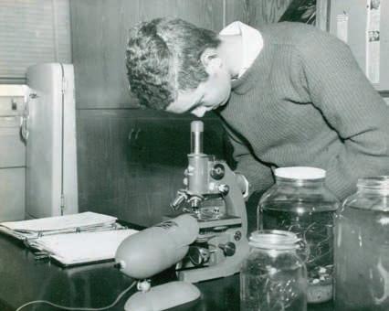
Ken Saladin’s penchant for writing began early. For his 10th-grade biology class, he wrote a 318-page monograph on hydras with 53 original India ink drawings and 10 original photomicrographs. We at McGraw-Hill think of this as Ken’s “first book.” At a young age, Ken already was developing his technical writing style, research habits, and illustration skills.
Some of Ken’s first pen-and-ink artwork
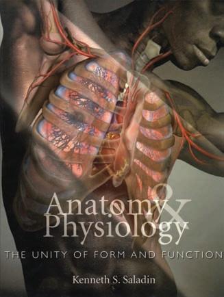
Ken served as an A&P textbook reviewer and testbank writer for several years and then embarked on his first book for McGraw-Hill in 1993. He published the first edition of Anatomy & Physiology: The Unity of Form and Function in 1997 and his first edition of Human Anatomy in 2004. The story continues with Human Anatomy, fifth edition.
Essentials book published in 2013
The story continues in 2016
Ken in 1964
Ken Saladin’s “first book,” Hydra Ecology (1965)
(1965)
Ken’s first textbook published in 1997
Courtesy of Ken Saladin
Courtesy of Ken Saladin
GUIDED TOUR
Instructive Artwork for Visual Learners
Saladin’s stunning illustrations and photos entice students who regard themselves as “visual learners.”
Vivid Illustrations with rich textures and shading and bold, bright colors bring anatomy to life.
and medulla, and leaves the node through one to three lymphatic vessels phatic organs have afferent lymphatic vessels; lymph nodes are the only organs that filter lymph as it flows along its course. With several afferent vessels but only a few efferent ones, the lymph node is a bottleneck that slows down lymph flow and allows time for cleansing it of foreign matter. The macrophages and reticular cells of the sinuses remove about 99% of the impurities before the lymph leaves the node. On its way to the bloodstream, lymph flows through one lymph node after another and thus becomes quite thoroughly cleansed of most impurities.
Blood vessels also penetrate the hilum of a lymph node. Arteries follow the medullary cords and give rise to capillary beds in the medulla and cortex. In the deep cortex near the junction with
in the neck, and monitor lymph coming from the head and neck.
Axillary lymph nodes are concentrated in the armpit (axilla) and receive lymph from the upper limb and the breast (see fig. 22.6b).
Process Figures relate numbered steps in the art with corresponding numbered text descriptions.
● Thoracic lymph nodes occur in the thoracic cavity and receive lymph from the lungs, airway, and mediastinum.
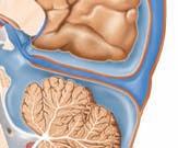
Figure 15.5 The Flow of Cerebrospinal Fluid.
CHAPTER ELEVEN The Muscular System II: Axial Musculature 281
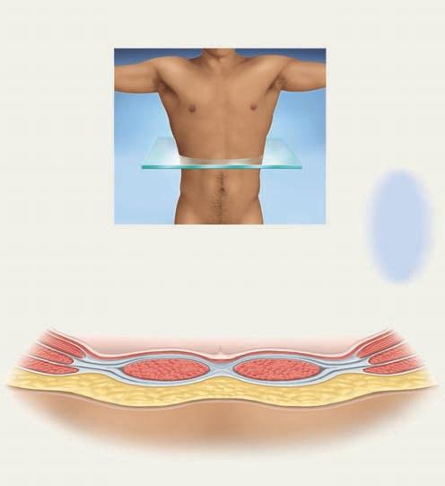
Abdominal Wall has little skeletal broad flat muscles strengthening the alternating layers of lumbar region and abdomen (fig. 11.10). abdominal oblique. Its deeper layer is pass upward and external oblique. (transversus pair of vertical to pubis. These tendinous bodybuilders muscles are continue medially and abdominis, they diverge enclosing the sheath. They meet between the rectus marks the lateral aponeurosis. The cordlike inguinal obliquely from the The linea alba, externally visible on a p. 338).
Orientation Tools, like dissection planes and a compass on the anatomical art, clarify the perspective from which a structure is viewed.
Muscles:
External abdominal oblique
Internal abdominal oblique
Transverse abdominal
Rectus abdominis
abdominal viscera against pull of vertebral column during heavy posture; compresses abdominal aiding in forceful expiration and loud singing and public speaking; aids urination, defecation, and vomiting. contraction causes contralateral rotation
oblique except that unilateral ipsilateral rotation of waist
abdominal contents, with same effects oblique, but does not contribute to vertebral column
region of vertebral column; flexes bending forward or doing sit-ups; region during walking; compresses
Aponeurosis of
Transverse abdominal
Internal abdominal oblique
External abdominal oblique
Skeletal Attachments Innervation
• Ribs 5–12
• Anterior half of iliac crest, pubic symphysis, and superior margin of pubis
Anterior rami of spinal nerves
T7–T12
The Psychology of Learning
Having taught human anatomy for 38 years, Saladin knows what works in the classroom and incorporates those approaches into the pedagogy of Human Anatomy.
Chapters Organized for Preview and Review
Chapter Outline provides a content preview and facilitates review and study.
Deeper Insights pique the interest of health-science students by showing the clinical relevance of the core science.
Brushing Up reminds students of the relevance of earlier chapters to the one on which they are currently embarking.
Anatomy & Physiology REVEALED® icons indicate which area of this interactive cadaver dissection program corresponds to the chapter topic.
• Inguinal ligament, iliac crest, and thoracolumbar fascia
• Linea alba, pubis, aponeurosis of internal oblique
• Pubic symphysis and superior margin of pubis
• Xiphoid process, costal cartilages 5–7
Anterior rami of spinal nerves T7–L1
Anterior rami of spinal nerves
T7–L1
Figure 11.10 Cross Section of the Anterior Abdominal Wall.
and sternum. The appendicular skeleton, studied in includes the bones of the upper limb and pectoral girdle, the lower limb and pelvic girdle.
Grand Total: 206 Bones
Anatomical Features of Bones
useful as you of reference other organ sysrelationships for example, muscle is attached artery travel the frontal, pariare named for understanding of how the depends on knowlpositions, shapes, for a clinician record a pulse, physical therapy
7.1
the Skeletal System
Overview of the Skeleton
stated that there are 206 bones in the skeleton, but typical adult count, not an invariable number. At about 270, and even more bones form during With age, however, the number decreases as separate example, each side of a child’s pelvic girdle has bones—the ilium, ischium, and pubis—but in adults, these single hip (coxal) bone on each side. The fusion bones, completed by late adolescence to the mid-20s, the average adult number of 206. These bones are 7.1. number varies even among adults. One reason is the sesamoid1 bones—bones that form within cerresponse to stress. The patella (kneecap) is the these; most of the others are small, rounded bones locations as the hands and feet (see fig. 8.14c, p. 198). for adult variation is that some people have extra skull called sutural (SOO-chure-ul), or wormian,2 7.6).
Expected Learning Outcomes
Bones exhibit a variety of ridges, spines, bumps, depressions, canals, pores, slits, cavities, and articular surfaces, often called bone markings. It is important to know the names of these features because later descriptions of joints, muscle attachments, and the routes traveled by nerves and blood vessels are based on this terminology. The terms for the most common of these features are listed in table 7.2, and several of them are illustrated in figure 7.2.
When you have completed this section, you should be able to a. define the two subdivisions of the skeleton; b. state the approximate number of bones in the adult body; c. explain why this number varies with age and from one person to another; and d. define several terms that denote surface features of bones.
Testing Your Recall
Reinforced Learning
CHAPTER THIRTEEN The Nervous System I: Nervous Tissue 369
As you study the skeleton, use yourself as a model. You can easily palpate (feel) many of the bones and some of their details through the skin. Rotate your forearm, cross your legs, palpate your skull and wrist, and think about what is happening beneath the surface or what you can feel through the skin. You will gain the most from this chapter (and indeed, the entire book) if you are conscious of your own body in relation to what you are studying.
Each section is a conceptually unified topic, framed between a pair of learning “bookends”—a set of learning objectives at the beginning and a set of review and self-testing questions at the end. Each section is numbered for easy reference in lecture, assignments, and ancillary materials.
Before You Go On
The skeleton (fig. 7.1) is divided into two regions: axial and appen dicular. The axial skeleton, studied in this chapter, forms the central supporting axis of the body and includes the skull, vertebral
1. The integrative functions of the nervous system are performed mainly by a. afferent neurons.
b. efferent neurons.
c. neuroglia.
d. sensory neurons.
e. interneurons.
6. Neurotransmitters are found in
a. the cell bodies of neurons.
b. the dendrites.
Answer the following questions to test your understanding of the preceding section:
c. the axon hillock.
1. Name the major components of the axial skeleton. Name those of the appendicular skeleton.
2. Neurons arise from embryonic a. endoderm.
b. epidermis.
c. mesoderm.
d. mesenchyme.
e. ectoderm.
Vocabulary Building
branches like a T, with a peripheral fiber carrying signals from the source of sensation and a central fiber continuing into the spinal cord. In most other neurons, a dendrite carries signals toward the soma, and an axon carries them away. In unipolar neurons, however, there is one long fiber that bypasses the soma and carries nerve signals directly to the spinal cord. The dendrites are the branching receptive endings in the skin or other place of origin, while the rest of the fiber is considered to be the axon (defined in these neurons by the presence of myelin and the ability to generate action potentials).
d. the presynaptic terminal.
Expected Learning Outcomes give the student a preview of key points to be learned within the next few pages.
11. Neurons that convey information to the CNS are called sensory, or _____, neurons.
e. the postsynaptic plasma membrane.
2. Explain why an adult does not have as many bones as a child does. Explain why one adult may have more bones than another adult of the same age.
7. Another name for the axon of a neuron is a. nerve fiber.
b. neurofibril.
3. Briefly describe each of the following bone features: condyle, epicondyle, process, tubercle, fossa, sulcus, and foramen.
c. neurilemma.
d. axoplasm.
e. endoneurium.
3. The soma of a mature neuron lacks a. a nucleus.
b. endoplasmic reticulum.
c. lipofuscin.
d. centrioles.
e. ribosomes.
● Anaxonic neurons have multiple dendrites but no axon. They communicate over short distances through their dendrites and do not produce action potentials. Anaxonic neurons are found in the brain, retina, and adrenal medulla. In the retina, they help in visual processes such as the perception of contrast.
4. The glial cells that destroy microorganisms in the CNS are a. microglia.
Before You Go On
b. satellite cells.
12. Motor effects that depend on repetitive output from a neural pool are most likely to use the _____ type of neural circuit.
Several features help build a student’s level of comfort with medical vocabulary.
Before You Go On prompts the student to pause and spot-check his or her mastery of the previous few pages before progressing to new material.
8. Nerves that directly control the motility of the stomach or rate of the heartbeat would belong to
a. the central nervous system.
Pronunciation Guides Knowing proper pronunciation is key to remembering and spelling terms. Saladin gives simple, intuitive “pro-NUN-see-AY-shun” guides to help students over this hurdle and widen the student’s comfort zone for medical vocabulary.
c. ependymal cells.
d. oligodendrocytes.
e. astrocytes.
13. Prenatal degeneration of the forebrain results in a birth defect called _____.
14. Neurons receive incoming signals by way of specialized processes called _____.
15. In the central nervous system, cells called _____ perform one of the same functions that Schwann cells do in the peripheral nervous system.
at least 10 to 1 by supportive cells called neuroglia (noo-ROG-leeuh), or glial (GLEE-ul) cells. Glial cells protect the neurons and aid their function. The word glia, which means “glue,” implies one of their roles—they bind neurons together. In the fetus, glial cells form a scaffold that guides young migrating neurons to their destinations. Wherever a mature neuron is not in synaptic contact with another cell, it is covered with glial cells. This prevents neurons from contacting each other except at points specialized for signal transmission, and thus lends precision to their conduction pathways.
Types of Neuroglia
b. the somatic sensory division.
c. the somatic motor division.
d. the visceral motor division.
16. A/An _____ synapse is formed when a presynaptic neuron synapses with the cell body of a postsynaptic neuron.
There are six major categories of neuroglia, each with a unique function (table 13.1). Four types occur only in the central nervous system (fig. 13.6):
e. the visceral sensory division.
9. The glial cells that guide migrating neurons in the developing fetal brain are a. astrocytes.
17. All of the nervous system except the brain and spinal cord is called the _____.
18. The _____ and _____ are necessary for regeneration of damaged nerve fibers in the peripheral nervous system.
Answer the following questions to test your understanding of the preceding section:
b. oligodendrocytes.
c. satellite cells.
Word Origins Accurate spelling and insight into medical terms are greatly enhanced by a familiarity with commonly used word roots, prefixes, and suffixes.
5. A friend takes a flash photograph of you, and you continue to see an image of the flash unit for several seconds afterward. This phenomenon is the result of a _____ circuit.
4. Explain why neurons could not function without the properties of excitability, conductivity, and secretion.
d. ependymal cells.
e. microglia.
5. Distinguish between sensory neurons, interneurons, and motor neurons.
a. diverging
1. Oligodendrocytes15 (OL-ih-go-DEN-dro-sites) somewhat resemble an octopus; they have a bulbous body with as many as 15 armlike processes. Each process reaches out to a nerve fiber and spirals around it like electrical tape wrapped repeatedly around a wire. This wrapping, called the myelin sheath, insulates the nerve fiber from the extracellular fluid and speeds up signal conduction in the nerve fiber.
10. Which of the following appears earlier than all the rest in prenatal development of the nervous system?
19. In the peripheral nervous system, the somas of the neurons are concentrated in enlarged, knotlike structures called _____ connected to the nerves.
20. At a given synapse, the _____ neuron has neurotransmitter receptors.
Footnotes throughout the chapters help build the student’s working lexicon of word elements. An end-of-book Glossary provides clear definitions of the most important or frequently used terms.
6. Define each of the following and explain its importance to neuronal function: dendrites, soma, axon, and presynaptic terminal.
b. converging
c. presynaptic
d. reverberating
a. the neural groove
b. a pair of primary vesicles
c. the neural plate
7. Sketch a multipolar, bipolar, unipolar, and anaxonic neuron; next to each sketch, state one place where such a neuron could be found.
e. parallel after-discharge
d. the neural crest
e. the neural tube
Building Your Medical Vocabulary An exercise at the end of each chapter helps students creatively use their knowledge of new medical word elements.
Building Your Medical Vocabulary
State a medical meaning of each of the following word elements, and give a term in which it is used.
1. -ic
2.
Answers in appendix A
Type LocationFunctions
OligodendrocytesCNSForm myelin in brain and spinal cord
Ependymal cellsCNSLine cavities of brain and spinal cord; secrete and circulate cerebrospinal fluid
Microglia CNSPhagocytize and destroy
c. describe how the speed of nerve signal conduction varies with nerve fiber diameter and the presence or absence of myelin; and
d. explain the relevance of neuroglia to the regeneration of damaged nerve fibers.
Answers in appendix A
of the extracellular fluid in the CNS; form scar tissue to replace damaged nervous tissue
Schwann cellsPNS Form neurilemma around all PNS nerve fibers and myelin around most of them; aid in regeneration of damaged nerve fibers
Parietal bone Occipital bone
Mandible
Humerus
Femur
Tibia
Fibula
Ulna Radius
Clavicle Scapula
Metatarsal bones Phalanges
Anterior view (b) Posterior view skeleton is colored green, and the rest is axial skeleton.
15oligo = few; dendro = branches; cyte = cell
TABLE 13.1 Types of Glial Cells
Self-Assessment Tools
Saladin provides students with abundant opportunities to evaluate their comprehension of concepts. A wide variety of questions from simple recall to analytical evaluation cover all six cognitive levels of Bloom’s Taxonomy of Educational Objectives.
Figure Legend Questions posed in many of the figure legends prompt the student to interpret the art and apply it to the reading.
MBefore You Go On questions test simple recall and lower-level interpretation of information read in the previous few pages.
Apply What You Know tests a student’s ability to think of the deeper implications or clinical applications of a point he or she just read.
● The placenta. This organ performs many functions in pregnancy, including fetal nutrition and waste removal. But it also secretes estrogen, progesterone, and other hormones that regulate pregnancy and stimulate development of the fetus and the mother’s mammary glands.
Apply What You Know Often, two hormones have opposite (antagonistic) effects on the same target organs. For example, oxytocin stimulates labor contractions, and progesterone inhibits premature labor. Name some other examples of antagonistic effects among the hormones in this chapter.
You can see that the endocrine system is extensive. It includes numerous discrete glands as well as individual cells in the tissues of other organs. The endocrine organs and tissues other than the hypothalamus and pituitary are reviewed in table 18.3
STUDY GUIDE
Assess Your Learning Outcomes
CHAPTER EIGHTEEN The Endocrine System 511
Before You Go On
To test your knowledge, discuss the following topics with a study partner or in writing, ideally from memory.
Answer the following questions to test your understanding of the preceding section:
9. Name two endocrine glands that are larger in children than in adults. What are their functions?
10. What hormone increases the body’s heat production in cold weather? What other functions does this hormone have?
11. Name the main hormone secreted by each layer of the adrenal cortex and one secreted by the adrenal medulla, and state the function of each.
12. What is the difference between a gonadal hormone and a gonadotropin?
13. What hormones are most important in regulating blood glucose concentration? What cells produce them? Where are these cells found?
14. Name one hormone produced by each of the following organs—the heart, liver, and placenta—and state the function of each hormone.
TABLE 18.3 Hormones from Sources Other Than the Hypothalamus and Pituitary
Source Hormone Target Organs and Tissues Principal Effects
Pineal glandMelatonin Brain Influences mood; may regulate the timing of puberty
uscles constitute nearly half of the body’s weight and occupy a place of central interest in several fields of health care and fitness. Physical and occupational therapists must be well acquainted with the muscular system to plan and carry out rehabilitation programs. Athletes and trainers, dancers and acrobats, and amateur fitness enthusiasts follow programs of resistance training to strengthen individual muscle groups through movement regimens based on knowledge of muscle, bone, and joint anatomy. Nurses employ their knowledge of the muscular system to give intramuscular injections correctly and to safely and effectively move patients who are physically incapacitated. Gerontological nurses are keenly aware of how deeply a person’s muscular condition affects the quality of life in old age. The muscular system is highly important to biomedical disciplines even beyond the scope of the movement sciences. It is the primary source of body heat in the moving individual, and through its absorption, storage, and use of glucose, it is a significant factor in blood glucose level and diabetes prevention. The next three chapters focus on the muscular system—the functional anatomy of muscular tissue in this chapter, muscles of the axial region of the body (head and trunk) in chapter 11, and muscles of the appendicular region (limbs and limb girdles) in chapter 12. These chapters draw on what we have covered in the preceding chapters—bone and joint structure—to flesh out our comprehension of body posture and movement. The current chapter also considers cardiac and smooth muscle and how they compare with skeletal muscle.
Thymus Thymopoietin, thymosin, thymulin T lymphocytes Stimulate T lymphocyte development and activity
8.1 The Pectoral Girdle and Upper Limb (p. 185)
1. The function of the pectoral girdle; the bones that compose it; and all points at which these bones articulate with each other, with the upper limb, and with the axial skeleton
2. The function of the pelvic girdle; the bones that compose it; and all points at which these bones articulate with each other, with the lower limb, and with the axial skeleton
3. The anatomical features of the hip bone, and the names and boundaries of the three childhood bones from which it arises
8.3
• What tissue characteristics evident in this photo distinguish this from cardiac and smooth muscle?
2. The anatomical features of the clavicle and scapula
3. The four segments (regions) of the upper limb
4. The names and locations of all 30 bones of the upper limb, and all points at which they articulate with each other
5. The anatomical features of the humerus, radius, ulna, carpal bones (especially the hamate), metacarpal bones, and phalanges
6. How the upper limb is anatomically adapted to the bipedalism of humans
8.2 The Pelvic Girdle and Lower Limb (p. 190)
1. The distinction between the pelvic girdle and pelvis
Testing Your Recall
b. articular cartilage.
Thyroid glandThyroxine (T4) and triiodothyronine (T3) Most tissues Elevate metabolic rate and heat production; promote alertness, quicker reflexes, enhanced absorption of dietary carbohydrates, protein synthesis, fetal and childhood growth, and CNS development
Calcitonin Bone Promotes net deposition of bone by inhibiting osteoclasts; reduces blood Ca2+ level
Parathyroid glandsParathyroid hormone (PTH)Bone, kidneys, small intestine Increases blood Ca2+ level by stimulating bone resorption, calcitriol synthesis, and intestinal Ca2+ absorption, and reducing urinary Ca2+ excretion
Muscle Types and Functions
Testing Your Recall sections at the end of each chapter offer 20 simple recall questions to test retention of terminology and basic ideas.
Adrenal medullaEpinephrine, norepinephrine, dopamine Most tissues Adaptive responses to arousal and stress
Adrenal cortexAldosterone Kidney Promotes Na+ retention and K+ excretion; maintains blood pressure and volume Cortisol and corticosteroneMost tissues Stimulate fat and protein catabolism, gluconeogenesis, stress resistance, and tissue repair
Expected Learning Outcomes
Androgens Bone, muscle, integument, many other tissues Growth of pubic and axillary hair, bone growth, sex drive, male prenatal development
True or False statements require students not merely to evaluate their truth, but also to concisely explain why the false statements are untrue, or rephrase them in a way that makes them true.
Pancreatic isletsGlucagon Primarily liver Stimulates glucose synthesis, glycogen and fat breakdown, release of glucose and fatty acids into circulation
Insulin Most tissues Stimulates glucose and amino acid uptake; lowers blood glucose level; promotes glycogen, fat, and protein synthesis
Amylin Stomach, gallbladderEnhances
1. The hip bone is attached to the axial skeleton through its a. auricular surface.
c. pubic symphysis.
d. conoid tubercle.
e. coronoid process.
a. ilium
b. pubis
c. femur
d. tibia e. talus
4. The meaning of greater pelvis, lesser pelvis, pelvic brim, pelvic inlet, and pelvic outlet, and the relationship of these structures to pregnancy and childbirth
5. How the pelvic girdle and gluteal muscles are anatomically adapted to the bipedalism of humans
6. Differences between the male and female pelvic girdles
Types of Muscle
7. The four segments (regions) of the lower limb
8. The names and locations of all 30 bones of the lower limb, and all points at which they articulate with each other
9. The anatomical features of the femur, patella, tibia, fibula, talus, calcaneus, navicular, cuboid, cuneiforms, metatarsals, and phalanges
10. The names and landmarks of the three foot arches
4. Compared to the male pelvis, the pelvis of a female
11. The Latin anatomical name for the thumb is _____, and the name for the great toe is _____.
a. has a less movable coccyx.
b. has a rounder pelvic inlet.
12. The acromion and coracoid process are parts of what bone?
2. Which of these bones supports the most body weight?
1. What portions of the appendicular skeleton are formed by intramembranous and endochondral ossification
2. Development of the limb buds and the manner in which they differentiate into the limbs, especially the processes of hand and foot development
3. The developmental processes that result in opposite directions of knee and elbow flexion and opposite orientations of the pollex and hallux
4. Developmental abnormalities of the appendicular skeleton including amelia, meromelia, polydactyly, syndactyly, and talipes
5. The most common noncongenital disorders of the appendicular skeleton
15. One of the wrist bones, the _____, is characterized by a prominent hook.
c. is narrower between the iliac crests.
d. has a narrower pubic arch. e. has a narrower sacrum.
13. How many phalanges, total, does the human body have?
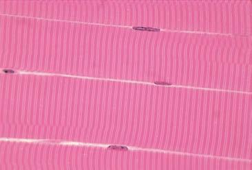
Skeletal muscle may be defined as voluntary striated muscle that is usually attached to one or more bones. It is called voluntary because it is usually subject to conscious control; we can decide when to contract a skeletal muscle. It is called striated because it exhibits a microscopic pattern of alternating light and dark bands, which result from the overlapping arrangement of the ). A typical skeletal muscle cell is about 100 μm in diameter and 3 cm (30,000 μm) long; some are as thick as 500 μm and as long as 30 cm. Because of their extraordinary length, skeletal muscle cells are usually —not
14. The bony prominences on each side of your elbow are the lateral and medial _____ of the humerus.
3. Which of these structures can be most easily palpated on a living person?
a. the deltoid tuberosity
b. the greater sciatic notch
c. the medial malleolus
Somatostatin Stomach, small intestine, pancreatic islets Inhibits digestion and nutrient absorption; inhibits glucagon and insulin secretion
When you have completed this section, you should be able to a. describe the distinctions between the three types of muscular tissue; and b. list the functions of muscular tissue and the properties it must have to carry out these functions.
7. The disc-shaped head of the radius articulates with the _____ of the humerus. a. radial tuberosity b. trochlea
16. The fibrocartilage pad that holds the pelvic girdle together anteriorly is called the _____.
5. The lateral and medial malleoli are most similar to a. the radial and ulnar styloid processes.
c. capitulum d. olecranon process e. glenoid cavity
17. The leg proper, between the knee and ankle, is called the _____ region.
b. the humeral capitulum and trochlea.
c. the acromion and coracoid process.
18. The _____ processes of the radius and ulna form bony protuberances on each side of the wrist.
d. the base and head of a metacarpal bone.
e. the anterior and posterior superior iliac spines.
State a medical meaning for each of the following word elements, and give a term in which it is used.
d. the coracoid process of the scapula
e. the glenoid cavity
sal03709_ch08_184-204.indd 203
Testing Your Comprehension questions are clinical application and other interpretive essay questions that require the student to apply the chapter’s basic science to clinical or other scenarios.
As we saw in chapter 3, there are three kinds of muscular tissue in the human body—skeletal, cardiac, and smooth. All types, however, are specialized for one fundamental purpose: to convert the chemical energy of ATP into the mechanical energy of motion. Muscle cells exert a useful force on other cells or tissues—either to produce desirable movements or to prevent undesirable ones. Although we examine all three muscle types in this chapter, most of our attention will focus on the muscular1 system, composed of the skeletal muscles only. The word muscle means “little mouse,” apparently referring to the appearance of muscles rippling under the skin. The study of the skeletal muscles is called myology.2
-icle
6. When you rest your hands on your hips, you are resting them on a. the pelvic inlet.
b. the pelvic outlet.
supra-
carpo-
19. Two massive protuberances unique to the proximal end of the femur are the greater and lesser _____.
20. The _____ arch of the foot extends from the heel to the great toe.
8. All of the following are carpal bones except the _____, which is a tarsal bone. a. trapezium d. triquetrum b. cuboid e. pisiform c. trapezoid
Answers in appendix A
9. The bone that supports your body weight when you are sitting down is a. the acetabulum. d. the coccyx. b. the pubis. e. the ischium. c. the ilium.
10. Which of these is the bone of the heel?
Building Your Medical Vocabulary 9. auro10. tarsoAnswers in appendix A
1. pect2. acro-
normally under conscious control. Its cells are not fibrous in shape, but relatively short and stumpy, somewhat like logs with myocytes rather than fibers. Cardiocytes are commonly about 80 μm long is also involuntary, and unlike skeletal and
True or False
c. the pelvic brim.
d. the iliac crests.
e. the auricular surfaces.
Determine which five of the following statements are false, and briefly explain why.
1. There are more carpal bones than tarsal bones.
2. The hands have more phalanges than the feet.
3. The upper limb is attached to the axial skeleton at only one point, the acromioclavicular joint.
4. On a living person, it would be possible to palpate the muscles in the infraspinous fossa but not those of the subscapular fossa.
Testing Your Comprehension
1. In adolescents, trauma sometimes separates the head of the femur from the neck. Why do you think this is more common in adolescents than in adults?
2. By palpating the hind leg of a cat or dog or examining a laboratory skeleton, you can see that cats and dogs stand on the heads of their metatarsal bones; the calcaneus does not touch the ground. How is this similar to the stance of a woman wearing highheeled shoes? How is it different?
a. cuboid d. trochlear b. calcaneus e. talus c. navicular
5. In strict anatomical terminology, the words arm and leg both refer to regions with only one bone.
If you rest your chin on your hands with your elbows on a table, the olecranon of the ulna rests on the table. 7. The most frequently broken bone in humans is the humerus. 8. The proximal end of the radius articulates with both the humerus and ulna.
9. The pisiform bone and patella are both sesamoid bones.
10. The pelvic outlet is the opening in the floor of the greater pelvis leading into the lesser pelvis.
Answers in appendix A
3. A deer hunter discovers a human skeleton in the woods and notifies authorities. A news report on the finding describes it as the body of an unidentified male between 17 and 20 years of age. What skeletal features would have been most useful for determining the sex and approximate age of the individual?
4. A surgeon has removed 8 cm of Joan’s radius because of osteosarcoma, a bone cancer, and replaced it with a graft taken from one of the bones of Joan’s lower limb. What bone do you think would most likely be used as the source of the graft? Explain your answer. 5. Andy, a 55-year-old, 75 kg (165-pound) roofer, is shingling the steeply pitched roof of a new house when he loses his footing and slides down the roof and over
I
various roles in communication—speech, writing, facial expressions, and other body language.
Figure 10.1 Skeletal Muscle Fibers.
CHAPTER EIGHT The Skeletal System III: Appendicular Skeleton 203
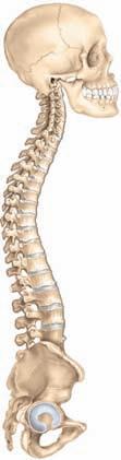
such as a chimpanzee; the head is balanced over the body’s center of gravity; and the eyes are directed straight forward (fig. 7.20). Abnormal lateral or anterior–posterior spinal curvatures are among the most common back problems (see Deeper Insight 7.3).
DEEPER INSIGHT 7.3
Abnormal Spinal Curvatures
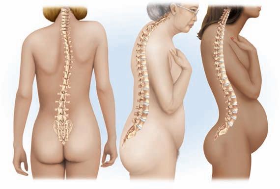
Abnormal spinal curvatures (fig. 7.21) can result from abdominal weight gain in obesity or pregnancy; poor posture; weakness or paralysis of the trunk muscles; some diseases; or congenital defects in vertebral anatomy. The most common deformity is an abnormal lateral curvature called scoliosis. It occurs most often in the thoracic region, particularly among adolescent girls. It sometimes results from a developmental abnormality in which the body and arch of a vertebra fail to develop on one side. If the person’s skeletal growth is not yet complete, scoliosis can be corrected with a back brace.
An exaggerated thoracic curvature is called kyphosis (hunchback, in lay language). It is usually a result of osteoporosis, but it also occurs in people with osteomalacia or spinal tuberculosis and in adolescents who engage heavily in such sports as wrestling and weight lifting. An exaggerated lumbar curvature is called lordosis (swayback). It can have the same causes as kyphosis, or it can result from added abdominal weight in pregnancy or obesity.
Figure 7.21 Abnormal Spinal Curvatures. (a) Scoliosis, an abnormal lateral deviation. (b) Kyphosis, an exaggerated thoracic curvature common in old age. (c) Lordosis, an exaggerated lumbar curvature common in pregnancy and obesity.
Making it Relevant
Deeper Insight essays cover the clinical application of basic science. Some Deeper Insight boxes highlight medical history and evolutionary interpretations of human structure and function.
Vertebral Columns. The S-shaped human vertebral column is an adaptation for bipedal locomotion.
Mc GRAW-HILL EDUCATION TEACHING AND LEARNING TOOLS
McGraw-Hill Education A&P: We Have Learning Down to a Science!
At McGraw-Hill Education, we work every day to unlock the full potential of each learner. Our mission is to accelerate learning through intuitive, engaging, efficient and effective experiences—grounded in research. MHE Anatomy & Physiology is your trusted, data-driven partner in A&P education. Since 2009, our adaptive programs in A&P have hosted 600,000 unique users who have answered more than 600 million probes, giving us the only data-driven solutions to help your students get from their first college-level course to program readiness.
LearnSmart® Prep is an adaptive learning tool that prepares students for college-level work in Anatomy & Physiology. Prep for Anatomy & Physiology now comes standard to students with Connect. The tool individually identifies concepts the student does not fully understand and provides learning resources to teach essential concepts so students enter the classroom prepared. Data-driven reports highlight areas where students are struggling, helping to accurately identify weak areas.
Anatomy
& Physiology∣
REVEALED®
is now mobile! Also, available in Cat and Fetal Pig versions.
Concept Overview Interactives combine multiple concepts into one big-picture summary. These striking, visually dynamic presentations offer a review of previously covered material in a creatively designed environment to emphasize how individual parts fit together in the understanding of a larger mechanism or concept. Concept Overview Interactive modules have assessable, auto-graded learning activities in Connect®, can be used as a self-study tool for students, and are also provided separately to instructors as classroom presentation tools.
Required=Results ®
McGraw-Hill Connect®
Learn Without Limits
Connect is a teaching and learning platform that is proven to deliver better results for students and instructors.
Connect empowers students by continually adapting to deliver precisely what they need, when they need it, and how they need it, so your class time is more engaging and effective.
88% of instructors who use Connect require it; instructor satisfaction increases by 38% when Connect is required.
Connect Insight®
Connect Insight is Connect’s new one-of-a-kind visual analytics dashboard—now available for both instructors and students—that provides at-a-glance information regarding student performance, which is immediately actionable. By presenting assignment, assessment, and topical performance results together with a time metric that is easily visible for aggregate or individual results, Connect Insight gives the user the ability to take a just-in-time approach to teaching and learning, which was never before available. Connect Insight presents data that empowers students and helps instructors improve class performance in a way that is efficient and effective.
Mobile
Students can view their results for any Connect course.
Connect’s new, intuitive mobile interface gives students and instructors flexible and convenient, anytime–anywhere access to all components of the Connect platform.
Adaptive
More students earn A’s and B’s when they use McGraw-Hill Education Adaptive products.
SmartBook®
Proven to help students improve grades and study more efficiently, SmartBook contains the same content within the print book, but actively tailors that content to the needs of the individual. SmartBook’s adaptive technology provides precise, personalized instruction on what the student should do next, guiding the student to master and remember key concepts, targeting gaps in knowledge and offering customized feedback, driving the student toward comprehension and retention of the subject matter. Available on smartphones and tablets, SmartBook puts learning at the student’s fingertips—anywhere, anytime.
THE FIRST AND ONLY ADAPTIVE READING EXPERIENCE DESIGNED TO TRANSFORM THE WAY STUDENTS READ
Over 4 billion questions have been answered, making McGraw-Hill Education products more intelligent, reliable, and precise.
ACKNOWLEDGMENTS
I wish to thank the hundreds of colleagues who have reviewed my writing over the years and tremendously contributed to the factual accuracy, scientific currency, and presentation style of the book before you. Much of this has come about through revising my flagship book, Anatomy & Physiology: The Unity of Form and Function, through seven editions. Human Anatomy and my book coauthored with Robin McFarland, Essentials of Anatomy & Physiology, have derived their own content improvements as they follow in the wake of the heavily reviewed two-semester textbook.
In addition to commissioned reviews of my chapters, spontaneous feedback from other instructors and from students all over the world has been enormously stimulating and helpful in the incessant effort to approach that elusive asymptote called textbook perfection. I’m deeply appreciative of all the encouragement, information, corrections, and suggestions these readers have sent, and I look forward to many more years of such productive correspondence.
My digital team—Steve Sullivan and Chris Gan—have greatly increased the educational value of these books through their work to create self-assessment tools and align McGraw-Hill’s Connect resources with the textbook. This has contributed greatly to student and instructor satisfaction with our overall package of learning media, and to the students’ success as they master A&P en route to their career aspirations. I am delighted to have them on my team—all for one, and one for all! Thank you so much for what you do to adapt my product to the needs of our students.
I would also like to extend appreciation to members of the Life Sciences Book Team at McGraw-Hill Education who have worked with me on this project, including Amy Reed, Senior Brand Manager; Chloe Bouxsein, Brand Manager; Donna Nemmers, Senior Product Developer; Vicki Krug, Senior Content Project Manager; Lori Hancock, Senior Content Licensing Specialist; Brent dela Cruz, Senior Content Project Manager; David Hash, Senior Designer Jeanne Patterson, freelance Copyeditor; and Julie De Adder, Photo Researcher. Their efforts have yielded another great edition of the text and its companion media suite of Connect products.
Ken Saladin
Georgia College & State University
LETTER TO STUDENTS
Dear Students,
When I was a young boy, I became interested in what I then called “nature study” for two reasons. One was the sheer beauty of nature. I reveled in children’s books with abundant, colorful drawings and photographs of animals, plants, minerals, and gems. It was this esthetic appreciation of nature that made me want to learn more about it and made me happily surprised to discover I could make a career of it. At a slightly later age, another thing that drew me still deeper into biology was to discover writers who had a way with words—who could captivate my imagination and curiosity with their elegant prose. Once I was old enough to hold part-time jobs, I began buying zoology and anatomy books that mesmerized me with their gracefulness of writing and fascinating art and photography. I wanted to write and draw like that myself, and I began learning from “the masters.” I spent many late nights in my room peering into my microscope and jars of pond water, typing page after page of manuscript, and trying pen and India ink as an art medium. My “first book” was a 318-page paper on some little pond animals called hydras, with 53 illustrations, that I wrote for my tenth-grade biology class when I was 16 (see p. ix).
Fast forward about 30 years to when I became a textbook writer, and I found myself bringing that same enjoyment of writing and illustrating to my own anatomy and physiology textbooks. Why? Not only for its intrinsic creative satisfaction, but because I’m guessing that you’re like I was—you can appreciate a book that does more than simply give you the information you need. You appreciate, I trust, a writer who makes it enjoyable for you through scientific, storytelling prose and a conceptualized way of illustrating things to spark interest and facilitate understanding. Some of you probably think of yourselves as “visual learners” and others as “verbal learners.” Either way, I hope this book will serve your learning style.
I know from my own students, however, that you need more than captivating illustrations and enjoyable reading. Let’s face it—A&P is a complex subject and it may seem a formidable task to acquire even a basic knowledge of the human body. It was difficult even for me to learn (and the learning never ends). So in addition to simply writing this book, I’ve given a lot of thought to what we call pedagogy—the art of teaching. I’ve designed my chapters to make them easier for you to study and to give you abundant opportunity to check whether you’ve understood what you read—to test yourself (as I advise my own students) before the instructor tests you. In later editions, we brought on a team of digital authors to produce online learning aids that students have commended as extremely helpful to them in learning human anatomy.
Each chapter is broken down into short, digestible bits with a set of learning goals (Expected Learning Outcomes) at the beginning of each section, and self-testing questions (Before You Go On) just a few pages later. Even if you have just 30 minutes to read during a lunch break or a bus ride, you can easily read or review one of these brief sections. There are also numerous self-testing questions at the end of each chapter, in some of the figure legends, and the occasional Apply What You Know questions dispersed through each chapter. The questions cover a broad range of cognitive skills, from simple recall of a term to your ability to evaluate, analyze, and apply what you’ve learned to new clinical situations or other problems.
The Guided Tour on page xii takes you through the learning aids we’ve created for you within the book itself and additional study aids available within Connect. I hope you will take a little time to look at the Guided Tour to see what we have to offer you. The Preface on page x goes a little deeper into my thinking behind the book’s design and content and will also help you get more out of your experience.
I hope you enjoy your study of this book, but I know there are always ways to make it even better. Indeed, what quality you may find in this edition owes a great deal to feedback I’ve received from students all over the world. If you find any typos or other errors, if you have any suggestions for improvement, if I can clarify a concept for you, or even if you just want to comment on something you really like about the book, I hope you’ll feel free to write to me. I correspond quite often with students and would enjoy hearing from you.
DEDICATION
This book is dedicated to my son Emory.
Ken Saladin
Georgia College & State University Milledgeville, GA 31061 (USA) ken.saladin@gcsu.edu
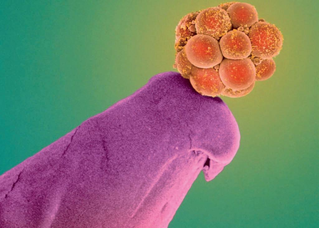
A new life begins—a human embryo on the point of a pin (scanning electron micrograph)
This book is an introduction to the structure of the human body. It is meant primarily to provide a foundation for advanced study in fields related to health and fitness. Beyond that purpose, however, the study of anatomy can also provide a satisfying sense of self-understanding. Even as children, we are curious about what’s inside the body. Dried skeletons, museum exhibits, and beautifully illustrated atlases of the body have long elicited widespread public fascination.
This chapter lays a foundation for our study of anatomy by considering some broad themes. We will consider what this science encompasses and what methods are used for the study of anatomy. We will lay out a general “road map” of the human body to provide a context for the chapters that follow. We will also get some insights into how a beginning anatomy student can become comfortable with medical terminology.
The Scope of Human Anatomy
Expected Learning Outcomes
When you have completed this section, you should be able to a. define anatomy and some of its subdisciplines; b. name and describe some approaches to studying anatomy; c. describe some methods of medical imaging; and d. discuss the variability of human anatomy.
Human anatomy is the study of the structural basis of body function. It provides an essential foundation for understanding physiology, the study of the functional relevance of that structure; anatomy and physiology together are the bedrock of the health sciences. You can study human anatomy from an atlas; yet as beautiful, fascinating, and valuable as atlases are, they teach almost nothing but the locations, shapes, and names of things. This book is different; it deals with what biologists call functional morphology1—not just the structure of organs, but the functional reasons behind it.
Anatomy and physiology complement each other; each makes sense of the other, and each molds the other in the course of human development and evolution. We cannot delve into the details of physiology in this book, but enough will be said of function to help you make sense of human structure and to more deeply appreciate the beauty of human form.
The Anatomical Sciences
Anatomy is an ancient human interest, undoubtedly older than any written language we know. We can only guess when people began deliberately cutting into human bodies out of curiosity, simply to
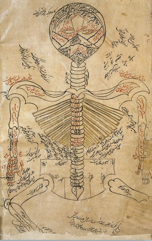
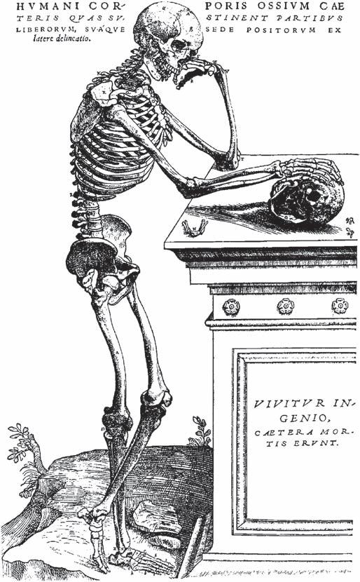
know what was inside. Some of the earliest and most influential books of anatomy were written by the Greek philosopher Aristotle (384–322 bce), the Greek physician Galen (129–c. 199 ce), and the Persian physician Avicenna (Ibn Sina, 980–1037 ce). For nearly 1,500 years, medical professors in Europe idolized these “ancient masters” and considered their works above reproach. Modern human anatomy, however, dates to the sixteenth century, when Flemish physician and professor Andreas Vesalius (1514–64) questioned the accuracy of the earlier authorities and commissioned the first accurate anatomical illustrations for his book, De Humani Corporis Fabrica (On the Structure of the Human Body, 1543) (fig. 1.1). The tradition begun by Vesalius has been handed down to us through such famous contemporary works as Gray’s Anatomy, Frank Netter’s Atlas of Human Anatomy, and many others, to the richly illustrated textbooks used by college students today.
For all its attention to the deceased body, or cadaver,2 human anatomy is hardly a “dead science.” New techniques of study continually produce exciting new insights into human structure and its functional relevance; anatomists have discovered far more about the human body in the last century than in the 2,500 years before. Anatomy now embraces several subdisciplines that study human structure from different perspectives. Gross anatomy is the study of structure visible to the naked eye, using methods such as surface observation, dissection, X-rays, and MRI scans. Surface anatomy is the external structure of the body, and is especially important in conducting a physical examination of a patient. Radiologic anatomy is the study of internal structure, using X-rays and other medical imaging techniques described in the next section.
Systemic anatomy is the study of one organ system at a time and is the approach taken by most introductory textbooks such as this one. Regional anatomy is the study of multiple organ systems at once in a given region of the body, such as the head or chest. (See the Atlas of Regional and Surface Anatomy on p. 329.) Medical schools and anatomy atlases typically teach anatomy from a regional perspective, because it is more practical to dissect all structures of the head and neck, the chest, or a limb, than it would be to try to dissect the entire digestive system, then the cardiovascular system, and so forth. Dissecting one system almost invariably destroys organs of other systems that stand in the way. Furthermore, as surgeons operate on a particular area of the body, they must think from a regional perspective and attend to the interrelationships of all structures in that area.
Ultimately, the structure and function of the body result from its individual cells. To see those, we usually take tissue specimens, thinly slice and stain them, and observe them under the microscope. This approach is called histology (microscopic anatomy). Histopathology3 is the microscopic examination of tissues for signs of disease. Cytology4 is the study of the structure and function of individual cells.
Anatomy, of course, is not limited to the study of humans, but extends to all living organisms. Even students of human structure
benefit from comparative anatomy—the study of more than one species in order to examine structural similarities and differences and analyze evolutionary trends. Anatomy students often begin by dissecting other animals with which we share a common ancestry and many structural similarities. Indeed, many of the reasons for human structure become apparent only when we look at the structure of other animals. In chapter 25, for example, you will see that physiologists had little idea of the purpose of certain tubular loops in the kidney (nephron loops) until they compared human kidneys with those of desert and aquatic animals, which have greater and lesser needs to conserve water. The greater an animal’s need to conserve water (the drier its habitat), the longer these loops are. Thus, comparative anatomy hinted at the function of the nephron loop, which could then be confirmed through experimental physiology. Such are the insights that can be gained by comparing different species with each other.
Methods of Study
There are several ways to examine the structure of the human body. The simplest is inspection—simply looking at the body’s appearance in careful detail, as in performing a physical examination or making a clinical diagnosis from surface appearance. Observations of the skin and nails, for example, can provide clues to such underlying problems as vitamin deficiencies, anemia, heart disease, and liver disease. Physical examinations involve not only looking at the body for signs of normalcy or disease, but also touching and listening to it. Palpation5 means feeling a structure with the hands, such as palpating a swollen lymph node or taking a pulse. Auscultation6 (AWS-cul-TAY-shun) is listening to the natural sounds made by the
Wellcome
Library, London
(a)
(b)
Figure 1.1 Evolution of Medical Art. Two illustrations of the skeletal system made about 500 years apart. (a) From an eleventh-century work attributed to Persian physician Avicenna. (b) From De Humani Corporis Fabrica (1543) by Andreas Vesalius.
body, such as heart and lung sounds. In percussion, the examiner taps on the body, feels for abnormal resistance, and listens to the emitted sound for signs of abnormalities such as pockets of fluid, air, or scar tissue.
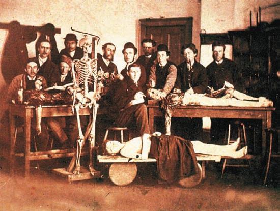
A deeper understanding of the body depends on dissection (dis-SEC-shun)—the careful cutting and separation of tissues to reveal their relationships. The very words anatomy7 and dissection8 both mean “cutting apart”; until the nineteenth century, dissection was called “anatomizing.” In many schools of health science, cadaver dissection is one of the first steps in the training of students (fig. 1.2).
Dissection, of course, is not the method of choice when studying a living person! Not long ago, it was common to diagnose disorders through exploratory surgery—opening the body and taking a look inside to see what was wrong and what could be done about it. Any breach of the body cavities is risky, however, and most exploratory surgery has now been replaced by medical imaging techniques—methods of viewing the inside of the body without surgery (fig. 1.3). The branch of medicine concerned with imaging is called radiology. Anatomy learned in this way is called radiologic anatomy, and those who use radiologic methods for clinical purposes include radiologists and radiologic technicians.
Some radiologic methods involve high-energy ionizing radiation such as X-rays or particles called positrons. These penetrate the tissues and can be used to produce images on X-ray film or through electronic detectors. The benefits of ionizing radiation must always be weighed against its risks. It is called ionizing because it ejects electrons from the atoms and molecules it strikes. This effect can cause mutation and trigger cancer. Thus, ionizing radiation cannot be used indiscriminately. Used judiciously, however, the benefits of a mammogram or dental X-ray substantially outweigh the small risk.
7ana = apart; tom = cut
8dis = apart; sect = cut
Some of the imaging methods to follow are considered noninvasive because they do not involve any penetration of the skin or body orifices. Invasive imaging techniques may entail inserting ultrasound probes into the esophagus, vagina, or rectum to get closer to the organ to be imaged, or injecting substances into the bloodstream or body passages to enhance image formation.
Any anatomy student today must be acquainted with the basic techniques of radiology and their respective advantages and limitations. Many of the images printed in this book have been produced by the following techniques.
Radiography
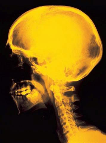
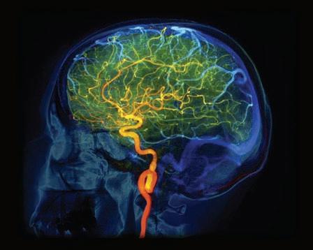
Radiography, first performed in 1895, is the process of photographing internal structures with X-rays. Until the 1960s, this was the only widely available imaging method; even today, it accounts for more than 50% of all clinical imaging. X-rays pass through the soft tissues of the body to a photographic film or detector on the other side, where they produce relatively dark images. They are absorbed, however, by dense tissues such as bones, teeth, tumors, and tuberculosis nodules, which leave the image lighter in these areas (fig. 1.3a). The term X-ray also applies to a photograph (radiograph) made by this method. Radiography is commonly used in dentistry, mammography, diagnosis of fractures, and examination of the chest. Hollow organs can be visualized by filling them with a radiopaque substance that absorbs X-rays. Barium sulfate, for example, is given orally for examination of the esophagus, stomach, and small intestine, or by enema for examination of the large intestine. Other substances are given by injection for angiography, the examination of blood vessels (fig. 1.3b). Some disadvantages of radiography are that images of overlapping organs can be confusing and slight differences in tissue density are not easily detected. In addition, X-rays present the aforementioned risks of ionizing radiation.
Computed Tomography
Computed tomography (a CT scan) is a more sophisticated application of X-rays. The patient is moved through a ring-shaped machine that emits low-intensity X-rays on one side and receives them with a detector on the opposite side. A computer analyzes signals from the detector and produces an image of a “slice” of the body about as thin as a coin (fig. 1.3c). The computer can “stack” a series of these images to construct a three-dimensional image of the body. CT scanning has the advantage of imaging thin sections of the body, so there is little organ overlap and the image is much sharper than a conventional X-ray. It requires extensive knowledge of cross-sectional anatomy to interpret the images. CT scanning is useful for identifying tumors, aneurysms, cerebral hemorrhages, kidney stones, and other abnormalities.
Magnetic Resonance Imaging
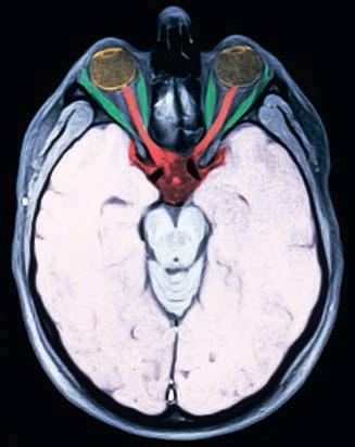
Magnetic resonance imaging (MRI) was conceived as a technique superior to CT for visualizing soft tissues (fig. 1.3d). The patient lies in a chamber surrounded by a large electromagnet that creates a very strong magnetic field. Hydrogen atoms in the tissues align themselves with the field. The technologist then activates a
National Library of Medicine
Figure 1.2 Early Medical Students in the Gross Anatomy Laboratory with Three Cadavers.
radio wave emitter, causing the hydrogen atoms to absorb additional energy and align in a different direction. When the radio waves are turned off, the hydrogen atoms abruptly realign to the magnetic field, giving off their excess energy at rates that depend on the type of tissue. A computer analyzes the emitted energy to produce an image of the body. MRI can “see” clearly through the skull and vertebral column to produce images of the nervous tissue. Moreover, it is better than CT for distinguishing between soft tissues such as the white and gray matter of the brain. It also avoids the harmful effects of X-rays. A disadvantage of MRI is that the patient must lie completely still in the enclosed space for about 45 minutes to scan one region of the body, and a complete procedure may entail 90 minutes to scan multiple regions such as the abdominal and pelvic cavities. Some patients find they cannot do this.
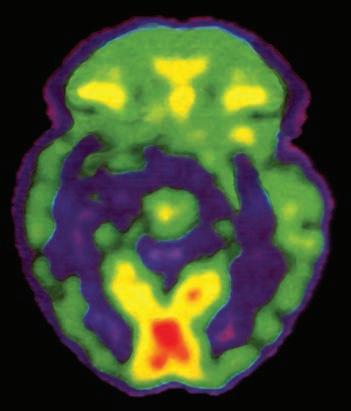
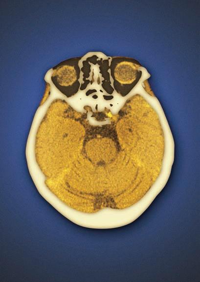
Figure 1.3 Radiologic Images of the Head. (a) X-ray (radiograph) showing the bones and teeth. (b) An angiogram of the cerebral blood vessels. (c) A CT scan at the level of the eyes. (d) An MRI scan at the level of the eyes. The optic nerves appear in red and the muscles that move the eyes appear in green. (e) A PET scan of the brain of an unmedicated schizophrenic patient. Red areas indicate regions of high metabolic rate. In this patient, the visual center of the brain at the rear of the head (bottom of photo) was especially active during the scan.
• What structures are seen better by MRI than by X-ray? What structures are seen better by X-ray than by PET?
MRI is also not very suitable for gastrointestinal imaging because it requires long exposures and the stomach and intestines move too much to create a sharp image. Functional MRI (fMRI) is a form of MRI that visualizes moment-to-moment changes in tissue function; fMRI scans of the brain, for example, show shifting patterns of activity as the brain applies itself to a specific sensory, mental, or motor task.
Apply What You Know
The concept of MRI was conceived in 1948 but could not be put into clinical practice until the 1970s. Speculate on a possible reason for this delay.
(a) X-ray (radiograph)
(b) Cerebral angiogram
(c) Computed tomographic (CT) scan
(e) Positron emission tomographic (PET) scan
(d) Magnetic resonance image (MRI)
Positron Emission Tomography
Positron emission tomography (the PET scan) is used to assess the metabolic state of a tissue and to distinguish which tissues are most active at a given moment (fig. 1.3e). The procedure begins with an injection of radioactively labeled glucose, which emits positrons (electron-like particles with a positive charge). When a positron and electron meet, they annihilate each other and give off gamma rays that can be detected by sensors and processed by computer. The result is a color image that shows which tissues were using the most glucose. In cardiology, PET scans can show the extent of tissue death from a heart attack. Since damaged tissue consumes little or no glucose, it appears dark. In neuroscience, PET scans can similarly reveal the extent of brain damage from stroke or trauma. PET scans are also widely used to diagnose cancer and evaluate tumor status. The PET scan is an example of nuclear medicine—the use of radioisotopes to treat disease or to form diagnostic images of the body.
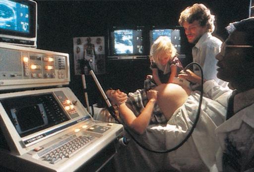
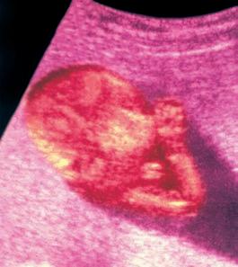
Sonography
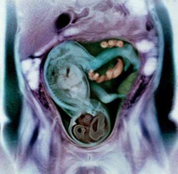
Sonography9 is the second oldest and second most widely used method of imaging. A handheld device pressed against the skin emits high-frequency ultrasound waves and receives the signals reflected back from internal organs. Sonography avoids the harmful effects of X-rays, and the equipment is relatively inexpensive and portable. Its primary disadvantage is that it does not produce a very sharp image. Although sonography was first used medically in the 1950s, images of significant clinical value had to wait until computer technology had developed enough to analyze differences in the way tissues reflect ultrasound. Sonography is not very useful for examining bones or lungs, but it is the method of choice in obstetrics, where the image (sonogram) can be used to locate the placenta and evaluate fetal age, position, and development (fig. 1.4). Sonography can also be used to view tissues in motion, such as fetal movements, a beating heart, and blood ejection from the heart. Sonographic imaging of the beating heart is called echocardiography.
Variation in Human Structure
A quick look around any classroom is enough to show that no two humans look exactly alike; on close inspection, even identical twins exhibit differences. Anatomy atlases and textbooks can easily give you the impression that everyone’s internal anatomy is the same, but this simply is not true. Books such as this one can teach you only the most common structure— the anatomy seen in approximately 70% or more of people. Someone who thinks that all human bodies are the same internally would make a very confused medical student or an incompetent surgeon.
Some people completely lack certain organs. For example, most of us have a palmaris longus muscle in the forearm and a plantaris muscle in the leg, but not everyone. Most of us have five lumbar vertebrae (bones of the lower spine), but some have four and some have six. Most of us have one spleen, but some people have two. Most have two kidneys, but some have only one. Most kidneys are supplied by a single renal artery and drained by one ureter, but in some people, a single kidney has two renal arteries or ureters. Figure 1.5 shows some common variations in human anatomy, and Deeper Insight 1.1 describes a particularly dramatic variation. 9sono = sound; graphy = recording process
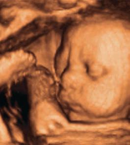
Figure 1.4 Fetal
Sonography. This threedimensional fetal image was made at 32 weeks of gestation.
• Why is sonography safer for the fetus than radiography or computed tomography?
DEEPER INSIGHT 1.1
Situs Inversus and Other Unusual Anatomy
In most people, the heart tilts toward the left, the spleen and sigmoid colon are on the left, the liver and gallbladder lie mainly on the right, the appendix is on the right, and so forth. This normal arrangement of the viscera is called situs (SITE-us) solitus
About 1 in 8,000 people is born, however, with a striking developmental abnormality called situs inversus—the organs of the thoracic and abdominal cavities are reversed between right and left. A selective left–right reversal of the heart is called dextrocardia. In situs perversus, a single organ occupies an atypical position, not necessarily a left–right reversal—for example, a kidney located low in the pelvic cavity instead of high in the abdominal cavity.
Some conditions, such as dextrocardia in the absence of complete situs inversus, can cause serious medical problems. Complete situs inversus, however, usually causes no functional problems because all of the viscera, though reversed, maintain their normal relationships to each other. Situs inversus is often diagnosed prenatally by sonography, but many people remain unaware of their condition for several decades until it is discovered by medical imaging, on physical examination, or in surgery. However, you can easily imagine the importance of such conditions in diagnosing appendicitis, performing gallbladder surgery, interpreting an X-ray, auscultating the heart valves, or recording an electrocardiogram.
People who are allergic to penicillin or aspirin often wear Medic Alert bracelets or necklaces that note this fact in case they need emergency medical treatment and are unable to communicate. Why would it be important for a person with situs inversus to have this noted on a Medic Alert bracelet?
Before You Go On
Answer the following questions to test your understanding of the preceding section:
1. How does functional morphology differ from the sort of anatomy taught by a photographic atlas of the body?
2. Why would regional anatomy be a better learning approach than systemic anatomy for a cadaver dissection course?
3. What is the difference between radiology and radiography?
4. What are some reasons that sonography would be unsuitable for examining the size and location of a brain tumor?
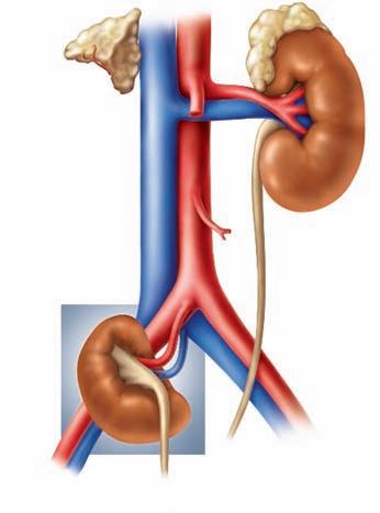
Variations in branches of the aorta
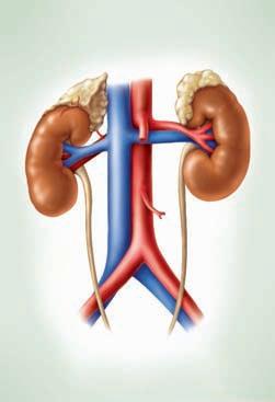
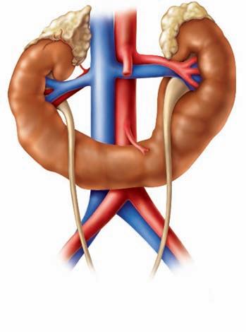
Horseshoe kidney
Figure 1.5 Variations in Anatomy of the Kidneys and Major Arteries near the Heart.
The Human Body Plan
Expected Learning Outcomes
When you have completed this section, you should be able to
a. list in proper order the levels of structural complexity of the body, from organism to atoms;
b. name the human organ systems and state the basic functions and components of each;
c. describe anatomical position and explain why it is important in medical language;
d. identify the three fundamental anatomical planes of the body;
e. define several terms that describe the locations of structures relative to each other;
f. identify the major body regions and their subdivisions;
g. name and describe the body cavities and the membranes that line them; and
h. explain what a potential space is, and give some examples.
The chapters that follow assume a certain core, common language of human structure. You will need to know what we mean by the names for the major body cavities and regions, know the difference between a tissue and an organ, and know where to look if you read that structure X is distal or medial to structure Y, for example. This section introduces this core terminology.
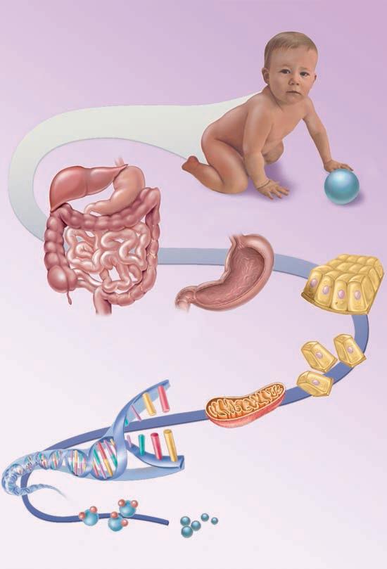
Levels of Human Structure
Although this book is concerned mainly with gross anatomy, the study of human structure spans all levels from the whole organism down to the atomic level. Consider for a moment an analogy to human structure: The English language, like the human body, is very complex, yet an endless array of ideas can be conveyed with a limited number of words. All words in the English language are, in turn, composed of various combinations of just 26 letters. Between the alphabet and a book are successively more complex levels of organization: syllables, words, sentences, paragraphs, and chapters. Humans have an analogous hierarchy of complexity (fig. 1.6), as follows:
The organism is composed of organ systems, organ systems are composed of organs, organs are composed of tissues, tissues are composed of cells, cells are composed (in part) of organelles, organelles are composed of molecules, and molecules are composed of atoms.
The organism is a single, complete individual, capable of acting separately from other individuals.
An organ system is a group of organs that carries out a basic function of the organism such as circulation, respiration, or digestion. The human body has 11 organ systems, defined and illustrated in the next section. Usually, the organs of a system are physically interconnected, such as the kidneys, ureters, urinary bladder, and urethra that compose the urinary system. The endocrine system, however, is a group of hormone-secreting glands and tissues that, for the most part, have no physical connection to each other.
An organ is any structure that has definite anatomical boundaries, is visually distinguishable from adjacent organs, and is composed of two or more tissue types working together to carry out a particular function. Most organs and higher levels of structure are within the domain of gross anatomy. However, there are organs within organs—the large organs visible to the naked eye contain smaller organs, some of which are visible only with the microscope. The skin, for example, is the body’s largest organ. Included within it are thousands of smaller organs: Each hair follicle, nail,
Organism
System Organ Tissue Cell
Organelle
Macromolecule
Molecule
Figure 1.6 The Body’s Structural Hierarchy from the Level of Organism to Atom. Each level depends on the structure and function of the level below it.
sweat gland, nerve, and blood vessel of the skin is an organ in itself.
A tissue is a mass of similar cells and cell products that forms a discrete region of an organ and performs a specific function. The body is composed of only four primary classes of tissue— epithelial, connective, nervous, and muscular tissue. Histology, the study of tissues, is the subject of chapter 3.
Cells are the smallest units of an organism considered to be alive. A cell is enclosed in a plasma membrane composed of lipids and protein, and it usually has one nucleus, an organelle that contains most of its DNA. Cytology, the study of cells and organelles, is the subject of chapter 2.
Organelles10 are microscopic structures in a cell that carry out its individual functions, much like organs such as the heart, liver, and kidneys carry out individual functions of the whole body. Organelles include the nucleus, mitochondria, lysosomes, centrioles, and others.
Organelles and other cellular components are composed of molecules—particles of at least two atoms joined by chemical bonds. The largest molecules, such as proteins, fats, and DNA, are called macromolecules.
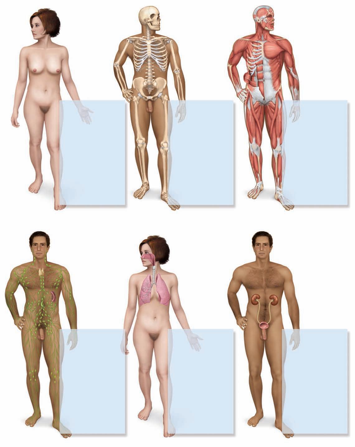
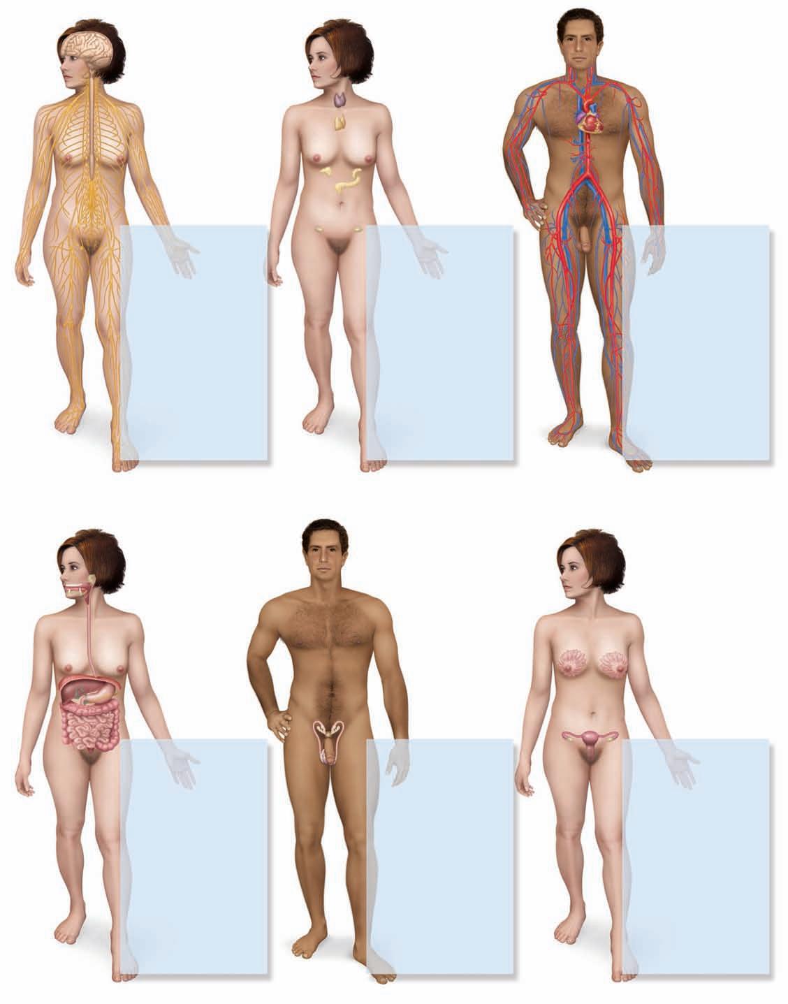
The Human Organ Systems
As remarked earlier, human structure can be learned from the perspective of regional anatomy or systemic anatomy. This book takes the systemic approach, in which we will fully examine one organ system at a time. There are 11 organ systems in the human body, as well as an immune system, which is better described as a population of cells that inhabit multiple organs rather than as an organ system. The organ systems are illustrated and summarized in figure 1.7 in the order that they are covered by this book. They are classified in the following list by their principal functions, although this is an unavoidably flawed classification. Some organs belong to two or more systems—for example, the male urethra is part of both the urinary and reproductive systems; the pharynx is part of the digestive and respiratory systems; and the mammary glands belong to both the integumentary and female reproductive systems.
Systems of Protection, Support, and Movement
Integumentary system
Skeletal system
Muscular system
Systems of Internal Communication and Integration
Nervous system
Endocrine system
Systems of Fluid Transport
Circulatory system
Lymphatic system
Systems of Intake and Output
Respiratory system
Digestive system
Urinary system
Systems of Reproduction
Male reproductive system
Female reproductive system
Some medical terms combine the names of two functionally related systems—for example, the musculoskeletal system, cardiopulmonary system, and urogenital (genitourinary) system. Such terms serve to call attention to the close anatomical or physiological relationship between two systems, but these are not literally individual organ systems.
The Terminology of Body Orientation
When anatomists describe the body, they must indicate where one structure is relative to another, the direction in which a nerve or blood vessel travels, the directions in which body parts move, and so forth. Clear communication on such points requires a universal terminology and frame of reference.
Anatomical Position
In describing the human body, anatomists assume that it is in anatomical position—that of a person standing upright with the feet flat on the floor and close together, arms at the sides, and the palms and face directed forward (fig. 1.8). Without such a frame of reference, to say that a structure such as the sternum, thymus, or aorta is “above the heart” would be vague, since it would depend on whether the subject was standing, lying face down, or lying face up. From the perspective of anatomical position, however, we can describe the thyroid gland as superior to the heart, the sternum as anterior(ventral) to it, and the aorta as posterior(dorsal) to it. These descriptions remain valid regardless of the subject’s position. Even if the body is lying down, such as a cadaver on the medical student’s dissection table, to say that the sternum is anterior to the heart invites the viewer to imagine the body standing in anatomical position and not to call it “above the heart” simply because that is the way the body happens to be lying.
Unless stated otherwise, assume that all anatomical descriptions refer to anatomical position. Bear in mind that if a subject is facing you in anatomical position, the subject’s left will be on your right and vice versa. In most anatomical illustrations, for example, the left atrium of the heart appears toward the right side of the page, and although the appendix is located in the right lower quadrant of the abdomen, it appears on the left side of most illustrations.
The forearm is said to be supinated when the palms face up or anteriorly and pronated when they face down or posteriorly (see fig. 9.13, p. 217); in anatomical position, the forearm is supinated. The words prone and supine seem similar to these but have an entirely different meaning. A person is prone if lying face down, and supine if lying face up.
Principal organs: Skin, hair, nails, cutaneous glands
Principal functions: Protection, water retention, thermoregulation, vitamin D synthesis, cutaneous sensation, nonverbal communication
Integumentary system
Principal organs: Lymph nodes, lymphatic vessels, thymus, spleen, tonsils
Principal functions: Recovery of excess tissue fluid, detection of pathogens, production of immune cells, defense against disease
Skeletal system
Principal organs: Bones, cartilages, ligaments
Principal functions: Support, movement, protective enclosure of viscera, blood formation, mineral storage, electrolyte and acid–base balance
Principal organs: Nose, pharynx, larynx, trachea, bronchi, lungs
Principal functions: Absorption of oxygen, discharge of carbon dioxide, acid–base balance, speech
Muscular system
Principal organs: Skeletal muscles
Principal functions: Movement, stability, communication, control of body openings, heat production
Principal organs: Kidneys, ureters, urinary bladder, urethra
Principal functions: Elimination of wastes; regulation of blood volume and pressure; stimulation of red blood cell formation; control of fluid, electrolyte, and acid–base balance; detoxification
Principal organs: Brain, spinal cord, ner ves, ganglia
Principal functions: Rapid internal communication, coordination, motor control and sensation
Principal functions: Hormone production; internal chemical communication and coordination
Principal organs: Heart, blood vessels
Principal functions:
Distribution of nutrients, oxygen, wastes, hormones, electrolytes, heat, immune cells, and antibodies; fluid, electrolyte, and acid–base balance
Nervous system
Principal organs: Teeth, tongue, salivary glands, esophagus, stomach, small and large intestines, liver, gallbladder, pancreas
Principal functions: Nutrient breakdown and absorption. Liver functions include metabolism of carbohydrates, lipids, proteins, vitamins, and minerals; synthesis of plasma proteins; disposal of drugs, toxins, and hormones; and cleansing of blood.
Principal functions: Production and delivery of sperm; secretion of sex hormones
Principal organs: Ovaries, uterine tubes, uterus, vagina, mammar y glands
Principal functions:
Production of eggs; site of fertilization and fetal development; fetal nourishment; birth; lactation; secretion of sex hormones
1.8 A Person Standing in Anatomical Position and Bisected Along the Three Principal Planes of Reference.
• What is another name for the specific sagittal plane shown here?
Anatomical Planes
Many views of the body are based on real or imaginary “slices” called sections or planes. Section implies an actual cut or slice to reveal internal anatomy, whereas plane implies an imaginary flat surface passing through the body. The three major anatomical planes are sagittal, frontal, and transverse (fig. 1.8).
A sagittal11 (SADJ-ih-tul) plane extends vertically and divides the body or an organ into right and left portions. The median (midsagittal) plane passes through the midline of the body and divides it into equal right and left halves. Other sagittal planes parallel to this (off center), called parasagittal12 planes, divide the body into unequal right and left portions. The head and pelvic organs are commonly illustrated on the median plane (fig. 1.9a).
A frontal (coronal13) plane also extends vertically, but it is perpendicular to the sagittal plane and divides the body into anterior (front) and posterior (back) portions. A frontal section of the head, for example, would divide it into one portion bearing the face and another bearing the back of the head. Contents of the thoracic and abdominal cavities are commonly shown in frontal section (fig. 1.9b).
A transverse (horizontal) plane passes across the body or an organ perpendicular to its long axis (fig. 1.9c); it divides the body into superior (upper) and inferior (lower) portions. CT scans are typically transverse sections (see fig. 1.3c), but not always.
Directional Terms
In “navigating” the human body and describing the locations of structures, anatomists use a set of standard directional terms (table 1.1). You will need to be very familiar with these in order to understand anatomical descriptions later in this book. The terms assume that the body is in anatomical position.
Most of these terms exist in pairs with opposite meanings: anterior versus posterior, superior versus inferior, medial versus lateral, proximal versus distal, and superficial versus deep. Intermediate directions are often indicated by combinations of these terms. For example, one structure may be described as anterolateral to another (toward the front and side).
The terms proximal and distal are used especially in the anatomy of the limbs, with proximal used to denote something relatively close to the limb’s point of attachment (the shoulder or hip joint) and distal to denote something farther away. These terms do have some
McGraw-Hill
Figure
(b) Frontal section
Sagittal section
Transverse section
Figure 1.9 Sections of the Body in the Three Primary Anatomical Planes. (a) Sagittal section of the pelvic region. (b) Frontal section of the thoracic region. (c) Transverse section of the head at the level of the eyes.
TABLE 1.1
Directional
Terms in Human Anatomy
Term Meaning
AnteriorToward the front of the body
PosteriorToward the back of the body
Ventral Toward the anterior side*
Dorsal Toward the posterior side*
SuperiorAbove
Inferior Below
CephalicToward the head or superior end
Rostral Toward the forehead or nose
Caudal Toward the tail or inferior end
Medial Toward the midline of the body
Lateral Away from the midline of the body
Examples of Usage
The sternum is anterior to the heart.
The esophagus is posterior to the trachea.
The abdomen is the ventral side of the body.
The scapulae are dorsal to the rib cage.
The heart is superior to the diaphragm.
The liver is inferior to the diaphragm.
The cephalic end of the embryonic neural tube develops into the brain.
The forebrain is rostral to the brainstem.
The spinal cord is caudal to the brain.
The heart is medial to the lungs.
The eyes are lateral to the nose.
ProximalCloser to the point of attachment or originThe elbow is proximal to the wrist.
Distal Farther from the point of attachment or originThe fingernails are at the distal ends of the fingers.
IpsilateralOn the same side of the body (right or left)The liver is ipsilateral to the appendix.
ContralateralOn opposite sides of the body (right and left)The spleen is contralateral to the liver.
SuperficialCloser to the body surface
Deep Farther from the body surface
The skin is superficial to the muscles.
The bones are deep to the muscles.
*In humans only; definition differs for other animals. In human anatomy, anterior and posterior are usually used in place of ventral and dorsal
applications to anatomy of the trunk of the body—for example, in referring to certain aspects of the intestines and the microscopic structure of the kidneys. But when describing the trunk and referring to a structure that lies above or below another in anatomical position, superior and inferior are the preferred terms. These terms are not usually used for the limbs. Although it may be technically correct, one would not generally say the elbow is superior to the wrist. Rather, it is proximal to the wrist. Because of the bipedal, upright stance of humans, some directional terms have different meanings for humans than they do for other animals. Anterior, for example, denotes the region of the body that leads the way in normal locomotion. For a fourlegged animal such as a cat, this is the head end of the body; for a human, however, it is the front of the chest and abdomen. What we call anterior in a human would be called ventral in a cat. Posterior denotes the region that comes last in normal locomotion—the tail end of a cat but the back of a human. In the anatomy of most other animals, ventral denotes the surface of the body closest to the ground and dorsal denotes the surface farthest away from the ground. These two words are too entrenched in human anatomy to completely ignore them, but we will minimize their use in this book to avoid confusion. You must keep such differences in mind, however, when dissecting other animals for comparison to human anatomy.
One vestige of the term dorsal is dorsum, used to denote the upper surface of the foot and the back of the hand. If you consider how a quadrupedal animal stands, the corresponding surfaces of its paws are both uppermost, facing the same direction as the dorsal side of its trunk. Although these surfaces of the human hand and foot face entirely different directions in anatomical position, the term dorsum is still used.
Major Body Regions
Knowledge of the external anatomy and landmarks of the body is important in performing a physical examination and many other clinical procedures. For purposes of study, the body is divided into two major regions called the axial and appendicular regions. Smaller areas within the major regions are described in the following paragraphs and illustrated in figure 1.10.
Axial Region
The axial region consists of the head, neck (cervical14 region), and trunk. The trunk is further divided into the thoracic region above the diaphragm and the abdominal region below it.
Upper limb:
Acromial r. (shoulder)
Axillary r. (armpit)
Brachial r. (arm)
Cubital r. (elbow)
Antebrachial r. (forearm)
Carpal r. (wrist)
Palmar r. (palm)
Lower limb:
Coxal r. (hip)
Patellar r. (knee)
Cephalic r. (head)
Facial r. (face)
Cervical r. (neck)
Thoracic r. (chest): Sternal r.
Pectoral r.
Umbilical r.
Abdominal r.
Inguinal r. (groin)
Pubic r.:
Mons pubis
External genitalia: Penis
Scrotum
Testes
Lower limb:
Femoral r. (thigh)
Crural r. (leg)
Tarsal r. (ankle)
Pedal r. (foot): Dorsum
Plantar surface (sole)
Cranial r.
Nuchal r. (back of neck)
Interscapular r.
Scapular r.
Vertebral r.
Lumbar r.
Sacral r.
Gluteal r. (buttock)
Dorsum of hand
Perineal r.
Femoral r.
Popliteal r.
Crural r.
Tarsal r.
Calcaneal r. (heel)
(a) Anterior (ventral)
(b) Anterior (ventral)
(c) Posterior (dorsal)
(d) Posterior (dorsal)
Figure 1.10 The Adult Female and Male Body Regions. (r. = region)