Veterinary Microbiology
Fourth Edition
Editors
D. Scott McVey, DVM, PhD, DACVM
Director and Professor, School of Veterinary Medicine and Biomedical Sciences
Associate Dean, Iowa/Nebraska Program for Professional Veterinary Medicine College of Agricultural Sciences and Natural Resources, Institute of Agriculture and Natural Resources University of Nebraska Lincoln Lincoln, NE, USA
Melissa Kennedy, DVM, PhD, DACVM
Professor Emeritus
Department of Biomedical and Diagnostic Sciences Department College of Veterinary Medicine University of Tennessee Knoxville, TN, USA
M.M. Chengappa, BVSc, MVSc, MS, PhD, DACVM
University Distinguished Professor Diagnostic Medicine Pathobiology College of Veterinary Medicine
Kansas State University Manhattan, KS, USA
Rebecca Wilkes, DVM, PhD, DACVM
Associate Professor and Section Head, Molecular Section
Animal Disease Diagnostic Laboratory
Department of Comparative Pathobiology College of Veterinary Medicine
Purdue University West Lafayette, IN, USA
This fifth edition first published 2022 © 2022 John Wiley & Sons, Inc.
All rights reserved. No part of this publication may be reproduced, stored in a retrieval system, or transmitted, in any form or by any means, electronic, mechanical, photocopying, recording or otherwise, except as permitted by law. Advice on how to obtain permission to reuse material from this title is available at http://www.wiley.com/go/permissions.
The right of Scott McVey, Melissa Kennedy, M.M. Chengappa, and Rebecca Wilkes to be identified as the author of the editorial material in this work has been asserted in accordance with law.
Registered Office
John Wiley & Sons, Inc., 111 River Street, Hoboken, NJ 07030, USA
Editorial Office
111 River Street, Hoboken, NJ 07030, USA
For details of our global editorial offices, customer services, and more information about Wiley products visit us at www.wiley.com. Wiley also publishes its books in a variety of electronic formats and by print-on-demand. Some content that appears in standard print versions of this book may not be available in other formats.
Limit of Liability/Disclaimer of Warranty
The contents of this work are intended to further general scientific research, understanding, and discussion only and are not intended and should not be relied upon as recommending or promoting scientific method, diagnosis, or treatment by physicians for any particular patient. In view of ongoing research, equipment modifications, changes in governmental regulations, and the constant flow of information relating to the use of medicines, equipment, and devices, the reader is urged to review and evaluate the information provided in the package insert or instructions for each medicine, equipment, or device for, among other things, any changes in the instructions or indication of usage and for added warnings and precautions. While the publisher and authors have used their best efforts in preparing this work, they make no representations or warranties with respect to the accuracy or completeness of the contents of this work and specifically disclaim all warranties, including without limitation any implied warranties of merchantability or fitness for a particular purpose. No warranty may be created or extended by sales representatives, written sales materials or promotional statements for this work. The fact that an organization, website, or product is referred to in this work as a citation and/or potential source of further information does not mean that the publisher and authors endorse the information or services the organization, website, or product may provide or recommendations it may make. This work is sold with the understanding that the publisher is not engaged in rendering professional services. The advice and strategies contained herein may not be suitable for your situation. You should consult with a specialist where appropriate. Further, readers should be aware that websites listed in this work may have changed or disappeared between when this work was written and when it is read. Neither the publisher nor authors shall be liable for any loss of profit or any other commercial damages, including but not limited to special, incidental, consequential, or other damages.
Library of Congress Cataloging-in-Publication Data applied for
[HB ISBN: 9781119650751]
Cover Design: Wiley
Cover Image: © Scott McVey
Set in 9.5/12.5pt STIXTwoText by Straive, Pondicherry, India
The editors appreciate the great support of our colleagues, friends and, most importantly, family members. Without your support, this book would not have been possible.
Contents
List of Contributors xii
Acknowledgments xviii
Preface xix
About the Companion Website xx
Part I Introduction 1
1 Microbial Infections of Animals 3
D. Scott McVey, Melissa Kennedy, and Charles Czuprynski
2 Basic Bacteriology 11
Tiruvoor G. Nagaraja
3 Basic Mycology 29
Charles Czuprynski and M.M. Chengappa
4 Basic Virology 35
Mohamed A. Abouelkhair and Melissa Kennedy
Part II Bacteriology 41
5 Family Enterobacteriaceae 43
Rodney A. Moxley
6 Enterobacteriaceae: Escherichia 56
Rodney A. Moxley
7 Enterobacteriaceae: Salmonella 75
Rodney A. Moxley
8 Family Yersiniaceae 88
Rodney A. Moxley
9 Enterobacteriaceae: Shigella 100
Rodney A. Moxley
10 Pasteurellaceae: Avibacterium, Bibersteinia, Mannheimia, and Pasteurella 108
William B. Crosby and Amelia R. Woolums
11 Pasteurellaceae: Actinobacillus 118
Bradley W. Fenwick and Andrew N. Rycroft
12 Pasteurellaceae: Glaesserella, Haemophilus, and Histophilus 129
Amelia R. Woolums
13 Bordetella 136
Bradley W. Fenwick
14 Brucella 151
S.C. Olsen and P. Boggiatto
15 Burkholderia mallei and Burkholderia pseudomallei 162
Sanjeev Narayanan
16 Francisella tularensis 168
Marilynn A. Larson and Peter C. Iwen
17 Moraxella 176
John Dustin Loy and Gabriele Maier
18 Pseudomonas 183
Deepti Pillai
19 Taylorella 187
Megan E. Jacob
20 Spirilla I: Borrelia 192
Roman R. Ganta
21 Spiral-Curved Organisms II: Brachyspira and Lawsonia 196
Gerald E. Duhamel
22 Spiral-Curved Organisms III: Campylobacter and Arcobacter 207
Gerald E. Duhamel
23 Spirilla IV: Helicobacter – the Spiral Microorganisms of the Gastrointestinal Tract and Liver 219
Megan E. Jacob
24 Spirochetes V: Leptospira 225
Sreekumari Rajeev
25 Staphylococcus 231
George C. Stewart
26 Streptococcus and Enterococcus 240
George C. Stewart
27 Trueperella 252
Tiruvoor G. Nagaraja
28 Bacillus 257
George C. Stewart
29 Corynebacterium 265
Tiruvoor G. Nagaraja
30 Erysipelothrix 273
Timothy Frana and Axel Neubauer
31 Listeria 280
Sanjeev Narayanan
32 Rhodococcus 286
Seth P. Harris and Joshua Daniels
33 Gram-Negative, Non-Spore-Forming Anaerobes 294
Tiruvoor G. Nagaraja
34 Clostridium 309
Iman Mehdizadeh Gohari and John F. Prescott
35 Filamentous Bacteria: Actinomyces, Nocardia, Dermatophilus, and Streptobacillus 335
Megan E. Jacob
36 Mycobacteria 345
Raul G. Barletta and David J. Steffen
37 Chlamydiaceae: Chlamydia 360
Roman R. Ganta
38 Mollicutes 364
Bonto Faburay and D. Scott McVey
39 Rickettsiaceae and Coxiellaceae: Rickettsia and Coxiella 377
Roman R. Ganta
40 Anaplasmataceae: Anaplasma 381
Roman R. Ganta
41 Anaplasmataceae: Ehrlichia and Neorickettsia 386
Roman R. Ganta
42 Bartonellaceae 392
Kathryn E. Reif
Part III Fungi 405
43 Yeasts: Cryptococcus, Malassezia, and Candida 407
Lisa M. Pohlman and M.M. Chengappa
44 Dermatophytes 418
M.M. Chengappa and Lisa M. Pohlman
45 Agents of Subcutaneous Mycoses 425
Lisa M. Pohlman and M.M. Chengappa
46 Agents of Systemic Mycoses 433
Lisa M. Pohlman and M.M. Chengappa
Part IV Viruses 449
47 Parvoviridae 451
Rebecca P. Wilkes
48 Circoviridae 469
Pablo Piñeyro and Sheela Ramamoorthy
49 Asfarviridae and Iridoviridae 478
Melissa Kennedy, Gustavo Delhon, D. Scott McVey, Hiep Vu, and Manuel Borca
50 Papillomaviridae and Polyomaviridae 484
Mohamed A. Abouelkhair and Melissa Kennedy
51 Adenoviridae 489
Yunjeong Kim and Kyeong-Ok Chang
52 Herpesviridae 496
Rebecca P. Wilkes and Jobin Kattoor
53 Poxviridae 522
Gustavo Delhon
54 Picornaviridae 533
Luis L. Rodriguez and Jonathan Arzt
55 Caliciviridae 543
Mohamed A. Abouelkhair and Melissa Kennedy
56 Togaviridae and Flaviviridae 552
Christopher C.L. Chase
57 Orthomyxoviridae 573
Wenjun Ma
58 Bunyavirales 589
William C. Wilson, Dana Mitzel, Lee W. Cohnstaedt, Leela Noronha, Barbara S. Drolet, and D. Scott McVey
59 Paramyxoviridae, Pneumoviridae, Filoviridae, and Bornaviridae 596
Stefan Niewiesk and Michael Oglesbee
60 Rhabdoviridae 609
Susan M. Moore and D. Scott McVey
61 Coronaviridae and Tobaniviridae 622
Udeni B.R. Balasuriya, Yun Young Go, and Mariano Carossino
62 Arteriviridae and Roniviridae 659
Udeni B. R. Balasuriya, Mariano Carossino, and Yun Young Go
63 Reoviridae 679
Barbara S. Drolet, Bethany L. McGregor, Lee W. Cohnstaedt, William C. Wilson, and D. Scott McVey
64 Birnaviridae 693
Melissa Kennedy and Donald L. Reynolds
65 Retroviridae 698
Jean-Pierre Frossard
66 Transmissible Spongiform Encephalopathies 728
Jürgen A. Richt and Nicholas Haley
Part V Control of Infectious Diseases 743
67 Immune Responses to Infectious Agents 745
Laurel J. Gershwin
68 Laboratory Diagnosis 760
D. Scott McVey, Bruce Brodersen, Duan Loy, and John Dustin Loy
69 Antimicrobial Chemotherapy and Antimicrobial Resistance 771
Michael D. Apley
70 Vaccines 803
D. Scott McVey, Jishu Shi, and Donald Reynolds
71 Disinfection and Sterilization 813
John Dustin Loy, D. Scott McVey, and M.M. Chengappa
72 Epidemiology of Infectious Diseases 818
Natalia Cernicchiaro, Ana R.S. Oliveira, and Lee W. Cohnstaedt
Index 829
List of Contributors
Mohamed A. Abouelkhair, DVM, MS, PhD, DACVM, CABMM
Assistant Professor of Virology and Immunology and Director, Virology Diagnostic Laboratory
University of Tennessee Veterinary Medical Center College of Veterinary Medicine
Biomedical and Diagnostic Sciences University of Tennessee Knoxville, Tennessee
United States
Michael D. Apley, DVM, PhD, DACVCP
Frick Professor
Department of Clinical Sciences College of Veterinary Medicine
Kansas State University Manhattan, Kansas United States
Jonathan Arzt, DVM, MPVM, PhD, DACVP Research Veterinary Medical Officer
Foreign Animal Disease Research Unit
Plum Island Animal Disease Center Agricultural Research Service, USDA Greenport, New York
United States
Udeni B.R. Balasuriya, BVSc, MS, PhD, FSLCVS
Professor of Virology, Department of Pathobiological Sciences
Director, Louisiana Animal Disease Diagnostic Laboratory (LADDL)
Director, LSU Biosafety Level 3 Core Facility
School of Veterinary Medicine
Louisiana State University
Baton Rouge, Louisiana United States
Raul G. Barletta, PhD Professor
School of Veterinary Medicine and Biomedical Sciences
University of Nebraska–Lincoln Lincoln, Nebraska United States
Brian Bellaire, PhD
Assistant Professor
Department of Veterinary Microbiology and Preventative Medicine
College of Veterinary Medicine
Iowa State University Ames, Iowa United States
Paola M. Boggiatto, DVM, PhD Veterinary Medical Officer
Infectious Bacterial Diseases Research Unit
National Animal Disease Center
USDA/Agricultural Research Service
Ames, Iowa United States
Manuel V. Borca, DVM, PhD
Lead Scientist and Research Veterinary Medical Officer
USDA Agricultural Research Service
Plum Island Animal Disease Center
Foreign Animal Disease Research Unit
Greenport , New York United States
Bruce W. Brodersen, DVM, PhD
Professor and Director
Nebraska Veterinary Diagnostic Center
School of Veterinary Medicine and Biomedical Sciences University of Nebraska–Lincoln Lincoln, Nebraska
United States
Mariano Carossino, DVM, PhD, DACVM
Assistant Professor of Veterinary Pathology Department of Pathobiological Sciences and Louisiana Animal Disease Diagnostic Laboratory School of Veterinary Medicine
Louisiana State University Baton Rouge, Louisiana
United States
Natalia Cernicchiaro, DVM, MS, PhD
Associate Professor of Epidemiology Center for Outcomes Research and Epidemiology Department of Diagnostic Medicine / Pathobiology College of Veterinary Medicine
Kansas State University Manhattan, Kansas
United States
Kyeong-Ok Chang, DVM, MS, PhD Professor Department of Diagnostic Medicine / Pathobiology College of Veterinary Medicine
Kansas State University Manhattan, Kansas
United States
Christopher C.L. Chase, DVM, MS, PhD, DACVM Professor Department of Veterinary and Biomedical Sciences
South Dakota State University
Brookings, South Dakota
United States
M.M. Chengappa, BVSc, MVSc, MS, PhD, DACVM
University Distinguished Professor Department of Diagnostic Medicine / Pathobiology College of Veterinary Medicine Kansas State University Manhattan, Kansas
United States
Lee W. Cohnstaedt, PhD
Research Epidemiologist
USDA Agricultural Research Service
Foreign Arthropod-Borne Animal Diseases Research Unit Manhattan, Kansas
United States
William B. Crosby, BS, DVM Graduate Research Assistant Department of Pathobiology and Population Medicine College of Veterinary Medicine Mississippi State University Starkville, Mississippi
United States
Charles Czuprynski, PhD, DACVM (hon.) Professor of Microbiology Department of Pathobiological Sciences School of Veterinary Medicine University of Wisconsin Madison, Wisconsin
United States
Joshua B. Daniels, DVM, PhD, DACVM Associate Professor Department of Veterinary Clinical Sciences College of Veterinary Medicine and Biomedical Sciences Colorado State University Fort Collins, Colorado
United States
Gustavo A. Delhon, MV, MS, PhD Research Professor School of Veterinary Medicine and Biomedical Sciences University of Nebraska–Lincoln Lincoln, Nebraska United States
Barbara Drolet, MS, PhD Research Microbiologist USDA Agricultural Research Service Center for Grain and Animal Health Research Arthropod-Borne Animal Diseases Research Unit Manhattan, Kansas United States
Gerald E. Duhamel, DVM, PhD, DACVP Professor of Anatomic Pathology New York State Animal Health Diagnostic Center and Section of Anatomic Pathology College of Veterinary Medicine Cornell University Ithaca, New York
United States
Bonto Faburay, DVM, MS, PhD
Section Head and Supervisory Veterinary Medical Officer
USDA Animal and Plant Health Inspection Service
Plum Island Animal Disease Center
Foreign Animal Disease Diagnostic Laboratory Greenport, New York
United States
Bradley W. Fenwick, DVM, MS, PhD, DACVM
Senior Vice President for Open Science and Innovation
Taylor and Francis London
United Kingdom
Timothy Frana, DVM, MS, MPH, PhD, DACVPM, DACVM
Boehringer Ingelheim Animal Health
St. Joseph , Missouri
United States
Jean-Pierre Frossard, BS (hons), MS, PhD
Emerging Virus Research Unit Head
Emerging Virus Research Unit
Animal and Plant Health Agency (APHA) Weybridge Surrey
United Kingdom
Roman R. Ganta, MSc, PhD
University Distinguished Professor
Director, Center of Excellence for Vector-Borne Diseases
Fellow of the Conference of Research Workers in Animals Diseases (CRWAD)
Fellow of the Association of Biotechnology and Pharmacy (FABAP)
Department of Diagnostic Medicine / Pathobiology College of Veterinary Medicine
Kansas State University Manhattan, Kansas
United States
Laurel J. Gershwin, DVM, PhD, DACVM
Distinguished Professor
Department of Pathology, Microbiology, and Immunology
College of Veterinary Medicine
University of California Davis, California
United States
Yun Young Go, DVM, MS, PhD, DACVM
Assistant Professor
Department of Infectious Diseases and Public Health
Jockey Club College of Veterinary Medicine
City University of Hong Kong
Hong Kong SAR
China
Iman Mehdizadeh Gohari, DVM, MSc, PhD
Department of Microbiology and Molecular Genetics
University of Pittsburgh
Pittsburgh , Pennsylvania
United States
Nicholas J. Haley, DVM, PhD
Associate Professor
Department of Microbiology and Immunology College of Graduate Studies
Midwestern University
Glendale, Arizona
United States
Seth P. Harris, DVM, PhD, DACVP
Professor
Nebraska Veterinary Diagnostic Center
School of Veterinary Medicine and Biomedical Sciences
University of Nebraska–Lincoln Lincoln, Nebraska
United States
Peter C. Iwen, MS, PhD, D(ABBM)
Director, Nebraska Public Health Laboratory and University Professor and Biosafety Officer University of Nebraska Medical Center
Omaha, Nebraska
United States
Megan E. Jacob, MS, PhD
Professor of Clinical Microbiology and Director, Clinical Microbiology Laboratory
Department of Population Health and Pathobiology College of Veterinary Medicine
North Carolina State University Raleigh, North Carolina
United States
Jobin J. Kattoor, BVSc & AH, MVSc, PhD
Post-Doctoral Research Associate
Animal Disease Diagnostic Laboratory and Department of Comparative Pathobiology College of Veterinary Medicine
Purdue University
West Lafayette, Indiana
United States
Melissa Kennedy, DVM, PhD, DACVM
Professor Emeritus
Department of Biomedical and Diagnostic Sciences College of Veterinary Medicine
University of Tennessee
Knoxville, Tennessee
United States
Yunjeong Kim, DVM, MS, PhD, DACVM
Associate Professor
Department of Diagnostic Medicine / Pathobiology
College of Veterinary Medicine
Kansas State University Manhattan, Kansas
United States
Peter W. Krug, PhD
Research Molecular Biologist
USDA Agricultural Research Service
Plum Island Animal Disease Center
Foreign Animal Disease Research Unit
Greenport, New York
United States
Marilynn A. Larson, BS, MSc, PhD
University Assistant Professor and BSL-3 Core Facility
Laboratory Director
University of Nebraska Medical Center Omaha, Nebraska
United States
Duan Loy, DVM, PhD, DACVM
Molecular Diagnostics Lab Manager
4040 East Campus Loop North Nebraska Veterinary Diagnostic Center, University of Nebraska -Lincoln Lincoln, Nebraska
United States
John Dustin Loy, DVM, PhD, DACVM
Associate Professor of Veterinary Microbiology
Nebraska Veterinary Diagnostic Center School of Veterinary Medicine and Biomedical Sciences
University of Nebraska–Lincoln Lincoln, Nebraska
United States
Wenjun Ma, BVSc, MVSc, PhD
Associate Professor
Department of Veterinary Pathobiology
College of Veterinary Medicine, and Molecular Microbiology and Immunology
School of Medicine
University of Missouri-Columbia Columbia , Missouri
United States
Gabriele Maier, DVM, MPVM, PhD, DACVPM
Assistant Professor of Cooperative Extension School of Veterinary Medicine
University of California Davis, California
United States
Bethany L. McGregor, PhD
Research Entomologist
USDA Agricultural Research Service
Center for Grain and Animal Health Research Arthropod-Borne Animal Diseases Research Unit
Manhattan, Kansas
United States
D. Scott McVey, DVM, PhD, DACVM
Professor and Director
School of Veterinary Medicine and Biomedical Sciences
University of Nebraska–Lincoln Lincoln, Nebraska
United States
Dana Mitzel, MS, PhD
Research Microbiologist and Molecular Biologist
USDA Agricultural Research Service Foreign Arthropod-Borne Animal Diseases Research Unit
Manhattan, Kansas
United States
Susan M. Moore, BS, MS, PhD, HCLD(ABB)
Adjunct Professor
Department of Diagnostic Medicine / Pathobiology College of Veterinary Medicine
Kansas State University
Manhattan, Kansas United States
Rodney Moxley, DVM, PhD, DACVM (Hon.)
Charles Bessey Professor
School of Veterinary Medicine and Biomedical Sciences
University of Nebraska–Lincoln Lincoln, Nebraska
United States
T.G. Nagaraja, BVSc, MVSc, PhD, DACVM (Hon.)
University Distinguished Professor and Dr. Roy Walter Upham Endowed Professor
Department of Diagnostic Medicine / Pathobiology College of Veterinary Medicine
Kansas State University
Manhattan, Kansas
United States
Sanjeev Narayanan, BVSc, MS, PhD, DACVM, DACVP
Professor and Head
Department of Comparative Pathobiology
College of Veterinary Medicine
Purdue University
West Lafayette, Indiana
United States
Axel Neubauer, Dr. med. vet, Fachtierarzt für Mikrobiologie, DACVM
Boehringer Ingelheim Vetmedica GmbH
Ingelheim am Rhein Germany
Stefan Niewiesk, DVM, PhD, DECLAM
Professor
Department of Veterinary Biosciences
College of Veterinary Medicine
The Ohio State University Columbus, Ohio
United States
Leela Noronha, DVM, PhD
Research Veterinary Medical Officer
USDA Agricultural Research Service
Foreign Arthropod-Borne Animal Diseases Research Unit
Manhattan, Kansas
United States
Michael Oglesbee, DVM, PhD, DACVP
Professor and Director, Infectious Diseases Institute
Department of Veterinary Biosciences
College of Veterinary Medicine
The Ohio State University Columbus, Ohio
United States
Ana R. S. Oliveira, DVM, MS
Research Fellow
International Livestock Research Institute (ILRI)
Addis Ababa
Ethiopia
Steven Olsen, DVM, PhD, DACVM
Veterinary Medical Officer
USDA Agricultural Research Service
National Animal Disease Center
Infectious Bacterial Disease Unit
Ames, Iowa
United States
Deepti Pillai, BVSc, MVSc, PhD, DACVM
Clinical Assistant Professor
Department of Comparative Pathobiology and Indiana
Animal Disease Diagnostic Laboratory
College of Veterinary Medicine
Purdue University
West Lafayette, Indiana
United States
Pablo Piñeyro, DVM, MVSc, DVSc, PhD
Associate Professor
Department of Veterinary Diagnostic and Production
Animal Medicine
College of Veterinary Medicine
Iowa State University
Ames, Iowa United States
Lisa M. Pohlman, DVM, MS, DACVP
Associate Professor of Clinical Pathology
Department of Diagnostic Medicine / Pathobiology
College of Veterinary Medicine
Kansas State University Manhattan, Kansas United States
John F. Prescott, MA, Vet MB, PhD University Professor Emeritus Department of Pathobiology University of Guelph Guelph , Ontario Canada
Sree Rajeev, BVSc, PhD, DACVM, DACVP Professor of Infectious Diseases
Department of Biomedical and Diagnostic Sciences College of Veterinary Medicine
The University of Tennessee Knoxville, Tennessee
United States
Sheela Ramamoorthy, BVSc, MS, PhD Associate Professor
Department of Microbiological Sciences
North Dakota State University Fargo, North Dakota United States
Kathryn E. Reif, MSPH, PhD
Associate Professor
Department of Diagnostic Medicine / Pathobiology
College of Veterinary Medicine
Kansas State University Manhattan, Kansas
United States
Donald L. Reynolds, DVM, PhD, DACVM Professor and Poultry Veterinarian
School of Veterinary Medicine and Biomedical Sciences
Nebraska Veterinary Diagnostic Center University of Nebraska–Lincoln Lincoln, Nebraska
United States
Juergen A. Richt, DVM, PhD, FAAAS Regents and University Distinguished Professor Department of Diagnostic Medicine / Pathobiology College of Veterinary Medicine Kansas State University Manhattan, Kansas
United States
Luis L. Rodriguez, DVM, PhD Research Leader and Supervisory Veterinary Medical Officer
Foreign Animal Disease Research Unit Plum Island Animal Disease Center Agricultural Research Service, USDA Greenport, New York
United States
Andrew N. Rycroft, PhD, FRSB, FRCPath Professor of Clinical and Veterinary Microbiology The Royal Veterinary College Hertfordshire United Kingdom
Jishu Shi, DVM, PhD Professor, Laboratory of Vaccine Immunology and Director, U.S.-China Center for Animal Health and Fellow, Biosecurity Research Institute Department of Anatomy and Physiology College of Veterinary Medicine Kansas State University Manhattan, Kansas United States
David J. Steffen, DVM, PhD, DACVP
Professor
School of Veterinary Medicine and Biomedical Sciences
Nebraska Veterinary Diagnostic Center University of Nebraska–Lincoln Lincoln, Nebraska
United States
George C. Stewart, PhD Professor Emeritus
Department of Veterinary Pathobiology College of Veterinary Medicine University of Missouri-Columbia Columbia , Missouri United States
Hiep Vu, BVSc, MS, PhD
Assistant Professor Department of Animal Science University of Nebraska–Lincoln Lincoln, Nebraska United States
Rebecca P. Wilkes, DVM, PhD, DACVM
Associate Professor Department of Comparative Pathobiology and Animal Disease Diagnostic Laboratory College of Veterinary Medicine Purdue University West Lafayette, Indiana United States
William Wilson, MS, PhD Research Microbiologist USDA Agricultural Research Service Foreign Arthropod-Borne Animal Diseases Research Unit Manhattan, Kansas United States
Amelia R. Woolums, DVM, MVSc, PhD, DACVIM, DACVM Professor
Department of Pathobiology and Population Medicine College of Veterinary Medicine
Mississippi State University
Mississippi United States
Acknowledgments
We wish to thank Drs Dwight C. Hirsh, N. James MacLachlan, and Richard L. Walker for allowing us to retain a significant portion of the second edition of the book. Also, we thank all of the chapter authors who contributed to the second and third editions, as we have retained some of their contributions in the revised edition. This book would not have been possible without the contributions of many outstanding research and
diagnostic microbiologists. We also thank the respective institutional support for making the book possible. Finally, we would like to acknowledge John Wiley & Sons, Inc., and its staff for their guidance and support in completing the book.
We appreciate the original artwork of Jordan Thatcher for the cover presentation. We appreciate the editing assistance from Kathryn Kauffman.
Preface
This collection of chapters and supporting materials is intended to provide a very broad overview of veterinary microbiology and infectious diseases. The writings represent a combination of the biology of the organisms that cause or are associated with disease and the diseases themselves. The scope of this book is intended to be general to appeal both to students of veterinary sciences and to seasoned veterinary practitioners and scientists. Like many textbooks, this book will hopefully serve as a sound foundation for the study of veterinary infectious diseases as well as a good reference text. The content emphasizes diseases that occur in North America, but many global, transboundary diseases are included.
Part I of the book contains chapters that deal with basic microbiology and microbial virulence and parasitism. This chapter is intended to convey a basic knowledge of bacteria, viruses, and fungi to provide a better understanding of specific pathogens and the diseases they cause, which are described in Parts II, III, and IV. Part II describes bacterial pathogens. This section covers a very diverse set of bacterial pathogens and many diseases, but yet the similarities of pathogenesis, virulence properties, and host responses among these organisms are striking. Part III of the book describes in detail mycotic diseases and the fungal pathogens responsible for them. We have tried to emphasize the consequences of fungal infections and the host responses. Part IV deals with important
viruses as well as diseases that they cause. We have described the veterinary significance of these diseases, along with methods of diagnosis, prevention and treatment. Part V, the last section of the book, deals with an overview of control of diseases and includes immune response to infectious agents, antimicrobial therapy and resistance, laboratory diagnosis, prophylactic measures with vaccines, and epidemiology and transmission of infectious agents. In the spirit of one medicine, the chapters take a comparative approach to describing both differences and similarities of diseases across many affected species. We have not included the clinical application section which exists in the third edition, as it did not fit well with the format as well as the scope of this book.
This edition contains numerous high-quality figures, which we believe will be very useful for veterinary students in their learning process. This edition will be very beneficial for veterinarians as they render their clinical services in a practice setting.
We have invited a group of outstanding microbiologists/ experts/scientists to contribute to this edition. We believe the contents are accurate and up to date. However, we welcome any comments or suggestions that you may have regarding the contents of this book.
D. Scott McVey, Melissa Kennedy, M.M. Chengappa, and Rebecca Wilkes
About the Companion Website
This book is accompanied by a companion website: www.wiley.com/go/mcvey/microbiology4
The website includes
● Additional instructional materials for selected chapters
● PowerPoint slides of all figures from the book for downloading
● Videos
Microbial Infections of Animals
Veterinary microbiology is the science of infectious agents that affect animals. These agents are categorized by their ecological associations with animals: (i) parasites live in permanent association with, and at the expense of, animal hosts; (ii) commensals are parasites that cause their host no discernible harm; (iii) saprophytes normally inhabit inanimate environments shared with animals; and (iv) symbiosis, or mutualism, usually refers to reciprocally beneficial associations of organisms. Pathogenic organisms can be either parasites or saprophytes and may cause disease in one or more animal species. The process by which organisms establish themselves in an individual host is termed colonization or infection. Some infections directly cause or induce deleterious outcomes in a host, whereas others do not and result in what might be called subclinical infection. The term virulence is sometimes used to express degrees of pathogenicity, often related to the severity of clinical illness and occurrence of deleterious outcomes (mortality or tissue damage) (Table 1.1).
Some Attributes of Host–Parasite Relationships
Many pathogenic microorganisms are host specific in that they parasitize only one or a few animal species. For example, the cause of equine strangles, Streptococcus equi subspecies equi, is essentially limited to primary infection of horses. Other microorganisms – for example, certain Salmonella serotypes have a broad host range. The basis for differences in host specificity is often incompletely understood but may in part be related to the need for specific attachment mechanisms between hosts (receptors) and pathogens (adhesins). Some agents infect several host species with varying effects. For example, the plague bacillus Yersinia pestis
behaves as a commensal parasite in many small rodent species, but causes fatal disease in cats and humans. Evolutionary pressure may explain some of these differences. For instance, Coccidioides immitis, a saprophytic fungus that requires no living host, infects cattle and dogs with equal ease. Yet it produces no clinical signs in cattle, but frequently causes progressive and fatal disease in dogs.
Potential pathogens also vary in their effects on different tissues in the same host. Escherichia coli strains that are commensals in the intestine can cause severe disease in the urinary tract and peritoneal cavity. Some microorganisms that are commensals in one habitat may be pathogenic in the same, or some other, habitat that is pathologically altered or otherwise compromised. For example, oral streptococci occasionally enter the bloodstream from which they can colonize a physically damaged heart valve and initiate bacterial endocarditis. In the absence of such a lesion, the streptococci do not colonize and are cleared uneventfully by the innate immune system. Similarly, the frequent translocation of intestinal bacteria across the intestinal mucosa, and into the vasculature channels, normally leads to their clearance by innate and adaptive defense mechanisms. However, in immunodeficient hosts or after overwhelming translocation of large numbers of bacteria, this translocation to the intestinal vasculature can lead to fatal septicemia.
Commensalism is a stable form of parasitic existence. But if a commensal gains entrance into a novel host or tissue, or there is a substantial reduction in host resistance, commensal parasites can become active pathogens that ensure the survival and multiplication of the microorganism. However, active disease can jeopardize pathogen survival by evoking an immune response that eliminates the microorganism, or by overwhelming defense mechanisms that kill the host and restrict further microbial
Veterinary Microbiology, Fourth Edition. Edited by Scott McVey, Melissa Kennedy, M.M. Chengappa, and Rebecca Wilkes. © 2022 John Wiley & Sons, Inc. Published 2022 by John Wiley & Sons, Inc. Companion website: www.wiley.com/go/mcvey/microbiology4
D. Scott McVey, Melissa Kennedy, and Charles Czuprynski
Table 1.1 Degrees of pathogenicity.
Saprophytes No disease – environmental microorganisms
Commensal organisms
Colonize host tissue – no disease
Symbiotic species Beneficial relationship for the host; colonize host tissue – mutually and parasitic microorganisms
Opportunistic parasites
Pathogenic microorganisms
Colonize host tissue (usually saprophyte or commensal), but under favorable conditions cause disease with tissue damage
Infection directly causes disease (although this may be host specific)
multiplication and transmission (Table 1.1). In general, evolutionary selective pressure tends to select for commensalism and generally eliminates host–parasite relationships that threaten the survival of either partner. Over time, less virulent strains of a pathogen, which permit survival of the host, tend to arise and replace the more lethal strain. Evolutionary selection also favors establishment of a resistant host population by eliminating highly susceptible individuals. One example is in Africa, where regionally adapted livestock are resistant or partially resistant to the protozoal pathogen of theileriosis. Most agents that cause serious disease have alternative modes of survival as commensals in tissues in which they do not cause damage (e.g. uropathogenic E. coli in the intestine), hosts less susceptible to disease (e.g. Y. pestis in small rodents), or the inanimate environment (e.g. Crabro immitis). Some pathogens cause chronic infections lasting months or years (e.g. tuberculosis and glanders disease), which increases the time and opportunities for their dissemination to other hosts that ensures their survival.
Criteria of Pathogenicity – Koch’s Postulates
The presence of a microorganism in diseased individuals does not prove its pathogenic significance. To formally demonstrate the causal role of an agent in a disease, the following qualifications or “postulates” of Robert Koch (1843–1910) should be satisfied:
1) The suspected agent is present in all cases of the disease.
2) The agent is isolated from such disease and propagated serially in pure culture, apart from its natural host.
3) Upon introduction into an experimental host, the isolate produces the original disease.
4) The agent can be reisolated from this experimental infection.
These postulates are ideals that cannot always be experimentally verified in all infectious diseases. The presence of some microorganisms cannot be demonstrated at the time of disease, especially in tissues affected by intoxication (e.g. tetanus and botulism). Some agents (e.g. Mycobacterium leprae) cannot be maintained in culture apart from their natural hosts. Other pathogens are difficult to isolate or die rapidly after isolation (e.g. Leptospira spp.). Still others, although clearly pathogenic, often require undetermined accessory factors to cause disease (e.g. Pasteurella-related pneumonias). In addition, contemporary molecular microbiological methods suggest that some infections involve more than one microbial species via interactions that, at this time, might not be understood.
Elements in the Transmission and Production of Infectious Disease
Effective transmission of a microbial agent occurs by ingestion, inhalation, or inoculation of a mucosal or cutaneous surface. Airborne infection takes place largely via droplet nuclei, which are 0.1–5 mm in diameter. Particles of this size stay suspended in air and can be inhaled. Larger particles settle but can be resuspended in dust, which might also harbor infectious agents from nonrespiratory sources (e.g. skin squames, feces, and saliva). Arthropods can serve as mechanical carriers of pathogens (e.g. equine arteritis or African swine fever) or play an indispensable part in the life cycles of disease-producing agents (e.g. plague, ehrlichiosis, and viral encephalitides or hemorrhagic fevers) before inoculating the organism into the skin.
Attachment to host surfaces requires interaction between the agent’s adhesins, which are usually proteins, and the host’s receptors, which are most often protein or carbohydrate residues. Examples of bacterial adhesins include fimbrial proteins (E. coli and Salmonella spp.), P-1 protein of Mycoplasma (Mycoplasma pneumoniae), and afimbrial surface proteins (some streptococci). Examples of host receptors include fibronectin for some streptococci and staphylococci, mannose for many E. coli strains, and sialic acid for M. pneumoniae
Pathogen attachment can be inhibited by commensal organisms that occupy or block available receptor sites or discourage colonization by other microbes via excretion of toxic metabolites, bacteriocins, and microcins. This “colonization resistance” is an important defense mechanism and may be augmented by host-derived antibacterial substances (e.g. defensins, lysozyme, lactoferrin, and organic acids) or by mucosal antibodies that prevent attachment of or damage the invader.
Penetration of an epithelial or mucosal host surface is a variable requirement among pathogens. Some agents, having reached their primary target cell or tissue, do not invade further (e.g. enterotoxigenic E. coli). Others traverse epithelial cells by inducing cytoskeletal rearrangements that result in “ruffles,” which entrap adhered bacteria or facilitate their passage between epithelial cells (e.g. Salmonella and Yersinia). Inhalation of facultative intracellular parasites such as Mycobacterium tuberculosis results in ingestion by pulmonary macrophages, in which the bacilli may survive, multiply, and travel via lymphatics to lymph nodes and other tissues. Percutaneous penetration by pathogens occurs through injuries, including minor trauma (scratches, etc.) and insect or arthropod bites. Dissemination within tissues, or among adjacent tissues, might involve microbial invasion of host cells, aided in part by bacterial enzymes, such as collagenase and hyaluronidase, that degrade the extracellular matrix and facilitate microbial movement. Once microorganisms breach an epithelial surface (cutaneous or mucosal), they can spread via various “highways” in the host including lymphatic and blood vessels, the bronchial tree, bile ducts, and nerve trunks. Migration within the host can occur both as extracellular microbial cells and after entering and hitching a ride in mobile phagocytes (macrophages and neutrophils).
Except for foodborne pathogens that produce toxins in foodstuffs prior to ingestion (e.g. Clostridium botulinum toxins, Staphylococcal enterotoxins), growth in or on a host is a prerequisite for all pathogenic organisms. To do so microbes must circumvent host defense mechanisms. Microbial strategies include firm attachment to prevent mechanical removal, avoidance of phagocytosis, and impairment of phagocytic function by release of toxins or other components that prevent ingestion or intracellular killing of microbes. Some pathogens degrade antibodies or deplete complement components that are important for host defense. Other pathogens alter the vascular supply to tissue, thus creating an environment that restricts defensive resources and impairs antimicrobial activity in the affected area.
When host defenses are significantly inhibited, microbial growth can proceed if nutritional supplies are adequate and the pH, temperature, and oxidation–reduction potential are appropriate. Host control of these parameters is important in host defense. Iron is often a limiting nutrient for microbial growth. The ability to appropriate iron from host iron-binding proteins (transferrin and lactoferrin) is an important factor in microbial virulence. Gastric acidity makes the stomach a harsh environment for many pathogenic bacteria, although in response to acid stress some bacteria express alternative sigma factors that result in transcription of genes whose products help the pathogen survive in an acidic environment (e.g.
Salmonella and enterohemorrhagic E. coli). The higher body temperature of birds may explain in part their resistance to some infectious diseases (e.g. anthrax and histoplasmosis). Requirements for a reduced oxygen environment allow anaerobic bacteria to multiply in tissues devitalized (i.e. nonoxygenated) as a result of injury, or in which prior or concomitant growth of facultative anaerobic organisms has sufficiently reduced the available oxygen.
Viral Infections
The outcome of any virus–host interaction will depend on various critical factors such as virus–cell interactions, susceptibility of the animal host, route of exposure, mode of virus dissemination, and host resistance. Although most virus-infected animals presented to veterinarians exhibit clinical signs, it is important to recognize that viral infections of animals often result in asymptomatic or subclinical infection. Nor can virulent viruses infect animals that are resistant to them. The potential consequences of virus–animal relationships are listed in Table 1.2.
Table 1.2 Consequences of virus–animal relationships.
No pathogenesis or disease
Some tissue damage and inflammation
Moderate clinical disease
Severe clinical disease
No disease – replication and transmission with no or minimal pathological changes
Mild clinical disease with rapid viral clearance and minimal transmission, limited to localized tissues (no dissemination)
Significant replication and shedding of virus to sustain transmission, some tissue replication, viremia – inflammation associated with some clinical signs (fever, malaise, tissue-specific signs)
Infect host tissues, significant viremia, and shedding − clinical signs associated with tissue damage (respiratory or neurological, for example) or systemic host responses (fever, malaise, muscle pain, and even shock)
Some viral infections may be limited to specific tissues with little viremia and no broad cross-tissue dissemination (feline immunodeficiency virus). Manifestations of disease may be the result of a prolonged perturbation of cellular function or cellular loss (immunodeficiency)
Fatal or other serious outcomes
Infection that results in severe disease leading to death from mechanisms described above for severe clinical disease. This could include permanent tissue damage resulting from viral replication or consequent inflammation
Interferon
Interferon signaling
Patter n Recognition Receptors (PRR)
RIG-like Receptors (RLR) Toll-Like Receptors (TLR)
viral RNA sensorsextracellular sensors
PKR
dsRNA and stress sensor
viral DNA sensors
Unfolded protein response
Innate defense genes transcription
Adaptive immunity
Immune response modulation
MHC T-Cell specificity
antigen presentation
Superantigen
NF-kappa B modulation
Cell cycle modulation
Apoptosis
Autophagy
Interferon-stimulated genes (ISG)
Host protein expression
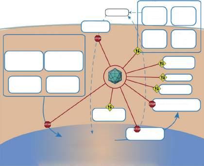
Vertebrate host–virus interactions
Figure 1.1 Interactions of viruses with host cells. Source: Courtesy of Creative Commons Corporation and ViralZone, 2021, https:// creativecommons.org/licenses/by/4.0/#; https://viralzone.expasy.org/.
There are several major routes of viral entry into a host: respiratory, alimentary, urogenital, and direct transmission via an insect or animal bite. Successful establishment of viral infection depends on the presence of appropriate cell receptors for viral attachment and internalization, and the physicochemical nature of the viral agent (Figure 1.1). To successfully initiate infection, a virus must survive until a susceptible host is encountered and access is gained to cells in which it can replicate (permissive cells). This requires the virus to overcome the host defense mechanisms at these sites. For example, viruses that infect animals via the alimentary tract are typically resistant to the low pH and potent enzymes that occur in the digestive tract.
Mechanisms of Pathogenesis
Microbial disease manifests itself either as the result of direct damage to host cells and cellular functions by exotoxins or products of microbial growth, or via collateral
damage due to host inflammatory or immune reactions that are triggered by the microbe or microbial components (e.g. endotoxin).
Direct Damage
Exotoxins are usually bacterial proteins that are freely excreted into the environment, whereas endotoxin (lipopolysaccharide) is an integral part of the gram-negative bacterial cell membrane that remains attached to the bacterial cell or is released in membrane vesicles. The different characteristics of endotoxins and exotoxins are described in Table 1.3 and Figure 1.2.
Exotoxins are encoded chromosomally, on plasmids or on bacteriophages. These toxins produce injury by destroying the cells in which they replicate or by altering cell function, appearance, and growth characteristics. There are several types of bacterial exotoxins. Some act extracellularly by damaging host cell membranes via enzymatic or
Table 1.3 Exotoxins and endotoxins compared.
Exotoxins
Often spontaneously diffusible
Proteins or polypeptides
Produced by gram-positive and gram-negative bacteria
Produce a single, pharmacologically specific effect
Each is distinct in structure and reactivity according to its bacterial species of origin
Lethal in minute amounts (mice = nanograms)
Labile to heat, chemicals, and storage
Convertible to toxoids (nontoxic, immunogenic toxin-derivatives); elicit antitoxin production
Endotoxins
Cell-bound as part of the cell wall
LPS (lipid A is a toxic component)
Limited to gram-negative bacteria
Produce a range of effects, largely due to host-derived mediators
Similar in structure and effect regardless of bacterial species of origin
Lethal in larger amounts (mice = micrograms)
Very stable to heat, chemicals, and storage
Not readily convertible to toxoids
Bacterium
Exotoxin
Receptor
Host cell
of host cell
Shed endotoxin
Receptor
Host cell
Endotoxin in bacterial cell wall
Secrection of host cell prouducts
Damage to neighboring host cells or tissue
Figure 1.2 A comparison of endotoxins and exotoxins. Source: Used courtesy of the Creative Commons and @Read/Study, 2021.
detergent-like mechanisms. Examples of these toxins include bacterial hemolysins, and leucocidins. Some exotoxins act as enzymes that degrade the extracellular matrix (e.g. collagenases and hyaluronidases) and play an ancillary role in facilitating bacterial spread in host tissue. One group of exotoxins consists of proteins or polypeptides that enter cells and enzymatically disrupt intracellular processes. Many, but not all, of these are bifunctional polypeptides
that consist of an A fragment with enzymatic activity and a B fragment that is responsible for toxin binding to target cells.
Endotoxins are lipopolysaccharides (LPS) that are part of the gram-negative cell wall and outer membrane. The LPS structure consists of a core polysaccharide, lipid A (which is the toxic moiety) and polysaccharide chains. The latter can act as an adhesin or virulence factor and contain
Exotoxin
Death
Endotoxin
the somatic (O) antigens recognized by the host immune response. LPS can bind directly to leukocytes, or to LPSbinding protein (a plasma protein), which in turn transfers the LPS to CD14. The CD14–LPS complex interacts with other receptor proteins (e.g. Toll-like receptor 4) on the surface of macrophages and other cells, triggering the release of proinflammatory cytokines and other mediators that elicit the manifestations of endotoxemia. These can include fever, headache, hypotension, leukopenia, thrombocytopenia, intravascular coagulation, inflammation, endothelial damage, hemorrhage, fluid extravasation, and circulatory collapse. Many of these outcomes result from LPS or cytokine stimulating: (i) activation of the complement cascade; and (ii) production of arachidonic acid metabolites (prostaglandins, leukotrienes, and thromboxanes). The clinical signs of endotoxemia closely resemble those of gram-negative septicemias. Although mediated by different bacterial components (lipoproteins) and host receptors (Toll-like receptor 2), similar manifestations can be induced by the cell walls (peptidoglycans) of grampositive bacteria.
Immune-Mediated Damage
Tissue damage due to immune reactions is considered in detail elsewhere in this volume (see Chapter 67). Innate immune and inflammatory responses are both essential to host defense and can cause substantial tissue damage and loss of function. For example, complement-mediated inflammation can occur in response to endotoxins or to peptidoglycan without preceding sensitization and can result in a vigorous local inflammatory response. Antigen- specific adaptive immune responses contribute to the pathogenesis of many infections, particularly those, such as tuberculosis, that elicit chronic granulomatous infection. Granulomas that form in response to infection with intracellular pathogens such as M. tuberculosis are the result of a cell-mediated immune response (Type IV delayed type hypersensitivity) by the host. These cell-mediated immune responses can damage tissue during the original infection, or upon subsequent encounters with the offending microorganisms or its antigens that stimulate T lymphocytes to release cytokines and other effector molecules. Other examples of immunemediated damage include anemia seen in anaplasmosis, and during infection with the hemotrophic mycoplasmas. These occur as the result of an antibody response to the hemoparasites that targets infected host erythrocytes for phagocytosis and C-mediated hemolysis.
Viral Dissemination within the Infected Host
Viruses cause two basic patterns of infection: localized or generalized (Figure 1.3). In localized infections, viral multiplication and cellular damage remain localized near the site of entry (e.g. the skin or the mucous membranes of the respiratory, gastrointestinal, or genital tract). The infecting virus spreads only to neighboring cells immediately adjacent to the original site of infection. For example, rhinovirus infections of animals are often restricted to the nasal epithelium and do not spread to the lower respiratory tract. Other respiratory viruses, such as parainfluenza and respiratory syncytial viruses, can replicate within the lungs of infected animals, but tissue injury does not extend beyond the respiratory tract. Generalized infections develop through several sequential steps: (i) the virus undergoes primary replication at the site of entry and in regional lymph nodes, (ii) progeny virus spreads through blood (primary viremia) and lymphatics to additional tissues, where (iii) further virus replication takes place, (iv) the virus is disseminated to the other target organs via a secondary viremia, and (v) it multiplies further in these target tissues where it causes cellular degeneration and/or necrosis, tissue injury, and clinical disease.
After the initial viral invasion, there is an asymptomatic incubation period before clinical signs are observed. In generalized viral infections, overt disease begins after the virus is widely disseminated in the body and replicates to significant levels. It is at this stage that the veterinarian typically is first alerted. Canine distemper provides an example of a generalized viral infection of animals. Canine distemper virus initiates infection at the site of entry, and then disseminates through the blood or lymphatic system to produce generalized infection that involves a variety of target organs (Figure 1.4). The sequence of events during the incubation period and clinical signs that occur in individual animals depend on which organ systems are infected by the virus. The virus is disseminated to these organs during the viremic period, in which viral dissemination occurs both via free virus particles in the blood and infected blood cells that serve as carriers to transport the virus to target organs (cell-associated viremia). Cellassociated viremias typically involve blood leukocytes, but some viruses, such as bluetongue virus, hog cholera virus, or parvovirus, can associate with and be disseminated by red blood cells in the infected host. For viruses that infect the central nervous system, viral dissemination can occur by viremia or, in the case of rabies, by centripetal transmission along peripheral nerves.