ELAINE MARIEB is the most trusted name in all of A&P. More than 3 million health care professionals started their careers with one of Elaine Marieb’s Anatomy & Physiology texts. Now, it’s your turn.
LEARN WHY THIS MATTERS
why this matters NEW! Chapter-opening Why This Matters videos describe how the material applies to your future career. Scan the QR codes to see brief videos of real health care professionals discussing how they use the chapter content every day in their careers.
SEE WHERE YOU
NEW! Every chapter opens with a Chapter Roadmap to give you a visual overview of all the key concepts in the chapter and how they fit together. The key concepts in the roadmap are linked to the section number in the chapter to make the connections clear.
5
The Integumentary System
In this chapter, you will learn that
The skin and its derivatives serve several (mostly protective) functions
why this matters
Chapter Roadmap
Key Concept
section header
W5.1 What is the structure of skin?
looking closer at looking closer at and asking by first asking
then learning about and next asking then asking
The appendages of the skin using
5.8 What are the functions of skin?
5.9 What happens when things go wrong?
5.7 Sweat and sebaceous glands
ould you be enticed by an ad for a coat that is waterproof, stretchable, washable, and air-conditioned, that automatically repairs small cuts, rips, and burns? How about one that’s guaranteed to last a lifetime? Sounds too good to be true, but you already have such a coat—your skin.
The skin and its derivatives (sweat and oil glands, hairs, and nails) make up a complex set of organs that serves several functions, mostly protective. Together, these organs form the integumentary system (in-teg″ u-men ′tar-e).
5.1 The skin consists of two layers: the epidermis and dermis
Learning Objective
List the two layers of skin and briefly describe subcutaneous tissue.
The skin receives little respect from its inhabitants, but architecturally it is a marvel. It covers the entire body, has a surface area of 1.2 to 2.2 square meters, weighs 4 to 5 kilograms (4–5 kg = 9–11 lb),
The variation in skin tone shown here is primarily due to varying concentrations of the pigment melanin.
ARE HEADED
NEW! Key concept organization presents the material in manageable chunks and helps you easily navigate the chapter. Each section header states the key concept of that section, and section-ending Check Your Understanding questions allow students to assess their understanding of the concept before moving on. Chapter 5 The Integumentary System 171
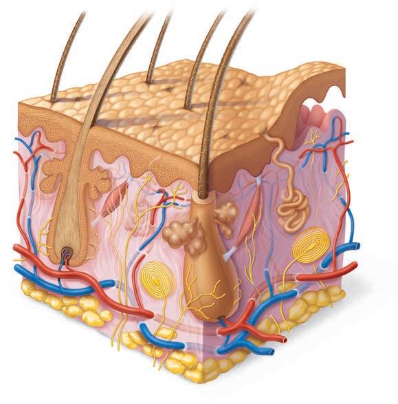
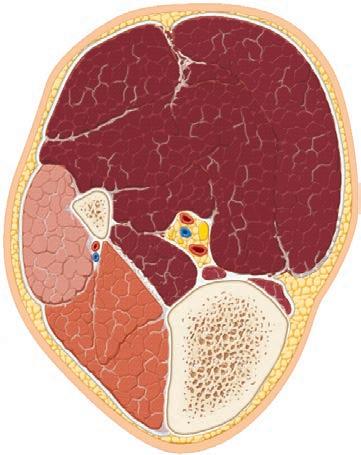
Hair shaft
Epidermis
Dermis Reticular layer Papillary layer
Hypodermis (subcutaneous tissue; not part of skin)
Nervous structures
• Sensory nerve fiber with free nerve endings
• Lamellar corpuscle
• Hair follicle receptor (root hair plexus)
Adipose tissue
Cutaneous plexus
Dermal papillae
Subpapillary plexus
Sweat pore
Appendages of skin
• Eccrine sweat gland
• Arrector pili muscle
• Sebaceous (oil) gland
• Hair follicle
• Hair root
Figure 5.1 Skin structure. Three-dimensional view of the skin and underlying subcutaneous tissue. The epidermal and dermal layers have been pulled apart at the upper right corner to reveal the dermal papillae.
and accounts for about 7% of total body weight in the average adult. Also called the integument (“covering”), the skin multitasks. Its functions go well beyond serving as a bag for body contents. Pliable yet tough, it takes constant punishment from external agents. Without our skin, we would quickly fall prey to bacteria and perish from water and heat loss.
Varying in thickness from 1.5 to 4.0 millimeters (mm) or more in different parts of the body, the skin is composed of two distinct layers (Figure 5.1):
● The epidermis (ep″ ĭ-der′mis), composed of epithelial cells, is the outermost protective shield of the body (epi = upon).
● The underlying dermis, making up the bulk of the skin, is a tough, leathery layer composed mostly of dense connective tissue.
Only the dermis is vascularized. Nutrients reach the epidermis by diffusing through the tissue fluid from blood vessels in the dermis.
The subcutaneous tissue just deep to the skin is known as the hypodermis (Figure 5.1). Strictly speaking, the hypodermis is not part of the skin, but it shares some of the skin’s protective functions. The hypodermis, also called superficial fascia because it is superficial to the tough connective tissue wrapping (fascia) of the skeletal muscles, consists mostly of adipose tissue. Besides storing fat, the hypodermis anchors the skin to the underlying structures (mostly to muscles), but loosely enough that the skin can slide relatively freely over those structures. Sliding skin protects us by ensuring that many blows just glance off our bodies. Because of its fatty composition, the hypodermis also acts as a shock absorber and an insulator that reduces heat loss.
Check Your Understanding
1. Which layer of the skin—dermis or epidermis—is better nourished?
For answers, see Answers Appendix.
compartment muscles
Anterior compartment muscles
816 UNIT 4 Maintenance of the Body
Fibularis muscles
Medial compartment muscles of thigh and lateral compartment muscles of leg
Posterior
TOOLS TO HELP YOU
Goblet cell
Mucosa
Pseudostratified ciliated columnar epithelium
Tibialis anterior
Lamina propria (connective tissue)
Lateral compartment of leg (plantar flexes and everts foot); innervated by superficial fibular nerve
NEW! Find study tools online with references to MasteringA&P®
Esophagus
Trachealis
Lumen of trachea
Practice art labeling
Visit MasteringA&P for self-study modules, interactive animations, virtual lab tools, and more!
Submucosa
(b) Muscles of the leg
Seromucous gland in submucosa
breakdown re ects some change in the capillary endothelial cells or their tight junctions.
>Study Area>Chapter 10
Hyaline cartilage
Check Your Understanding
Adventitia
Figure 10.26 Summary: Actions of muscles of the thigh and leg.
Anterior
19. What is CSF? Where is it produced? What are its functions?
(a) Cross section of the trachea and esophagus
20. A brain surgeon is about to make an incision. Name all the tissue layers that she cuts through from the skin to the brain.
For answers, see Answers Appendix.
Cerebrovascular
Anterior compartment of (dorsiflexes foot, extends toes); innervated by deep fibular nerve
NEW! Easily find clinical examples to help you see how A&P concepts apply to your future career. The clinical content— Homeostatic Imbalance sections, A Closer Look boxes, At the Clinic sections, and Critical Thinking and Clinical Application questions at the end of the chapter—has a unified new look and feel.
M10_MARI7040_09_SE_CH10_321-387.indd
Figure 22.7 Tissue composition of the tracheal wall In the scanning electron micrograph in (c), the cilia appear as yellow, grasslike projections. Mucus-secreting goblet cells (orange) with short microvilli are interspersed between the ciliated cells
View histology slides >Study Area>
(b) Photomicrograph of the tracheal wall (320)
C LI NICAL
12.9 Brain injuries and disorders have devastating consequences
Learning Objectives
Describe the cause (if known) and major signs and symptoms of cerebrovascular accidents, Alzheimer’s disease, Parkinson’s disease, and Huntington’s disease. List and explain several techniques used to diagnose brain disorders.
length of the pharynx acts as a resonating chamber, to amplify and enhance the sound quality. e oral, nasal, and sinus cavities also contribute to vocal resonance. In addition, good enunciation depends on muscles in the pharynx, tongue, so palate, and lips that “shape” sound into recognizable consonants and vowels.
Brain dysfunctions are unbelievably varied and extensive. We have mentioned some of them already, but here we will focus on traumatic brain injuries, cerebrovascular accidents, and degenerative brain disorders.
C LI NICAL
HOMEOSTATIC
IMBALANCE 22.3
(c) Scanning electron micrograph of cilia in the trachea (2500)
Traumatic Brain Injuries
In ammation of the vocal folds, or laryngitis, causes the vocal folds to swell, interfering with their vibration. is changes the vocal tone, causing hoarseness, or in severe cases limiting us to a whisper. Laryngitis is most o en caused by viral infections, but may also be due to overusing the voice, very dry air, bacterial infections, tumors on the vocal folds, or inhalation of irritating chemicals. ✚
Sphincter Functions of the Larynx
The Trachea
Head injuries are a leading cause of accidental death in North America. Consider, for example, what happens if you forget to fasten your seat belt and then rear-end another car. Your head is moving and then stops suddenly as it hits the windshield. Brain damage is caused not only by localized injury at the site of the blow, but also by the ricocheting e ect as the brain hits the opposite end of the skull.
A concussion is an alteration in brain function, usually temporary, following a blow to the head. e victim may be dizzy or lose consciousness. Although typically mild and short-lived, even a seemingly mild concussion can be damaging, and multiple concussions over time produce cumulative damage.
Under certain conditions, the vocal folds act as a sphincter that prevents air passage. During abdominal straining associated with defecation, the glottis closes to prevent exhalation and the abdominal muscles contract, causing the intra-abdominal
More serious concussions can bruise the brain and cause per-
e trachea (tra′ke-ah), or windpipe, descends from the larynx through the neck and into the mediastinum. It ends by dividi into the two main bronchi at midthorax (see Figure 22.1). humans, it is 10–12 cm (about 4 inches) long and 2 cm (3/4 inch) in diameter, and ver y exible and mobile.
e tracheal wall consists of several layers that are common to many tubular body organs—the mucosa, submucosa, adventitia—plus a layer of hyaline cartilage (Figure 22.7). mucosa has the same goblet cell–containing pseudostrati epithelium that occurs throughout most of the respiratory tract.
e single most leading cause accidents (C CVAs occur and brain tiss blood supply e most a cerebral artery. the heart, for of a brain artery strokes are cause Many who body (hemiplegia or have di culty the picture is of their lost branches that lost functions. to prevent musc cles due to groups). Not all strokes reversible ce (TIAs), are characterized speech. ese tute “red ags A CVA is blor that does the coast later. stroke is not vessels in the neuron-killi wreak the mos Experimental glutamate, an key role in lear functions. H of oxygen begin
Tibia
ON YOUR JOURNEY
Stunning 3-D art with vibrant colors appears on every page to help you better visualize and understand key anatomical structures and their functions.
and Movement of the Body
ws passive heat loss from er 24 discusses body tem-
Excretion
cutaneous sensory receptors, nervous system. e cutaneous tors (ek″ster-o-sep′torz) ng outside the body. For les (in the dermal papilbecome aware of a caress or in, whereas lamellar (also deeper dermis or hypoderlving deep pressure. Hair blowing through our hair and nerve endings that meander stimuli (irritating chemicals, defer detailed discussion er 13.
cutaneous receptors mentioned ch are found only in skin n in Figure 5.2b.
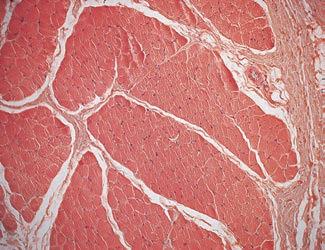
9.1 Connective tissue sheaths of skeletal muscle: epimysium, perimysium, and endomysium. (b) Photomicrograph of a cross section of part of a skeletal muscle (30×). (For a related image, see A Brief Atlas of the Human Body, Plate 29.)
Let’s consider these connective tissue sheaths from external to internal (see Figure 9.1 and the top three rows of Table 9.1).
Attachments
NEW! Making Connections questions in each chapter ask you to apply what you’ve learned across different body systems and chapters so that you build a cohesive understanding of the body.
● Epimysium. The epimysium (ep″ ĭ-mis′e-um; “outside the muscle”) is an “overcoat” of dense irregular connective tissue that surrounds the whole muscle. Sometimes it blends with the deep fascia that lies between neighboring muscles or the superficial fascia deep to the skin.
e body eliminates limited amounts of nitrogen-containing wastes (ammonia, urea, and uric acid) in sweat, although most such wastes are excreted in urine. Profuse sweating is an important avenue for water and salt (sodium chloride) loss.
Check Your Understanding
● Perimysium and fascicles. Within each skeletal muscle, the muscle fibers are grouped into fascicles (fas′ĭ-klz; “bundles”) that resemble bundles of sticks. Surrounding each fascicle is a layer of dense irregular connective tissue called perimysium (per″ ĭ-mis′e-um; “around the muscle”).
● Endomysium. The endomysium (en″do-mis′e-um; “within the muscle”) is a wispy sheath of connective tissue that surrounds each individual muscle fiber. It consists of fine areolar connective tissue.
21. What chemicals produced in the skin help provide barriers to bacteria? List at least three and explain how the chemicals are protective
22. Which epidermal cells play a role in body immunity?
23. How is sunlight important to bone health?
As shown in Figure 9.1, all of these connective tissue sheaths are continuous with one another as well as with the tendons that join muscles to bones. When muscle fibers contract, they pull on these sheaths, which transmit the pulling force to the bone to be moved. The sheaths contribute somewhat to the natural elasticity of muscle tissue, and also provide routes for the entry and exit of the blood vessels and nerve fibers that serve the muscle.
Recall from Chapter 8 that most skeletal muscles span joints and attach to bones (or other structures) in at least two places. When a muscle contracts, the movable bone, the muscle’s insertion, moves toward the immovable or less movable bone, the muscle’s origin. In the muscles of the limbs, the origin typically lies proximal to the insertion.
Muscle attachments, whether origin or insertion, may be direct or indirect.
● In direct, or fleshy, attachments, the epimysium of the muscle is fused to the periosteum of a bone or perichondrium of a cartilage.
● In indirect attachments, the muscle’s connective tissue wrappings extend beyond the muscle either as a ropelike tendon (Figure 9.1a) or as a sheetlike aponeurosis (ap″ onu-ro ′sis). The tendon or aponeurosis anchors the muscle to the connective tissue covering of a skeletal element (bone or cartilage) or to the fascia of other muscles.
Indirect attachments are much more common because of their durability and small size. Tendons are mostly tough collagen fibers which can withstand the abrasion of rough bony projections that would tear apart the more delicate muscle tissues. Because of their relatively small size, more tendons than
24. MAKING co nne ctio ns When blood vessels in the dermis constrict or dilate to help maintain body temperature, which type of muscle tissue that you learned about (in Chapter 4) acts as the effector that causes blood vessel dilation or constriction? For answers, see Answers Appendix.
Figure
PRACTICE MAKES PERFECT
NEW! Concept Maps are fun and challenging activities that help you solidify your understanding of a key course concept. These fully mobile activities allow you to combine key terms with linking phrases into a free-form map for topics such as protein synthesis, events in an action potential, and excitation-contraction coupling.
NEW! Interactive Physiology
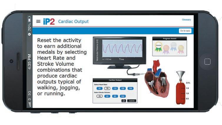
help you understand the hardest part of A&P: physiology. Fun, interactive tutorials, games, and quizzes give you additional explanations to help you grasp difficult concepts. IP 2.0 includes topics that have been updated for today’s technology, such as Resting Membrane Potential, Cardiac Output, Electrical Activity of the Heart, Factors Affecting Blood Pressure, and Cardiac Cycle.
WITH MasteringA&P
A&P Flix™ are 3-D movie-quality animations with self-paced tutorials and gradable quizzes that help you master the toughest topics in A&P.
<<Practice
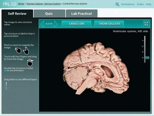
Anatomy Lab™ (PAL™) 3.0 is a virtual anatomy study and practice tool that gives you 24/7 access to the most widely used lab specimens, including the human cadaver, anatomical models, histology, cat, and fetal pig. PAL 3.0 is easy to use and includes built-in audio pronunciations, rotatable bones, and simulated fill-in-the-blank lab practical exams.
STUDY ON THE GO WITH THESE MOBILE TOOLS
NEW! Dynamic Study Modules offer a mobile-friendly, personalized reading experience of the chapter content. As you answer questions to master the chapter content, you receive detailed feedback with text and art from the book itself. The Dynamic Study Modules help you acquire, retain, and recall information faster and more efficiently than ever before.
The PAL 3.0 App lets you access PAL 3.0 on your iPad or Android tablet. Enlarge images, watch animations, and study for your lab practicals with multiple-choice and fill-in-the-blank quizzes—all while on the go!
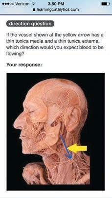
Learning Catalytics is a “bring your own device” (laptop, smartphone, or tablet) engagement, assessment, and classroom intelligence system. Use your device to respond to open-ended questions, and then discuss your answers in groups based on responses.
Human Anatomy & Physiology
Tenth Edition
Global Edition
Elaine N. Marieb, R.N., Ph.D.
Holyoke Community College
Katja Hoehn, M.D., Ph.D.
Mount Royal University
Editor-in-Chief: Serina Beauparlant
Sr. Acquisitions Editor: Brooke Suchomel
Production and Design Manager: Michele Mangelli
Program Manager: Shannon Cutt
Development Editor: Tanya Martin
Art Development Manager: Laura Southworth
Art Development Editors: Laura Southworth, Elisheva Marcus
Editorial Assistant: Arielle Grant
Text Permissions Project Manager: Timothy Nicholls
Director of Development: Barbara Yien
Program Management Team Lead: Michael Early
Project Management Team Lead: Nancy Tabor
Copyeditor: Anita Hueftle
Senior Acquisitions Editor, Global Edition: Priyanka Ahuja
Associate Project Editor, Global Edition: Binita Roy
Acknowledgments of third party content appear on pages 1195 and 1196 , which constitutes an extension of this copyright page.
PEARSON, ALWAYS LEARNING, MasteringA&P®, A&PFlixTM, and PALTM are exclusive trademarks in the U.S. and/or other countries owned by Pearson Education, Inc. or its affiliates.
Pearson Education Limited
Edinburgh Gate Harlow Essex CM20 2JE England
and Associated Companies throughout the world
Visit us on the World Wide Web at: www.pearsonglobaleditions.com
The rights of Elaine N. Marieb and Katja Hoehn to be identified as the authors of this work have been asserted by them in accordance with the Copyright, Designs and Patents Act 1988.
All rights reserved. No part of this publication may be reproduced, stored in a retrieval system, or transmitted in any form or by any means, electronic, mechanical, photocopying, recording or otherwise, without either the prior written permission of the publisher or a license permitting restricted copying in the United Kingdom issued by the Copyright Licensing Agency Ltd, Saffron House, 6–10 Kirby Street, London EC1N 8TS.
All trademarks used herein are the property of their respective owners. The use of any trademark in this text does not vest in the author or publisher any trademark ownership rights in such trademarks, nor does the use of such trademarks imply any affiliation with or endorsement of this book by such owners.
Paperback
ISBN 10: 1-292-09697-7
ISBN 13: 978-1-292-09697-1
Hardback
ISBN 10: 1-292-10042-7
ISBN 13: 978-1-292-10042-5
British Library Cataloguing-in-Publication Data
A catalogue record for this book is available from the British Library
10 9 8 7 6 5 4 3 2 1
Typeset in 10.5 Minion Pro by Cenveo® Publisher Services
Printed and bound by Courier Kendallville in the United States of America.
About the Authors
We dedicate this work to our students both present and past, who always inspire us to “push the envelope.”
Elaine N. Marieb
For Elaine N. Marieb, taking the student’s perspective into account has always been an integral part of her teaching style. Dr. Marieb began her teaching career at Springfield College, where she taught anatomy and physiology to physical education majors. She then joined the faculty of the Biological Science Division of Holyoke Community College in 1969 after receiving her Ph.D. in zoology from the University of Massachusetts at Amherst. While teaching at Holyoke Community College, where many of her students were pursuing nursing degrees, she developed a desire to better understand the relationship between the scientific study of the human body and the clinical aspects of the nursing practice. To that end, while continuing to teach full time, Dr. Marieb pursued her nursing education, which culminated in a Master of Science degree with a clinical specialization in gerontology from the University of Massachusetts. It is this experience that has informed the development of the unique perspective and accessibility for which her publications are known.
Dr. Marieb has partnered with Benjamin Cummings for over 30 years. Her first work was Human Anatomy & Physiology Laboratory Manual (Cat Version), which came out in 1981. In the years since, several other lab manual versions and study guides, as well as the softcover Essentials of Human Anatomy & Physiology textbook, have hit the campus bookstores. This textbook, now in its 10th edition, made its appearance in 1989 and is the latest expression of her commitment to the needs of students studying human anatomy and physiology.
Dr. Marieb has given generously to colleges both near and far to provide opportunities for students to further their education. She contributes to the New Directions, New Careers Program at Holyoke Community College by funding a staffed drop-in center and by providing several full-tuition scholarships each year for women who are returning to college after
a hiatus or attending college for the first time and who would be unable to continue their studies without financial support. She funds the E. N. Marieb Science Research Awards at Mount Holyoke College, which promotes research by undergraduate science majors, and has underwritten renovation and updating of one of the biology labs in Clapp Laboratory at that college. Dr. Marieb also contributes to the University of Massachusetts at Amherst where she generously provided funding for reconstruction and instrumentation of a cutting-edge cytology research laboratory. Recognizing the severe national shortage of nursing faculty, she underwrites the Nursing Scholars of the Future Grant Program at the university.
In 1994, Dr. Marieb received the Benefactor Award from the National Council for Resource Development, American Association of Community Colleges, which recognizes her ongoing sponsorship of student scholarships, faculty teaching awards, and other academic contributions to Holyoke Community College. In May 2000, the science building at Holyoke Community College was named in her honor.
Dr. Marieb is an active member of the Human Anatomy and Physiology Society (HAPS) and the American Association for the Advancement of Science (AAAS). Additionally, while actively engaged as an author, Dr. Marieb serves as a consultant for the Benjamin Cummings Interactive Physiology® CD-ROM series.
When not involved in academic pursuits, Dr. Marieb is a world traveler and has vowed to visit every country on this planet. Shorter term, she serves on the scholarship committee of the Women’s Resources Center and on the board of directors of several charitable institutions in Sarasota County. She is an enthusiastic supporter of the local arts and enjoys a competitive match of doubles tennis.
Katja Hoehn
Dr. Katja Hoehn is a professor in the Department of Biology at Mount Royal University in Calgary, Canada. Dr. Hoehn’s first love is teaching. Her teaching excellence has been recognized by several awards during her 20 years at Mount Royal University. These include a PanCanadian Educational Technology Faculty Award (1999), a Teaching Excellence Award from the Students’ Association of Mount Royal (2001), and the Mount Royal Distinguished Faculty Teaching Award (2004).
Dr. Hoehn received her M.D. (with Distinction) from the University of Saskatchewan, and her Ph.D. in Pharmacology from Dalhousie University. In 1991, the Dalhousie Medical Research Foundation presented her with the Max Forman (Jr.) Prize for excellence in medical research. During her Ph.D. and postdoctoral studies, she also pursued her passion for teaching by presenting guest lectures to first- and second-year medical students at Dalhousie University and at the University of Calgary.
Dr. Hoehn has been a contributor to several books and has written numerous research papers in Neuroscience and Pharmacology. She oversaw a recent revision of the Benjamin Cummings Interactive Physiology® CD-ROM series modules, and coauthored the newest module, The Immune System
Following Dr. Marieb’s example, Dr. Hoehn provides financial support for students in the form of a scholarship that she established in 2006 for nursing students at Mount Royal University.
Dr. Hoehn is also actively involved in the Human Anatomy and Physiology Society (HAPS) and is a member of the American Association of Anatomists. When not teaching, she likes to spend time outdoors with her husband and two sons, compete in triathlons, and play Irish flute.
Preface
As educators we continually make judgments about the enormous amount of information that besets us daily, so we can choose which morsels to pass on to our students. Yet even this refined information avalanche challenges the learning student’s mind. What can we do to help students apply
Unifying Themes
Three unifying themes that have helped to organize and set the tone of this textbook continue to be valid and are retained in this edition. These themes are:
Interrelationships of body organ systems. This theme emphasizes the fact that nearly all regulatory mechanisms have interactions with several organ systems. The respiratory system, for example, cannot carry out its role of gas exchange in the body if there are problems with the cardiovascular system that prevent the normal delivery of blood throughout the body. The unique System Connections feature is a culmination of this approach and helps the student think of the body as a community of dynamic parts instead of a number of independent units.
Homeostasis. Homeostasis is the normal and most desirable condition of the body. Its loss is always associated with past or present pathology. This theme is not included to emphasize pathological conditions but rather to illustrate what happens in the body when homeostasis is lost.
Whenever students see a red balance beam symbol accompanied by an associated clinical topic, their understanding of how the body works to stay in balance is reinforced.
Complementarity of structure and function. This theme encourages students to understand the structure of some bodily part (cell, bone, lung, etc.) in order to understand the function of that structure. For example, muscle cells can produce movement because they are contractile cells.
the concepts they are faced with in our classrooms? We believe that this new edition of our textbook addresses that question by building on the strengths of previous editions while using new, innovative ways to help students visualize connections between various concepts.
Changes Past and Present
Many of the changes made to the 9th edition have been retained and are reinforced in this 10th edition.
• There are more step-by-step blue texts accompanying certain pieces of art (blue text refers to the instructor’s voice).
• The many clinical features of the book have been clearly identified to help students understand why this material is important.
• The “Check Your Understanding” questions at the end of each module reinforce understanding throughout the chapter.
• We have improved a number of our Focus Figures. (Focus Figures are illustrations that use a “big picture” layout and dramatic art to walk the student through difficult processes in a step-by-step way.)
• MasteringA&P continues to provide text-integrated media of many types to aid learning. These include Interactive Physiology (IP) tutorials that help students to grasp difficult concepts, A&PFlix animations that help students visualize tough A&P topics, and the PAL (Practice Anatomy Lab) collection of virtual anatomy study and practice tools focusing on the most widely used lab specimens. These are by no means all of the helpful tools to which students have access. It’s just a smattering.
New To The Tenth Edition
So, besides these tools, what is really new to this textbook this time around? Each chapter begins with a “Chapter Roadmap” diagram that indicates the topics covered by the modules in the chapter and shows how these topics relate to each other. Another nicety on each chapter’s first page is the “Why This Matters” icon and QR code that links to a video of a health-care professional telling us why the chapter’s content is important for his or her work.
In this edition, we have taken great pains to ensure that the text and associated art are almost always covered on the same two-page spread. This sounds simple, but the fact that this type of presentation has not usually been achieved in textbooks until now tells you that it is not. How many times have you heard complaints about having to flip back and forth between a figure on one page and text on another? Accomplishing this type of text-art correlation is extremely difficult, yet invaluable to student learning.
Other new features include (1) declarative headers at the beginning of each chapter module so that the student can quickly grasp the “big idea” for that module, (2) more modularization (chunking) of the text so that students can tackle manageable pieces of information as they read through the material, (3) increased readability of the text as a result of more bulleted lists and shorter paragraphs, (4) more summary tables to help students connect information, (5) improvements to many of the figures so that they teach even more effectively, and (6) “Making Connections” questions in each chapter that ask students to incorporate related information from earlier chapters or earlier modules in the same chapter, helping students to see the forest, not just the trees, as they study.
Chapter-by-Chapter Changes
Chapter 1 The Human Body: An Orientation
• Updated Figure 1.8 for better teaching effectiveness.
Chapter 2 Chemistry Comes Alive
• Updated Figure 2.18 for better teaching effectiveness.
Chapter 3 Cells: The Living Units
• Updated statistics on Tay-Sachs disease.
• Updated information about riboswitches and added information about small interfering RNAs (siRNAs).
• Added summary text to Figure 3.3 for better pedagogy.
• Updated Focus Figure 3.4.
Chapter 4 Tissue: The Living Fabric
• Multiple updates to A Closer Look feature on cancer reflect new understanding of cancer mechanisms.
• New photos of simple columnar epithelium, pseudostratified ciliated columnar epithelium, cardiac muscle tissue, and smooth muscle tissue (Figures 4.3c, d and 4.9b, c).
Chapter 5 The Integumentary System
• Added information about the role of tight junctions in skin.
• New photo of stretch marks (Figure 5.5).
• New photo of cradle cap (seborrhea) in a newborn (Figure 5.9).
• New photo of malignant melanoma (Figure 5.10).
Chapter 6 Bones and Skeletal Tissues
• Revised Figure 6.9 for improved teaching effectiveness.
• New X rays showing Paget’s disease and normal bone (Figure 6.16).
Chapter 7 The Skeleton
• Illustrated the skull bone table to facilitate student learning (Table 7.1).
• Added three new Check Your Understanding figure questions asking students to make anatomical identifications.
• New photos of humerus, radius, and ulna (Figures 7.28 and 7.29).
• New photo showing the outcome of cleft lip and palate surgery (Figure 7.38b).
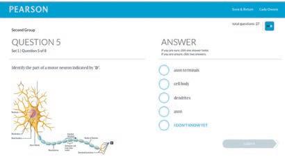
• New image of a motor neuron based on a computerized 3-D reconstruction of serial sections.
• Converted Figure 11.17 to tabular head style to teach better.
Chapter 12 The Central Nervous System
• Updated mechanisms of Alzheimer’s disease to include propagation of misfolded proteins.
• Updated information about gender differences in the brain.
• Streamlined discussion of sleep, memory, and stroke.
• New figure to show distribution of gray and white matter (Figure 12.3).
• Functional neuroimaging of the cerebral cortex (Figure 12.6).
• Improved reticular formation figure with “author’s voice” blue text (Figure 12.18).
• New figure showing decreased brain activity in Alzheimer’s (Figure 12.26).
Chapter 13 The Peripheral Nervous System and Reflex Activity
• Updated and expanded description of axon regeneration (in Figure 13.5).
Chapter 14 The Autonomic Nervous System
• Improved teaching effectiveness of Figure 14.3 (differences in the parasympathetic and sympathetic nervous systems).
• New summary table for autonomic ganglia (Table 14.2).
Chapter 15 The Special Senses
• Updated description of cytostructure of human cochlear hair cells (they have no kinocilia).
• New data on the number of different odors that humans can detect.
• Added a new part to the figure teaching eye movements made by extrinsic eye muscles (Figure 15.3).
• Reorganized discussion of sound transmission to the inner ear. New numbered text improves text-art correlation.
• New figure teaches the function of the basilar membrane (Figure 15.31).
• New figure on how the hairs on the cochlear hair cells transduce sound (Figure 15.32).
• New figure shows the structure and function of the macula (Figure 15.34).
• New photo of a boy with a cochlear implant (Figure 15.37).
Chapter 16 The Endocrine System
• Updated statistics on pancreatic islet transplant success in A Closer Look and added new information on artificial pancreases.
• New information on actions of vitamin D and location of its receptors.
• New summary table showing differences between watersoluble and lipid-soluble hormones (Table 16.1).
• New summary flowchart shows the signs and symptoms of diabetes mellitus (Figure 16.19).
Chapter 17 Blood
• Improved teaching effectiveness of Figure 17.14 (intrinsic and extrinsic clotting factors).
Chapter 18 The Cardiovascular System: The Heart
• Rearranged topics in this chapter for better flow.
• New section and summary table (Table 18.1) teach key differences between skeletal muscle and cardiac muscle.
• New Making Connections figure question (students compare three action potentials).
• Rearranged material so that all electrical events are presented in one module.
• Added tabular headers, a photo, and bullets to more effectively teach ECG abnormalities (Figure 18.18).
• Streamlined figure showing effects of norepinephrine on heart contractility (Figure 18.22).
Chapter 19 The Cardiovascular System: Blood Vessels
• New information about pericytes (now known to be stem cells and generators of scar tissue in the CNS).
• New information that the fenestrations in fenestrated capillaries are dynamic structures.
• Rearranged topics in the physiology section of this chapter for better flow.
• New micrograph of artery and vein (Figure 19.2).
• Revised Figure 19.3 (the structure of different types of capillaries), putting all of the information in one place.
• New figure summarizes the major factors determining mean arterial pressure to give a “big picture” view (Figure 19.9).
• New figure illustrating active hyperemia (Figure 19.15).
• Updated Focus Figure 19.1 (Bulk Flow across Capillary Walls).
• New Homeostatic Imbalance feature on edema relates it directly to the preceding Focus Figure 19.1) and incorporates information previously found in Chapter 26.
• New photos of pitting edema (Figure 19.18).
Chapter 20 The Lymphatic System and Lymphoid Organs and Tissues
• Updated statistics on survival of non-Hodgkin’s lymphoma patients.
• Updated figure to improve teaching of primary and secondary lymphoid organs (Figure 20.4).
Chapter 21 The Immune System: Innate and Adaptive Body Defenses
• Updated information on aging and the immune system, particularly with respect to chronic inflammation.
• Added a new term, pattern recognition receptors, to help describe how our innate defenses recognize pathogens.
• Provided new research results updating the number of genes in the human genome to about 20,000.
Chapter 22 The Respiratory System
• New Check Your Understanding question with graphs reinforces concepts learned in Focus Figure 22.1 (The OxygenHemoglobin Dissociation Curve).
• New figure illustrating pneumothorax (Figure 22.14).
Chapter 23 The Digestive System
• Updated information about the treatment of peptic ulcers.
• Updated information about the types and locations of epithelial cells of the small intestine.
• New information about roles of our intestinal flora.
• Updated hepatitis C treatment to include the new FDAapproved drug sofosbuvir.
• Added discussion of non-alcoholic fatty liver disease.
• New information about fecal transplants to treat antibioticassociated diarrhea.
• Updated figure that compares and contrasts peristalsis and segmentation (Figure 23.3) for improved teaching effectiveness.
• Updated Figure 23.4 explaining the relationship between the peritoneum and the abdominal organs to improve teaching effectiveness.
• Enteric nervous system section rewritten and rearranged with new figure (Figure 23.6).
• Improved teaching effectiveness of Figure 23.14 (the steps of deglutition).
• Streamlined Figure 23.19 to enhance teaching of regulation of gastric secretion.
• Updated Figure 23.20 (the mechanism of HCl secretion by parietal cells) for improved teaching effectiveness.
• Improved the text flow by moving discussion of the liver, gallbladder, and pancreas before the small intestine.
• Improved teaching effectiveness of Figure 23.28 (mechanism promoting secretion and release of bile and pancreatic juice).
• Updated and revised sections about motility of the small and large intestines.
• Rearranged text to discuss digestion and absorption together for each nutrient. The figures for digestion and absorption of carbohydrates (Figure 23.35) and proteins (Figure 23.36) now parallel each other and appear together for easy comparison.
• Rearranged and rewrote lipid digestion and absorption text and updated Figure 23.37.
Chapter 24 Nutrition, Metabolism, and Energy Balance
• Chapter title changed from Nutrition, Metabolism, and Body Temperature Regulation in order to emphasize the concept of energy balance.
• Updated shape and mechanism of action of ATP synthase to reflect new research findings.
• Updated hypothalamic control of food intake per new research findings.
• Updated the description of gastric bypass surgery and its effect on metabolic syndrome.
• Updated information on weight-loss drugs.
• Added new clinical term “protein energy malnutrition” incorporating both kwashiorkor and marasmus.
• Revised Figure 24.4 to enhance the ability of students to compare and contrast the mechanisms of phosphorylation that convert ADP to ATP.
• Revised figure describing ATP synthase structure and function (Figure 24.10).
• Revised Figure 24.13 to help students compare and contrast glycogenesis and glycogenolysis (Figure 24.12).
• Three new figures help students grasp the terms for key pathways in carbohydrate, protein, and fat metabolism (Figures 24.12, 24.14, and 24.18).
• New text and figure about metabolic syndrome (Figure 24.29).
Chapter 25 The Urinary System
• New cadaver photo of urinary tract organs (Figure 25.2).
• New Check Your Understanding question for nephron labeling.
• Added new illustrations to improve teaching effectiveness of Figure 25.19 (the effects of ADH on the nephron).
Chapter 26 Fluid, Electrolyte, and Acid-Base Balance
• New Check Your Understanding figure question requires students to integrate information.
Chapter 27 The Reproductive System
• Updated screening recommendations for prostate cancer, as well as updated information on detection and treatment.
• Updated screening guidelines for cervical cancer.
• Updated breast cancer statistics.
• New Check Your Understanding figure labeling question.
• New figure teaches independent assortment (Figure 27.8).
• New photo of female pelvic organs (Figure 28.15c)
• New photos of mammograms showing normal and cancerous breast tissues (Figure 27.19).
• Revised Figure 27.23 to reflect recent research about follicular development in humans.
• Revised section describing the stages of follicle development to facilitate student learning and to incorporate recent research.
Chapter 28 Pregnancy and Human Development
• Updated the details of fertilization, including zinc “sparks.”
• New information about the membrane block to polyspermy in humans (also incorporated in Focus Figure 28.1, Sperm Penetration and the Blocks to Polyspermy).
• Updated Figure 28.7 (relationship between the fetal and maternal circulation).
Chapter 29 Heredity
• Updated text on fetal genetic screening to include testing of maternal blood for fetal DNA.
• New Figure 29.7 teaches pedigree analysis.
Appendix E
• Updated periodic table to reflect naming of two new elements.
• Added a table of the genetic code (Appendix G).
Acknowledgments
Each time we put this textbook to bed, we promise ourselves that the next time will be easier and will require less of our time. Now hear this! This is its 10th edition (and 30 years more or less) and fulfillment of this promise has yet to materialize. How could there be so much going on in physiology research and so many new medical findings? Winnowing through these findings to decide on the updates to include in this edition has demanded much of our attention. Many people at Pearson have labored with us to produce another fine text. Let’s see if we can properly thank them.
As Katja and I worked on the first draft of the manuscript, Tanya Martin (our text Development Editor) worked tirelessly to improve the readability of the text, all the while trying to determine which topics could be shortened or even deleted in the 10th edition. After we had perused and acted on some of Tanya’s suggestions, we forwarded the manuscript to Shannon Cutt, the highly capable and still-cheery Program Manager, who oversees everything having to do with getting a clean manuscript to production. Aided by Editorial Assistant Arielle Grant (and before her, Daniel Wikey), Shannon reviewed the entire revised manuscript. Nothing escaped her attention as she worked to catch every problem.
At the same time the text was in revision, the art program was going through a similar process. Laura Southworth, our superb Art Development Editor (aided briefly by Elisheva Marcus), worked tirelessly to make our Focus Figures and other art even better. Needing a handshake and a heartfelt “thank you” in the process are Kristin Piljay (Photo Researcher) and Jean Lake, who handled the administrative aspects of the art program. This team ensured that the artists at Imagineering had all the information they needed to produce beautiful final art products.
As the manuscript made the transition from Editorial to Production, Michele Mangelli, the Production and Design Manager, made her appearance known. The head honcho and skilled handler of all aspects of production, everyone answered to her from this point on. In all previous editions, the manuscript would simply go directly into production once the writing and editing phases were over, but our new modular design required extra steps to make the art-text correlation a reality—the electronic page layout. Working closely with Katja
and her husband Larry Haynes, Michele’s small but powerful team “yanked” the new design to attention, fashioning two-page spreads, each covering one or more topics with its supporting art or table. This was our Holy Grail for this edition and the ideal student coaching device. They made it look easy (which it was not). Thank you Katja, Larry, and Michele—you are the ideal electronic page layout team. This was one time I felt fortunate to be the elder author.
The remaining people who helped with Production include David Novak (our conscientious Production Supervisor), Martha Ghent (Proofreader), Betsy Dietrich (Art Proofreader), Kathy Pitcoff (Indexer), Alicia Elliot (Project Manager at Imagineering), and Tim Frelick (Compositor). Copyeditor Anita Hueftle (formerly Anita Wagner) is the unofficial third author of our book. We are absolutely convinced that she memorizes the entire text. She verified the spelling of new terms, checked the generic and popular names of drugs, confirmed our grammar, and is the person most responsible for the book’s consistency and lack of typographical errors. We are grateful to Izak Paul for meticulously reading each chapter to find any remaining errors, and to Yvo Riezebos for his stunning design work on the cover, chapter opening pages, and the text.
Finally—what can we say about Brooke Suchomel, our Acquisitions Editor? She loved playing with the modular design and the chapter road maps and advising on Focus Figures, but most of her time was spent out in the field talking to professors, demonstrating the book’s changes and benefits. She spent weeks on the road, smiling all the time—no easy task. Finally, we are fortunate to have the ongoing support and friendship of Serina Beauparlant, our Editor-in-Chief.
Other members of our team with whom we have less contact but who are nonetheless vital are: Barbara Yien (Director of Development), Michael Early (Program Manager Team Lead), Nancy Tabor (Project Manager Team Lead), Stacey Weinberger (our Senior Manufacturing Buyer), Allison Rona (our topnotch Senior Marketing Manager), and Derek Perrigo (Senior Anatomy & Physiology Specialist). We appreciate the hard work of our media production team headed by Liz Winer, Aimee Pavy, and Lauren Hill and also wish to thank Eric Leaver.
Kudos to our entire team. We feel we have once again prepared a superb textbook. We hope you agree.
There are many people who reviewed parts of this text— both professors and students, either individually or in focus groups, and we would like to thank them. Input from the following reviewers has contributed to the continued excellence and accuracy of this text:
Matthew Abbott, Des Moines Area Community College
Lynne Anderson, Meridian Community College
Martin W. Asobayire, Essex Community College
Yvonne Baptiste-Szymanski, Niagara County Community College
Claudia Barreto, University of New Mexico–Valencia
Diana Bourke, Community College of Allegheny County
Sherry Bowen, Indian River State College
Beth Braun, Truman College
C. Steven Cahill, West Kentucky Community and Technical College
Brandi Childress, Georgia Perimeter College
William Michael Clark, Lone Star College–Kingwood
Teresa Cowan, Baker College of Auburn Hills
Donna Crapanzano, Stony Brook University
Maurice M. Culver, Florida State College at Jacksonville
Smruti A. Desai, Lone Star College–CyFair
Karen Dunbar Kareiva, Ivy Tech Community College
Elyce Ervin, University of Toledo
Martha Eshleman, Pulaski Technical College
Juanita A. Forrester, Chattahoochee Technical College
Reza Forough, Bellevue College
Dean Furbish, Wake Technical Community College
Emily Getty, Ivy Tech Community College
Amy Giesecke, Chattahoochee Technical College
Abigail Goosie, Walters State Community College
Mary Beth Hanlin, Des Moines Area Community College
Heidi Hawkins, College of Southern Idaho
Martie Heath-Sinclair, Hawkeye Community College
Nora Hebert, Red Rocks Community College
Nadia Hedhli, Hudson County Community College
D.J. Hennager, Kirkwood Community College
Shannon K. Hill, Temple College
Mark Hollier, Georgia Perimeter College
H. Rodney Holmes, Waubonsee Community College
Mark J. Hubley, Prince George’s Community College
Jason Hunt, Brigham Young University–Idaho
William Karkow, University of Dubuque
Suzanne Keller, Indian Hills Community College
Marta Klesath, North Carolina State University
Nelson H. Kraus, University of Indianapolis
Steven Lewis, Metropolitan Community College–Penn Valley
Jerri K. Lindsey, Tarrant County College–Northeast
Chelsea Loafman, Central Texas College
Paul Luyster, Tarrant County College–South
Abdallah M. Matari, Hudson County Community College
Bhavya Mathur, Chattahoochee Technical College
Tiffany Beth McFalls-Smith, Elizabethtown Community and Technical College
Todd Miller, Hunter College of CUNY
Regina Munro, Chandler-Gilbert Community College
Necia Nicholas, Calhoun Community College
Ellen Ott-Reeves, Blinn College–Bryan
Jessica Petersen, Pensacola State College
Sarah A. Pugh, Shelton State Community College
Rolando J. Ramirez, The University of Akron
Terrence J. Ravine, University of South Alabama
Laura H. Ritt, Burlington County College
Susan Rohde, Triton College
Brian Sailer, Central New Mexico Community College
Mark Schmidt, Clark State Community College
Amy Skibiel, Auburn University
Lori Smith, American River College
Ashley Spring-Beerensson, Eastern Florida State College
Justin R. St. Juliana, Ivy Tech Community College
Laura Steele, Ivy Tech Community College
Shirley A. Whitescarver, Bluegrass Community and Technical College
Patricia Wilhelm, Johnson and Wales University
Luann Wilkinson, Marion Technical College
Peggie Williamson, Central Texas College
MaryJo A. Witz, Monroe Community College
James Robert Yount, Brevard Community College
Interactive Physiology 2.0 Reviewers
Lynne Anderson, Meridian Community College
J. Gordon Betts, Tyler Junior College
Mike Brady, Columbia Basin College
Betsy Brantley, Valencia College
Tamyra Carmona, Cosumnes River College
Alexander G. Cheroske, Mesa Community College at Red Mountain
Sondra Dubowsky, McLennan Community College
Paul Emerick, Monroe Community College
Brian D. Feige, Mott Community College
John E. Fishback, Ozarks Technical Community College
Aaron Fried, Mohawk Valley Community College
Jane E. Gavin, University of South Dakota
Gary Glaser, Genesee Community College
Mary E. Hanlin, Des Moines Area Community College
Mark Hubley, Prince George’s Community College
William Karkow, University of Dubuque
Michael Kielb, Eastern Michigan University
Paul Luyster, Tarrant County College–South
Louise Millis, North Hennepin Community College
Justin Moore, American River College
Maria Oehler, Florida State College at Jacksonville
Fernando Prince, Laredo Community College
Terrence J. Ravine, University of South Alabama
Mark Schmidt, Clark State Community College
Cindy Stanfield, University of South Alabama
Laura Steele, Ivy Tech Community College
George A. Steer, Jefferson College of Health Sciences
Shirley A. Whitescarver, Bluegrass Community and Technical College
Harvey Howell, my beloved husband and helpmate, died in August of 2013. He is sorely missed.
Katja would also like to acknowledge the support of her colleagues at Mount Royal University (Trevor Day, Sarah Hewitt, Tracy O’Connor, Izak Paul, Michael Pollock, Lorraine Royal, Karen Sheedy, Kartika Tjandra, and Margot Williams) and of Ruth Pickett-Seltner (Chair), Tom MacAlister (Associate Dean), and Jeffrey Goldberg (Dean). Thanks also to Katja’s husband, Dr. Lawrence Haynes, who as a fellow physiologist has provided invaluable assistance to her during the course of
the revision. She also thanks her sons, Eric and Stefan Haynes, who are an inspiration and a joy.
We would really appreciate hearing from you concerning your opinion—suggestions and constructive criticisms—of this text. It is this type of feedback that will help us in the next revision, and underlies the continued improvement of this text.
Elaine N. Marieb
Katja Hoehn
Elaine N. Marieb and Katja Hoehn
Anatomy and Physiology
Pearson Education 1301 Sansome Street San Francisco, CA 94111
Pearson wishes to thank and acknowledge the following people for their work on the Global Edition:
Contributor
Karen Vipond, Bangor University
Eva Strandell, Halmstad University
Christiane Van den Branden, Vrije Universiteit Brussel
Reviewers
Marjorie L Wilson, Teesside University
Steven Fenby, Teesside University
Snezana Kusljic, Florey Institute of Neuroscience and Mental Health
Contents
1 The Human Body: An Orientation 21
1.1 Form (anatomy) determines function (physiology) 21
1.2 The body’s organization ranges from atoms to the entire organism 23
1.3 What are the requirements for life? 34
1.4 Homeostasis is maintained by negative feedback 28
1.5 Anatomical terms describe body directions, regions, and planes 31
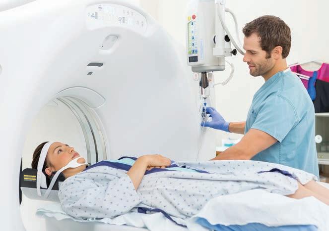
A C LOSER L OO k Medical Imaging: Illuminating the Body 34
1.6 Many internal organs lie in membrane-lined body cavities 37
2 Chemistry Comes Alive 43
PART 1 B ASIC C HEMISTRY 43
2.1 Matter is the stuff of the universe and energy moves matter 43
2.2 The properties of an element depend on the structure of its atoms 45
2.3 Atoms bound together form molecules; different molecules can make mixtures 48
2.4 The three types of chemical bonds are ionic, covalent, and hydrogen 50
2.5 Chemical reactions occur when electrons are shared, gained, or lost 55
PART 2 B IOCHEMISTRY 58
2.6 Inorganic compounds include water, salts, and many acids and bases 58
2.7 Organic compounds are made by dehydration synthesis and broken down by hydrolysis 61
2.8 Carbohydrates provide an easily used energy source for the body 62
2.9 Lipids insulate body organs, build cell membranes, and provide stored energy 64
2.10 Proteins are the body’s basic structural material and have many vital functions 67
2.11 DNA and RNA store, transmit, and help express genetic information 72
2.12 ATP transfers energy to other compounds 74
3
Cells: The Living Units 80
3.1 Cells are the smallest unit of life 81
PART 1 P LASMA M EMBRANE 83
3.2 The fluid mosaic model depicts the plasma membrane as a double layer of phospholipids with embedded proteins 83
3.3 Passive membrane transport is diffusion of molecules down their concentration gradient 88
3.4 Active membrane transport directly or indirectly uses ATP 93
F OCUS F I g URE 3.1 Primary Active Transport: The Na+-K+ Pump 94
3.5 Selective diffusion establishes the membrane potential 99
3.6 Cell adhesion molecules and membrane receptors allow the cell to interact with its environment 101
F OCUS F I g URE 3.2 G Proteins 102
PART 2 T HE C YTOPLASM 103
3.7 Cytoplasmic organelles each perform a specialized task 103
3.8 Cilia and microvilli are two main types of cellular extensions 110
PART 3 N UCLEUS 111
3.9 The nucleus includes the nuclear envelope, the nucleolus, and chromatin 111
3.10 The cell cycle consists of interphase and a mitotic phase 116
3.11 Messenger RNA carries instructions from DNA for building proteins 118
F OCUS F I g URE 3.3 Mitosis 120
F OCUS F I g URE 3.4 Translation 126
3.12 Apoptosis disposes of unneeded cells; autophagy and proteasomes dispose of unneeded organelles and proteins 129
Developmental Aspects of Cells 129
4 Tissue: The Living Fabric 135
4.1 Tissue samples are fixed, sliced, and stained for microscopy 136
4.2 Epithelial tissue covers body surfaces, lines cavities, and forms glands 137
4.3 Connective tissue is the most abundant and widely distributed tissue in the body 146
4.4 Muscle tissue is responsible for body movement 157
4.5 Nervous tissue is a specialized tissue of the nervous system 159
A C LOSER L OO k Cancer—The Intimate Enemy 160
4.6 The cutaneous membrane is dry; mucous and serous membranes are wet 161
4.7 Tissue repair involves inflammation, organization, and regeneration 163
Developmental Aspects of Tissues 165
UNIT 2
Covering, Support, and Movement of the Body
5 The Integumentary System 170
5.1 The skin consists of two layers: the epidermis and dermis 170
5.2 The epidermis is a keratinized stratified squamous epithelium 172
5.3 The dermis consists of papillary and reticular layers 174
5.4 Melanin, carotene, and hemoglobin determine skin color 176
5.5 Hair consists of dead, keratinized cells 177
5.6 Nails are scale-like modifications of the epidermis 180
5.7 Sweat glands help control body temperature, and sebaceous glands secrete sebum 181
5.8 First and foremost, the skin is a barrier 182
5.9 Skin cancer and burns are major challenges to the body 184
Developmental Aspects of the Integumentary System 187
S YSTEM C ONNECTIONS 188
6 Bones and Skeletal Tissues 193
6.1 Hyaline, elastic, and fibrocartilage help form the skeleton 193
6.2 Bones perform several important functions 195
6.3 Bones are classified by their location and shape 195
6.4 The gross structure of all bones consists of compact bone sandwiching spongy bone 197
6.5 Bones develop either by intramembranous or endochondral ossification 203
6.6 Bone remodeling involves bone deposit and removal 207
6.7 Bone repair involves hematoma and callus formation, and remodeling 209
6.8 Bone disorders result from abnormal bone deposition and resorption 212
Developmental Aspects of Bones 213
S YSTEM C ONNECTIONS 215
7 The Skeleton 219
PART 1 T HE Ax IAL Sk ELETON 219
7.1 The skull consists of 8 cranial bones and 14 facial bones 221
7.2 The vertebral column is a flexible, curved support structure 238
7.3 The thoracic cage is the bony structure of the chest 244
PART 2 T HE A PPENDICULAR Sk ELETON 247
7.4 Each pectoral girdle consists of a clavicle and a scapula 247
7.5 The upper limb consists of the arm, forearm, and hand 250
7.6 The hip bones attach to the sacrum, forming the pelvic girdle 256
7.7 The lower limb consists of the thigh, leg, and foot 260
Developmental Aspects of the Skeleton 266
8 Joints 271
8.1 Joints are classified into three structural and three functional categories 271
8.2 In fibrous joints, the bones are connected by fibrous tissue 272
8.3 In cartilaginous joints, the bones are connected by cartilage 273
8.4 Synovial joints have a fluid-filled joint cavity 274
8.5 Five examples illustrate the diversity of synovial joints 280
F OCUS F I g URE 8.1 Synovial Joints 282
8.6 Joints are easily damaged by injury, inflammation, and degeneration 291
A C LOSER L OO k Joints: From Knights in Shining Armor to Bionic Humans 293
Developmental Aspects of Joints 294
9 Muscles and Muscle Tissue 298
9.1 There are three types of muscle tissue 299
9.2 A skeletal muscle is made up of muscle fibers, nerves, blood vessels, and connective tissues 300