Sonography
Principles and Instruments
Frederick W. Kremkau, PhD
Professor of Radiologic Sciences
Director, Program for Medical Ultrasound Center for Applied Learning
Wake Forest University School of Medicine
Winston-Salem, North Carolina
with contributions by: Flemming Forsberg, PhD
Professor of Radiology
Jefferson Medical College
Thomas Jefferson University Philadelphia, Pennsylvania
3251 Riverport Lane
St. Louis, Missouri 63043
SONOGRAPHY: PRINCIPLES AND INSTRUMENTS, Ninth Edition
Copyright © 2016 by Elsevier Inc. All rights reserved.
Previous editions copyrighted 2011, 2006, 2002, 1998, 1993, 1989, 1984, and 1980.
ISBN: 978-0-323-32271-3
No part of this publication may be reproduced or transmitted in any form or by any means, electronic or mechanical, including photocopying, recording, or any information storage and retrieval system, without permission in writing from the publisher. Details on how to seek permission, further information about the Publisher’s permissions policies, and our arrangements with organizations such as the Copyright Clearance Center and the Copyright Licensing Agency can be found at our website: www.elsevier.com/permissions
This book and the individual contributions contained in it are protected under copyright by the Publisher (other than as may be noted herein).
Notices
Knowledge and best practice in this field are constantly changing. As new research and experience broaden our understanding, changes in research methods, professional practices, or medical treatment may become necessary.
Practitioners and researchers must always rely on their own experience and knowledge in evaluating and using any information, methods, compounds, or experiments described herein. In using such information or methods they should be mindful of their own safety and the safety of others, including parties for whom they have a professional responsibility.
With respect to any drug or pharmaceutical products identified, readers are advised to check the most current information provided (i) on procedures featured or (ii) by the manufacturer of each product to be administered, to verify the recommended dose or formula, the method and duration of administration, and contraindications. It is the responsibility of practitioners, relying on their own experience and knowledge of their patients, to make diagnoses, to determine dosages and the best treatment for each individual patient, and to take all appropriate safety precautions.
To the fullest extent of the law, neither the Publisher nor the authors, contributors, or editors assume any liability for any injury and/or damage to persons or property as a matter of products liability, negligence or otherwise, or from any use or operation of any methods, products, instructions, or ideas contained in the material herein.
Library of Congress Cataloging-in-Publication Data
Kremkau, Frederick W., author.
Sonography : principles and instruments / Frederick W. Kremkau ; with contributions by Flemming Forsberg. -- Ninth edition. p. ; cm.
Includes bibliographical references and index.
ISBN 978-0-323-32271-3 (hardcover : alk. paper)
I. Forsberg, Flemming, author. II. Title.
[DNLM: 1. Ultrasonography--methods. 2. Ultrasonography--instrumentation. WN 208]
RC78.7.U4
616.07’543--dc23
Publisher: Loren Wilson
Executive Content Strategist: Sonya Seigafuse
Content Development Manager: Billie Sharp
Associate Content Development Specialist: Sarah Vora
Publishing Services Manager: Catherine Jackson
Senior Project Manager: Clay S. Broeker
Design Direction: Julia Dummitt
2015024189
To the next generation of professionals—Myra, Donna, Shelley, Cara, and Olivia.
Debra Krukowski, BS, RT(R), RDMS
Program Coordinator/Faculty Triton College River Grove, Illinois
Cherie Pohlmann, MS, RT(R), RDMS
Senior Instructor
Department of Radiologic Science University of South Alabama Mobile, Alabama
David Sloan, BA, RDMS, RVT
Program Director
Diagnostic Medical Sonography Degree Program
School of Health and Patient Simulation
Springfield Technical Community College
Springfield, Massachusetts
This book is intended for sonography students, allied-health personnel, and physicians who seek understanding of the principles and instrumentation of diagnostic sonography. Applying these underlying principles in practice improves the quality of medical care involving sonography. The best sonographers and image interpreters understand these principles and apply them in their practice.
The purpose of this book is to explain how contemporary diagnostic sonography works. It serves as a principles textbook in sonography educational programs and helps readers handle artifacts properly, scan safely, and prepare for registry and board examinations. The content of the book is driven by the author’s assessment of contemporary technology in the field and his experience in teaching this material in the medical-school classroom and at conferences and seminars. The book does not describe how to perform diagnostic examinations or how to interpret the results. Other Elsevier books cover these topics.
Although this latest edition includes newer developments in the field, the emphasis is on the fundamentals. For the sake of beginners, the text is simplified, yet at the same time it maintains its integrity and usefulness for more experienced users. Although the book is designed for nonphysicist and non-engineering readers, digestion of the material will require some effort. Admittedly, for such readers, the material can be difficult. It cannot be made easy for everyone and still maintain the necessary level for appropriate application in practice. However, 40 years of lecturing and publication experience have convinced the author that the material can be understood with reasonable preparation and effort on the part of the student. It is assumed that the student has completed courses in basic physics (including mechanics, waves, and electricity) and mathematics (including algebra, trigonometry, and statistics), which are normal prerequisites in sonography programs. The following topics are not covered in this textbook: the history of the development of sonography, therapy applications, and investigational techniques. They are covered in other books and journal articles.
DIFFERENCES WITH EARLIER EDITIONS AND NATIONAL EXAMINATIONS
There are several differences between each new edition compared with earlier editions and compared with the content of registry and specialty-board examinations. This is because this text is up to date with current technology, whereas examinations change more slowly because of the necessarily thorough and time-consuming process, which requires practice surveys; committee decisions; and item generation, review, and approval. Outmoded descriptions of technology and instrument features that are no longer largely present in the field are eliminated with each new edition. Thus the book
tends to change more rapidly than the examinations. The philosophy of the book is to be, with each new edition, as consistent as possible with contemporary sonographic technology and usage.
FEATURES
• Comprehensive coverage of the principles of sonography
• Preparation for the ARDMS SPI examination
• Latest developments in commercially available sonographic technology
• Hundreds of color illustrations and images
• Hundreds of exercises with answers
• Comprehensive multiple-choice examination with annotated answers
• Consistent pedagogy, including learning objectives, chapter outlines, and key terms
• Key points set off by icons
• Descriptive subheadings
• B oxes and tables
• Math review
• Glossary
NEW TO THIS EDITION
• New illustrations and images demonstrating the latest and best images from the newest equipment
• Expanded content on volume imaging, shear-wave and acoustic-radiation-force elastography, and sophisticated echo acquisition techniques, keeping students up to date on the latest technology
• The latest instrument output data and official safety statements
• Alignment with the ARDMS examination specifications, making this a useful text for preparing for the SPI examination
FOR THE STUDENT
This book should be read in sequential chapter order, as each chapter builds on material previously presented. Key terms are listed at the beginning of each chapter, are highlighted in blue in the chapter, and are defined in the Glossary at the back of the book.
After studying this text, the student should be able to:
• Describe what ultrasound is
• Explain how ultrasound is sent into the body
• Explain how ultrasound detects and locates anatomic structures
• Discuss how echoes are received from the body and processed in the instrument
• Describe how anatomic information is presented on the display
Preface
• Explain how ultrasound detects and measures tissue motion and blood flow
• List the ways motion and flow information are presented
• Explain how flow detection is localized to a specific site in tissue
• List the common artifacts that can occur in diagnostic sonography
• Discuss how performance of sonographic instruments is tested
• Describe the risk and safety issues associated with diagnostic sonography
FOR THE INSTRUCTOR
The material in Chapter 4 has been rearranged to enable treatment of more fundamental aspects first, followed by more advanced features.
The following resources are available at http://evolve. elsevier.com/Kremkau/ultrasound
• Instructor’s Electronic Resource containing an instructor’s manual, PowerPoint slides, a test bank, and an image collection
• The instructor’s manual includes outlines and summaries of textbook chapters, visual learning exercises, lab and learning assignments, and review questions.
• The PowerPoint presentation includes notes for instructors.
• The test bank, available in Examview or Word, includes over 400 questions.
• The image collection can be downloaded in PowerPoint or as jpeg files.
• Real-time videos of the following:
• Use of a sonographic phantom as a patient surrogate
• Effect of frequency on attenuation and penetration
• Image formats of various transducer types
• Impact of output and gain controls on the image
• Color-Doppler displays and control effects
• Spectral-Doppler displays and control effects
• Aliasing artifact and ways to correct it
Any questions?
For assistance with illustrations, the author thanks:
Amy Lex Philips Healthcare
Heather Mareth and Neeta Mhatra
Sherri Pyron
Jake Zeimantz
Siemens Healthcare
GE Healthcare
Spencer Technologies
For their cooperation and assistance, he thanks:
The American Institute of Ultrasound in Medicine (AIUM)
The American Registry for Diagnostic Medical Sonography (ARDMS)
Sonya Seigafuse, Sarah Vora, and Billie Sharp at Elsevier Inc.
1 Introduction, 1 Sonography, 1 Doppler Ultrasound, 7 Review, 10 Exercises, 10
2 Ultrasound, 13 Sound, 14 Pulsed Ultrasound, 19 Attenuation, 23 Echoes, 28 Review, 36 Exercises, 36
3 Transducers, 42 Construction and Operation, 43 Beams and Focusing, 47 Arrays, 51 Detail Resolution, 59 Review, 66 Exercises, 67
4 Instruments, 73 Beam Former, 75 Signal Processor, 85 Image Processor, 87 Display, 104
Contrast and Temporal Resolutions, 106 Contemporary Features, 112 Review, 125 Exercises, 125
5 Doppler Principles, 133 Flow, 134 Doppler Effect, 139
Color-Doppler Displays, 142 Spectral-Doppler Displays, 159 Review, 176 Exercises, 176
6 Artifacts, 183 Propagation, 184 Attenuation, 193 Spectral Doppler, 196 Color Doppler, 204 Review, 210 Exercises, 210
7 Performance and Safety, 217 Performance Measurements, 217 Output Measurements, 219 Bioeffects, 220 Safety, 233 Review, 236 Exercises, 236
8 Review, 240 ARDMS SPI Examination Content Outline, 242 Comprehensive Examination, 242
Glossary, 255 Answers to Exercises, 259
APPENDICES
A Lists of Symbols, 268
B Compilation of Equations, 270
C Mathematics Review, 271
References, 281 Index, 283
Introduction
LEARNING OBJECTIVES
After reading this chapter, the student should be able to do the following:
• Explain the pulse-echo principle used in sonographic imaging.
• Describe the image formats used in sonography.
OUTLINE
Sonography
Doppler Ultrasound
KEY TERMS
Color-Doppler display Doppler effect
Gray-scale image
Image
Linear image
Review Exercises
Pulse-echo technique Scan line Sector image Sonography
• Explain how the Doppler effect is applied in sonography.
• List the ways in which Doppler information is presented.
Transducer
Ultrasound
Volume imaging
Spectral-Doppler display
Bats, dolphins, and other animals used ultrasound long before humans adopted it for their needs. These animals use ultrasound to detect, locate, determine the motion of, and capture prey; to avoid obstacles; to detect and avoid predators; and to court their mates. One way humans have applied ultrasound techniques is by using sonography in diagnostic medicine. Sonography is the use of ultrasound in medical anatomic and flow imaging. Diagnostic ultrasound encompasses sonography and Doppler ultrasound. Doppler ultrasound includes the detection, quantization, and evaluation of tissue motion and blood flow by using the Doppler effect with ultrasound. This chapter presents an overview of the principles of sonography and Doppler ultrasound. Here, we are water skiing over the principles. In subsequent chapters we will scuba dive into the details.
SONOGRAPHY
The word sonography comes from the Latin sonus (sound) and the Greek graphein (to write). Diagnostic sonography is medical two-dimensional (2D) and three-dimensional
(3D) anatomic and flow imaging with the use of ultrasound. Ultrasound is sound that is higher in pitch than the range of human hearing. Ultrasound imaging is not a passive push-button activity; rather, it is an interactive process that involves a sonographer (an allied health professional who acquires the images), a patient, an ultrasound transducer, an instrument, and a sonologist (a physician who interprets the images). Understanding and application of the underlying physical and electronic principles presented in this book will strengthen the expertise of the sonographer and the sonologist, and thus improve the quality of medical care that involves diagnostic sonography.
Medical imaging with ultrasound is called sonography.
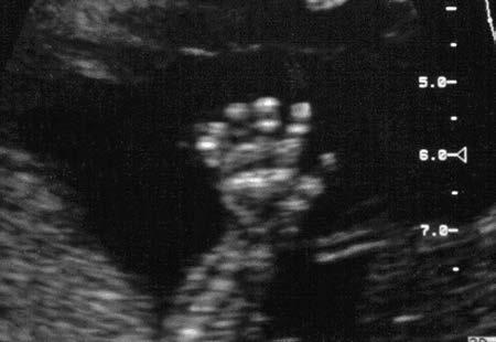
An image (from the Latin term for imitate ) is a reproduction, representation, or imitation of the physical form of a person or object. An ultrasound image is the visible counterpart of an invisible object, produced in an
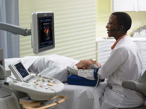
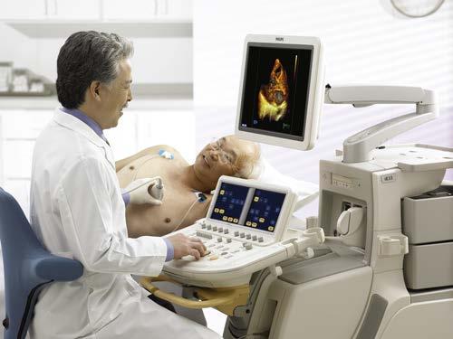
electronic instrument by the interaction of ultrasound with the object. Ultrasound provides a noninvasive way of looking inside the human body (Figure 1-1 ) to image otherwise hidden anatomy. Anatomic imaging with ultrasound is accomplished with a pulse-echo technique . Pulses of ultrasound generated by a transducer are sent into the patient ( Figure 1-2 ), where they produce echoes at organ boundaries and within tissues. These echoes return to the transducer,
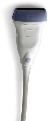
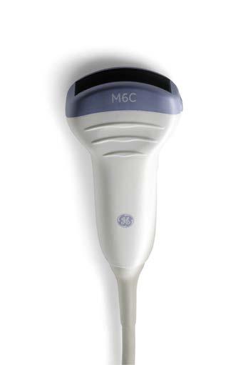
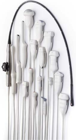
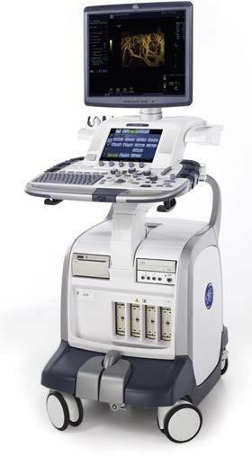
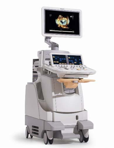
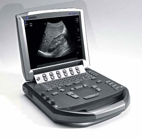
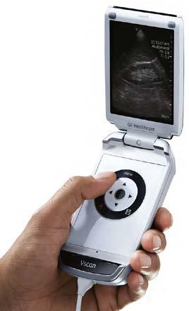
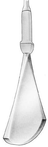
where they are detected and presented on the display of a sonographic instrument. The transducer ( Figure 1-3 ) generates the ultrasound pulses and receives the returning echoes. Sonography requires knowledge of the location of origin of each echo and its strength as it returns from the patient. The ultrasound instrument (Figure 1-4 ) processes the echoes and presents them as visible dots, which form the anatomic image on the display. The brightness
1-1 Ultrasound provides a window into the human body, allowing us to see what would otherwise be hidden from view (A-B). Images shown are as follows: C, abdominal; D, cardiac; E, obstetric, and F, vascular.
FIGURE
FIGURE 1-2 Pulse-echo technique. A, In diagnostic ultrasound, ultrasound pulses are sent into the tissues to interact with them and to obtain information about them. B, Echoes return from the tissues, providing information that enables anatomic imaging and observation of motion and flow, thus contributing to diagnosis.
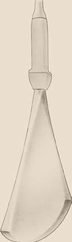
FIGURE 1-3 A-C, Transducers.
FIGURE 1-4 Sonographic instruments by: A and D, GE Healthcare, B, Philips Healthcare, and C, Sonosite, Inc.
FIGURE 1-5 One pulse of ultrasound generates a single scan line (series of echoes) as it travels through tissue. Echoes are presented in sequence on a scan line while they return from tissue during pulse travel. A, The first echo is displayed.B, The second echo is added. C, Three more echoes are added. D, All the echoes from a single pulse have been received and displayed as a completed scan line. E, A complete scan line results from one emitted pulse. In practice, this is accomplished in less than one-thousandth of a second. F, According to the pulse-echo imaging principle, one pulse traveling through tissues produces a stream of echoes that become one scan line on the display.

FIGURE 1-6 A single rectangular image or scan (also called a frame) is composed of many vertical parallel scan lines. Each scan line represents a series of echoes returning from a pulse traveling through the tissues. A, One scan line from one pulse, as generated in Figure 1-5 B, A second scan line is added. C-D, Five and ten scan lines, respectively. E, A complete frame consisting of (in this example) 100 scan lines.
FIGURE 1-4, cont’d
of each dot corresponds to the echo strength, producing what is known as a gray-scale image . The location of each dot corresponds to the anatomic location of the echo-generating structure. Positional information is determined by knowledge of the direction of the pulse when it enters the patient and by measurement of the time it takes for each echo to return to the transducer. The proper location to present the echo can then be determined from a starting point on the display (usually at the top). With knowledge of the sound speed, the instrument uses the echo arrival time to determine the depth of the structure that produced the echo.
If one pulse of ultrasound is sent into tissue, a series of dots (one line of echo information, specifically, an echo line, data line, or scan line) is displayed (Figure 1-5).


Not all of the ultrasound pulse is reflected back from any structure. Rather, most of the original pulse continues on to be reflected back from deeper structures. The echoes from one pulse appear as one scan line (see Figure 1-5, EF ). If the process is repeated, but with different starting points for each subsequent pulse, a cross-sectional image of the anatomy is constructed (Figure 1-6). Pulses travel in the same direction from different points and yield vertical parallel scan lines and a rectangular image, as shown in Figure 1-7. These cross-sectional images are produced with vertical parallel scan lines that are so close together they cannot be identified individually. The rectangular display resulting from this procedure often is called a linear scan, or linear image, referring to the linear-array transducer that is used to produce it. A second approach to sending ultrasound pulses through the anatomy to be imaged is shown in Figure 1-8. With this method, each pulse originates from the same starting point, but subsequent pulses go out in slightly different directions. This approach results in a sector scan, or sector image, which has a shape similar to a slice of pie (Figure 1-9). Figure 1-10 shows a format that is a combination of the two just described; that is, pulses (and scan lines) originate from


FIGURE 1-7 A, Ultrasound sent through a thin rectangular volume of tissue produces a rectangular image, commonly called a linear image or linear scan. B, Clinical linear (rectangular) scan. C, Poor-quality (by current standards) fetal image from the late 1970s, revealing the 120 vertical parallel scan lines of which it is composed.
Sonography is accomplished with a pulse-echo technique.
Echoes from anatomic structures represent these structures in a sonographic image.
FIGURE 1-8 A single sector frame is progressively built up with 1 (A), 2 (B), 5 (C), 10 (D), and 100 (E) scan lines in sequence. All originate from a common origin and travel out in different directions.
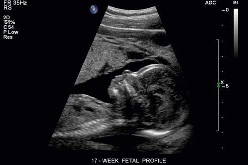
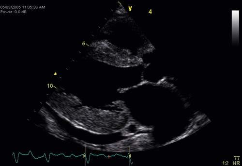
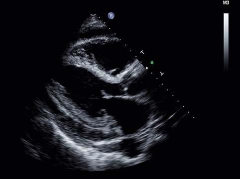
FIGURE 1-9 A, Ultrasound sent through a thin pie-slice–shaped volume of tissue produces an image commonly called a sector image or sector scan B, Sector scan of adult heart.
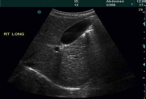
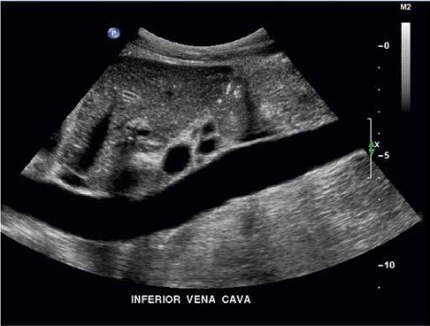
FIGURE 1-10 A, A modified form of a sector scan is produced when pulses and scan lines originate from different points across the curved top of a sector display. B, Abdominal scan with use of the scan format shown in A
different starting points (as in a linear image), but each pulse (and scan line) travels in a slightly different direction from that of the previous one (as in a sector image). In this example, the starting points form a curved line across the top of the scan, rather than a straight line, as in the linear scans shown in Figure 1-7.
Sonographic images are composed of many scan lines.
Sonographic scan formats commonly are limited to three types: (1) linear, (2) sector, and (3) a combination of the two. Other formats may be used occasionally, but in any case, what is required is that ultrasound pulses be sent through all portions of the anatomy that are to be imaged. Each pulse generates a series of echoes, resulting in a series of dots (a scan line) on the display. The resulting cross-sectional image is composed of many (typically 96 to 256) of these scan lines. The scan format determines the starting points and paths for individual scan lines, according to the starting point and path for each pulse used to generate each scan line. The clinical cross-sectional gray-scale sonographic images produced are sometimes called B scans. This term implies that the images are produced by scanning the ultrasound through the imaged cross-section (i.e., sending pulses through all regions of the cross-section) and converting the echo strength into the brightness of each represented echo on the display (hence, B [brightness] scan). The terms B scan and gray-scale scan have the same meaning.
2D images are presented in linear (rectangular) and sector forms.
For decades, sonography was limited to 2D cross-sectional scans (or “slices”) through the anatomy. Today, 2D imaging,
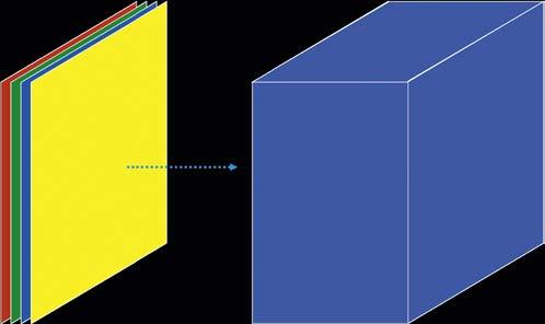
while still being used extensively, has been extended to 3D scanning and imaging, also called volume imaging. This method requires scanning the ultrasound through many adjacent 2D tissue cross-sections to compose a 3D volume of echo information similar to a loaf of sliced bread (Figure 1-11). This 3D volume of echoes can then be processed and accessed to present 2D or 3D images of the anatomy.
Sonographic images are of 2D and 3D types.
DOPPLER ULTRASOUND
Echoes produced by moving objects have frequencies that are different from the pulses sent into the body. This phenomenon is called the Doppler effect, which is put to use in detecting and measuring tissue motion and blood flow. The Doppler effect is named after Christian Andreas Doppler, the Austrian physicist who conducted an extensive investigation into its nature.
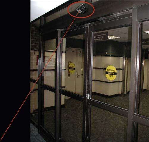
The use of Doppler radar in weather forecasting, aviation safety, and vehicle speed detection (police radar) has made the Doppler effect a household term. In addition to its observation in everyday life (as demonstrated by the changing pitch of a siren or horn heard as the vehicle passes by), the Doppler effect has been applied to automatic door openers in public buildings (Figure 1-12) and to other motion-detecting devices.
The Doppler effect is a change in frequency caused by moving objects.
Doppler ultrasound has been used in diagnostic medicine for decades. Long-standing applications include monitoring the fetal heart rate during labor and delivery and evaluating blood flow in the heart and in the arteries and veins of circulation. Rapid scanning and processing of Doppler data
BC
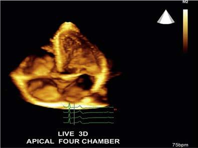
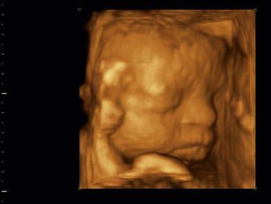
FIGURE 1-11 Three-dimensional (volume) sonographic images. A, Three-dimensional echo data acquired by obtaining many two-dimensional sections of echo information (colored slices) from the imaged anatomy, forms a three-dimensional volume of stored echo information (blue box). B, Cardiac four-chamber view. C, Fetal head.
FIGURE 1-12 An ultrasonic automatic door opener (circle).
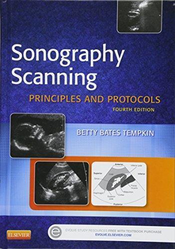
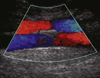
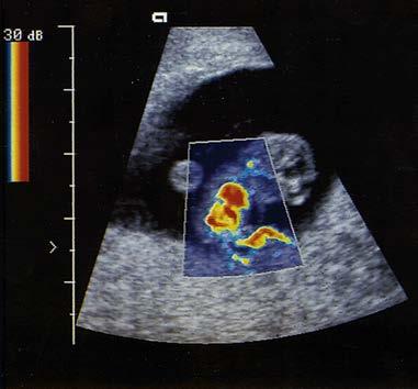
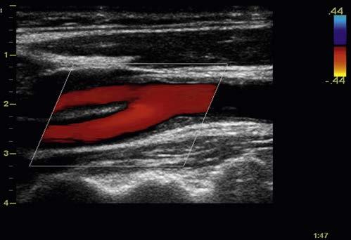
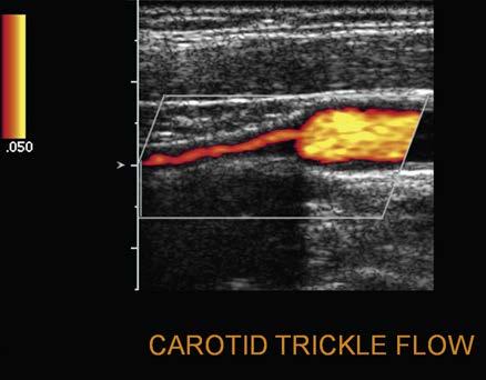
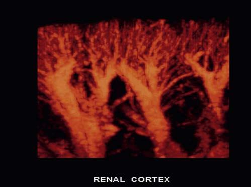
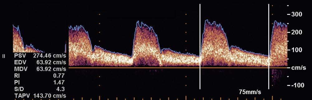
enable color-coded 2D and 3D presentations of Doppler information (color-Doppler displays) to be superimposed on gray-scale anatomic images (Figure 1-13). Doppler information is applied to loudspeakers for audible evaluation and to spectral-Doppler displays for quantitative analysis (Figure 1-14). The spectral-Doppler operation includes
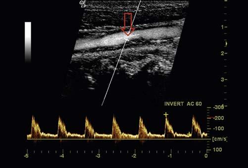
anatomic imaging to determine the location(s) from which the spectral information is acquired (Figure 1-15).
Doppler information is presented in audible, colorDoppler, and spectral-Doppler forms.
FIGURE 1-13 Color-Doppler displays of blood flow. Presented in forms called (A) colorDoppler shift, (B) color-Doppler power, and (C) three-dimensional color-Doppler power displays.
FIGURE 1-14 Spectral-Doppler display of arterial blood flow with presentation of calculated flow velocity data.
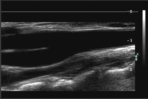
FIGURE 1-15 Spectral-Doppler display of blood flow in the carotid artery. The anatomic image shows the location (arrow) from which the spectral-Doppler information was acquired.