Large Animal Review
ORIGINAL ARTICLES
BOVINE
ISSN: 1124-4593
LARGE ANIMAL REVIEW is ranked in Citation Index (SciSearch®) Journal Citation Reports/Science Edition and CAB ABSTRACTS
• Effect of fermented concentrated potato protein on milk yield and fertility parameters in dairy cows in the prepartum and postpartum periods
• The Association between the STAT1 g.3141C>T Polymorphism and Reproductive Performance in High-yielding Holstein-Friesian Dairy Cows
• Evaluation of diagnostic accuracy of flexible borescope diagnosing digital dermatitis in milking parlor
• Effect of rumen-protected fat and/or vitamin C supplementation on growth performance, carcass characteristics and meat composition in Hanwoo steers during late fattening period
• Field trial of the effect of vaccination against Bovine herpesvirus 1 on milk yield and rumination time: comparison between two live marker vaccines
OVINE
• Biological activities of Juniperus phoenicea essential oil and impact on in vitro ruminal fermentation in sheep
• Does Isoxsuprine HCl Facilitate The Passage Of The Cervix in Sheep?: A Case Series
SWINE
• African Swine Fever: risk factors and biosecurity measures in backyard holdings
BOVINE
• Salmonella enterica serovar Dublin infection in dairy cattle: a case study on the management of an outbreak in Italy
LAR Bimonthly, Year 29, Number 2, April 2023
02/23
SOCIETÀ
VETERINARI
ANIMALI
ASSOCIAZIONE FEDERATA ANMVI
ITALIANA
PER
DA REDDITO
ERAY AKTUĞa*, EROL BAYTOKb, BURÇİN TÜRKMENOĞLUc
a Department of Animal Nutrition and Nutritional Diseases, Tekirdağ Namık Kemal University, Faculty of Veterinary Medicine, Tekirdağ, Türkiye
b Department of Animal Nutrition and Nutritional Diseases, Erciyes University, Faculty of Veterinary Medicine, Kayseri, Türkiye
c Department of Basic Pharmaceutical Sciences, Faculty of Pharmacy, Erzincan Binali Yıldırım University, Erzincan, Türkiye
SUMMARY
This study aims to determine the effects of fermented concentrated potato protein (FCPP) which showed very high levels of indole acetic acid (IAA) on milk yield, fertility, and level of insulin-like growth factor 1 (IGF-1) parameters in pregnant dairy cows and pregnant heifers.
In total, sixty Holstein cattle were enrolled in the study. The animals were divided into three groups, as control group (n=20), 25 g FCPP group (n=20), and 50 g FCPP group (n=20). Besides, these main groups were also divided into two sub-groups, as primiparous (n=10) and multiparous cows (n=10). Oral administration of FCPP started two weeks (14±4 days) before expected parturition and continued until postpartum day 100. The affinity of IAA found in FCPP pellets to 5HT1 and JAK2 receptors, which is thought to be related to IGF-1 release, was determined by the molecular docking method that receptor affinities were found as -5.8637 kcal/mol and -4.3857 kcal/mol, respectively.
Blood IGF-1 profile was followed at 7 different time points throughout the study. It was detected that the IGF-1 concentrations have significant difference in terms of both time and groups (P<0.05). Furthermore, there was a significant difference in interaction of time and parity (P<0.05).
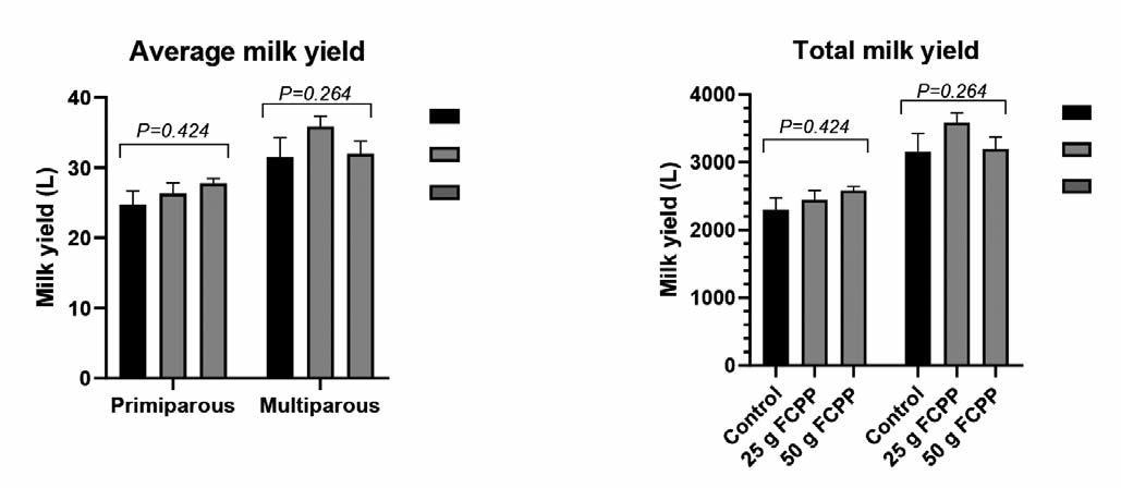
The results showed that average and total 100-day milk yield was not affected by FCCP supplementation (P>0.05). FCPP supplementation generally has improved the mathematical data of fertility parameters, but no statistical significance was detected except for calving-conception interval. It was found that calving-conception interval reduce by 16.8% in primiparous cows supplemented with 25 g FCPP. The pregnancy rates in control, 25 g and 50 g FCPP were found as 72.2 %, 78.9 % and 88.9 %, respectively (P>0.05).
This study has concluded that fermented concentrated potato protein (which has indole acetic acid-indole compounds) may improve the productivity of dairy cows supplemented in transition period and it has suggested that further research must be done for its usage and beneficial effects in dairy cows.
KEY WORDS
Cow, fermented concentrated potato protein, fertility parameters, indole acetic acid, molecular docking.
INTRODUCTION
Dairy cattle are a sub-branch of animal husbandry. In addition to providing a regular income with the milk obtained from dairy cattle, the calf obtained every year is also an added value. Milk yield and fertility are closely related to IGF-1 and is thought to be used as a genetic predictor. For this reason, studies on feed additives that support IGF-1 production, and thus increase fertility and milk yield, gain importance.
In high-yielding cows, plasma IGF-1 concentration decreases after parturition, however, upregulation of liver growth hormone (GH) receptors stimulates IGF-1 production and con-
Corresponding Author: Eray Aktuğ (eaktug@nku.edu.tr).
sequently increase IGF-1 levels in the blood1. There are cases in which the IGF-1 level cannot be increased due to various stress factors. It is essential not to prolong this situation2-6, as IGF-1 affects follicle-stimulating hormone (FSH) and luteinizing hormone (LH), which have an impact on ovarian follicles. The IGF-1 is active in the cows’ genital system and is useful in the formation and continuation of pregnancy7,8.
In most species, follicular granulosa cells synthesize IGF-1, but this is not seen in cows. In ruminants, IGF-1, which is in follicular fluid, comes from blood circulation. The concentration of IGF-1 varies with age, breed, and lactation period9. Consequently, there are studies regarding the increase the release of IGF-1 in animals. There are also studies regarding the increase of IGF-1 level in the blood by using fermented concentrated potato protein (FCPP), but these are few. There were no studies conducted on the active substance IAA10-15.
Tryptophan derivatives are bioactive compounds found in FCPP
E. Aktuğ et al. Large Animal Review 2023; 29: 51-5751
N
Effect of fermented concentrated potato protein on milk yield and fertility parameters in dairy cows in the prepartum and postpartum periods
of fermented concentrated potato protein on milk yield and fertility parameters in dairy cows
that can stimulate tissue growth in both animals and plants16
Plant growth is stimulated with tryptophan derivatives (e.g., indole acetic acid). Growth and cell proliferation in animal tissues are stimulated by serotonin, tryptamine, and indole, whose molecular structures are similar. Indole also stimulates liver regeneration16. There is no study suggesting that indole acetic acid increases IGF-1 secretion in cows. However, Gillessen and Rebiere17 in 2011 (Patent Issue 13/064,818,) reported that there was increased IGF-1 in catfish, piglet, and laying hens. Therefore, the molecular docking method used in the study is an application that predicts the preferential orientation of a molecule to a second molecule when bound to form a stable compound18. In this study, molecular docking compatibility is discussed to elucidate the mechanism of interaction between ligand-receptor19
The present study aims to find the effect of FCPP (indole acetic acid-indole compounds) on IGF-1 release and its effect on milk yield and fertility parameters in dairy cattle.
MATERIALS AND METHODS
Animal Housing and Care
The study was conducted in a commercial enterprise in the central Anatolia Region (Bünyan, Kayseri, Türkiye). In this farm, approximately 600 dairy cows were bred and milking was performed twice a day with a rotating milking system. Animal health care and herd management were under veterinary supervision. The animals were housed in groups in free-stall barns bedded with plastic and equipped with overhead fans and a sprinkler system.
Experimental Design
The study was carried out with 60 Holstein Friesian cattle consisting of 30 pregnant cows (multiparous cows) and 30 pregnant heifers (primiparous cows), which were randomly assigned to one of the three groups. They were paired based on similarities; lactation number (parity), milking performance in the previous lactation and BCS (3.5) to provide three groups (10 in each group). Also, pregnant heifers (primiparous cows) were
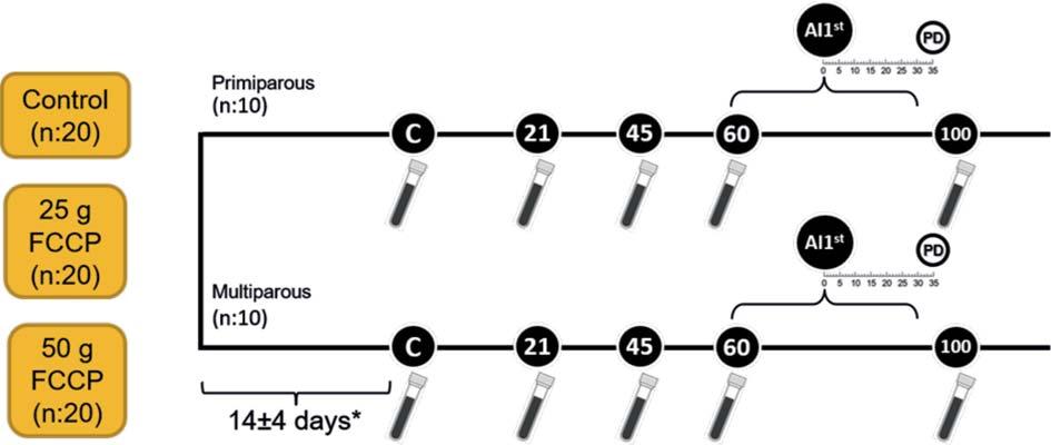
divided into three groups as pregnant heifers (10 in each group) with similar BCS values (3.5) and from the same father. The FCPP (Lianol® Dairy) used in the study was obtained from ANC Animal Nutrition and Health Services Inc. Feed was in pellet and suitable for consuming by cattle. The recommended dose, according to product instructions, was 25 g daily per animal. The nutritional analysis of the product (label values of the product) was the following: calcium carbonate 46%, dicalcium phosphate 10%, fermented potato protein 15%, potato protein 10%, wheat (carrier) 13%, molasses (carrier) 4%, soybean oil (carrier) 2%. The nutrient content of FCPP (label values of the product) was: Crude Protein 13.60%, Crude Fat 2.80%, Ash 52.00%, Crude Cellulose 1.00%. The FCPP was administered once per day to each animal orally, mixed in equal parts with water under human supervision. The administration started about two weeks (14±4 days) before expected time of calving and continued until postpartum (pp) day 100. As far as the rest of animal nutrition is concerned, a vertical TMR mixer was used, which, twice a day (at 09:00 and 17:00), was distributing the rations mentioned in Table 1 in equal amounts.
Data Collection and Sample Analysis
The schematic diagram of the study is presented in Figure 1. Milk yield data of the cows were continuously obtained from the farm management software for the whole 100-day period and were recorded for each animal. The milking process was carried out in different milking systems for the first seven days in order to get used to milking the heifers after parturition. Subsequent data were recorded up to pp day 100 as in cows. Blood samples (~8 ml) were collected from each animal by venipuncture of the coccygeal vessel at the beginning of the experiment, at parturition, on Day pp 21, 45, 60, at first insemination time (AI1st) and on Day pp 100. Once the samples were collected, the serum was separated by centrifugation (Hettich Universal 320, Germany) at 3000 rpm for 10 min and then frozen at -80°C for subsequent analysis.
Insemination and Fertility Parameters
In the postpartum period, oestrus symptoms were detected by pedometers and experienced farm personnel. Routine gynae-
52Effect
Figure 1 - The schematic diagram of the study.
PERIODWheat strawAlfalfa hayCorn SilageConcentrated Concentrated Corn grainMagnesiumSodiumBypass fat Pellet FeedPellet Feedbicarbonate (21% HP) *(19% HP)**
* As fed basis (%): Razmol 21%, 46% HP soybean meal 20%, broken grain 20%, 28% HP sunflower meal 12.5%, rice bran 13%, corn 7.7%, molasses 2.6%, soybean oil 0.6%, marble powder 1.8%, salt 0.7%, vitamin-mineral mix 0,1%***
** As fed basis (%): Razmol 24%, 46% soybean meal 13.7%, broken grain 20%, 28% HP sunflower meal 12,5, 28% HP cotton seed meal 1.8%, rice bran 12.6%, corn 10.3%, molasses%
2.5, marble powder 1.8%, salt 0.7%, vitamin-mineral mix 0.1% ***
*** Each kilogram of vitamin-mineral mix; 13.000,000 IU of vitamin A, 3.500,000 IU of vitamin D, 40.000 mg of vitamin E, 50.000 mg of zinc, 50.000 mg of manganese, 50.000 mg of iron, 10.000 mg of copper, 150 mg of cobalt, 800 mg of iodine, 300 mg of selenium.
cological examinations were weekly performed to evaluate the healthy voluntary waiting period and to diagnose likely pp disease in the scope of the reproductive management procedure. Thus, it was confirmed that the genital tract of the animals to be included in the reproductive program were health. In addition, cows were followed up to the pp day 150 to evaluate the pregnancy rates obtained after the AI2nd and AI3rd. Artificial inseminations were performed by the same herd veterinarian. The animals were inseminated no more than three times during this study. Pregnancy diagnosis was performed with a portable ultrasound (MINDRAY DP-10 Vet, China) equipped with 5-8 MHz linear probe on Day 35 after AI.
The fertility parameters including calving to AI1st; calving to conception interval; first oestrus; conception rate in AI1st, AI2nd, AI3rd and total pregnancy rate were calculated as described by Ata20 (2013) and Tekin and Daskin21 (2016).
IGF-1 Analysis
The total serum IGF-1 concentration was determined by a commercial IGF-1 ELISA kit (Sunred Bio Bovine IGF-1 Elisa Kit). The absorbance values of the samples were determined by a 96well microplate reader (µQuant, BIO-TEK) with a spectral waveband of 400-750 nm. The microplate was measured at 450 nm with this instrument.
Indole Acetic Acid (IAA) Analysis
The amount of IAA in FCPP pellet was analysed by AGILENT 1260 model high-pressure liquid chromatography (HPLC) with a DAD (Diode Array Detector) at Erciyes University Technology Research and Application Center (TAUM). Indole acetic acid, 87-51-4 CAS number, was used as a standard analyte in the analysis. The extracted samples were analysed by high-performance liquid chromatography (HPLC)22.
Molecular Docking Method
The molecular docking method was used to determine the receptor-ligand relationship of IAA, which was contained in the FCPP pellet. The optimal molecular docking calculations were selected, based on Türkmenoğlu and Güzel19 (2018). In this study, the molecular docking process was applied to two different receptors, which were predicted to interact. Proteins acting on JAK2 and 5HT1 receptors from the protein data bank were examined, and the individual RMSD (square root of the standard deviation) value was calculated. The binding site between the most stable L-R was observed by finding the binding energies by the FlexX docking programme.
Molecular docking results were found from the protein databank (www.rcsb.org). In the molecular docking programme,
IAA was used as ligand and interacted with two different proteins (PDB ID: 2XDG and 3UGC).
Statistical Analysis
The appropriateness of the data to the normal distribution was evaluated by the Q-Q plot, histogram, and Shapiro Wilk test. The homogeneity of the variances was examined by the Levene test. Descriptive statistics were shown as “Mean ± SEM,” and percentages.
One-Way ANOVA and Kruskal Wallis were used to compare the importance between groups (Control, 25 g FCPP, and 50 g FCPP) in terms of milk yield and fertility parameters. Analyses of significant differences were performed using analysis of variance followed by the Tukey post hoc test.
The effect of the group on pregnancy rate was investigated by the Chi-Square test. The effect of groups, time, and lactation (primiparous-heifer and multiparous-cow) on the IGF-1 level was calculated with repeated measures ANOVA. Statistical analysis of the data was performed by the SPSS (version 20.0, SPSS Inc, USA) programme. The significance level was accepted as P <0.05.
RESULTS
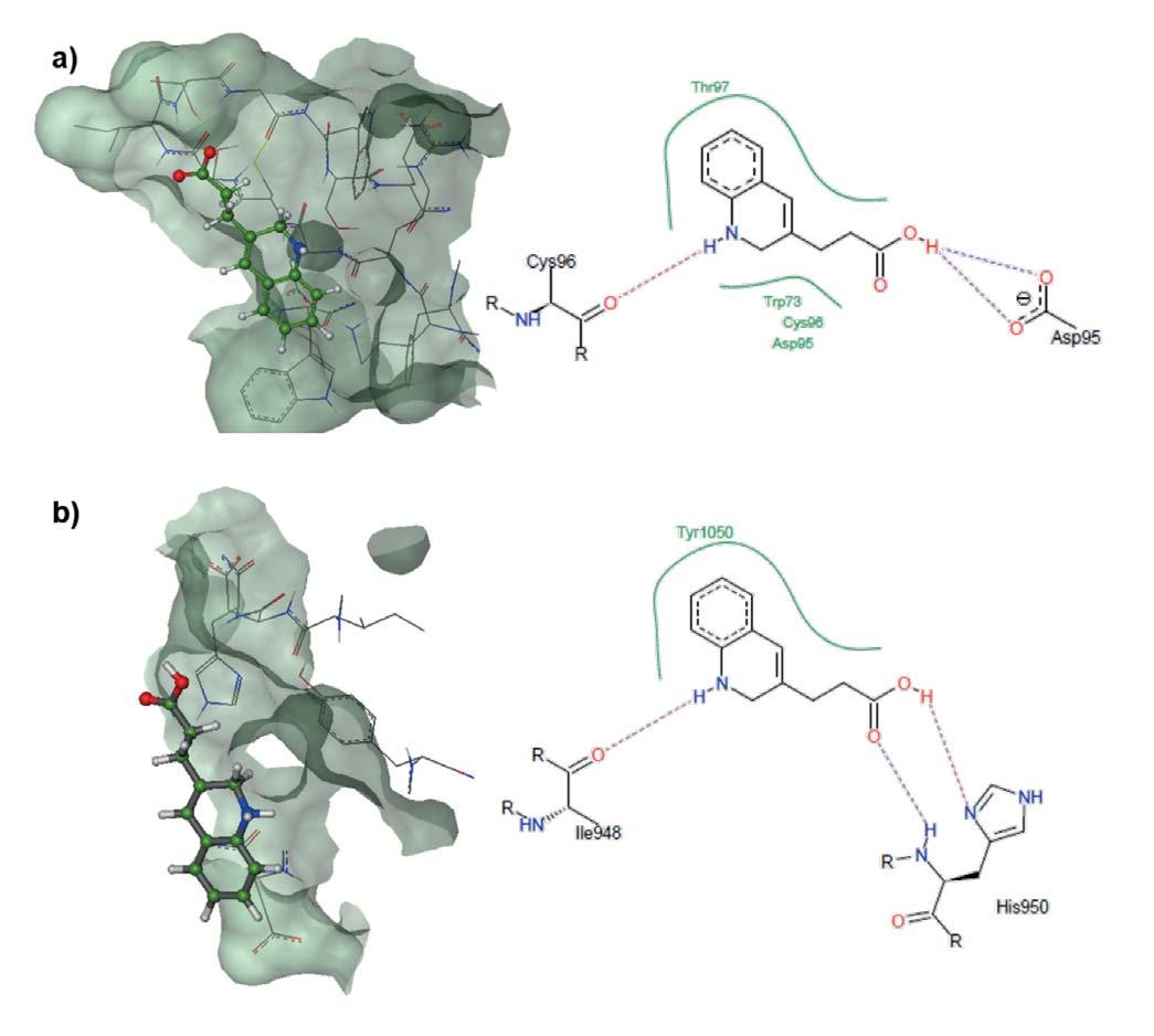
The interaction between the ligand-receptor shown in Figure 2a and 2b was found to be binding affinity G = -5.8637 kcal/mol using the FlexX docking programme. Between the ligand IAA and the amino acids, Asp 95 and Cys 96, there is a hydrogen bond. These bonds also indicate the interaction between the ligand and the receptor.
The interaction between the ligand-receptor shown in Figure 2 was found to be binding affinity G = -4.3857 kcal/mol using the FlexX docking programme. Between the ligand IAA and the amino acids, Ile 948 and His 950, there is a hydrogen bond. In this case, too, these bonds also indicate the interaction between the ligand and the receptor. Based on these results, it can be stated that IAA is theoretically active relative to the receptors. Results for IGF-1 levels, fertility parameters, milk yield are given in Table 2, Table 3 and Figure 3, respectively. Five animals were removed from the farm against our will due to the farm’s business policies at different times.
DISCUSSION
In literature, there are few studies showing the effect of FCCP on IGF-1 levels23,24. In these studies, it was observed that the
E. Aktuğ et al. Large Animal Review 2023; 29: 51-5753
Close up2.23.518.54.63.520.080.3Early Lactation1.5523.513.5-20.080.30.2 Peak Lactation1.55234.62.520.080.3-
Table 1 - The amount of raw material (kg) of TMR used in the farm.
amount of IGF-1 was increased by the addition of FCCP to pigs as a feed additive. It has been reported that FCCP supplementation positively affected various yield parameters in those animals23 and these effects of FCCP is associated with its tryptophan and derivatives ingredients25,26. However, indole derivatives (particularly IAA) are not mentioned in these studies. Indole derivatives are also known to be produced by various microorganisms in the intestine. These bioactive molecules also play a role in providing communication between intestinal microorganisms27. Currently, the intestinal flora is considered as the second brain of the body and the mechanisms of action of some bioactive substances produced by flora are still being studied. Indole derivatives, which are also bioactive substances (AhR ligand), are reported to stimulate the immune system28,29. It has been shown that different ligands such as 7,8Tetrachlorodibenzo-p-dioxin (TCDD), flavonoids, carotenoids, and indoles can be bound to AhR30. Based on the previous studies, that different ligands can be bound to the same receptor,
suggest that IAA may also be bound and activates a structure which triggers IGF-1 production. Furthermore, the molecular structure of the IAA included in FCPP was studied using the molecular docking method and computer-mediated examination, and showed that it can be bound to structures that can activate IGF-1 production.
In this study, it was determined that IGF-1 levels were significantly higher in the groups consuming 25 g and 50 g FCPP. Similarly, studies in pigs have shown that the use of FCPP significantly increases IGF-1 levels10-13,15,23. Similar results have been reported by Gillessen and Rebiere17 (Patent Issue 13/064,818, 2011). In the early lactation period in dairy cattle, IGF-1 was in low concentration when GH levels were high31,32. In the third week after calving, IGF-1 concentration started to increase with the upregulation of liver GH receptors1. This finding is in line with the results of this study. Although there was a generally rapid increase of IGF-1 levels in all three groups within 100 days in milk, generally the higher IGF-1 levels were observed in the
54Effect of fermented concentrated potato protein on milk yield and fertility parameters in dairy cows
Figure 2 - a) Molecular docking between the receptor-ligand and the interaction diagram of the ligand inserted within the active site of the 5HT1 enzyme (PDB ID: 2XDG) of the GHRH receptor. (b) Molecular docking between receptor-ligand and the interaction diagram of the inserted ligand within the active site of the JAK2 receptor (PDB ID: 3UGC).
Table 2 - The effects of FCCP on blood serum IGF-1 levels (ng / ml) in the prepartum and postpartum periods (x ± Sx). Control25
FCPP50 g FCPP
a, b: Different letters on the same line show the difference between groups.
A, B, C, D, E, F, G, H, I, M, N, P: Different letters in the same column show the difference between groups. (x): Arithmetic mean (Sx): Standard error
FCPP groups (except prepartum -14±4 and parturition day in cows; 60th day in heifers)
It is thought that the effect of FCCP on IGF-1 can be explained in two ways. First, IAA, which is found in the FCPP, acts like serotonin, affects the GHRH release. Indole acetic acid such as serotonin can be found in the central nervous system33. According to recent studies, it has been reported that similar molecules can activate the same receptor30. Docking method results showed that, like serotonin, IAA had been found to affect GHRH (Figure 2a). In this case, the GH level may increase and indirectly make it possible to increase IGF-1. A second possibili-
ty is that the affinity of the IAA to JAK2, as shown by the docking method, is likely to activate the signal in the liver (Figure 2b).
The effect of IGF-1 on body functions throughout the lifespan of the animal is significant34. Therefore, the insufficiency of IGF1 may adversely affect growth and productivity. The IGF-1 also affects many parameters, such as milk yield and fertility9,35. In the study, there was no significant difference between average daily milk yields among groups. Although FCPP did not significantly affect milk yield, a mathematical increase was observed in both primiparous and multiparous cows. The fact that milk yield lev-
E. Aktuğ et al. Large Animal Review 2023; 29: 51-5755
-14±4200.00±19.57 CF 219.46±15.13 DF 211.36±14.37CE P value Within 24 hours124.48±17.51 DI 154.63±13.74 EI 155.20±13.72DF Group 0.031 21st day208.94±20.44 CG 227.18±12.81CD 225.33±16.19C Heifer 45th day238.09±10.14 BM 245.47±36.38BG 247.79±16.76BH Lactation0.106 60th day250.24±26.27B 241.86±26.02BCM 258.30±27.53AB First Insemination Day197.42±7.61 CN 236.59±16.12BCN 267.79±7.12AG Time <0.001 100th day 261.97±12.44 AP 278.37±23.30AP 264.98±5.69A Group * Lactation0.641 -14±4275.81±19.07AF 263.49±19.63BF 256.36±33.51BE Within 24 hours160.78±13.59FI 135.31±14.70DI 164.40±17.48DF Time * Group0.679 21st day169.27±14.40aDG 220.52±28.51bC 207.62±15.48bC Cow 45th day251.05±27.98CM 319.37±35.60AG 285.70±22.93AH Time * Lactation 0.035 60th day217.35±25.80B 315.74±44.87AM 269.71±20.51A First Insemination Day183.14±20.44EN 245.87±27.03CN 239.00±5.62BG Time * Group * Lactation0.475 100th day 265.36±28.89BP 280.04±24.80BP 287.68±18.41A
g
Service Period (Day) (x± Sx)Heifer80.60 ± 3.3772.10 ± 4.0576.75 ± 3.990.282 Cow81.87 ± 7.3180.33 ± 3.5475.80 ± 3.660.654 Calving-Conception Interval (Day) (x± Sx) Heifer116.00 ± 5.82a 96.42 ± 6.20b 111.42 ± 3.56ab 0.045 Cow119.83 ± 8.12117.37 ± 7.56113.33 ± 8.310.853 First Oestrus (Day) (x± Sx)Heifer43.00 ± 3.2045.50 ± 3.7145.50 ± 1.750.792 Cow51.42 ± 5.2541.75 ± 3.2842.12 ± 1.270.119 Pregnancy Rate in AI1st (%)Heifer30.040.037.5 Cow25.033.340.0 Total27.836.838.90.756 Pregnancy Rate in AI2nd (%)Heifer57.150.060.0 Cow33.350.033.3 Total46.250.045.50.972 Pregnancy Rate in AI3rd (%)Heifer0.00.050.0 Cow50.066.775.0 Total28.633.366.70.333 Overall Pregnancy Rate (%)Heifer70.070.087.5 Cow75.088.990.0 Total72.278.988.90.453
in 25 g FCPP, 50 g FCPP and control
ParametersParityControl25 g FCPP50 g FCPPP value a, b: The
(x): Arithmetic mean, Sx: Standard error
Table 3 - Postpartum fertility parameters of primiparous and multiparous cows
groups.
difference between the mean values of groups bearing different letters on the same line is significant (P<0.05).
els in primiparous cows are more homogeneous than in multiparous cows could be because the primiparous cows used in the study have the same father line. This may be one reason why standard deviations in primiparous cow groups are lower than in multiparous cow groups. In previous studies conducted in lactating goats infused with IGF-1 into the mammary artery, it was observed that blood flow was accelerated, and milk production was increased36,37. However, an increase in milk yield was not detected in sheep injected with growth hormone into the mammary artery38. Peel and Bauman38 have reported that growth hormone might indirectly affect breast tissue with IGF-1. When the fertility parameters were assessed, it was found that the first oestrus after calving observed earlier for multiparous cows, to which FCPP was administered, than in the control group. As far as the Calving-First Insemination Interval for both primiparous and multiparous cows in the 25 g and 50 g FCPP groups is concerned, although there was no statistically significant difference. This period was shorter, even for a lot of days, compared to the control group. In parallel, the CalvingConception Interval was statistically shorter in primiparous cows. Although there is no statistically significant difference, the pregnancy rate of all animals in the 25 g and 50 g FCPP groups was found to be higher by 6.7% and 16.7%, respectively, when compared to the control group. Plasma IGF-1 level in dairy cattle during the periparturient period has a positive impact on insemination and has been stated to be a useful parameter for reproduction39. Likewise, it is considered as an essential indicator for the fertility management of dairy animals in the postpartum period39. IGF-1 is seen as an important factor for the resumption of the oestrus cycle in the early postpartum period40. Scaramuzzi et al.35 showed that exogenous administration of IGF-1 in vivo is a potent stimulator of both follicle growth and estradiol secretion in sheep. However, Falkenberg et al.41 argued that IGF-1 is not an essential factor. The results of the study have shown that supplementation with FCPP increases the total pregnancy rate in primiparous and multiparous cows, shortens the service period in primiparous cows and increases the milk yield in primiparous cows. Based on the results obtained from this study, it can be concluded that
addition of FCCP to the diet has beneficial effects on fertility and milk yield by supporting blood IGF-1 concentration in transition period (pre- and postpartum period) in dairy cows and overcome problems resulted in fermentation process by its antibiotic resistance. However, to clarify its features on productivity and metabolism, more detailed studies should be done in dairy cows. It is thought that the results will contribute to herd management and give further insight into how molecules produced by bacteria affect the liver, brain, and, in particular, specific body functions, reproduction and milk production.
Welfare Statement
Trial was completely non-invasive. No animal was displaced from its home farm. The feed additive used was an already commercially available and legally approved additive. All experimental procedures involving the use of animals were in accordance with the animal welfare legislation and approved by Erciyes University Local Ethics Committee for Animal Experiments (HADYEK) (Kayseri, Turkey; date: 13.01.2016 protocol no: 16/007).
Acknowledgments
This article was produced from the first author’s PhD thesis entitled “Investigation of the Effect of Fermented Concentrated Potato Protein to Milk Yield and Fertility Parameters on Dairy Cows in Prepartum and Postpartum Period”. This study is summarized from the thesis supported by the Teaching Staff Training Programme (ÖYP).
References
1. Wathes D.C., Taylor V.J., Cheng Z., Mann G.E. (2003). Follicle growth, corpus luteum function and their effects on embryo development in postpartum dairy cows. Reprod Suppl, 61:219-237.
2. Thissen J.P., Underwood L.E., Ketelslegers J.M. (1999). Regulation of insulin-like growth factor-I in starvation and injury. Nutr Rev, 57(6):167176.
3. Kerr D.E., Manns J.G., Laarveld B., Fehr M.I. (1991). Profiles of serum IGF-I concentrations in calves from birth to eighteen months of age and in cows throughout the lactation cycle. Can J Anim Sci, 71(3): 695-705.
56Effect of fermented concentrated potato protein on milk yield and fertility parameters in dairy cows
Figure 3 - The average (kg/day/animal) and total milk yields (kg/animal) of multiparous between 0-100 days and primiparous between 8100 days.
4. Wathes D.C.C., Cheng Z., Bourne N., Taylor V.J.J., Coffey M.P.P., Brotherstone S. (2007). Differences between primiparous and multiparous dairy cows in the inter-relationships between metabolic traits, milk yield and body condition score in the periparturient period. Domest Anim Endocrinol, 33(2): 203-225.
5. Muthuramalingam P., Kennedy A.D., Berry R.J. (2006). Plasma melatonin and insulin-like growth factor-1 responses to dim light at night in dairy heifers. J Pineal Res, 40(3): 225-229.
6. Pushpakumara P.G.A., Gardner N.H., Reynolds C.K., Beever D.E., Wathes D.C. (2003). Relationships between transition period diet, metabolic parameters and fertility in lactating dairy cows. Theriogenology, 60(6): 1165-1185.
7. Spicer L.J., Alpizar E., Echternkamp S.E. (1993). Effects of insulin, insulinlike growth factor I, and gonadotropins on bovine granulosa cell proliferation, progesterone production, estradiol production, and(or) insulinlike growth factor I production in vitro. J Anim Sci, 71(5): 1232–1241.
8. Lucy M.C. (2000). Regulation of ovarian follicular growth by somatotropin and insulin-like growth factors in cattle. J Dairy Sci, 83(7): 1635-1647.
9. Taylor V.J., Cheng Z., Pushpakumara P.G.A., Beever D.E., Wathes D.C. (2004). Relationships between the plasma concentrations of insulin-like growth factor-I in dairy cows and their fertility and milk yield. Vet Rec, 155(19): 583-588.
10. Kanora A., Smulders D., Wavreille J., Planchon V., Robert R., Forrier R, et al. (2011). The effect of Lianol Solapro on sow milk production. Page 127 in Proceedings of 5th Asian Pig Veterinary Society Congress, Thailand
11. Kanora A., Smulders D., Forier R. (2011). The effects of Lianol Colostro on piglet survivability. Page 128 in Proceedings of 5th Asian Pig Veterinary Society Congress, Thailand.
12. Kanora A., Scollo A., Mazzoni C., Avanzini C., Depondt W., Smulders D. (2014). The effect of supplying Lianol® Colostro to just born piglets: Mortality and medicine consumption. Page 347 in Proceedings of the 23rd IPVS Congress, Cancun, Mexico.
13. Wavreille J., Planchon V., Renaville R., Forier R., Agneessens R., Kanora A., et al. (2010). Influence on fertility of Lianol® Solapro incorporation in lactation diet. Page 717 in Proceedings of the 21st IPVS Congress, Vancouver, Canada.
14. Kanora A., Smulders D., Thinh T.N. (2011). The effect of Lianol Solapro on sow fertility. Page 129 in Proceedings of 5th Asian Pig Veterinary Society Congress, Thailand.
15. Wavreille J., Planchon V., Renaville R., Forier R., Agneessens R., Kanora A., et al. (2010). Influence of Lianol® Solapro on sow milk production and piglet weight gain. Page 718 in Proceedings of the 21st IPVS Congress, Vancouver, Canada.
16. Konyshev V.A. (1976). Chemical nature and systematization of substances regulating animal tissue growth. Int Rev Cytol, 47:195-224.
17. Gillessen F.H.J.M., Rebiere C. (2011).Animal feed composition. Available from: https://patentimages.storage.googleapis.com/61/62/c2/ 111ba2d1381685/US20110196013A1.pdf
18. Lengauer T., Rarey M. (1996). Computational methods for biomolecular docking. Curr Opin Struct Biol, 6(3):402-406.
19. Türkmenoğlu B., Güzel Y. (2018). Molecular docking and 4D-QSAR studies of metastatic cancer inhibitor thiazoles. Comput Biol Chem, 76: 327337.
20. Ata A. (2013). Current assessments of fertility parameters in dairy cows. MAKU J Health Sci Inst, 1(1): 30-41.
21. Tekin K, Dakin A. (2016). The reproductive parameters affecting fertility in cattle livestock enterprises. Kocatepe Vet J, 9(1):43-50.
22. Arora P.K., Bae H. (2014). Identification of new metabolites of bacterial transformation of indole by gas chromatography-mass spectrometry and high performance liquid chromatography. Int J Anal Chem, 2014:239641.
23. Poltep K., Tantawet S., Chanapiwat P., Korchunjit J., Kaeoket K.,
Wongtawan T. (2016). Effect of feeding a fermented potato extract protein on piglet growth and immunity. Thai J Vet Med Suppl, 46:215-216.
24. Li P.F., Xue L.F., Zhang R.F., Piao X.S., Zeng Z.K., Zhan J.S. (2011). Effects of fermented potato pulp on performance, nutrient digestibility, carcass traits and plasma parameters of growing-finishing pigs. Asian-Australasian J Anim Sci, 24(10):1456-1463.
25. Dukes A., Davis C., El Refaey M., Upadhyay S., Mork S., Arounleut P., et al. (2015). The aromatic amino acid tryptophan stimulates skeletal muscle IGF1/p70s6k/mTor signaling in vivo and the expression of myogenic genes in vitro. Nutrition, 31(7-8):1018-1024.
26. Musumeci G., Trovato F., Avola R., Imbesi R., Castrogiovanni P. (2013). Serotonin/growth hormone/insulin-like growth factors axis on pre- and post-natal development: a contemporary review. OA Anat, 1(2): 1-7.
27. Lee J-H., Lee J. (2010). Indole as an intercellular signal in microbial communities. FEMS Microbiol Rev, 34(4): 426-444.
28. Cervantes-Barragan L., Chai J.N., Tianero M.D., Di Luccia B., Ahern P.P., Merriman J., et al. (2017). Lactobacillus reuteri induces gut intraepithelial CD4 + CD8 + T cells. Science, 357(6353):806-810.
29. Gutiérrez-Vázquez C., Quintana F.J. (2018). Regulation of the immune response by the aryl hydrocarbon receptor. Immunity, 48(1):19-33.
30. Busbee P.B., Rouse M., Nagarkatti M., Nagarkatti P.S. (2013). Use of natural AhR ligands as potential therapeutic modalities against inflammatory disorders. Nutr Rev, 71(6): 353-369.
31. Butler S.T., Marr A.L., Pelton S.H., Radcliff R.P., Lucy M.C., Butler W.R. (2003). Insulin restores GH responsiveness during lactation-induced negative energy balance in dairy cattle: effects on expression of IGF-I and GH receptor 1A. J Endocrinol, 176(2): 205-217.
32. Radcliff R.P., McCormack B.L., Keisler D.H., Crooker B.A., Lucy M.C. (2006). Partial feed restriction decreases growth hormone receptor 1A mRNA expression in postpartum dairy cows. J Dairy Sci, 89(2): 611619.
33. Young S.N., Anderson G.M., Gauthier S., Purdy W.C. (1980). The origin of indoleacetic acid and indolepropionic acid in rat and human cerebrospinal fluid. J Neurochem, 34(5):1087-1092.
34. Hellström A., Ley D., Hansen-Pupp I., Hallberg B., Ramenghi L., Löfqvist C., et al. (2016). Role of insulin like growth factor 1 in fetal development and in the early postnatal life of premature infants. Am J Perinatol, 33(11): 1067-1071.
35. Scaramuzzi R.J., Murray J.F., Downing J.A., Campbell B.K. (1999). The effects of exogenous growth hormone on follicular steroid secretion and ovulation rate in sheep. Domest Anim Endocrinol, 17(2-3): 269-277.
36. Prosser C.G., Fleet I.R., Corps A.N., Froesch E.R., Heap R.B. (1990). Increase in milk secretion and mammary blood flow by intra-arterial infusion of insulin-like growth factor-I into the mammary gland of the goat. J Endocrinol, 126(3): 437-443.
37. Prosser C.G., Davis S.R., Farr V.C., Moore L.G., Gluckman P.D. (1994). Effects of close-arterial (external pudic) infusion of insulin-like growth factor-II on milk yield and mammary blood flow in lactating goats. J Endocrinol, 142(1): 93-99.
38. Peel C.J.J., Bauman D.E.E. (1987). Somatotropin and lactation. J Dairy Sci, 70(2): 474-486.
39. Patton J., Kenny D.A., McNamara S., Mee J.F., O’Mara F.P., Diskin M.G., et al. (2007). Relationships among milk production, energy balance, plasma analytes, and reproduction in holstein-friesian cows. J Dairy Sci, 90(2): 649-658.
40. Thatcher W.W., Bilby T.R., Bartolome J.A., Silvestre F., Staples C.R., Santos J.E.P. (2006). Strategies for improving fertility in the modern dairy cow. Theriogenology, 65(1): 30-44.
41. Falkenberg U., Haertel J., Rotter K., Iwersen M., Arndt G., Heuwieser W. (2008). Relationships between the concentration of insulin-like growth factor-1 in serum in dairy cows in early lactation and reproductive performance and milk Yield. J Dairy Sci, 91(10): 3862-3868.
E. Aktuğ et al. Large Animal Review 2023; 29: 51-5757

The Association Between the STAT1 g.3141C>T Polymorphism and Reproductive Performance in High-yielding Holstein-Friesian Dairy Cows
N
1 Bursa Uludag University, Faculty of Veterinary Medicine, Department of Obstetrics and Gynecology, Bursa, Turkey
2 Atasancak Dairy Farm, Acipayam, Denizli, Turkey
3 Bursa Uludag University, Faculty of Veterinary Medicine, Department of Genetics, Bursa, Turkey
SUMMARY
In dairy cattle, selection programs have mainly focused on high milk production which led to significant improvements in yield. However, it has also caused serious problems in bovine fertility. Reproductive performance is increasing in popularity worldwide. Therefore, this study aimed to evaluate the effects of g.3141C>T polymorphism of bovine STAT1 gene on reproductive traits in high-yielding Holstein-Friesian cows. The data of 4800 cows were used and the initial experimental population consisted of 500 purebred cows housed in three free-stall barns. All animals were fed the same diets and had the same management procedures. The phenotypic traits analyzed in this study were total milk yield, 305-day milk yield, days open, the number of inseminations, and culling rates based on repeat-breeding. Body condition scores, lactation season, and lactation rank were also evaluated in statistical models. Initially, all of the cows were ranked by a selection index based on individual milk yield records and health traits. Next, a total of 75 cows were selected and genotyped for the STAT1 marker located in 3’UTR by the PCR-RFLP method. Genotype-phenotype association analysis was carried out by the least-squares method as applied in a general linear model (GLM) procedure with Tukey’s test as a post-hoc comparison. The association between the cull rates and the genotypes was evaluated by Pearson’s chi-square test. Population genetics parameters including heterozygosity (He), homozygosity (Ho), number of effective alleles (Ne), and the polymorphic information content (PIC) were evaluated and the deviation from Hardy-Weinberg Equilibrium (HWE) was tested. Results revealed that g.3141C>T polymorphism exhibited admissible levels of population parameters (He=0.4801; Ne=1.9231) indicating that this marker is moderately informative for the selected population (PIC=0.3648). There was a deviation from HWE (P<0.001). In GLM, the association between the STAT1 marker and the number of inseminations was found to be statistically significant (P<0.05). The TT animals were characterized by the highest number of inseminations (3.71±0.73). On the other hand, heterozygous animals were shown to be associated with desirable reproduction performance. This is a critical result because the STAT1 g.3141C>T marker is included in many SNP-panels or SNP-chips for its previously reported effects on milk yield. To the best of our knowledge, this study has shown a novel effect of this STAT1 marker on the number of inseminations per conception. Considering the TT genotype has a frequency of 26.67%, ignoring this association can lead to a significant reproduction performance decrease on a herd basis. Moreover, there was a significant association between the STAT1 and cull rates (P<0.01). There was no association between the STAT1 and any other traits analyzed. This study demonstrates novel effects of the STAT1 gene, and hence, may contribute to the adequate genotypic evaluation of dairy cattle reproduction performance.
KEY WORDS
Fertility, high-yielding dairy cow, Holstein-Friesian, STAT1, PCR-RFLP.
INTRODUCTION
The large improvement in milk production traits, especially milk yield, over the last 40 years has resulted in a decrease in fertility trait1. It is well known that there is an antagonism between high yield and fertility2. Selection programs mainly focused on milk yield have led to significantly high milk yield accompa-
Corresponding Author: Sena
Ardicli (sardicli@uludag.edu.tr)
nied by remarkable decreases in fertility and health traits. Recently, lifetime productivity and longevity are increasing in popularity day-to-day and current selection programs are gradually adopting this approach more commonly compared to conventional high yield-oriented management systems3,4. This novel scheme contributes to profitability by increasing the profit from an individual cow and decreasing the heifer replacement costs5
Genetic evaluation has enabled effective dairy cattle breeding and selection schemes that offer options according to different breeding purposes. In this context, numerous candidate genes associated with functional traits have been defined in dairy
OZGUR ALDEVIR1, SEVKET GUCLU2, SERDAR DURSUN2, ISMAIL ILKER KOCAER2, AHMET GUMEN1, SENA ARDICLI1*
O. Aldevir et al. Large Animal Review 2023; 29: 59-6359
cattle. Among them, the signal transducer and activator of the transcription 1 gene (STAT1) is one of the most important genes associated with improved milk yield and content 6. It is located on bovine chromosome 2 (BTA2) and encodes a cytoplasmic transcription factor that plays a major role in the regulation of cytokine signaling pathways and cellular functions7-9 STAT proteins are phosphorylated by Janus kinases (JAK) and thus they regulate the transcriptions of various genes10. These proteins comprise a family of seven structurally and functionally related proteins based on a cell- and tissue-specific distribution as follows: STAT1, STAT2, STAT3, STAT4, STAT5A, STAT5B, and STAT610,11. It is important to note that the JAKSTAT pathway regulates the lactation and phosphatidylinositol-3 kinases (PI3K/Akt) within the JAK-STAT overexpress in lactating cows12. The corresponding mechanism regulates many gene expressions and pathways related to important cellular pathways involving proliferation, differentiation, and apoptosis. The bovine STAT1 gene maps to BTA2 at intervals 60 to 63 cM6. This genomic region has been associated with production traits by whole-genome scans13,14. The current knowledge on the genetic background of reproductive efficiency is rather limited compared to yield traits. Many important genetic markers have been evaluated focusing only on their production effects in dairy cattle. Notably, improvement applications in the reproductive status of cows, along with profitability in production, are preferred trends in sustainable dairy cattle breeding programs3. The STAT1 has been studied in dairy cattle because of its relationship with improved milk yield and composition traits6,9,10,15. However, the effects of this gene on reproductive traits are insufficient in high-yielding dairy cows. Therefore, this study was aimed to evaluate the effects of a C/T single nucleotide polymorphism located in the 3’ UTR region of bovine STAT1 on certain reproductive traits in elite high-yielding Holstein-Friesian cows.
MATERIALS AND METHODS
Ethical considerations
All procedures performed complied with worldwide ethical considerations. Blood samples were taken from the animals only once, and no invasive procedure was applied other than this application. The study was approved by Bursa Uludag University Local Ethics Committee for Animal Research (approval number: 2022-02/04).
Animals and management
A total of 500 purebred Holstein-Friesian cows raised in the same commercial farm, located in the east part of the Aegean region of Turkey (Atasancak Acıpayam Dairy Farm, Acipayam/Denizli), were used in this study. The total herd size was 4800 cows. All animals were fed the same diets and were raised in free-stall barns with sand bedding. They had full access to water throughout the experiment. Automatic dipping and flushing systems were used in barns. All animals were milked three times per day in a parlor where 200 cows can be milked at the same time. On the farm, 950 cows can be milked per hour and 187 tons of milk is obtained per day. Blood samples (~4 mL obtained) were obtained from the vena jugularis of each cow.
Phenotypic traits
The phenotypic traits analyzed in this study were total milk yield,
305-d milk yield, days open, and the number of inseminations. A large dataset of 4800 animals was evaluated in this study. Cull rates, body condition scores, and lactation season were also evaluated in statistical models. Initially, all of the cows were ranked (G1-G100) by a selection index based on individual milk yield records (rate in the index: 40%), health traits (rate in the index: 45%), and feed conversion (15%). 305-d milk yield was calculated based on the dataset obtained from individual daily milk yield records. The herd-management software (Delpro, DeLaval) were used to record data. A total of 500 cows were selected by using this category system and high-value cows were determined based on their records. The number of inseminations was determined as the number of inseminations required for conception3. A cow was considered a repeat breeder if she had at least three artificial inseminations and no subsequent calving16. Some different causes of culling with relatively low incidences, including laminitis and enterotoxemia were excluded from the analysis. From the cows with the highest milk yields in the herd, 75 cows were selected (11408±130 kg, 305-d milk yield) for the STAT1 genotyping.
Genomic DNA extraction and the genotyping
DNA extraction from blood samples was performed using the phenol-chloroform method as described by Green and Sambrook17 with some minor modifications applied by the authors. The concentrations and the purity of DNA were determined using a NanoDrop 2000c spectrometer (Thermo Scientific, Wilmington, DE, USA). The genotyping of the SNP located in the 3’ UTR region of bovine STAT1 (GenBank Acc. No: AW289395) was performed by the PCR-RFLP method. The primer sequences (from 5’ to 3’) were as follows:
F: 5’GCCTCAAGTTTGCCAGTGGC3’
R: 5’GGCTCCCTTGATAGAACTGT3’
PCR reaction mixtures consisted of 1 μL of forward and reverse primers-each (0.5 μM) based on the study by Cobanoglu et al.6, 12.50 μL PCR master mix (OneTaq Quick-Load 2x MM, New England BioLabs Inc., Ipswich, MA, USA), ~2.5 µL of total purified DNA, 8 μL DNase and RNase-free molecular grade water (Thermo Fisher Scientific) were mixed to make a total volume up to 25 μL. MyGenie 96 thermal block (Bioneer Corporation, South Korea) was used for the DNA amplification reactions. The PCR condition was as follows: 95°C for 5 min, followed by 30 cycles of 94°C for 45 s, touchdown annealing from 65 to 50°C for 45 s (−2°C/cycle), 72°C for 45 s, and a final extension at 72°C for 7 min.
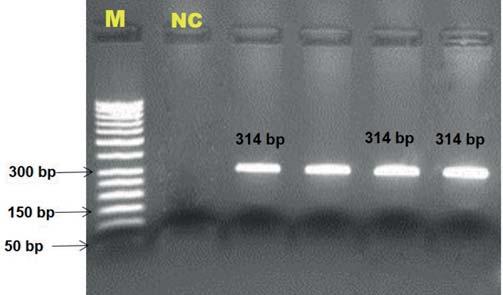
The fragments of the PCR product (314 bp) were digested by the BspHI endonuclease. PCR and restriction products were controlled using 2% and 3%, respectively, agarose gel electrophoresis (migration for ~1 h at 100 V) and were visualized by a gel documentation system with UV transillumination (DNR-Minilumi, DNR Bio-Imaging Systems, Israel). SafeView Classic (Applied Biological Materials Inc., Richmond, Canada) was used as a DNA-intercalating dye (~7 µL). To obtain the fragment size, a 100-1000 bp DNA ladder (Biomatik Co., Canada) was used in gels.
Statistical analysis
Genotype and allele frequencies were estimated according to Falconer and Mackay18. The deviation from Hardy-Weinberg
Association
60The
Between the STAT1 g.3141C>T Polymorphism and Reproductive Performance
Equilibrium (HWE) was tested using a standard chi-squared goodness-of-fit. Population genetics parameters including heterozygosity (He), homozygosity (Ho), number of effective alleles (Ne), and the polymorphic information content (PIC) were calculated by the formulas demonstrated by Botstein et al.19 and Nei and Roychoudhury20. Anderson-Darling test was used to evaluate the normality of data. Genotype-phenotype association analysis was carried out by the least-squares method as applied in a general linear model (GLM) procedure of Minitab software (Minitab Inc., Pennsylvania, USA, v17.1.0). Body condition scores, milk yield, lactation season, and lactation rank were included in the models when appropriate. Tukey’s test was used as a post-hoc comparison. The association between the cull rates and the genotypes was evaluated by Pearson’s chi-square test.
RESULTS
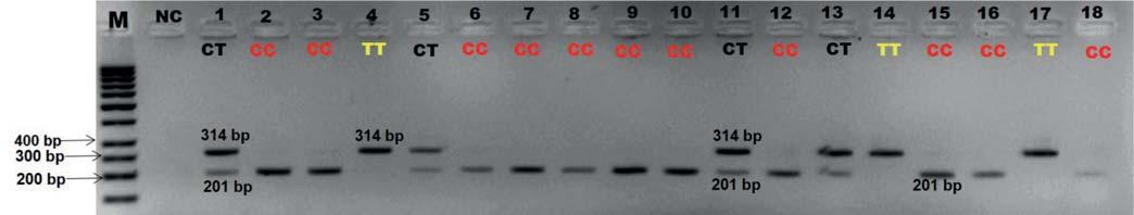
The electrophoresis pattern of STAT1 PCR amplification of the 314 bp fragment is shown in Figure 1. Concerning the results of BspHI enzyme digestion, the amplicon was cleaved into three fragments (314 bp, 201 bp, and 113 bp). Two bands (314 bp and 201 bp) were distinctive for genotype determination as shown in Figure 2. Undigested fragment of the 314 bp was diagnostic for the TT genotype in the STAT1 assay. Heterozygous genotype was indicated by two distinctive bands of 314 bp and 201 bp while 201 bp fragment was diagnostic for the CC genotype (Figure 2).
Two alleles and three genotypes for the STAT1 g.3141C>T marker were found in the present study. Table 1 shows the genotype and allele frequencies. The predominant genotype was the CC (~47%). Nevertheless, the genotypic distribution seemed to be balanced with 20 cows each for the TT and heterozygous genotypes. Concerning population genetics parameters, admissible variability results were observed for the STAT1 marker (Table 1). Results indicated that the Ne value approached 2.00. The STAT1 g.3141C>T polymorphism is a moderately informative marker for the tested population. In the chi-square test, a deviation from HWE was observed in the studied population (P<0.001).
The least-squares means and their respective standard errors obtained for the effects of STAT1 marker on reproductive traits in Holstein-Friesian cows are presented in Table 2. The mark-
Locus STAT1
GenotypeCCCTTT
n 352020
Genotypic frequency (%)46.6626.6726.67
AlleleCT
Allelic frequency0.600.40
Theoretical Heterozygosity (Hthe)1 0.4801
Number of effective alleles (Ne)1.9231
Polymorphism information content (PIC)0.3648
χ2(HWE)14.8146***
er STAT1 g.3141C>T affected the number of inseminations (P<0.05). The TT animals were characterized by the highest number of inseminations (3.71±0.73) which is indicating a potential negative effect of this genotype on reproductive performance. Moreover, there was a significant association between the STAT1 marker and cull-rates (P<0.01). There was no association between the STAT1 and any other traits analyzed.
DISCUSSION
The remarkable dominance of milk yield in current selection indexes has been started to be depleted gradually by non-production traits, including reproduction performance and health characteristics. The selective breeding for economically important traits was traditionally based on phenotypic recordings and it has been quite successful to some extent. From the 1990s, molecular genetics and its wide applications in animal breeding have led to more genetic improvement than using only phenotypic records. The detection and fine mapping of genes underlying the traits of interest, which can be termed quantitative traits loci (QTL), combination of QTL information and the best linear unbiased prediction-estimates of breeding values (BLUP-EBV), and marker-assisted selection (MAS) had provided promising improvement in information at the DNA level21. But the quantitative traits are much more complex than expected. In this context, numerous genotypic interactions make the genotype-phenotype association considerably hard to predict22. Antagonistic relationships among particular phenotypic traits generally cause failure to achieve the initially anticipated progress. Breeding strategies that mainly focus on the high production of dairy cattle have resulted in ignoring reproductive traits. Thus, the evaluation of the effects of widely used genetic markers on the reproductive performance of cows is a critical subject.
Here, we present a potential negative effect of the STAT1 g.3141C>T polymorphism on the number of inseminations per conception in high-yielding Holstein-Friesian cows. Furthermore, the favorable genotype seemed to be the heterozygous genotype with the lowest number of inseminations (1.15±0.08). In this respect, the TT genotype was characterized by a higher number of inseminations compared to alternative genotypes (P<0.05). And more important, TT animals had +2.56 and +1.44 higher means for the number of inseminations compared to heterozygous and the CC animals, respectively. The geno-
O. Aldevir et al. Large Animal Review 2023; 29: 59-6361
Figure 1 - The electrophoresis pattern of PCR amplification for the g.3141C>T polymorphism within the bovine STAT1 gene.
M: Marker; NC: Negative control.
Table 1 - Genotypic, allelic frequencies (%), population genetics parameters, and Hardy-Weinberg Equilibrium (HWE) test results in the STAT1 g.3141C>T polymorphism.
1In a diallelic locus, 1 - theoretical heterozygosity (Hthe) = locus homozygosity (Ho). ***P<0.001; not consistent with HWE.
types CC and CT were associated with significant increases in milk, fat, and protein yields as demonstrated by Cobanoglu et al.6. These authors indicated that the C allele of the STAT1 marker was also associated with an increase in milk protein and fat percentages. Similarly, Rychtářová et al.9 have shown that significant associations were observed for the CC and CT genotypes in estimated breeding value for protein and fat percentages. In their study, animals with the TT genotype showed the lowest values for fat and protein percentage (although not statistically significant, P>0.05). Concerning the Jersey breed, the TT genotype was characterized by significantly higher means for test day milk yield (+2.07 kg and +1.29 kg), fat yield (+0.13 kg and +0.09 kg), and protein yield (+0.07 kg and +0.05) compared to those with CC and heterozygous genotypes, respectively, in contrast to Holstein cows23. On the other hand, Ardicli et al.3 found no significant association between the STAT1 g.3141C>T polymorphism and any of the reproductive performance traits. The frequency of the genotype TT in HolsteinFriesian cows has been reported in the range of 2.15-18.19% in various studies on the STAT1 g.3141C>T marker6,9,10,15,23. This genotype seems to be rare in Jersey cattle23. In this study, the TT genotype frequency was remarkably high (26.67%) in highyielding Holstein-Friesians. It is important to note thatthe STAT1-TT genotype has highly undesirable properties for both milk production and reproduction performance traits based on previously published papers and the present study. From another point of view, the heterozygous genotype has been characterized by the higher milk production trait means (based on previous association studies as discussed above) and the lowest number of inseminations (this study). It is conceivable to interpret that it is a positive and beneficial approach to decrease the frequency of the TT genotype and increase the number of CT animals at the herd level regarding the studied STAT1 marker. However, as mentioned before, economically important
quantitative traits are very complex. For instance, Khatib et al.10 reported that the interaction between STAT1 g.3141C>T and STAT3 SNP19069 (as they designated) was highly significant for early embryonic survival rate. Furthermore, Cobanoglu et al.6 indicated that the C allele seems to be associated with an increase in somatic cell counts compared with the T allele. There is plenty of room for a better understanding of the genotypic background of complex traits, such as bovine reproduction performance. But first, the genetics studies should focus more on non-production characteristics because the recent knowledge on the effects of many genetic markers on reproductive traits is rather limited compared to production traits in dairy cattle. Although MAS results can provide limited efficiency on the traits of interest in livestock production21, the present results may be useful in the evaluation of popular genetic markers, such as STAT1, influences on non-production traits in dairy cattle. Notably, further studies are needed to confirm these findings and to discuss the other novel effects in different Holstein populations.
The most important problem caused by insufficient reproduction performance is the early culling of dairy cows. This leads to significant economic losses and prevents sustainability in dairy cattle farms. In this context, repeat breeding is defined as failure to conceive from three or more regularly spaced services in the absence of detectable abnormalities and is a substantial problem in cattle breeding resulting in increased calving interval and increased culling rates16. In this study, the relationship between the culling status of cows and the STAT1 g.3141C>T genotypes was evaluated based on repeat-breeding. Consistent with the results in ANOVA, the TT cows had higher culling rates compared to CC and CT genotype carriers (P<0.05). It is important to note that TT cows were characterized by higher days open (250.81±44.70 d) compared to the heterozygous (130.62±46.20 d) and CC (180.01±31.10 d)
62The Association Between the STAT1 g.3141C>T Polymorphism and Reproductive Performance
Figure 2 - The electrophoresis pattern of BspHI restriction enzyme digestion of PCR products the g.3141C>T polymorphism within the bovine STAT1 gene.
305-d milk yield (kg)10958±29611291±44010879±441NS Average daily milk yield (last 7d)1 39.57±0.9540.93±1.4039.39±1.36NS Days open (d)180.00±31.10130.60±46.20250.80±44.70NS3 Number of inseminations2 2.27±0.52b 1.15±0.08b 3.71±0.73a P<0.05
M: Marker; NC: Negative control.
Traits analyzed Genotypes Significance CCCTTT a,bDifferent superscripts within a raw indicate a statistical significance in Tukey’s post hoc comparison. 1Milk yield average in the week of sampling. 2Number of inseminations per conception. 3P<0.1
Table 2 - Least-square means and their corresponding standard errors for the effects of the STAT1 g.3141C>T polymorphism on the phenotypic traits analyzed.
animals. But this effect was not substantiated in statistical analysis (Table 2). Since the STAT1 TT is not a preferred genotype for milk yield, its effect on reproductive traits about this genotype has not been reported in the literature. Indeed, the effects of the STAT1 gene on bovine reproduction are interestingly low. Taken together, we suggest that the TT is an undesirable genotype and the heterozygous genotype is significantly associated with superior characteristics in reproduction traits. It was also observed that most of the cows conceived by single artificial insemination are the CT genotype carriers (data not shown). In the present statistical analyses, the significant effects of lactation season and body condition score on some reproduction traits were observed but these are widely studied environmental factors in previously published papers, and hence, these factors will not be discussed further. It is well known that an increase in milk yield negatively affects dairy cow fertility24. We thus think that evaluation of the STAT1 gene effects on the selected reproduction traits in high-yielding Holstein-Friesians can provide more confidential and consistent interpretations from an applicable perspective at the herd level. The mean of 305-d milk yield ranges from 6608 kg to 7871.51 kg in the most of previously published papers regarding the STAT1 g.3141C>T polymorphism9,15,23. In the present study, the selected cows had 11408±130 kg of 305-d milk yield (min: 9092 kg; max: 14358.52 kg). In this respect, the results demonstrated in this paper may reveal critical points in dairy cattle management concerning reproduction traits. The JAK-STAT pathway plays a major role in controlling cytokine signals and has an association with mammary gland development and milk production. Moreover, JAK-STAT signaling along with the lactogenic hormones regulates the processes of lactation and reproduction in mammals12. Hence, the STAT1 g.3141C>T marker deserves a higher level of focus on bovine fertility.
CONCLUSIONS
This paper focuses on the effects of the STAT1 g.3141C>T marker on certain reproduction traits in high-yielding HolsteinFriesian cows. Novel significant differences were found among the genotypes of the STAT1 locus. In this context, the TT genotype was characterized by the highest number of inseminations and high values for the culling rates related to repeat-breeding. On the other hand, heterozygous animals were shown to be associated with desirable reproduction performance. The broadening of selection aims with the fertility traits may be more useful than conventional production-focused approaches to achieve sustainable and profitable dairy cattle management.
ACKNOWLEDGEMENTS
The authors gratefully thank Atasancak Acıpayam Dairy Farm (Acipayam/Denizli).
References
1. Cochran S.D., Cole J.B., Null D.J., Hansen P.J. (2013). Discovery of single nucleotide polymorphisms in candidate genes associated with fertility and production traits in Holstein cattle. BMC genetics, 14(1): 1-23.
2. Laben R., Shanks R., Berger P., Freeman A. (1982). Factors affecting milk yield and reproductive performance. J Dairy Sci, 65(6): 1004-1015.
3. Ardicli S., Samli H., Soyudal B., Dincel D., Balci F. (2019). Evaluation of candidate gene effects and environmental factors on reproductive performance of Holstein cows. S Afr J Anim Sci, 49(2): 379-374.
4. Berglund B. (2008). Genetic improvement of dairy cow reproductive performance. Reprod Domest Anim, 43: 89-95.
5. Török E., Komlósi I., Sznyi V., Béri B., Mészáros G., Posta J. (2021). Combinations of Linear Type Traits Affecting the Longevity in Hungarian Holstein-Friesian Cows. Animals, 11(11): 3065-3076.
6. Cobanoglu O., Zaitoun I., Chang Y., Shook G., Khatib H. (2006). Effects of the signal transducer and activator of transcription 1 (STAT1) gene on milk production traits in Holstein dairy cattle. J Dairy Sci, 89(11): 44334437.
7. Kisseleva T., Bhattacharya S., Braunstein J., Schindler C. (2002). Signaling through the JAK/STAT pathway, recent advances and future challenges. Gene, 285(1-2): 1-24.
8. Bromberg J.F. (2001). Activation of STAT proteins and growth control. Bioessays, 23(2): 161-169.
9. Rychtářová J., Sztankoova Z., Kyselova J., Zink V., Stipkova M., Vacek M., Stolc L. (2014). Effect of DGAT1, BTN1A1, OLR1, and STAT1 genes on milk production and reproduction traits in the Czech Fleckvieh breed. Czech J Anim Sci, 59(2): 45-53.
10. Khatib H., Huang W., Mikheil D., Schutzkus V., Monson R. (2009). Effects of signal transducer and activator of transcription (STAT) genes STAT1 and STAT3 genotypic combinations on fertilization and embryonic survival rates in Holstein cattle. J Dairy Sci, 92(12): 6186-6191.
11. Darnell Jr. J.E. (1997). STATs and gene regulation. Science, 277(5332): 16301635.
12. Khan M.Z., Khan A., Xiao J., Ma Y., Ma J., Gao J., Cao Z. (2020). Role of the JAK-STAT pathway in bovine mastitis and milk production. Animals, 10(11): 2107.
13. Ashwell M, Heyen D, Sonstegard T, Van Tassell C.P., Da Y., Van Raden P.M., Ron M., Weller, J.I., Lewin H.A. (2004). Detection of quantitative trait loci affecting milk production, health, and reproductive traits in Holstein cattle. J Dairy Sci, 87(2): 468-475.
14. Ron M, Feldmesser E, Golik M, Tager-Cohen I., Kliger D., Reiss V., Domochovsky R., Alus O., Seroussi E., Ezra E., Weller J.I. (2004). A complete genome scan of the Israeli Holstein population for quantitative trait loci by a daughter design. J Dairy Sci, 87(2): 476-490.
15. Ardicli S, Soyudal B, Samli H, Dincel D, Balci F. (2018). Effect of STAT1, OLR1, CSN1S1, CSN1S2, and DGAT1 genes on milk yield and composition traits of Holstein breed. Rev Bras de Zootec, 47: e20170247.
16. Gustafsson H., Emanuelson U. (2002). Characterisation of the repeat breeding syndrome in Swedish dairy cattle. Acta Vet Scand, 43(2): 1-11.
17. Green M.R., Sambrook J. (2012). Isolation of high-molecular-weight DNA from mammalian cells using proteinase K and phenol, in: Molecular Cloning: A Laboratory Manual, 4, Cold Spring Harbor Laboratory Press, Cold Spring Harbor, New York, USA, 47- 48.
18. Falconer D.S., Mackay T.F.C. (1996). Introduction to quantitative genetics, Pearson Education Ltd, Harlow, England.
19. Botstein D., White R.L., Skolnick M., Davis R.W. (1980). Construction of a genetic linkage map in man using restriction fragment length polymorphisms. Am J Hum Genet, 32: 314-331.
20. Nei M., Roychoudhury A.K. (1974). Sampling variances of heterozygosity and genetic distance. Genetics, 76(2): 379-390.
21. Meuwissen T, Hayes B, Goddard M. Genomic selection: A paradigm shift in animal breeding. Animal frontiers. 2016;6(1):6-14.
22. Ardicli S., Samli H., Vatansever B., Soyudal B., Dincel D., Balci F. (2019). Comprehensive assessment of candidate genes associated with fattening performance in Holstein-Friesian bulls. Arch Anim Breed, 62(1): 9-32.
23. Cobanoglu O., Gurcan E.K., Soner C., Ertugrul K., Samet A.H. (2016). The detection of STAT1 gene influencing milk related traits in Turkish Holstein and Jersey cows. J Agric Sci Technol A,6: 261-269.
24. Loeffler S.H., de Vries M.J., Schukken Y.H. (1999). The effects of time of disease occurrence, milk yield, and body condition on fertility of dairy cows. J Dairy Sci, 82(12): 2589-2604.
O. Aldevir et al. Large Animal Review 2023; 29: 59-6363
SALVATORE FERRARO1,2,*, MARJOLAINE ROUSSEAU1, SIMON DUFOUR3, JOCELYN DUBUC1, JEAN-PHILIPPE ROY1, ANDRÉ DESROCHERS1
1 Département de sciences cliniques, Faculté de médecine vétérinaire, Université de Montréal, 3200 rue Sicotte, St-Hyacinthe, Québec, J2S 2M2, Canada
2 Department of Clinical Sciences, Swedish University of Agricultural Sciences, P.O. Box 7054, SE-750 07 Uppsala, Sweden.
3 Département de pathologie et microbiologie, Faculté de médecine vétérinaire, Université de Montréal, 3200 rue Sicotte, St-Hyacinthe, Québec, J2S 2M2, Canada
SUMMARY
Digital dermatitis causes lameness, discomfort, and economic losses worldwide. The test considered the ‘gold standard’ for diagnosis of digital dermatitis is the visual inspection of the feet into a trimming chute. However, this test is challenging to perform in daily operations. For this reason, several alternative methods to diagnose digital dermatitis in the milking parlor have been explored in the last years. The use of a rigid borescope was one of them but its use has been limited because of its labor ious use in the milking parlor and its high cost. Several and affordable models of flexible borescope are now available on the market. Our study objective was to quantify the accuracy of a flexible borescope for diagnosing digital dermatitis in a milking parlor. The study was conducted in a commercial free-stall herd milking approximately 200 cows. The borescope evaluation of hind feet was performed in the milking parlor 24-48 hours before the routine preventive hoof trimming. The same observer was used to diagnose the disease. The lesions at both evaluations were scored using the classification described by Döpfer and modified by Berry (2001).
Data were analyzed using two statistical approaches. In the first one, data were dichotomized as digital dermatitis lesions (M1, M2, M3, M4, M4.1) vs. no lesions (M0). In the second one, data were dichotomized as active lesions of digital dermatitis (M1, M2, M4.1) vs. chronic lesions/no lesions of digital dermatitis (M3, M4, M0). Sensitivity, specificity, and predictive values of both models were computed. Data from 870 hind feet were analyzed. Using the first approach, sensivity, specificity, positive and negative values were 64% (95%C.I.: 57%-69%), 91% (95%C.I.: 88%-93%), 97% (95%C.I.: 91%-99%) and 80% (95%C.I.: 78%-83%), respectively. Using the second approach, the values were 39% (95%C.I.: 32%-45%), 99% (95%C.I.: 98%-100%), 97% (95%C.I.: 91%-99%) and 80% (95%C.I: 77%-83%), respectively.
Based on these results, it was concluded that a flexible borescope can be used to diagnose the digital dermatitis lesions in the milking parlor. However, the ability of this test to diagnose active lesions of digital dermatitis appears to be limited. This means that it could result in an underestimation of the true prevalence of active lesions compared with the gold standard technique.
KEY WORDS
Dairy cattle, digital dermatitis, flexible borescope, lameness, milk parlor.
INTRODUZIONE
La dermatite digitale (DD) del bovino è una malattia infettiva diagnosticata per la prima volta in Italia nel 1974; da allora, questa malattia si è diffusa in tutto il mondo [1]. La prevalenza della DD varia nei vari Paesi e nelle diverse realtà zootecniche ed è stimata essere attorno al 20-30% [1, 2]. I fattori di rischio dell’insorgenza della DD sono molteplici e tra di essi vi sono lo stadio di lattazione, l’età degli animali, la nutrizione, l’igiene dell’allevamento e la biosicurezza [1]. L’eziologia della DD non è stata ancora completamente chiarita, ma è certo che i batteri appartenenti al genere Treponema giochino un ruolo importan-
Corresponding Author:
Salvatore Ferraro (salvatore.ferraro@slu.se).
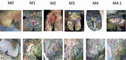
te nell’insorgenza della stessa [3]. L’aspetto delle lesioni della DD è solitamente vario (le lesioni possono variare da necrotico-ulcerativo a papilliforme-proliferativo). In letteratura sono descritte diverse classificazioni della DD, ma quella più usata è la classificazione descritta da Dopfer nel 1997 e che è stata successivamente modificata da Berry nel 2001[4, 5]. Secondo questa classificazione si riconoscono sei stadi della DD e cinque tipi di lesioni (Figura 1):
• La lesione M0 rappresenta la cute integra senza traccia di DD;
• M1 è una lesione ulcerativa di colore rossastro, dolorosa avente un diametro inferiore a 2 cm;
• M2 è una lesione ulcerativa, di colore rossastro, dolorosa avente un diametro superiore a 2 cm;
• M3 è caratterizzata da una lesione indolore che ha l’aspetto di una crosta nerastra
• M4 è una lesione indolore cronica, proliferativa, dall’aspetto a cavolfiore e di colore grigiastro;
S.
et al. Large Animal Review 2023; 29: 65-7065
Ferraro
Valutazione dell’accuratezza diagnostica di un boroscopio flessibile per la diagnosi della dermatite digitale in sala di mungitura
N
Figura 1 - Classificazione delle lesioni come descritta da Dopfer (1997) e successivamente modificata da Berry (2001). Nella prima linea della foto si può vedere l’aspetto delle lesioni usando la visualizzazione diretta nel travaglio. Nella seconda linea si può vedere l’aspetto delle lesioni quando viene usato il borescopio flessibile in sala mungitura (le immagini della prima linea sono state gentilmente concesse dal Dr. André Desrochers)
• M4.1 è una lesione indolore cronica, proliferativa dall’aspetto a cavolfiore di colore grigiastro in cui sono presenti dei punti rossastri di riattivazione [3, 6].

Il 94% delle lesioni è localizzato nei piedi posteriori e l’85% di esse è localizzato nella faccia plantare del piede [1, 7]. Le lesioni della DD sono causa di zoppie dei bovini e di conseguenza, la presenza della DD ha un impatto negativo sul benessere degli animali. La DD ha anche dei costi che sono dovuti al suo controllo, alla riduzione della produzione lattea e della fertilità associate alla malattia [1, 8-10]. Il trattamento della DD è basato sulla terapia individuale delle bovine che ne sono affette e sull’utilizzo dei pediluvi collettivi per il suo controllo [2] .
L’ispezione visiva delle lesioni nel travaglio per il pareggio è considerata il test ‘gold standard’ per la diagnosi della DD [7, 11].
Ma l’ispezione visiva delle lesioni nel travaglio è un esame diagnostico laborioso e, inoltre, può inficiare il benessere e la produttività delle bovine [12]. Per questi motivi esso è difficilmente integrabile nelle operazioni quotidiane dell’allevamento delle bovine da latte [12].
A causa delle difficoltà di sottoporre frequentemente le bovine all’ispezione visiva nel travaglio, negli ultimi anni sono stati descritti diversi metodi alternativi per la diagnosi della DD. Tra i diversi metodi diagnostici proposti vi è la visualizzazione delle lesioni in sala di mungitura [13], che può essere diretta o indiretta tramite l’aiuto di uno specchietto [7, 11]. La valutazione in sala mungitura può anche essere fatta tramite l’utilizzo di un boroscopio rigido [14, 15]. L’utilizzo del boroscopio rigido in sala mungitura in passato ha dato dei buoni risultati ma il suo uso è stato limitato dalla sua scarsa manegevolezza e dal suo alto costo [7, 11, 13]. Ma negli ultimi anni sul mercato sono presenti diversi modelli di boroscopi dai prezzi contenuti e che hanno la possibilità di essere collegati, tramite wireless, con un cellulare o un tablet. Inoltre, alcuni modelli di boroscopio presenti sul mercato sono flessibili e quindi poten-
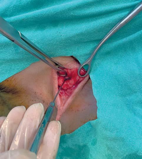
zialmente il loro utilizzo potrebbe migliorare la visualizzazione delle lesioni in sala di mungitura (Figura 2).
L’obbiettivo del presente studio è stato quello di valutare l’accuratezza diagnostica del boroscopio flessibile per la diagnosi della DD nelle bovine da latte in sala mungitura. La nostra ipotesi è stata quella di ritenere il boroscopio flessibile un valido metodo diagnostico della DD delle bovine da latte in alternativa all’utilizzo dell’ispezione visiva delle lesioni nel travaglio.
MATERIALI E METODI
Management degli animali arruolati
I dati dello studio sono stati tratti da un più ampio progetto di ricerca sulla diagnosi e controllo della DD nelle bovine da latte. Il progetto è stato approvato dal Comitato Etico per l’Utilizzazione degli Animali per la Ricerca Scientifica dell’Université de Montréal (protocollo CÉUA: # 16-RECH-1826). Lo studio è stato condotto in un allevamento commerciale di bovine da latte. Le bovine presenti in allevamento erano tutte di razza Frisona Canadese. In allevamento erano presenti, al momento dello studio, circa 200 vacche in lattazione. Le bovine erano munte tre volte al giorno in una sala mungitura rotativa obliqua. La produzione media delle bovine per una lattazione standard di 305 giorni era di 11800 kg. L’allevamento era a stabulazione libera con cuccette. Le cuccette erano provviste di lettiera fatta di paglia e calce. La lettiera era aggiunta giornalmente e cambiata completamente ogni due settimane, le corsie di alimentazione e di movimentazione erano costituite da pavimento fissurato e pulite tramite robot semovibile. Il pareggio funzionale era effettuato regolarmente sulle vacche in lattazione e in asciutta tre volte l’anno. Le manze, invece, erano sottoposte a pareggio funzionale due volte l’anno. Le bovine che presentavano una zoppia erano trattate individualmente secondo la causa della pa-
66Valutazione dell’accuratezza diagnostica di un boroscopio flessibile per la diagnosi della dermatite digitale
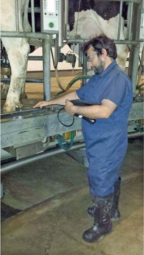
Foto 1 - Nella foto 1 é riportato il modello del boroscopio flessibile che é stato usato durante lo studio. Da notare che questo modello di borescopio é formato da una base solida, in cui sono contenute delle batterie e in cui é possibile inserire uno smartphone per la visualizzazione delle immagini, e da un cavo flessibile alla cui estremitá é presente una videocamera e una sorgente di luce LED (foto tratta da www.amazon.ca).
tologia specifica. Le bovine affette da DD erano trattate con tetraciclina in polvere. Il controllo della DD delle bovine in lattazione era effettuato tramite pediluvio (due volte a settimana) contenente una soluzione al 5% di solfato di rame. La soluzione veniva cambiata regolarmente dopo ogni giornata di trattamento. L’unità di campionamento dello studio era la faccia plantare dei piedi posteriori [7, 11].
Campionamento
La valutazione delle lesioni in sala mungitura è stata fatta tramite l’utilizzo di un boroscopio flessibile (WF200 WiFi Endoscope, Teslong Technology Ltd., Shenzhen, Guangdong, China (foto 1), connesso tramite wireless ad un tablet (iPad, Apple Inc, Cupertino, CA, USA). Le immagini derivate dal boroscopio sono state visualizzate sul tablet e valutate da due veterinari (SF e AD). Durante la valutazione in sala mungitura è stata utilizzata la luce LED prodotta dal boroscopio per visualizzare le lesioni (foto 2). La valutazione in sala mungitura è stata fatta 24-48 ore prima del pareggio funzionale. I piedi non sono stati lavati prima della valutazione, ne sono stati manipolati in alcun modo. I risultati della valutazione sono stati raccolti in fogli di carta prestampati e successivamente copiati su un foglio di calcolo Excel. La valutazione delle lesioni durante il pareggio funzionale è stata fatta dal primo autore (SF) ponendosi a circa 50 cm dalla superficie plantare del piede posteriore della bovina contenuta nel travaglio. Durante la valutazione i piedi non sono stati lavati né sono stati sottoposti ad alcuna manipolazione. La valutazione è stata fatta con la luce naturale. I dati sono stati raccolti su fogli di carta prestampati e successivamente copiati su un foglio di calcolo Excel.

Analisi statistiche
I dati provenienti dallo studio sono stati analizzati utilizzando due modelli. Nel primo modello è stata valutata la capacita’ del
Foto 2 - Utilizzo di un boroscopio flessibile in sala mungitura fatta durante lo studio. Da notare la presenza sulle braccia dell’operatore dell’ Ipad usato come schermo per la visualizzazione delle lesioni della dermatite digitale. L’utilizzo dell’Ipad puo’ essere sostituito da uno smartphone montato direttamente sul boroscopio flessibile (vedi foto 1).
borescopio di diagnosticare le lesioni della DD in sala mungitura. Per questo motivo i dati sono stati dicotomizzazati in due categorie: lesioni della DD (M1, M2, M3, M4, M4.1) versus assenza delle lesioni (M0) [16]. Nel secondo modello è stata valutata la capacita’ del borescopio di diagnosticare, in sala mungitura, le lesioni attive della DD. Per questo motivo i dati sono stati dicotomizzati in due categorie: lesioni attive della dermatite (M1, M2, M4.1) versus assenza delle lesioni e/o lesioni croniche (M0, M3, M4) [16]. I dati raccolti sono stati sintetizzati due tavole di contigenza 2x2 (Tabella 1 e 2). I dati provenienti dalla valutazione fatta tramite il boroscopio in sala mungitura (index test) sono stati comparati ai dati della valutazione effettuata nel travaglio (reference test). La valutazione delle lesioni nel travaglio è stata considerata come il test ‘gold standard’. L’ accordo tra la valutazione effettuata in sala mungitura tramite boroscopio e la valutazione fatta nel travaglio è stato valutando il Kappa di Cohen (k) [17]. I risultati sono stati interpretati seguendo le linee guida riportate da Doho e colle-
S. Ferraro et al. Large Animal Review 2023; 29: 65-7067
Tabella 1 - Tavola di contingenza 2x2 dei risultati dell’esame fatto con il boroscopio flessibile e la visualizzazione diretta delle lesioni nel travaglio fatta su 870 piedi secondo il primo modello: lesioni della dermatite digitale (M1, M2, M3, M4, M4.1) versus assenza delle lesioni (M0).
Boroscopio +Boroscopio -Totale
Visualizzazione 183105288
nel travaglio +
Visualizzazione 52530582
nel travaglio -
Totale235635870
Tabella 3 - Sintesi dei risultati dell’accuratezza diagnostica dell’esame con boroscopio flessibile in sala mungiture. Modello 1: lesioni della dermatite digitale (M1, M2, M3, M4, M4.1) vs assenza delle lesioni della dermatite digitale (M0).
MisuraRisultato (%)Intervallo di confidenza (%)
Prevalenza apparente27,0%24,0-30,0%
Prevalenza reale33,0%29,0-36,0%
Sensibilità boroscopio64,0%57,0-69,0%
Specificità boroscopio91,0%88,0-93,0%
Valore predittivo positivo77,0%72,0-83,0%
Valore predittivo negativo83,0%80,0-86,0%
Tabella 2 - Tavola di contingenza 2x2 dei risultati dell’esame fatto con il boroscopio flessibile e la visualizzazione diretta delle lesioni nel travaglio fatta su 870 piedi secondo il primo modello: lesioni attve della dermatite digitale (M1, M2, M4.1) versus assenza delle lesioni o lesioni croniche (M0, M3, M4).
Boroscopio +Boroscopio -Totale
Likehood ratio positivo7,1%5,4-9,3%
Likehood ratio negativo4,0%3,4-4,6%
Indice di Youden54,0%46,0-62,0%
Accuratezza diagnostica81,9%-
Area sotto la curva (AUC)77,3%-
Visualizzazione
nel travaglio +94148242
Visualizzazione
nel travaglio +3625628
Totale97773870
ghi (2009) [17]: ≤0 = accordo molto scarso, 0.01 to 0.2= leggero accordo, 0.21 to 0.4 = accordo equo, 0.41 to 0.6 = accordo moderato, 0.61 to 0.8 = accordo sostanziale, 0.81 to 1 = accordo quasi perfetto[17]. La concordanza tra la valutazione fatta in sala mungitura utilizzando il boroscopio flessibile e la valutazione fatta nel travaglio è stato calcolato usando SAS 9.4. L’accuratezza diagnostica del boroscopio flessibile è stata valutata usando Rstudio 4.0[19] utilizzando il package «epiR» (Package epiR 2.0.19) e sono stati calcolati i seguenti parametri: la prevalenza, la sensibilità, la specificità, i valori predittivi (positivo e negativo) e rapporti di verosimiglianza (positivo e negativo). L’area sotto la curva è stata calcolata utilizzando Rstudio (R version 4.0.4)[20] con la funzione «accuracy» del package «rfutilities». I dati sono stati analizzati utilizzando un intervallo di confidenza del (I.C.) del 95%.
RISULTATI
Un totale di 1064 piedi posteriori è stato valutato nel travaglio durante il pareggio funzionale. Un totale di 904 piedi posteriori è stato valutato utilizzando il boroscopio flessibile in sala mungitura. Un totale di 870 osservazioni è stato utilizzato per l’analisi dell’accuratezza diagnostica del boroscopio flessibile. Trenta quattro valutazioni sono state escluse perche non era stato possibile fare la valutazione della faccia plantare dei piedi posteriore. I risultati della analisi statistiche dell’accuratezza diagnostica del boroscopio flessibile in sala munitura per la diagnosi delle DD sono sintetizzati nelle tabelle 3 e 4.
L’ accordo (k) tra i risultati della valutazione delle lesioni in sala di mungitura con il boroscopio e la valutazione nel travaglio durante il pareggio funzionale riguardo la detenzione delle lesioni della DD (M1, M2, M3, M4 e M4.1 versus M0) si è attestata a 0.57 (95% CI: 00.51, 0.63). La concordanza (k) tra i risul-
Cohen’s Kappa0,57-
Tabella 4 - Sintesi dei risultati dell’ accuratezza diagnostica dell’ esame con boroscopio flessibile in sala mungiture. Modello 1: lesioni della dermatite digitale (M1, M2, M4.1) vs assenza delle lesioni della dermatite digitale (M0, M3, M4).
MisuraRisultato (%)Intervallo di confidenza (%)
Prevalenza apparente11,0%9,0-13,4%
Prevalenza reale27,0%25,0-30,0%
Sensibilità boroscopio39,0%33,0-45,0%
Specificità boroscopio99,0%98,0-100%
Valore predittivo positivo96,0%91,0-99,0%
Valore predittivo negativo80,0%77,0-83,0%
Likehood ratio positivo81,0%26,0-254%
Likehood ratio negativo61,0%55,0-67,0%
Indice di Youden38,0%31,0-45,0%
Accuratezza diagnostica82,6%-
Area sotto la curva (AUC)69,1%-
Cohen’s Kappa0,47-
tati della valutazione dei piedi in sala mungitura con il boroscopio flessibile e la valutazione nel travaglio durante il pareggio funzionale riguardo la detenzione delle lesioni attive (M1, M2 e M4.1 versus M0, M3 e M4) era di 0.47 (95% IC: 0.40, 0.54). Utilizzando il primo modello (presenza di almeno una lesione dovuta alla DD (M1, M2,M3,M4, M4.1) versusassenza delle lesioni da DD (M0)), la prevalenza apparente delle lesioni della DD era di del 27% (I.C.95%=24%-30%). La prevalenza reale delle lesioni della DD era del 33% (I.C.95%=30%-36%). La sensibilità della valutazione dei piedi posteriori in sala di mungitura utilizzando il boroscopio flessibile era del 64% (I.C.95%=57%-69%) e la specificità era del 91% (I.C.95%=88%93%). Il valore predittivo positivo era del 97% (I.C.95%=91%-
68Valutazione dell’accuratezza
diagnosi della
digitale
diagnostica di un boroscopio flessibile per la
dermatite
99%) e il valore predittivo negativo era del 80% (I.C.95%=78%83%). L’ accuratezza diagnostica delle lesioni in sala mungitura era del 82%. L’Area sotto la curva era del 77%.
Utilizzando il secondo modello (lesioni attive della DD (M1, M2, M4.1)) versuslesioni croniche della DD/assenza delle lesioni della DD (M3, M4, M0) la prevalenza apparente delle lesioni attive era del 11% (I.C.95%=9%-13%) e la prevalenza reale delle lesioni attive era del 27% (I.C.95%=25%-30%). La sensibilità del boroscopio flessibile per la detenzione delle lesioni attive in sala mungitura era del 39% (I.C.95%=32%-45%). La specificità era del 99% (I.C.95%= 98%-100%). Il valore predittivo positivo era del 97% (I.C.95%=91%-99%) e il valore predittivo negativo 80% (I.C.95%=77%-83%). L’accuratezza diagnostica del boroscopio flessibile per la detenzione delle lesioni era del 82%. L’area sotto la curva era del 69%.
DISCUSSIONE
I risultati del nostro studio dimostrano che il boroscopio ha una buona accuratezza diagnostica per la diagnosi delle lesioni della DD in sala mungitura, ma l’accuratezza diagnostica del boroscopio flessibile per quanto rigiarda la diagnosi delle lesioni attive della DD è limitata.
Per discutere i risultati del nostro studio bisogna considerare tre fattori che possono aver avuto un impatto su questi: la qualità del boroscopio, la distribuzione delle lesioni e il design dello studio.
Sul mercato sono presenti diversi tipi di boroscopio che sono utilizzati in idraulica e in meccanica, ai diversi tipi di boroscopio corrispondono diversi prezzi e una diversa qualità tra i borescopi che sono disponibili in commercio. Nel nostro studio abbiamo scelto un modello con un costo medio-basso (circa 70 euro) e questo può avere avuto un impatto sui risultati del nostro studio. Nel concetto di qualità di un boroscopio flessibile bisogna anche considerare la capacità di risoluzione delle immagini. La risoluzione delle immagini del boroscopio flessibile che abbiamo usato nel nostro studio dipendeva anche dal dispositivo portatile (cellulare, tablet) che veniva usato come schermo per la visualizzazione delle immagini (Figura 2). Nel nostro caso bisogna considerare che abbiamo usato un vecchio modello di iPad che aveva una minore risoluzione che i nuovi modelli iPadpro (2,048 × 1,536 vs 3,264 × 2,448 pixels) [16]. La qualità della risoluzione delle immagini può aver influito sui risultati del nostro studio. Anche la distribuzione delle lesioni può aver avuto un impatto dui risultati del nostro studio. In particolare, la detenzione delle lesioni classificate come M1 può essere molto difficile [16]. La definizione delle lesioni M1 è vaga poiché include delle lesioni con un diametro inferiore a 2 cm e questo significa che in questa categoria sono comprese anche lesioni che possono avere un diametro di qualche millimetro e che dunque sono difficili da diagnosticare [7, 11,19]. Questi primi due punti ci portano al terzo punto di discussione che è lo study design. Come é stato riportato nei materiali e metodi, i dati analizzati fanno parte di un più ampio progetto di ricerca sul controllo della DD. Nello studio, oltre alla valutazione dell’accuratezza diagnostica del boroscopio flessibile si è valutata anche l’efficacia di un prodotto per il suo utilizzo nei pediluvi collettivi (dati non publicati). Per non influire sui risultati della valutazione di questo prodotto per i pediluvi si é deciso di non lavare i piedi prima della valutazione, cosa che invece è stato fatto in altri studi nella quale sono stati valutati altri metodi dia-
gnostici della DD in sala mungitura [7,11]. Ma questa decisione é stata dettata anche dalla riluttanza degli allevatori a lavare i piedi delle bovine durante la mungitura per paura di una possibile contaminazione dei gruppi di mungitura, e quindi un possibile aumento del rischio di mastite nelle bovine. Un altro timore del propietario della stalla dove é stato fatto lo studio era che il lavaggio dei piedi in sala mungitura avrebbe potuto rallentare le operazioni di mungitura.
La scelta di non lavare i piedi delle bovine prima della valutazione è a nostro avviso la parte dello study design che probabilmente ha avuto il maggiore impatto sui risultati dello studio. In uno studio neozelandese è stato riportato che lavare i piedi prima della valutazione in sala mungitura quasi raddoppia la capacita’ di detenzione delle lesioni della DD. Però bisogna anche considerare che la scelta di non lavare i piedi prima della loro valutazione ha reso più realistico il nostro studio e di conseguenza ne ha aumentato la validità esterna. Quindi questo può essere considerato uno dei punti di forza del nostro studio. Un altro punto di forza dell’utilizzo del boroscopio flessibile è la sua semplicità di utilizzo e la possibilità di acquisire delle foto delle lesioni. La possibilità di acquisire delle foto delle lesioni, e potenzialmente di conservarle, potrebbe permettere la valutazione a distanza delle stesse e questo potrebbe giustificare l’utilizzo del boroscopio sia in ambito della ricerca sia per la pratica quotidiana. Inoltre, è stato riportato che l’utilizzo frequente del boroscopio (cinque valutazioni in sala mungitura a frequenza settimanale) incrementa la sensibilità della boroscopia sino all’85% (95% CrI: 50, 99) [16].
CONCLUSIONI
Possiamo concludere dicendo che l’utilizzo di un borescopio flessibile permette la valutazione delle lesioni della DD nelle bovine da latte in sala mungitura quindi puo’ essere un valido strumento per la valutazione e il monitoraggio della prevalenza della DD in stalla. Ma bisogna anche considerare che la sua limitata capacita’ di diagnosi delle lesioni attive puo’ ritardare il trattamento di questo tipo di lesioni.
Ulteriori studi, con un differente «study design » e un diverso modello di boroscopio flessibile, sarebbero auspicabili per la valuatazione dell’accuratezza diagnostica di questo utensile per la diagnosi delle lesioni della DD in sala mungitura.
Bibliografia
1. Evans NJ, Murray RD, Carter SD. (2016). Bovine digital dermatitis: Current concepts from laboratory to farm. Vet J, 211: 3-13. https://doi.org/10.1016/j.tvjl.2015.10.028.
2.Plummer PJ, Krull A. (2017). Clinical Perspectives of Digital Dermatitis in Dairy and Beef Cattle. Vet Clin North Am Food Anim Pract, 33: 165181. https://doi.org/10.1016/j.cvfa.2017.02.002.
3. Orsel K, Plummer P, Shearer J, et al.(2018). Missing pieces of the puzzle to effectively control digital dermatitis. Transbound Emerg Dis, 65 Suppl 1: 186-198. https://doi.org/10.1111/tbed.12729.
4. Dopfer D, Koopmans A, Meijer FA, et al. (1997) Histological and bacteriological evaluation of digital dermatitis in cattle, with special reference to spirochaetes and Campylobacter faecalis. Vet Rec, 140: 620-623. https://doi.org/10.1136/vr.140.24.620.
5. Berry SL, Read DH, Famula TR, et al. (2012).Long-term observations on the dynamics of bovine digital dermatitis lesions on a California dairy after topical treatment with lincomycin HCl. Vet J, 193: 654-658. http://dx.doi.org/10.1016/j.tvjl.2012.06.048.
6. Biemans F, Bijma P, Boots NM, et al. (2018). Digital Dermatitis in dairy cattle: The contribution of different disease classes to transmission. Epi-
S. Ferraro et al. Large Animal Review 2023; 29: 65-7069
70Valutazione dell’accuratezza diagnostica di un boroscopio flessibile per la diagnosi della dermatite digitale
demics, 23: 76-84. https://doi.org/10.1016/j.epidem.2017.12.007.
7. Solano L, Barkema HW, Jacobs C, et al. (2017). Validation of the M-stage scoring system for digital dermatitis on dairy cows in the milking parlor. J Dairy Sci, 100:1592-1603. https://doi.org/10.3168/jds.2016-11365.
8. Cha E, Hertl JA, Bar D, et al. (2010). The cost of different types of lameness in dairy cows calculated by dynamic programming. Prev Vet Med, 97: 1-8. https://doi.org/10.1016/j.prevetmed.2010.07.011.
9. Relun A, Lehebel A, Chesnin A, et al. (2013). Association between digital dermatitis lesions and test-day milk yield of Holstein cows from 41 French dairy farms. J Dairy Sci, 96: 2190-2200. https://doi.org/10.3168/ jds.2010-4054.
10. Gomez A, Cook NB, Socha MT, et al. (2015). First-lactation performance in cows affected by digital dermatitis during the rearing period. J Dairy Sci, 98: 4487-4498. http://dx.doi.org/10.3168/jds.2014-9041.
11. Relun A, Guatteo R, Roussel P, et al. (2011). A simple method to score digital dermatitis in dairy cows in the milking parlor. J Dairy Sci, 94: 54245434. https://doi.org/10.3168/jds.2010-4054.
12. Van Hertem T, Parmet Y, Steensels M, et al. (2014). The effect of routine hoof trimming on locomotion score, ruminating time, activity, and milk yield of dairy cows. J Dairy Sci, 97: 4852-4863. https://doi.org/10.3168/ jds.2013-7576.
13. Thomsen PT, Klaas IC, Bach K. (2008). Short communication: scoring of digital dermatitis during milking as an alternative to scoring in a hoof trimming chute. J Dairy Sci, 91: 4679-4682. https://doi.org/10.3168/ jds.20081342.
14. Stokes JE, Leach KA, Main DC, et al. (2012). The reliability of detecting digital dermatitis in the milking parlour. Vet J, 193: 679-684. https://doi.org/10.1016/j.tvjl.2012.06.053.
15. Laven RA. (1999).The environment and digital dermatitis. Cattle Practice, 7: 349-354.
16. Ferraro S, Buczinski S, Dufour S, et al. (2020). Bayesian assessment of diagnostic accuracy of a commercial borescope and of trimming chute exams for diagnosing digital dermatitis in dairy cows. J Dairy Sci; 103: 3381-3391. https://doi.org/10.3168/jds.2019-17129.
17. Dohoo IR, Martin SW, Stryhn H. (2009). Veterinary epidemiologic research. 2nd ed. Charlottetown, P.E.I.: VER, Inc.
18. R Core Team. (2020). A Language and Environment for Statistical Computing. https://www.R-project.org/.
19. Cramer G, Winders T, Solano L, et al. Evaluation of agreement among digital dermatitis scoring methods in the milking parlor, pen, and hoof trimming chute. J Dairy Sci 2018; 101: 2406-2414. https://doi.org/10.3168/ jds.2017-13755.
1 Department of Animal Science, Kangwon National University, Chuncheon 24341, Korea
SUMMARY
High-energy diets to steers during the late fattening period, the consumption of large amounts of an energy-rich diet is liable to cause metabolic problems such as ruminal dysfunction and impaired liver-related immune function in beef cattle, as well as an increase in inedible fat. In ruminants, supplementation of rumen-protected fat is an effective approach for increasing energy density and enhancing productivity. In addition, Vitamin C, which is a strong antioxidant, is known to improve meat color by inhibiting phospholipid oxidation and reactive oxygen formation, and has also been found to improve marbling score and meat quality by enhancing adipocyte differentiation. However, studies on Hanwoo steers following the supplementation of rumen protected fat and vitamin C are insufficient.
Thus, this study was conducted to investigate the effect of rumen-protected fat and/or vitamin C supplementation on growth performance, carcass characteristics, and meat composition in Hanwoo steers during late period. Twenty-eight Hanwoo steers (initial body weight 723.0 ± 59.2 kg, approximately 27 months of age) were allotted to 4 treatments as follow: CON = basal diet, T1 = basal diet + rumen-protected fat (RPF) at the level of 0.1% of diet, T2 = basal diet + rumen-protected vitamin C (RPVC) at the level of 0.1% of diet, T3 = basal diet + RPF + RPVC. Final weight, average daily gain and dry matter intake were higher in T1 than in CON (p < 0.05). Plasma metabolites were similar among the treatments. Carcass weight and rib eye area was higher in all treatment group than in CON (p < 0.05). However, there was no effect according to the treatment on the chemical and fatty acid composition of longissimus muscle.
The present results suggest that rumen-protected fat and/or vitamin C supplementation has positive effects on concentrate feed intake. In particular, rumen-protected fat was founded to be effective in increasing in dry matter intake, average daily gain, carcass weight and rib eye area of Hanwoo steers. However, there was no complementary effect of rumen-protected fat and vitamin C supplementation.
KEY WORDS
Rumen-protected fat; Vitamin C; Growth performance; Carcass characteristics; Hanwoo steers.
INTRODUCTION
Although it is necessary to feed high-energy diets to steers during the late fattening period (1), the consumption of large amounts of an energy-rich diet is liable to cause metabolic problems such as ruminal dysfunction and impaired liver-related immune function in beef cattle, as well as an increase in inedible fat (2). In addition, given that feed efficiency and metabolic diseases are influenced by abnormal rumen and liver function, it is important to manage energy intake (of carbohydrate and fat) and use antioxidants in during the late fattening period of
*Corresponding Author: Byung Ki Park(animalpark@hanmail.net).
†These authors contributed equally to this work.
beef cattle.
In ruminants, supplementation of rumen-protected fat is an effective approach for increasing energy density and enhancing productivity. In particular, it has been established that the provision of rumen-protected fat is an effective means of enhancing milk yield and composition, by increasing energy intake without affecting rumen fermentation characteristics (3, 4). In beef cattle, rumen-protected fat has been used to enhance body weight gain and marbling score by increasing energy intake, and in recent years, studies have been conducted with the aim of increasing particular fatty acids, such as polyunsaturated fatty acids, oleic acid, and -linolenic acid, in the diets of beef cattle (5, 6).
Vitamin C, which is a strong antioxidant, is known to improve meat color by inhibiting phospholipid oxidation and reactive oxygen formation (7), and has also been found to improve marbling score and meat quality by enhancing adipocyte differ-
MIN JI KIM1,2†, GI HWAL SON1†, JONG SUH SHIN1, AND BYUNG KI PARK1*
2 Laboratory of Animal Physiology, Graduate School of Agricultural Science, Tohoku University, Aoba, Sendai 9808572, Japan
M.J. Kim et al. Large Animal Review 2023; 29: ??-??71
N
Effect of rumen-protected fat and/or vitamin C supplementation on growth performance, carcass characteristics and meat composition in Hanwoo steers during late fattening period
72Effect of rumen-protected fat and/or vitamin C supplementation on growth performance
entiation (8). For example, Yano et al. (9) reported that vitamin C and high-energy feed improve marbling score and fat color in Japanese black steers, whereas Park. (10) has demonstrated that vitamin C (2 g/d) influences meat quality in Hanwoo steers. It has been established that in addition to having antioxidant effects, vitamin C is also associated with the synthesis of immune substances such as prostaglandins and cytokines (11). However, given that ruminants have lower levels of vitamin C compared with monogastric animals, they tend to be prone to vitamin C deficiency under conditions of dietary and environmental stress (9). Although there have been some studies that have investigated supplementation of vitamin C for the purpose of improving meat quality in Hanwoo steers,
Table
most studies in Japan apply top dressing levels [40 mg/kg body weight (BW)], and it tends to be difficult to supply constant amounts of feed additives for individual steers when using the top dressing method. Accordingly, in order to efficiently use and supply constant amounts of vitamin C, it is necessary to supply this in a composite feed concentrate in Hanwoo steers. To date, however, there have been no studies that have investigated the effects of the combined provision of supplemental fat and vitamin C.
Consequently, in this study, we sought to investigate the effect of rumen-protected fat and/or vitamin C supplementation on the growth performance, carcass characteristics, and meat composition of Hanwoo steers.
Ingredient and chemical composition of experimental diets for late fattening Hanwoo steers.
Ingredient composition (%) Corn grain26.0426.0226.0426.00 Wheat grain8.008.008.008.00 Barley grain2.002.002.002.00 Cane molasses5.505.505.305.50 Tapioca residue8.757.528.887.00 Wheat bran-2.32-2.84 Wheat flour2.00-2.00Corn gluten feed18.3520.0018.3620.00 Rapeseed meal-1.62-1.48 Coconut meal10.0010.0010.0010.00 Palm kernel meal12.0012.0012.0012.00 Cottonseed hull1.001.001.001.00 Rumen protected fat (Ca-salt)0.101.000.101.00 Lupin3.00-3.00Emulsifier0.100.060.100.10 Salt dehydrated0.500.500.500.50 Limestone (1mm)1.631.421.581.44 Sodium bicarbonate0.500.500.500.50 Premix1) 0.200.200.200.20 Rumen protected vitamin C--0.100.10 Others (feed additives)0.340.340.340.34 Chemical composition (%) Dry matter88.4788.3988.4988.39 Crude protein12.2012.2012.2012.20 Ether extract3.864.533.864.54 Crude fiber7.677.637.697.61 Crude ash6.236.066.176.05 NFC38.9139.1440.6940.66 TDN73.2473.2373.2373.29
ItemsTreatments ControlT1T2T3 NFC, non-fiber carbohydrate; TDN, total digestible nutrients (calculated value). 1) Premix provided the following quantities of vitamins and minerals per kilogram of diet: vitamin A, 10,000 IU; vitamin D3, 1,500 IU; vitamin E, 25 IU; Fe, 50 mg; Cu, 7mg; Zn, 30 mg; Mn, 24 mg; I, 0.6 mg; Co, 0.15 mg; Se, 0.15 mg.
1 -
MATERIALS AND METHODS
Animal care
All procedures on animals were carried out in compliance with South Korea regulations (Animal and Plant Quarantine AgencyMinistry of Food and Drug Safety Joint Animal Testing and/or Laboratory Animal Related Committee (IACUC; 2020) Standard Operating Guidelines).
Animals, treatments, and management
Twenty-eight Hanwoo steers (initial body weight, 723.0 ± 59.2 kg; approximately 27 months of age) were randomly assigned to one of the following four dietary treatments: CON = basal diet, T1 = basal diet + rumen-protected fat (RPF) at the level of 0.1% of the diet, T2 = basal diet + rumen-protected vitamin C (RPVC) at the level of 0.1% of the diet, and T3 = basal diet + 0.1% RPF + 0.1% RPVC.
The steers were allotted by treatment group into four pens (5 m × 10 m) and fed 5 kg (as-fed basis) of concentrate three times daily (08:30, 13:00, and 18:00), along with a daily ration of 1.5 kg rice straw (as-fed basis). Basal diets were formulated for the late fattening period of steers according to National Research Council recommendations (12). Water was freely available at all times, and other feeding management was conducted in accordance with the practices of the farm. The chemical compositions of the experimental diets were analyzed according to the methods of the Association of Official Analytical Chemists (AOAC, 13) and are shown in Table 1. Neutral detergent fiber (NDF) and acid detergent fiber (ADF) were analyzed according to the methods of Van Soest (14) using a filter bag (Ankom F57; Ankom Technology, NY, USA).
Growth performance and plasma metabolism
Feed intake was measured daily prior to morning feeding, and the amount of feed consumed was determined by measuring the orts. Average daily gain (ADG) was calculated based on the number of days in each month, and the feed conversion ratio (FCR) was calculated using dry matter intake (DMI) and ADG. For the analysis of plasma metabolites, 10-mL blood samples were collected from the jugular vein of the experimental animals using an 18-gauge needle and heparin-coated blood collection tubes (Vacutainer, Becton-Dickinson, NJ, USA). The col-
lected blood samples were stored in an icebox and transferred to the laboratory within 6 h of collection, wherein they centrifuged at 1,250 × g for 10 min to separate the plasma, which was subsequently analyzed using an automatic blood analyzer (Hitachi 7020; Hitachi Ltd., Tokyo, Japan). The samples were analyzed for albumin, calcium, creatinine, glucose, total cholesterol, total protein, triglyceride, and blood urea nitrogen (BUN).
Carcass characteristics and chemical composition of the longissimus muscle
At the end of the experimental period, the steers were slaughtered at a local slaughterhouse to determine the effects of treatment on carcass traits (yield and quality). Carcass evaluation was performed at the 13th rib section from the left side of each carcass by meat graders, using criteria provided by the Korean carcass grading system (15). To analyze the composition of the longissimus muscle, the fat and connective tissues of meat samples were removed in a low temperature room (5°C). The chemical composition of the longissimus muscle was determined according to the standard methods of the AOAC (13). For the measurement of meat pH, approximately 10 g of longissimus muscle was cut into small pieces and homogenized with 90 mL of distilled water (PolyTron PT-2500 E; Kinematica, Lucerne, Switzerland). pH values were measured immediately after homogenization using an Orion 230A pH meter (Thermo Fisher Scientific Inc., MA, USA).
The fatty acid composition of the longissimus muscle was determined according to the methods of Folch et al. (16). Lyophilized samples (0.5 g) were homogenized in chloroformmethanol (2:1) and 0.88% NaCl solution, after which the bottom layer was separated by centrifugation (1,250 × g, 4°C, 30 min) and transferred to a fresh tube, wherein the organic solvent was flushed with nitrogen gas. Following the addition of 1 mL of 0.5 N methanolic NaOH to the tube, the mixture was heated for 15 min and then cooled. Thereafter, 2 mL of 14% BF3-methanol was added to the tube followed by heating and subsequent cooling, after which, a heptane-NaCl solution (1:2 v/v) was added, and the mixture was allowed to stand at room temperature for 40 min. The supernatant was transferred to a vial, and fatty acids were analyzed by gas chromatography (Shimadzu-17A; Shimadzu, Kyoto, Japan).
M.J. Kim et al. Large Animal Review 2023; 29: ??-??73
Initial BW (kg)713.69±21.61729.79±21.00734.53±22.57713.83±21.61 Final BW (kg)764.13±25.41b 818.86±22.99a 807.79±24.57ab 790.65±23.84ab ADG (kg/d)0.36±0.09b 0.68±0.08a 0.58±0.08ab 0.54±0.08ab Intake (DM, kg/steer/day) Concentrate6.53±0.47b 8.68±0.63a 8.48±0.51a 8.17±0.56a Rice straw0.59±0.020.62±0.010.62±0.010.62±0.01 DMI7.13±0.51b 9.31±0.71a 9.11±0.63ab 8.79±0.60ab Feed conversion ratio19.81±7.1213.69±3.5415.71±4.2216.28±5.51
ItemsTreatments ControlT1T2T3 BW, body weight; ADG, average daily gain; DM, dry matter; DMI, dry matter intake. a,bMeans with difference superscript in the same
are significantly different (P<0.05).
Table 2 - Effects of rumen protected fat and vitamin C on growth performances of late fattening Hanwoo steers.
row
Table 3 - Effects of rumen protected fat and vitamin C on plasma metabolite concentrations of late fattening stage Hanwoo steers.
Statistical analysis
The least squares method was used to estimate the environmental effects on body weight, ADG, carcass traits, and chemical composition of the longissimus muscle. We used the following linear model:
yijkl = + TRTi + 1X1ij + 2X2ik + eijkl,
where is the overall average, TRTi is the effect of treatment (1–4), X1 and X2 are the covariation of castration age and measurement month, respectively, 1, and 2 are regression coefficients, and eijkl is the random error effect. The least squares method was used to estimate environmental effects on feed intake and the FCR for each treatment using the following equation:
yij = µ + TRTi + eij,
where µ is the overall average, TRTi is the effect of treatment (1–4), and eij is the random error effect.
The linear model was analyzed using the SAS 9.1 (SAS Institute, Cary, USA) software package and variance analysis was performed using a Type III squared fit for unbalanced data among the four squares obtained in the SAS/generalized linear model analysis. Statistically significant differences for the treatments between the least squares averages were determined
using the following null hypothesis at a significance level of 5%:
H0: LSM(i) = LSM(j),
where LSM (i(j)) is the least squares average of the I (j) and the effects (I ≠ j).
The two-way analysis of variance (ANOVA) was applied to determine significant differences of blood metabolites between treatment groups (experimental time and feed additives) using general linear mixed model.
RESULTS
Growth performance and plasma metabolism
The effects of rumen-protected fat and/or vitamin C supplementation on the growth performance of Hanwoo steers are shown in Table 2. We found that the final BW and ADG were higher in the T1 group steers than in those of the control group (p < 0.05). Similarly, the final BW and ADG of T2 and T3 steers were slightly higher than those of steers in the control group, although in this case the differences were not statistically significant. Furthermore, concentrate intake was higher in the treat-
Table 4 - Effects of rumen protected fat and vitamin C on carcass characteristics of Hanwoo steers.
ItemsTreatments ControlT1T2T3
a,bMeans with difference superscript in the same row are significantly different (P<0.05).
1) Area and back fat thickness were measured from the longissimus muscle taken at the 13th rib. Yield index was calculated using the following equation: [68.184 − (0.625 × back fat thickness (mm)) + (0.130 × rib eye area (cm2)) − (0.024 × dressed weight amount (kg))] + 3.23.
2) Grading ranges are 1 to 9 for marbling score, where higher numbers indicate better quality (1 = devoid, 9 = abundant); meat color (1 = bright red, 7 = dark red); texture (1 = soft, 3 = firm); and maturity (1 = youthful, 9 = mature).
74Effect of rumen-protected fat and/or vitamin C supplementation on growth performance
Yield traits1) Carcass weight (kg)452.16±16.71b 495.55±15.29a 476.14±16.11ab 463.15±15.91b Back fat thickness (mm)12.72±2.88b 20.54±2.63a 19.56±2.77a 17.84±2.74a Rib eye area (cm2)83.77±3.62b 94.55±3.31a 90.08±3.49ab 86.83±3.45b Yield index63.49±2.0158.96±1.8459.46±1.9460.43±1.91 Quality traits2) Marbling score4.79±0.695.21±0.666.02±0.705.45±0.72 Meat color4.77±0.154.89±0.144.64±0.154.93±0.15 Fat color2.79±0.103.02±0.092.90±0.102.96±0.10 Texture1.21±0.151.10±0.141.06±0.151.21±0.15 Maturity2.51±0.162.83±0.152.94±0.162.48±0.15
Albumin (g/dl)3.90±0.083.79±0.083.94±0.084.00±0.083.95±0.063.90±0.064.01±0.073.85±0.064.02±0.094.00±0.084.07±0.094.07±0.080.241 0.0350.766 BUN (mg/dl)13.82±0.8315.97±0.8117.41±0.8714.09±0.8312.89±0.9812.81±0.9412.14±1.0212.93±0.9716.77±1.0115.41±0.9116.90±0.9716.30±0.950.4060.0010.260 Total protein (g/dl)7.13±0.146.60±0.137.10±0.147.12±0.146.93±0.166.62±0.166.86±0.176.70±0.167.20±0.127.09±0.117.25±0.127.20±0.110.0800.0010.337 Glucose (mg/dl)65.74±3.0661.76±2.9756.59±3.1965.31±3.0657.20±1.7555.05±1.6856.13±1.8155.45±1.7269.55±2.2070.72±1.9974.01±2.1372.92±2.070.5280.0010.122 Cholesterol (mg/dl)153.60±10.48159.28±10.18159.08±10.94155.31±10.48169.58±14.00210.76±13.49166.62±14.53183.02±13.81187.81±14.45 200.77±15.98183.32±14.99221.04±15.440.4030.0010.470 Triglyceride (mg/dl)15.80±2.9116.70±2.8316.82±3.0415.94±2.9119.62±2.3722.19±2.2820.94±2.4627.93±2.3417.89±2.5019.72±2.7619.28±2.5926.63±2.670.0260.3440.124 Creatinine (mg/dl)1.39±0.051.57±0.051.54±0.051.48±0.051.52±0.061.45±0.051.42±0.061.34±0.061.49±0.071.30±0.061.51±0.071.37±0.070.6320.1750.057 Calcium (mg/dl)8.93±0.168.88±0.158.95±0.169.20±0.169.27±0.169.01±0.168.87±0.179.12±0.169.08±0.139.03±0.119.18±0.129.23±0.120.3400.3740.755
ItemsInitial (0 day)Mid (60 days)Final (120 days)P-value ControlT1T2T3ControlT1T2T3ControlT1T2T3TRTTimeTRT * Time TRT, Treatment; BUN, blood urea nitrogen.
ment groups than in the control group (p < 0.05), and FCR was slightly, although non-significantly, higher in T1 steers than in those of the other treatment groups.
The effects of rumen-protected fat and/or vitamin C supplementation on the concentrations of plasma metabolites in Hanwoo steers are shown in Table 3. In blood collected at the midpoint (60 d) and end (120 d) of the study period, Blood albumin, BUN, total protein, glucose, cholesterol, and triglyceride were significantly changed according to the experimental period. Triglyceride was higher in the treated groups than in the control group, especially in the T3 group.
Collectively, our findings indicated that blood metabolites late fattening Hanwoo steers are thought to have a greater variation depending on the time of blood collected rather than the experimental treatment. However, supplementation of protective fat or/and vitamin C appears to have an effect on the increase in triglyceride concentrations.
Carcass characteristics and chemical composition of the longissimus muscle
The effects of rumen-protected fat and/or vitamin C supplementation on the carcass characteristics of Hanwoo steers are shown in Table 4. We found that carcass weight and rib eye area were higher in T1 steers than in those in the control group (p < 0.05), whereas they tended to be higher in T2 and T3 steers than in the controls. The back fat thickness of steers was higher in all three treatment groups than in the control group (p < 0.05), and marbling score was slightly higher in treatment group steers than in control group steers, although the differences were not significant. Contrastingly meat color, fat color, texture, and maturity varied little among the four groups. The effects of rumen-protected fat and/or vitamin C supplementation on the chemical and fatty acid composition of the longissimus muscleare shown in Tables 5 and 6, respectively.
The pH of the longissimus muscle was observed to be slightly, but not significantly, higher in the treatment groups than in the control. The order of moisture and crude protein content of the longissimus muscle, from highest to lowest, was control, T1, T3, and T2, whereas the order of the ether extract of the longissimus muscle was the exact opposite (T2, T3, T1, and control). However, in all instances, the differences were not statistically significant.
Oleic and palmitic acid contents were similarly found to be comparable among all treatment groups, and likewise, there were
no significant differences among groups with respect to individual fatty acids, saturated fatty acids, and monounsaturated fatty acids, or polyunsaturated fatty acid contents.
DISCUSSION
Growth performance and plasma metabolites
In this study, we found that rumen-protected fat and/or vitamin C supplementation had a positive effect on ADG and FCR, which is similar to the findings of previous studies (17). In addition, the increase in fat intake, as a consequence of supplementation with rumen-protected fat (Table 1), was considered to have been effective in improving the ADG of Hanwoo steers during the late fattening period. Consistently, (5) has reported that ADG was enhanced in Hanwoo steers (24 months of age) fed diets supplanted with rumen-protected fat, whereas Park et al. (17) have reported that there was no significant difference in the ADG of Hanwoo steers receiving diets supplemented with rumen-protected vitamin C (1–3 g/d). In this study, our observation that ADG was lower in steers fed a diet supplemented with both rumen-protected fat and vitamin C than in those receiving a diet supplement with only rumen-protected fat or vitamin C, indicates that there was no complementary effect of combined rumen-protected fat and vitamin C supplementation.
Plasma cholesterol is generally known as a fat-related indicator, and the concentration in Korean cow is 170–230 mg/dl at previous study (18). Plasma cholesterol concentration has been shown to be related to fat content (marbling score) of the longissimus muscle in beef cattle, and although we detected no statistically significant differences among the four experimental groups in the present study, we observed higher concentrations of cholesterol in the plasma of treatment group steers, along with a higher marbling score (Table 4) and fat content (Table 5), than in the control steers.
These findings are consistent with those reported in previous studies, in which plasma cholesterol concentrations were observed to increase with an increase energy (lipid) intake (19). Similarly, (20) have reported higher blood cholesterol concentrations in crossbreeds with high carcass fat content than in Chianina cattle, and that there was a correlation between the concentration of cholesterol and carcass fat content (longissimus muscle).
ItemsTreatments
a,bMeans with difference superscript in the same row are significantly different (P<0.05).
1) Area and back fat thickness were measured from the longissimus muscle taken at the 13th rib. Yield index was calculated using the following equation: [68.184 − (0.625 × back fat thickness (mm)) + (0.130 × rib eye area (cm2)) − (0.024 × dressed weight amount (kg))] + 3.23.
2) Grading ranges are 1 to 9 for marbling score, where higher numbers indicate better quality (1 = devoid, 9 = abundant); meat color (1 = bright red, 7 = dark red); texture (1 = soft, 3 = firm); and maturity (1 = youthful, 9 = mature).
M.J. Kim et al. Large Animal Review 2023; 29: ??-??75
pH5.37±0.235.58±0.185.44±0.215.46±0.15 Dry matter (%)35.82±2.1136.74±1.2937.92±0.6837.15±1.24 Moisture (%)64.18±0.6563.26±0.7962.08±0.3362.85±0.54 Crude protein (%)28.85±1.1228.14±1.0926.65±1.7727.74±2.07 Ether extract (%)14.13±.0.7615.87±0.3117.49±0.8616.48±0.20 Ash (%)2.64±0.092.48±0.212.51±0.232.25±0.33
Table 5 - Effects of rumen protected fat and vitamin C on chemical composition in longissimus muscle of Hanwoo steers.
ControlT1T2T3
Table 6 - Effects of rumen protected fat and vitamin C on fatty acid composition in longissimus muscle of Hanwoo steers.
Carcass characteristics and chemical composition of the longissimus muscle
It has been established that rumen-protected fat supplementation influences body fat and marbling score, whereas rumenprotected vitamin C has been reported to influence the deposition of intramuscular fat. Back fat thickness is known to increase concomitantly with an increase in energy intake (21), and although we detected no significant differences in the TDN content of steers receiving the different treatments, back fat thickness tended to increase in the rumen-protected fat supplementation groups fed concentrates with a high fat content, which is consistent with previously reported observations (22). Our observations that marbling score in Hanwoo steers was improved by supplementation with rumen protected fat is consistent with the findings of Park et al. (17), whereas in contrast, Ryu. (23) reported a reduction in marbling score in response to an increase in the fat content of dietary concentrate. However, in the results of this study, the marbling score showed a tendency to increase in the treatment groups, but there was no statistical significance. In addition, there was a study that reported increase in adipocyte differentiation in a previous study in which vitamin C was fed (8), but in this study, there was no statistical difference on marbling score between the treatments. This is thought to be due to the difference in fattening stage and period of supplementation.
pH, which is an important measure in meat quality assessment, affects meat color, hardness, rancidity, and water-holding capacity (24), and in beef cattle, meat pH values are typically lower than 5.75 (25). In the present study, we found that the pH of the longissimus muscle among all treatments ranged from approximately 5,4 to 5,6, thereby indicating that neither rumenprotected fat nor vitamin C had any substantial effects on longis-
simus muscle pH.
In general, marbling score and meat quality grade are known to be related to the chemical composition of the carcass, and in this regard, our results are consistent with those reported in a previous study (26), in which it was found that marbling score and meat quality grade increased with an increase in longissimus muscle fat content, whereas there were concomitant reductions moisture and protein content.
The fatty acid composition of beef has been found to be affected by feed type, fattening period, and meat quality grade (27). The fatty acid characteristics of feed in particular have been shown to be associated with changes in carcass fatty acid composition (28). However, in the present study, we found that rumen-protected fat and vitamin C had little effect on the fatty acid composition of the longissimus muscle, a finding that has similarly been reported by (29). Consistently, Lee. (30) established that there was no difference in the fatty acid composition of the longissimus muscle with respect to dietary energy level (low, medium, and high) in Hanwoo steers slaughtered at 30 months of age and that in terms of individual fatty acids, the proportions were approximately 50% oleic acid, 30% palmitic acid, and 10% stearic acid; this is consistent with the observations in our study.
CONCLUSION
The results obtained in this study revealed that supplementation of cattle feed with rumen-protected fat and/or vitamin C has positive effects on ADG, FCR, carcass weight, rib eye area, and marbling score in Hanwoo steers during the late fattening period. In contrast, supplementation appeared to have no significant effects on plasma metabolites or meat chemical com-
76Effect of rumen-protected fat and/or vitamin C supplementation on growth performance
C12:0 (Lauric, %)1.36±0.111.35±0.121.05±0.121.35±0.12 C14:0 (Myristic, %)3.33±0.223.25±0.223.22±0.223.25±0.22 C14:1 (Myristoleic, %)0.71±0.061.00±0.081.10±0.080.90±0.08 C16:0 (Palmitic, %)26.78±1.0227.18±0.4526.10±0.4527.18±0.45 C16:1 (Palmitoleic, %)5.12±0.654.51±0.554.09±0.554.51±0.55 C18:0 (Stearic, %)10.91±0.3211.91±1.2213.02±1.2212.01±1.22 C18:1 (Oleic, %)47.63±0.3846.35±1.6448.09±1.8146.35±1.84 C18:2 (Linoleic, %)2.25±0.042.06±0.062.00±0.062.06±0.06 C18:3 (Linolenic, %)0.18±0.010.19±0.020.20±0.020.19±0.02 C20:0 (Arachidic, %)0.10±0.000.11±0.010.11±0.010.11±0.01 C20:1 (Eicosenoic, %)0.11±0.010.12±0.010.10±0.010.12±0.01 Others1.52±0.121.97±0.130.92±0.131.97±0.13 SFA42.50±0.4243.90±0.4343.50±0.4343.90±0.43 MUFA53.66±0.4551.88±0.4453.38±0.4451.88±0.44 UFA56.00±0.5154.13±0.5455.58±0.5454.13±0.54 MUFA/SFA1.18±0.021.18±0.071.23±0.071.18±0.07
ItemsTreatments ControlT1T2T3
SFA, saturated fatty acid; MUFA, mono-unsaturated fatty acid; UFA, unsaturated fatty acid.
position, and we failed to detect any complementary effect of combined rumen-protected fat and vitamin C supplementation at a 0.1% level in the basal diet.
These findings indicate that supplementation of feed with rumen-protected fat and/or vitamin C can improve feed efficiency and meat carcass traits in Hanwoo steers during the fattening period, and would accordingly be beneficial with respect to enhancing the productivity of these cattle. Further studies should, however, be conducted to evaluate the changes in carcass traits and meat chemical composition in response to supplementing feed with higherlevels of rumen-protected fat and vitamin C.
Acknowledgments
This study was supported by 2020 Research Grant from Kangwon National University.
References
1. Chung, K. Y., Chang, S. S., Lee, E. M., Kim, H. J., Park, B. H., & Kwon, E. G. (2015). Effects of high energy diet on growth performance, carcass characteristics, and blood constituents of final fattening Hanwoo steers. Korean J Agric Sci, 42(3), 261-268.
http://doi.org/10.7744/cnujas.2015.42.3.261
2. Cho, W., Lee, S., Ko, Y., Chang, I., Lee, S., & Moon, Y. (2013). Effects of dietary type during late fattening phase on the growth performance, blood characteristics and carcass traits in Hanwoo steers. J Anim Sci Technol, 55(5), 443-449.
https://doi.org/10.5187/JAST.2013.55.5.443
3. Jenkins, T. C., & McGuire, M. A. (2006). Major advances in nutrition: impact on milk composition. J Dairy Sci, 89(4), 1302-1310.
http://doi.org/10.3168/jds.S0022-0302(06)72198-1
4. Rico, D. E., & Harvatine, K. J. (2013). Induction of and recovery from milk fat depression occurs progressively in dairy cows switched between diets that differ in fiber and oil concentration. J Dairy Sci, 96(10), 6621-6630. http://doi.org/10.3168/jds.2013-6820
5. Kim, C. (2004). Effects of fat sources and vitamin C and E on weight gain and carcass characteristics of Hanwoo steer (Doctor’s dissertation). Konkuk University, Seoul, Korea.
6. Gillis, M. H., Duckett, S. K., & Sackmann, J. R. (2004). Effects of supplemental rumen-protected conjugated linoleic acid or corn oil on fatty acid composition of adipose tissues in beef cattle. J Anim Sci, 82(5), 1419-1427.
http://doi.org/10.2527/2004.8251419x
7. Imik, H., Atasever, M. A., Koc, M., Atasever, M., & Ozturan, K. (2010). Effect of dietary supplementation of some antioxidants on growth performance, carcass composition and breast meat characteristics in quails reared under heat stress. Czech J Anim Sci, 55(5), 209-220. http://doi.org/10.17221/147/2009-CJAS
8. Matsui, T. (2012). Vitamin C nutrition in cattle. Asian-Australas J Anim sci, 25(5), 597. http://doi.org/10.5713/ajas.2012.r.01
9. Yano, H., Tanaka, S., Torli, S. S., Ohyama, M., Hino, N., & Matsui, T. (2000). Some factors to regulate adipocyte differentiation in beef cattle in relation to intramuscular fat accumulation. Asian-Australas J Anim sci, 13(Special iss.), 219-226.
10. Park, MS. (2003). Effect of vitamin C supplementation level on carcass characteristics of Hanwoo steers [master’s thesis]. Konkuk University, Seoul, Korea.
11. Ha, A. W., & Kim, W. K. (2017). Erratum: Antioxidant mechanism of black garlic extract involving nuclear factor erythroid 2-like factor 2 pathway. Nutr Res Pract, 11(4), 347.
http://doi.org/ 10.4162/nrp.2017.11.3.206
12. National Academies of Sciences, Engineering, and Medicine. (2016). Nutrient requirements of beef cattle. 8th ed., National Academies Press, Washington, DC.
13. Latimer, G. W. (2012). Official methods of analysis of AOAC International. 19th ed., AOAC International. Gaithersburg, MD.
14. Van Soest, P. V., Robertson, J. B., & Lewis, B. A. (1991). Methods for di-
etary fiber, neutral detergent fiber, and nonstarch polysaccharides in relation to animal nutrition. J Dairy Sci, 74(10), 3583-3597.
http://doi.org/10.3168/jds.S0022-0302(91)78551-2
15. MAFRA (Ministry of Agriculture, Food and Rural Affairs) Official Announcement. (2018). Standards for fabrications of saleable meat. Seoul, Korea.
16. Folch, J., Lees, M., & Stanley, G. S. (1957). A simple method for the isolation and purification of total lipides from animal tissues. J Biol Chem, 226(1), 497-509.
https://doi.org/10.1016/S0021-9258(18)64849-5
17. Park, B. K., Choi, N. J., Kim, H. C., Kim, T. I., Cho, Y. M., Oh, Y. K., ... & Jang, H. Y. (2010). Effects of amino acid-enriched ruminally protected fatty acids on plasma metabolites, growth performance and carcass characteristics of Hanwoo steers. Asian-Australas J Anim sci, 23(8), 1013-1021. http://doi.org/10.5713/ajas.2010.90559
18. Lee, S. M., Chang, S. S., Chung, K. Y., Kim, H. C., Kwon, E. G., Park, B. K., Lee, E. M., Kim, H. S., Kang, H. S., Lee, S. S., Cho, Y. M. (2013). Studies on Growth Performance and Carcass Characteristics of Fattening Hanwoo Heifers Slaughtered at Different Ages. Ann Anim Resour Sci, 24(1), 52-59.
https://doi.org/10.12718/AARS.2013.24.1.52
19. Kim, MJ. (2018). A Study on relationship of blood metabolites and carcass traits in Hanwoo steers [master’s thesis]. Kangwon National University, Chuncheon, Korea.
20. Wheeler, T. L., Davis, G. W., Stoecker, B. J., & Harmon, C. J. (1987). Cholesterol concentration of longissimus muscle, subcutaneous fat and serum of two beef cattle breed types. J Anim Sci, 65(6), 1531-1537.
http://doi.org/10.2527/jas1987.6561531x
21. Park, B. K., Ahn, J. S., Park, J. K., Kim, Y. S., Song, K. S., Lim, H., Seo, J. K., Choi, Y. J., Kwon, E. G., Shin, J. S. (2018). Effect of Supplementation Levels of Copra and Palm Meal on Growth Performance and Carcass Characteristics of Hanwoo Steers. Ann Anim Resour Sci, 29(2), 67-76.
https://doi.org/10.12718/AARS.2018.29.2.67
22. Mir, P. S., McAllister, T. A., Zaman, S., Morgan Jones, S. D., He, M. L., Aalhus, J. L., & Mir, Z. (2003). Effect of dietary sunflower oil and vitamin E on beef cattle performance, carcass characteristics and meat quality. Can J Anim Sci, 83(1), 53-66.
http://doi.org/10.4141/A02-014
23. Ryu CH. 2017. Investigation of relationship dietary crude protein and energy levels, and growth performance and carcass characteristics of Hanwoo using statistical Meta-analysis. [Ph. D. Thesis]. Chonbuk National University, Jeonju, Korea.
24. Węglarz, A. (2010). Meat quality defined based on pH and colour depending on cattle category and slaughter season. Czech J. Anim. Sci, 55(12), 548-556.
http://doi.org/ 10.17221/2520-CJAS
25. Wulf, D. M., & Page, J. K. (2000). Using measurements of muscle color, pH, and electrical impedance to augment the current USDA beef quality grading standards and improve the accuracy and precision of sorting carcasses into palatability groups. J Anim Sci, 78(10), 2595-2607.
http://doi.org/10.2527/2000.78102595x
26. Lee, Y. J., Kim, C. J., Park, B. Y., Seong, P. N., Kim, J. H., Kang, G. H., ... & Cho, S. H. (2010). Chemical composition, cholesterol, trans-fatty acids contents, pH, meat color, water holding capacity and cooking loss of Hanwoo beef (Korean native cattle) quality grade. Korean J Food Sci Ani Resour, 30(6), 997-1006.
https://doi.org/10.5851/kosfa.2010.30.6.997
27. Smith, S. B., Gill, C. A., Lunt, D. K., & Brooks, M. A. (2009). Regulation of fat and fatty acid composition in beef cattle. Ann Anim Resour Sci, 22(9), 1225-1233.
http://doi.org/10.5713/ajas.2009.r.10
28. Kang, S. M., Muhlisin., Shin, J. S., Cho, S. H., Park, B. Y., Jung, S. G., Lee, S. K., (2012). Effect of Dietary Cottonseed Meal Supplementation on Carcass and Meat Quality Characteristics of Hanwoo (Korean Cattle) Bulls. Ann Anim Resour Sci, 23(1), 19-25.
29. Kim, H. C., Lee, C. W., Park, B. K., Lee, S. M., Kwon, E. G., Im, S. K., ... & Hong, S. K. (2010). Studies on growth performance and meat quality improvement of the unselected Hanwoo bulls in the performance test. J Anim Sci Technol, 52(5), 427-434.
http://doi.org/10.5187/JAST.2010.52.5.427
30. Lee, C. R. (2017). Effects of energy-enriched concentrate on growth performance and carcass characteristics of Hanwoo strees [master’s thesis]. Kyungpook National University, Sangju, Korea.
M.J. Kim et al. Large Animal Review 2023; 29: ??-??77
IL PORTALE DEL VETERINARIO DI FIDUCIA
Il portale del Veterinario di Fiducia è una piattaforma multimediale rivolta a Medici Veterinari che svolgono attività clinica e manageriale negli allevamenti italiani.
È gestito dalla SIVAR (Società Italiana Veterinari per Animali da Reddito - Federata ANMVI) che ne è anche proprietaria.
Tutti i dati vengono trattati ai sensi della normativa sulla privacy. Il portale contiene: –notizie
–materiali didattici
–DES (Database Epidemiologico Sanitario)
–DDD (Database per il Monitoraggio degli Antibiotici)
–forum di discussione
AREA LIBERA DEL PORTALE
Alcune sezioni e funzioni (es. notizie e materiali didattici sono di libero accesso e non richiedono l’inserimento di credenziali (né password né username EGO).
AREA RISERVATA DEL PORTALE
Alcune sezioni e funzioni sono accessibili solo utilizzando il proprio Codice Ego (username e password) dopo averne richiesto l’attivazione alla casella: vetdifiducia@anmvi.it oppure info@sivarnet.it.
Sono in area riservata le seguenti funzionalità:
–DES (Database Epidemiologico Sanitario)
–DDD (Database per il Monitoraggio degli Antibiotici)
–forum di discussione
–alcuni materiali didattci
DATABASE EPIDEMIOLOGICO SANITARIO
Raccoglie dati sanitari ed epidemiologici di cui dispone solo il veterinario d’azienda
DEFINED
DAlLY DOSE
Software sperimentale che consente di calcolare le quantità di antibiotici somministrati sui propri allevamenti
Se sei un Socio SIVAR in regola con la quota annuale, puoi richiedere di essere abilitato scrivendo a: vetdifiducia@anmvi.it oppure info@sivarnet.it
DES DDD CODICE EGO
➜ ➜ ➜
ISCRIVITI ALLA SIVAR PER IL 2020 E RICHIEDI L’ABILITAZIONE AI SERVIZI DEL PORTALE VETERINARIO DI FIDUCIA: AVRAI ACCESSO A DES, DDD E FORUM INVIO RICHIESTA A info@sivarnet.it o vetdifiducia@anmvi.it
Field trial of the effect of vaccination against Bovine herpesvirus 1 on milk yield and rumination time: comparison between two live marker vaccines
GIUSEPPE STRADAIOLI1*, EMANUELA SORGIA2, ADRIANO SORIOLO3, MIRCO CORAZZIN1
1 Dipartimento di Scienze Agroalimentari, ambientali e animali, via delle Scienze 206, 33100 Udine
2 Libero professionista, Via San Petronio Vecchio 24, 40125 Bologna
3 Bio98 S.r.l., Viale E. Forlanini 15, 20134 - Milano
SUMMARY
N
In a dairy farm using a voluntary milking system (VMS), two homogeneous groups of dairy cows were formed. For each group the VMS automatically determined daily data related to individual milk yield (MY) and rumination time (RT). Data were analyzed from 3 days before vaccination to 6 days after vaccination, which was performed with 2 live attenuated IBR marker vaccines regularly marketed in Italy (group A: BioBos IBR marker live and group B: Bovilis IBR marker live). Data from groups A and B were statistically compared to determine differences within the group before and after vaccination, as well as differences between the groups. The between group analysis failed to find a difference in MY, but A had higher RT than B the first day after vaccination (P<0.05). In group A, vaccination had no effect on both MY and RT, conversely, in group B, MY did not change after vaccination and RT decreased on the day after vaccination, but returned to pre-vaccination values on day there. Rumination time is known to be one of the indicators of animal welfare, i.e., absence of stressors. This study showed that vaccination against IBR with the selected products had no effect on milk yield and had a very little effect on RT limited to the first day after vaccination. The latter is mainly due to reduction of RT in group B and indicated a slightly altered physiological homeostasis, whereas group A maintained the same rumination behavior as before vaccination.
KEY WORDS
Dairy cow; BHV-1 vaccination; Milk yield; Rumination time.
INTRODUCTION
Bovine herpesvirus 1 (BHV-1) is a cattle pathogen that causes infectious bovine rhinotracheitis (IBR) and infectious pustulous vulvovaginitis/balanoposthitis (IPV/IPB) in adult cow. After recovery from acute BHV-1 infection, animals retain a lifelong latency of the virus, which can be reactivated under unfavourable conditions (1,2). Italy is characterized by a fragmented epidemiological situation, with few regions officially free of BHV-1 and most others where the virus still circulates, justifying systematic prophylactic vaccination with an attenuated marker live vaccine. Veterinarians have frequently pointed out that one of the obstacles to proper planning of vaccination prophylaxis is the fear of farmers that vaccination may be «stressful» for animals and associated with a reduction in milk yield and heath status of vaccinated animals. This fear is particularly pronounced for vaccinations that affect the entire herd at the same time, such as vaccinations to prevent respiratory disease or for diseases that have not been diagnosed on the farm for some time, such as prophylaxis against IBR. Such
Corresponding Author:
Giuseppe Stradaioli (giuseppe.stradaioli@uniud.it).
behaviour can become a serious obstacle to the correct implementation of the vaccination programme proposed by the veterinarian and can significantly compromise the effectiveness of the established programme and, most importantly, the immune protection of the herd (3).
The evaluation of stress intensity in animals can be done by different methods, many of which are expensive and complicated. One of the behavioural changes observed in the cow during malaise or in the course of overt disease is the slowing or cessation of rumination. Thus, the idea of using this behaviour to indirectly measure animal well-being is not new, although it initially clashed with data collection difficulties. Direct observation, video recording, or the use of halters equipped with pressure sensors were the first systems used, but their complexity limited their use to research purposes. The advent of automatic measurement systems, mainly based on microphones (4) or three-dimensional accelerometers (5), has allowed the recording of data in real time and for indefinite periods, making the recording of rumination time available in herd management software. Using this approach, Soriani (6) has shown how measuring the duration of rumination negatively correlates with heat stress, which not only reduces the total time spent ruminating but also alters the daily distribution by reducing the frequency during the day and increasing it at night. Researchers from
G. Stradaioli et al. Large Animal Review 2023; 29: 79-8279
80Field trial of the effect of vaccination against Bovine herpesvirus 1 on milk yield and rumination time
the Catholic University of Piacenza also later found (7) that rumination time increases when data from a few weeks before calving are compared with those from the first weeks of lactation; they also found that animals with metabolic and clinical problems are also characterised by shorter rumination times, and concluded that rumination times may be early indicators of incipient discomfort in cows in the first weeks after calving. Malasauskeie and coworkers (8) also pointed out that in the first month of lactation, decreases in rumination times are associated with increases in blood cortisol levels. A recent bibliographic review (9) reported that there is solid scientific basis for considering the measurement of rumination time as a reliable and objective parameter that allows the identification of stressed animals that are about to become ill or are already affected by clinically manifest diseases. The automatic monitoring systems that record rumination time, when connected to a milking system that also records milk yield and other characteristics, such as the voluntary milking system (VMS - milking robot), make it possible to obtain these data in a simple, precise and continuous way.
Based on these scientific observations, in addition to measuring daily milk yield, we chose to measure rumination time as an indicator of the level of discomfort/stress caused by vaccination for our study. The experiment involves the comparison of live IBR virus vaccines produced by two different companies. Infectious rhinotracheitis vaccination was chosen because it meets two of the main characteristics that increase farmers’ reluctance to vaccinate: it is a vaccination that is usually performed simultaneously on the whole herd, and it is often a disease that has not been diagnosed on the farm for a long time, so the breeder has a different perception of the risk-benefit ratio of vaccination.
The first objective of this work was to verify, with objective data, if prophylaxis with live IBR vaccines has negative effects on milk production and if it is a stress factor for the animals. The second objective was to determine any differences between two different commercially available vaccines.
MATERIALS AND METHODS
The experiment was conducted in a dairy farm with about 200 dairy cows in the Po Valley, Reggio Emilia province. All animals are milked with 4 Lely Astronaut voluntary milking systems (VMS; Lely, Maassluis, The Netherlands). The farm routinely performs prophylaxis against infectious rhinotracheitis with a marker live vaccine.
Daily milk yield (MY, kg/day) and rumination time (RT, min/day) were recorded directly from the VMS management software. Data collection began 3 days before vaccination and continued 6 days after (DpV).
The vaccines used for the experiment were two products regularly licensed and marketed in Italy, whose characteristics are described in the respective summaries of product characteristics available online (10):
Group A: BioBos IBR Marker live® (Bio98 s.r.l., Italy).
Group B: Bovilis IBR Marker live® (Intervet International B.V., The Netherlands).
The vaccines were administered intramuscularly at the same volume (2 ml) according to the manufacturer’s instructions; all animals received booster vaccination as part of a vaccination program that required administration at 6-month inter-
vals. For the experiment, 197 dairy cows were selected, divided into two groups that were paired according to parity, milk production, and days in milk. At the beginning of the trial, 40% and 45% of the cows in groups A and B were primiparous and, in general, there was no significant association between vaccine group and parity (P>0.05). Days in milk were also similar between the vaccine group (mean ± se; 157.1 ± 10.47, 160.5 ± 9.93 for A and B, respectively; P>0.05).
On the day of vaccination (day 0, DpV0), the cows were vaccinated by two experienced veterinarians who performed the vaccinations and recorded the animals’ identification data. The two groups of animals were housed in the same place, fed the same ration, and milked by the same VMS.
STATISTICAL ANALYSIS
Data were analysed using R software, vers. 4.0.4 (R Core Team, 2021) and SPSS vers. 17 software (SPSS Inc., Illinois). Normality of the data distribution and the residuals of the statistical model adopted was tested using Shapiro-Wilk test. At the beginning of the trial, the difference between vaccine group (A vs. B) in days in milk was tested using Mann-Whitney U test, while the association between vaccine group (A, B) and parity (primiparous, multiparous) was tested using Chi-Square Test. In order to assess differences between vaccines, milk yield and rumination time were analysed using generalized estimating equations applied on a covariance model with vaccine group as fixed and day after vaccination (DpV1, DpV2, DpV3, DpV4, DpV5, DpV6) as repeated factor. The interaction vaccine group × day after vaccination was also considered. The mean performance (MY, RT) of three days preliminary period was used as covariate factor. Sequential Bonferroni was used as post-hoc test. In order to assess the effect of the vaccine over time on milk yield and rumination time, the two vaccines were analysis separately using generalized estimating equations. These equations were applied on a model with the day (the mean performance of the of three days preliminary period, DpV, and six days after vaccination, DpV1, DpV2, DpV3, DpV4, DpV5, DpV6) treated as repeated factor. Sequential Bonferroni was used as post-hoc test.
RESULTS
The effects of vaccine group and day after vaccination on MY and RT are shown in Table 1. MY was similar between vaccine groups (P>0.05), and changed during post vaccination period (P<0.05). In particular, MY increased from DpV1 to DpV2 (P<0.05), remained constant from DpV2 to DpV3 (P>0.05) and decreased from DpV3 to DpV6 (P<0.05). RT was affected by both vaccine group (P<0.05) and DpV (P<0.05). However, as the interaction vaccine group × DpV was statistically significant (P<0.05), the main effects can not be discussed separately. Considering the results of interaction from the perspective of the vaccine factor. A had higher RT than B at DpV1 (estimated marginal means adjusted to initial values ± se; 445 ± 3.6 vs. 404 ± 5.1 min/d for B and R, respectively; P<0.05; data not reported in Tables), but not in all the other days post vaccination (DpV2: 449 ± 3.9 vs. 442 ± 4.4 min/d; P>0.05; DpV3: 447 ± 4.4 vs. 442± 4.7 min/d; P>0.05; DpV4: 435 ± 4.2 vs. 440± 4.5 min/d; P>0.05; DpV5: 466 ± 4.3 vs. 466± 4.7 min/d; P>0.05; DpV6: 457 ± 4.5 vs. 454± 5.1 min/d; P>0.05; data not report-
Table 1 - Estimated marginal means adjusted to initial values (average of three days before vaccination) of milk yield (MY) and rumination time (RT) in the six days after vaccination.
post vaccination (DpV)SEM
BioBos IBR Marker live® (Bio98 s.r.l., Italy); B: Bovilis IBR Marker live® (Intervet International B.V., The Netherland); a,b,c,d: Within the same row with unlike letters differ significantly at P<0.05.
Table 2 - Estimated marginal means of milk yield (MY) and rumination time (RT) before vaccination (DbV) and in the six days after vaccination (DpV1, DpV2, DpV3, DpV4, DpV5, DpV6) in animals treated with A vaccine.
Within the same row with unlike letters differ significantly at P<0.05.
Table 3 - Estimated marginal means of milk yield (MY) and rumination time (RT) before vaccination (DbV) and in the six days after vaccination (DpV1, DpV2, DpV3, DpV4, DpV5, DpV6) in animals treated with B vaccine.
Within the same row with unlike letters differ significantly at P<0.05.
ed in Tables).
The effects of the vaccine A over time are shown in Table 2. MY before vaccination (DbV) was similar to the values observed during the six days after vaccination (P>0.05) with the only exception of day 3 (DpV3), which showed a higher value (P<0.05). RT values were similar before (DbV) and after 1 (DpV1), 3 (DpV3), 4 (DpV4) days after vaccination (P>0.05); higher values were observed at 2 (DpV2), 5 (DpV5) and 6 (DpV6) days after vaccination (P<0.05). The effects of the vaccine B over time are shown in Table 3. MY did not change after vaccination (P>0.05), on the contrary, RT decreased the day post vaccination (DpV1; P<0.05), then increases from DpV1 to DpV2 (P<0.05), remained constant from DpV2 to DpV4 (P>0.05) and reached the highest values at DpV5 and DpV6 as observed for vaccine A.
DISCUSSION
The economic impact of BHV-1 infection in dairy cows is well established and justifies vaccine prophylaxis. The objective of this experiment was to demonstrate that vaccination against BHV-1 virus does not cause significant economic or animal welfare problems in dairy cows. The beneficial effects of vaccination in seropositive herds have been documented (11); however, the effects of vaccination on herds where the virus is not circulating are less clear. The results reported here show that milk yield does not decrease significantly in the days after vaccine administration. Bosch et
al (12), in a field trial in the Netherlands with a BHV-1 genedeleted vaccine administered to naive dairy cows, reported a slight but significant decrease in milk production in the six days after vaccination, especially after the second dose. They concluded that vaccination had a significant but negligible negative effect on average milk production over six days. However, unlike our protocol, that of Bosch and colleagues (12) used a control group of animals receiving PBS injection to compare the effect of vaccination, and this control group produced 1 litre more than the treated group before treatment, although the animals were paired for calving date. In addition, data for the second injection, which had a significant effect on milk production (0.6-0.7 kg less milk in the vaccinated animals), were collected four weeks after the first vaccination. The animals were from five different farms with different health status (BHV positivity) and different milk recording systems. The vaccine used was also different. Overall, these differences could explain the discrepancies between our results and those of Bosch et al (12). Recently, Dubovi et al. (13), who used a multivalent virus vaccine that included IBR viruses in dairy cows, found no change in milk production, which is consistent with our results. Rumination time was not changed in group A, whereas a slight but significant decrease was observed in group B the day after vaccine administration (P < 0.05). However, also the mean value recorded on the first day after vaccination in group B was within the range reported by Beauchemin (14) for lactating dairy cows. The effects of vaccination on rumination time have not been studied in dairy cows, so comparison with literature data is not possible. However, in a recent article, Munoz
G. Stradaioli et al. Large Animal Review 2023; 29: 79-8281
MY, kg39.038.838.1a 39.4bc 39.6c 38.8abc 38.9abc 38.6ab 0.230.63<0.010.313 RT, min/d450441425a 445bc 444b 437b 466d 455c 2.10.04<0.01<0.01
Vaccine (V)Day
P-value AB123456VDpVV×DpV A:
MY, kg39.0ab 38.5a 39.6bc 39.8c 39.3abc 39.3abc 38.7ab 0.98<0.01 RT, min/d 439a 449ab 452bd 450ab 439ad 470c 461bc 4.5<0.01
Days of experimental periodSEM P-value DbVDpV1DpV2DpV3DpV4DpV5DpV6 a,b,c,d:
MY, kg38.5ab 37.6a 39.2b 39.4b 38.4ab 38.6ab 38.4ab 0.94<0.01 RT, min/d 429b 400a 438bc 438bc 436b 462d 450cd 4.9<0.01
Days of experimental periodSEM P-value DbVDpV1DpV2DpV3DpV4DpV5DpV6 a,b,c,d:
82Field trial of the effect of vaccination against Bovine herpesvirus 1 on milk yield and rumination time
et al (15) reported that there was no difference in rumination time in newly housed beef cattle subjected to pentavalent vaccination with modified live viruses that also contained live BHV1 virus. Our observation confirmed the negligible effect of vaccination against BHV-1 on animal welfare. The significant difference observed between the two vaccine types on the day after vaccination could be due to differences in formulation (e.g., adjuvant used) or antigen type.
CONCLUSION
This study showed that vaccination against IBR with the selected products had no statistically detectable effect on animal production under the experimental conditions. With respect to the stress-inducing effect of vaccination, which was assessed by measuring rumination time, the two vaccines were found to be slightly different under the experimental conditions only the first day after vaccination. Thus, in group A, there was no statistically significant difference in rumination time after vaccination, while in group B, rumination time decreased significantly the day after vaccination, indicating a moderately effect of the vaccine on the animals physiological homeostasis. However, further experiments are recommended to confirm the result in a larger group of animals before final conclusions are drawn.
Thanks to the information provided by the milking robots, this test is repeatable in different livestock situations, so that objective data on tolerance for a given farm can be obtained relatively quickly and the risk-benefit ratio of a given treatment or procedure can be evaluated.
Conflict of Interest: Dr. A. Soriolo declares that he has a conflict of interest because he is an employee of the company that manufactures one of the vaccines tested. However, he confirms that he was not involved in the selection of animals between the two groups, nor in the collection and statistical processing of the data. The other authors declare that they have no conflicts of interest.
References
1.Kaashoek M.J., Straver P.H., Van Rooij E.M., Quak J., Van Oirschot J.T. (1996). Virulence, immunogenicity and reactivation of seven bovine herpesvirus 1.1 strains: clinical and virological aspects. Vet. Rec., 139: 416421. doi.org/10.1136/vr.139.17.416
2.Jones C., Chowdhury S. (2007). A review of the biology of bovine herpesvirus type 1 (BHV-1), its role as a cofactor in the bovine respiratory disease complex and development of improved vaccines. Anim. Health Res. Rev., 8: 187-205. doi.org/10.1017/S146625230700134X
3.Cresswell E., Brennan M.L., Barkema H.W., Wapenaar W. (2014). A questionnaire-based survey on the uptake and use of cattle vaccines in the UK. Vet Rec Open, 1:e000042. doi.org/10.1136/vropen-2014-000042
4.Schirmann K., von Keyserlingk M.A.G., Weary D.M., Veira D.M., Heuwieser W. (2009). Technical note: Validation of a system for monitoring rumination in dairy cows. J Dairy Sci, 92: 6052-6055. doi.org/10.3168/jds.2009-2361
5.Borchers M.R., Chang Y.M., Tsai I.C., Wadsworth B.A., Bewley J.M. (2016). A validation of technologies monitoring dairy cow feeding, ruminating, and lying behaviors. J Dairy Sci, 99: 7458-7466. doi.org/10.3168/jds.201510843
6.Soriani N., Panella G., Calamari L. (2013). Rumination time during the summer season and its relationships with metabolic conditions and milk production. J Dairy Sci, 96: 5082-5094. doi.org/10.3168/jds.2013-6620
7.Calamari L., Soriani N., Panella G., Petrera F., Minuti A., Trevisi E. (2014). Rumination time around calving: An early signal to detect cows at greater risk of disease. J Dairy Sci, 97: 3635-3647. doi.org/10.3168/jds.2013-7709
8.Malašauskienė D., Televičius M., Juozaitienė V., Antanaitis R. (2019). Rumination time as an indicator of stress in the first thirty days after calving. Pol J Vet Sci, 22: 363-368.
9.Paudyal S. (2021). Using rumination time to manage health and reproduction in dairy cattle: a review. Vet Q, 41: 292-300. doi.org/10.1080/01652176.2021.1987581
10.VETINFO, https://www.vetinfo.it/j6_prontuario/public/
11.Raaperi K., Orro T., Viltrop A. (2015). Effect of vaccination against bovine herpesvirus 1 with inactivated gE-negative marker vaccines on the health of dairy cattle herds. Prev Vet Med, 118: 467-76. doi: 10.1016/j.prevetmed.2015.01.014
12.Bosch J.C., Frankena K., van Oirschot J.T. (1997). Effect on milk production of vaccination with a bovine herpesvirus 1 gene-deleted vaccine. Vet Rec, 140: 196-199
13.Dubovi E.J., Gröhn Y.T., Brunner M.A., Hertl J.A. (2000). Response to modified live and killed multivalent viral vaccine in regularly vaccinated, fresh dairy cows. Vet Ther, 1: 49-58.
14.Beauchemin K.A. (2018). Current perspectives on eating and rumination activity in dairy cows. J Dairy Sci, 101: 4762-4784. doi: 10.3168/jds.201713706.
15.Munoz V.I., Samuelson K.L., Tomczak D.J., Seiver H.A., Smock T.M., Richeson J.T. (2020). Comparative efficacy of metaphylaxis with tulathromycin and pentavalent modified-live virus vaccination in high-risk, newly received feedlot cattle. Applied Animal Science, 36:799-807. doi.org/10.15232/aas.2020-02054
Biological activities of Juniperus phoenicea essential oil and impact on in vitro ruminal fermentation in sheep l
SELMI HOUCINE1*, DHIFALLAH AMENI1, JEDIDI SABER1, BAHRI AMANI2, ABBES CHAABANE1 AND ROUISSI HAMADI2
1 Laboratory of Sylvo-Pastoral Resources, University of Jendouba, Sylvo-Pastoral Institute of Tabarka, Tunisia
2 Higher School of Agriculture of Mateur, University of Carthage, Tunisia
SUMMARY
Juniperus phoenicea L is a medicinal plant belonging to the Cupressaceae family. The present study was conducted to evaluate the in vitro antioxidant and antimicrobial activities of Juniperus phoenicea essential oils (JPEO) and its effects on fermentation kinetics in sheeps. Our result firstly indicated that the use of GC-MS allowed to the identification of 48 compounds in JPEO and the principal compound is α-pinene. JPEO is characterized by an excellent antioxidant (IC50 = 93.23 µg/mL) against the DPPH radical, but still lower when compared to that of ascorbic acid (IC50 = 61.30 µg/mL), used as reference antioxidant molecule. Importantly, the JPEO showed a significant effect against the entire tested bacterial flora and the highest zone of inhibition was found against Bacillus subtilis (zone of inhibition 2.85 ± 0.02 mm). A variation in the antimicrobial properties of JPEO according to the microorganism species was observed. Indeed, the zone of inhibition showed a great effect against Gram+ strains compared to the Gram-. Interestingly, the different doses of JPEO with sheep ruminal fluid exert a significant effect, but in a way that results in inhibiting ruminal gas production (GP) after 24 h incubation, and therefore the volatile fatty acids (AGV), organic matter digestibility (OMD), and metabolizable energy (ME) significantly decreased with increasing essential oil level. We concluded that the incorporation of essential oils in the ration did not improve the digestibility parameters of the sheep.
KEY WORDS
Juniperus phoenicea, essential oils, antioxidant capacity, antibacterial activity, digestibility.
INTRODUCTION
Improving digestibility, ruminal fermentation parameters as well as the production performance levels of farm animals requires an upgrade of the quality of feed for the latter [1]. Indeed, rations traditionally composed of a single raw material do not meet the needs of the herd [2]. This mismatch between inputs and needs significantly influences production levels. On the other hand, the uses of feed additives, antibiotics and synthetic antioxidants have adverse effects on animal health and the microbial ecosystem in the rumen [3]. Moreover, their uses were prohibited in the European Union (Regulation 1831/2003 / EC). This has led to the reappearance of pathogens responsible for diseases and losses [4] or by the quantity and quality of Nitrate released into the environment (Nitrate Directive, 91/676 / EU) and gas emissions [5].
So we resort to the search for other natural antioxidants to improve ruminal conditions and decrease the production of greenhouse gases in the first place and consequently the growth of the productivity of farm animals.
Historically, humans have used essential oils extracted from medicinal and aromatic plants daily for perfume, cooking and healing. The current craze for essential oils is not just scientific. Es-
sential oils are therefore gaining ground in many fields: cosmetics, agrifood, and well-being and of course health and animal feed.
Several studies on the use of essential oils in animal nutrition and their impacts on products of animal origin have been set up [6,7] while their effects on ruminal fermentation and greenhouse gas emissions remain negligible.
Juniperus phoenica (Cupressaceae family) is a shrub or tree that grows in the northern hemisphere and has a typical Mediterranean distribution [8]. The Phytochemical investigation of this plant revealed a richness in essential oil, carbohydrates, glycosides, sterols, terpenes, and flavonoids [9,10]. Due to its richness in phenolic compounds J. phoenicea is characterized by a significant diuretic action as its use in folk medicine [11], antibacterial and antidiabetic properties [9, 12].
Hence, the current investigation aimed to evaluate the antioxidant and antibacterial capacities of J. phoenicea essential oils as well as their effects on in vitro ruminal fermentation of oat hay and gas production in sheep.
MATERIAL AND METHODS
Plant collection
Corresponding
Author:
Houcine Selmi (houcine_selmi@live.fr).
The aerial part of Juniperus phoenicea was sampled in February 2020 from the region of Tabarka (North West Tunisia) characterized by a Pluvio thermal Quotient of Emberger (Q2 = 2000P / M² − m²) of the order of 158.8 with an altitude 108
S. Houcine et al. Large Animal Review 2023; 29: 83-8883
84Biological activities of Juniperus phoenicea essential oil and impact on in vitro ruminal fermentation in sheep
m, a longitude w = 36 * 55’48 .4 and a latitude E = 008 * 48 ‘04.5.
Essential oil extraction
The extraction of essential oil is carried out according to the method of distillation with the training in water vapor for 3 hours at a temperature of 90 °C. The plant material was immersed in water and heated to a boil, according to the method recommended in the European Pharmacopoeia [13]. After which the essential oils were evaporated with water vapor and finally collected in a condenser and stored in a dark glass bottle (sealed brown vials) at 4 °C until chemical analysis [14].
Gas chromatography-mass spectrometry (GC - MS)
Juniperus phoenicea essential oils (JPEO) were subjected to GCMS analysis using Trace GC ULTRA / Polaris Q (GC - MS, Thermo Electron). The column was a VB-5 (5% phenyl / 95% dimethylpolysiloxane) with film thickness of 0.25 m, a length of 30 m and an internal diameter of 0.25 m was used with helium as carrier gas. The GC oven temperature was kept at 50 C for 5 min and programmed to 250 C for 3 min at rate of 4 ° C / min and programmed to 300 C at rate of 25 C / min. The injector temperature was set at 250 C. Split flow was adjusted at 50 mL / min. MS were taken at 70 eV. Mass range was from m 20-350. A library search was carried out using the “Wiley GC / MS Library”, Nist and Pmw. The sample was dissolved in Hexane.
Identification of volatile compounds in JPEO
The volatile compounds of essential oils have been identified by calculating their retention index (IR) from a range of linear alkanes (C8- C25) injected into the same analytical conditions [15]. The calculation of the indices of retention of volatile compounds is given by the following formula:
IR = [n + (TRi- TRn) / (TR (n + 1) - TRn)] * 100
IR: unidentified peak retention index
n: number of carbon atoms of the aliphatic hydrocarbon eluted just before the peak at identify
TRi: retention time of unidentified peak.
TRn: retention time of the eluted aliphatic hydrocarbon just before the peak to be identified
TRn + 1: retention time of the eluted aliphatic hydrocarbon just after the peak to be identified.
DPPH radical-scavenging
The antioxidant capacity of JPEO was performed using 2, 2diphenyl-1-picrylhydrazyl (DPPH) radical scavenging activity. Briefly, various concentrations of JPEO (10, 20, 50, 75, 150, 200 250 and 300 µg/mL) were added to 1 mL of methanol solution of DPPH (0.1 mM) and incubated at 27 °C for 30 min [16]. The absorbance of the sample was measured at 517 nm.
DPPH radical-scavenging activity (RSA), expressed as percentage was calculated using the following formula:
RSA (%) = ADPPH− (Asample−Acontrol) / ADPPH × 100.
Antibacterial activity
Bacterial strains and growing conditions
Antibacterial activity of JPEO was tested according to [17] against certain bacterial strains provided by the microbiolo-
gy laboratory of the Sylvo-pastoral institute of Tabarka. These strains are composed of two Gram-positive bacteria Listeria monocytogenes (foodstuff 2132) and Bacillus subtilis ATCC 6633 and two Gram-negative bacteria Salmonella enterica (foodstuff) and Escherichia coli ATCC 25922.
The pre-cultures of the strains are composed of 20 mL of liquid NB (Nutrim Broth) and 100 L of the bacterial strain, the mixture is incubated at 37 °C. overnight in a water bath with shaking.
Preparation of culture medium
The culture medium used is NB (Nutrient Broth). To prepare the solid medium, 25 g of powder NB and 15 g of agar were dissolved in 1 L of distilled water. Once the medium is well stirred, it is autoclaved at 121 °C. for 1 h. Finally, 20 mL of the mixture was put in each Petri dish (diameter = 90 cm) [17].
Well method preparation
This method has been described by Guven [17]. It is based on the solid medium diffusion technique, which consists in inoculating the bacteria at a rate of 100 L/dish from a pre-culture prepared. Let stand for 2 hours at room temperature. Then, 6 mm diameter wells are dug using a sterile Pasteur pipette and are filled with 60 L of the corresponding essential oil and two control wells, one positive (the antibiotic: Gentamicin) and the other negative (ethanol). The petri dishes thus prepared are incubated at 4 ° C for 3 to 4 hours to allow the diffusion of the essential oils present in the wells. Lastly, the dishes will be incubated in an oven at 37 ° C for 48 hours. The Antibacterial activity of the essential oil is measured in terms of the diameter of the zone of inhibition that surrounds the wells using a caliper.
Ruminal fermentation and kinetics of gas production
The rumen content is then homogenized and filtered to remove the solid phase. The contents of the flask were emptied into an industrial mixer and purged simultaneously with CO2 to maintain anaerobic conditions [18]. After mixing, the rumen fluid was transferred to a 100 ml glass syringe. In each syringe we mixed: 10 mL of filtered rumen juice, 20 mL of artificial saliva and 300 mg of crushed substrate (3 replicates per sample). Then the syringe were stored in a water bath at 39 °C, purged with CO2 and continued as recommended by Goering and Van Soest [19]. The digestibility of organic matter (DOM) is calculated using the formula proposed by Menke and Steingass [20]. The metabolizable energy (ME) content as well as the volatile fatty acids produced, was calculated according to the method of Makkar [21].
DOM = 14.88 + 0.889 GP + 0.45 CP + 0.0651 MM
ME (MJ / kg DM) = 2.20 + 0.136GP + 0.057CP
VFA (mmol / syringe) = 0.0239GP - 0.0601
Statistical analysis
The results of the effects of doses of Juniperus phoenicea EO on the measured parameters (antiradical activity, anti microbial and ruminal fermentation parameters) were subjected to analysis of variance according to the procedure GLM of SAS [22] and compared by the multiple range test of Duncan [23]. The characteristic parameters of the gas production kinetics were predicted according to the non-linear regression model of Orskov and McDonald [24]: Y = a + b * (1-e-ct).
Table 1 - Phytochemical composition of Juniperus phoenicea essential oils (JPEO): 48 compounds were identified in the essential oils as a result of Gas Chromatography-Mass Spectrometry Analysis. RT: Retention time.
RESULT AND DISCUSSION
Chemical composition of Juniperus phoenicea essential oils
The chemical composition of Juniperus phoenicea essential oils (JPEO), reported in the Table 1, allowed to the identification of 48 components and the α-pinene is the major compound. Indeed, this oil is characterized by a high level of a hydrocarbon monoterpene (α-pinene). The study of the chemical composition of the oil from this same plant, but from other origins, has been the subject of several researches works [25]. The results obtained for the various works show that this oil is formed mainly of α-pinene. However, in the hydrodistillation from dried leaves showed that the germacrene D (12.6 %), (E)caryophyllene (7.2 %) are the major component [9]. The other compounds differ from one region to another and even from one country to another and this variation are linked to pedoclimatic, environmental and genetic parameters [26].
Antioxidant activity of Juniperus phoenicea essential oils
The antioxidant activity of JPEO was tested by the radical method using a spectrophotometer. Result obtained is presented in the form of a straight line whose equation is presented in Table 2 and Figure 1. The JPEO is endowed with an important antioxidant activity. It is almost balanced with the antioxidant activity of ascorbic acid used as a reference molecule. So the JPEO can be used as a natural antioxidant and can even replace other synthetic antioxidants that have adverse effects. The JPEO has antioxidant activity which may be related to its chemical composition. It is difficult to attribute this activity to a single compound since a synergistic effect between the different compounds can take place. Our results are in line with those of Meddini et al [27]. In addition, Mansouri et al [28] have shown that the leaves methanolic extract of this plant is endowed with a low antioxidant activity (IC50 = 12.57 µg/ mL) compared to essential oils.
Antibacterial activity of Juniperus phoenicea essential oils
The results recorded in Table 3 show that JPEO exhibits significant antimicrobial activity against the four strains tested despite their morphology and their Gram. This activity depends on the essential oil (p<0.0001) and the strain (p <0.0001). The results show that the Gram negative bacteria are more resistant than the Gram positive bacteria. Indeed, Escherichia coli were inhibited from the 30µl concentration, Bacillusis inhibited from 10 µl. Gram positive bacteria have been shown to be more sensitive than Gramnegative. This result corroborates with that found by Bouzouita et al [25]. This sensitivity of Grampositive bacteria to essential oils is linked in particular to the nature of the membrane of the bacterium which is hydrophobic lipopolysaccharide, which causes a destabilization of the
S. Houcine et al. Large Animal Review 2023; 29: 83-8885
1Androst-4-en-3-one708 0.215 2Hexadecanoic acid717 0.587 3 δ-Cadinene727 2.944 4Podocarp-7-en-3-one7300.289 5Caryophyllene oxide7320.106 6 β-Elemene7332,152 71,5,5-Trimethyl-6-methylene7454,702 8Androstan-3-ol7500.016 9Retinol 7530.370 10Octadecanoic acid7570.248 11Ledene alcohol7580.237 12Bornyl chloride7590.123 13 Aristolen epoxide7680.245 141-Naphthalenol 7710.885 15 Terpinolene7734.227 16Tricyclene7760.357 17Longifolene-(V4)7832,880 18Eucayptol7870.403 19 τ-Muurolol7910.805 20 Murolan7920.119 21 Himachala-2,4-diene 7960.141 22Cyclohexane 802 3.332 23Humulan-1,6-dien-3-ol 8030.373 24 δ- Silienne8050.010 252,3-Dihydroxydroxypropyl elaidate8080.411 27p-Cymene8142.001 26Cubenol8160.486 282-Cyclohexen-1-ol 8250.151 29Bornyl acetate8270.131 30D-Limonene 8280.440 311R, 4S, 7S, 11R-2,2,4,8-Tetrame 8313,791 32Santolinatriene8360.265 33Isopulegol acetate8420.337 34Bicyclosesquiphellandrene8451,770 35Cyclohexene 8461,024 361,3,8-p-Menthatriene 8470.039 37 Azulene8493.366 38 α-Pinene85120,245 39 Camphene 8520.256 40Ylangene8600.021 41Myrcene8613,253 42 α-Cubebene8620.253 43trans-linalool oxide8653,727 44Copaene8700.973 45 β-Pinene8805.683 46Naphthalene8856,659 473-Carene 8870.091 48Thujopsene8960.330 Total identified 99.86
Pic ComponentsLaughedCompositions No. (%) Juniperus
essential oils93.23ª ± 2.41 Ascorbic acid61.30b ± 0
F0.0022
phoenicea
P>
IC50 (µg/ml)
Table 2 - IC50 values of the DPPH radical inhibition of Juniperus phoenicea essential oils (JPEO)and ascorbic acid.
structure and an increase in membrane permeability [29]. These changes lead to the leakage of ions and intracellular compounds [30,31].
This antimicrobial activity observed for essential oils Juniperus phoenicea can be attributed to their predominant compounds. It has been reported that α-pinene, which is the major compound of Juniperus oxycedrus, exhibits several biological activities, it is antibacterial, anti-inflammatory, antiviral, expectorant, sedative, herbicide and insect repellent [32,28]. This suggests that it seems to be the determining element of the activity observed against the microorganisms tested in this study.
Ruminal fermentation and kinetics of gas production Kinetics of gas production
The effect of Juniperus phoenicea essential oil on the kinetics of gas production was totally in contradiction with what is mentioned in the bibliography. Indeed, the doses of the latter exert a significant effect, but in a way, which leads to inhibit the production of ruminal, gas, by the fact that the values of con-
trol (C0) are the largest compared to the others in the three tests (Table 4). It can be seen from the figure that gas production is rapid, increasing but from 24 hours the fermentation rate becomes more stable, hence obtaining a plateau.
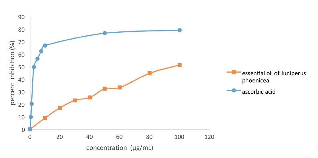
The curve of C0 is the largest in value and the fastest to reach its stability, followed by the dose of essential oil at the same rate but in value lower than that of C0.
Gas production after 24 hours of incubation was significantly reduced. These results are in agreement with those obtained by Arhab et al [33] with the essential oil of Juniperus phoenicea
The low gas production of the ration incubated with different levels of EO is thought to be due to the antimicrobial activity of the EO compounds, according to Derwich et al [34], the components can act individually or synergistically to selectively inhibit on the activity of microorganisms and limit fermentation.
Prediction of the digestibility of Organic Matter (d MO), the production of Volatile Fatty Acids (VFA), and Metabolizable Energy (ME)
An effect is observed at the levels of the decrease in VFAs, MO
86Biological activities of Juniperus phoenicea essential oil and impact on in vitro ruminal fermentation in sheep
1001.7d ± 0.122.17c ± 0.032.32b± 0.452.85a ± 0.02 801.3c ± 0.142.05b ± 0.212.07b ± 0.672.2a ± 0.01 601.07d ± 0.071.95b ± 0.771.77c ± 0.172.15a ± 0.11 300.95d ± 0.171.77b ± 0.0351.6c ± 0.242.1a ± 0.07 100.75d ± 0.081.6b ± 0.171.32c ± 0.881.75a ± 0.11 Gentamicin2.64 ± 0.0073.64 ± 0.0983.67 ± 0.154.08 ± 0.25 Bacteria effect<0.0001 Dose effect <0.0001
Figure 1 - A Free radical-scavenging activity of Juniperus phoenicea essential oils (JPEO) and ascorbic acid on 2,2-diphenyl-1- picrylhydrazyl (DPPH).
Doses(µl)
Table 3 - Inhibition zone diameter (IZD) of Juniperus phoenicea essential oils (JPEO) against four reference strains.
Escherichia coliSalmonella entericaListeria monocytogenesBacillus subtilis
a, b,c and d: The means in the same column, for the same test and bearing different letters are significantly different (α=0.01)
a, b and c: The means in the same column, for the same test and bearing different letters are significantly different.(α=0.01)
Table 5 - Prediction of the digestibility of Organic Matter (d MO), the production of Volatile Fatty Acids (VFA), and Metabolizable Energy (ME) according to different doses.
a, b and c: The values assigned the same letter on the same column do not differ significantly; d OM: digestibility of Organic Matter; EM: Metabolisable Energy; AGV: Volatile Fatty Acids
gas production in sheep according to different doses of Juniperus phoenicea essential oils (JPEO).
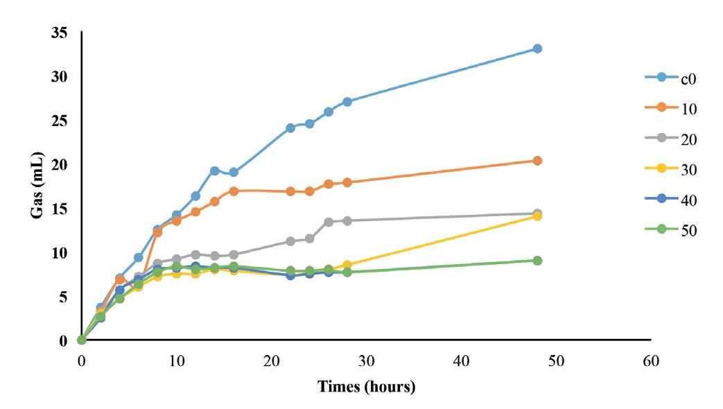
and EM of control (C0) compared to the different doses of JPEO (Table 5).
These results on AGV production and d MO are in agreement with those of [3] who observed that increasing the dose of es-
sential oil decreased the production of AGV and d MO, this due to the fact that at high concentrations, the compounds of essential oils would cause defaunation, which would be at the origin of the decrease in the fermentation activity of mi-
S. Houcine et al. Large Animal Review 2023; 29: 83-8887
C0 0.3597 ± 0.00135.466ª ± 0.2560.0492 ± 0.002 10-0.092 ± 0.39220.840 ± 0.2480.0968 ± 0.002 200.7324 ± 0.00112.920 ± 0.0020.0977 ± 0 302.4911ª ± 0.00111.134 ± 0.0030.0388 ± 0 40-0.3880 ± 0.0018.513 ± 0.0050.3011ª ± 0 50-0.0991 ± 0.3458.594 ± 0.0020.2504 ± 0.005 P> F<0.0001<0.0001<0.0001
Doses(µl)abc
Table 4 - Gas production kinetics according to different doses.
C038.272ª ± 8.03795.676ª ± 1.2290.525ª ± 0.216 1031.456ªb ± 9.8634.634ªb ± 1.5080.342ªb ± 0.265 2026.715 ± 7.15353.908 ± 1.0940.215 ± 0.192 3023.306 ± 3.3953.386 ± 0.5190.123 ± 0.091 4023.158 ± 1.3343.364 ± 0.2040.119 ± 0.036 5023.455 ± 1.7963.409 ± 0.2750.127 ± 0.048 P> F0.06080.06080.0608
Dosesd MO (%)ME (MJ / kg DM)AGV (mmol / syringe)
Figure 2 - Kinetics of
88Biological activities of Juniperus phoenicea essential oil and impact on in vitro ruminal fermentation in sheep
croorganisms and, consequently, of the decrease in digestibility and the production of AGVs.
CONCLUSION
Our result of chemical composition of Juniperus phoenicea essential oils indicated that the α-pinene is the major compound. It has a very interesting antioxidant activity, the IC50 of which proves that this property is close to that of the reference molecule, ascorbic acid. Its antibacterial activity on all 4 strains corroborate with other studies which prove that gram negative bacterial strains are more resistant than gram positive ones. In addition, the effect of JPEO has exceeded all expectations that any essential oil improves ruminal fermentation, as this is not the case in this work. We also demonstrate that JPEO inhibited gas production and decreased the ruminal fermentation parameters. So it can be concluded that JPEO is not recommended for ruminants even at low doses.
Acknowledgements
The financial support of the Institution of Agricultural Research and Higher Education IRESA Tunisia is appreciatively acknowledged.
References
1.Selmi H., Hasnaoui M., Bahri A., Abbès C., Dallali D., Jedidi S., Rouissi H. (2018). Chemical properties, antioxidant activities and in vitro fermentation profiles of some shrubs of North Western Tunisia.Indian J. Anim. Research, 54(7): 851-855.
2.McIntosh F. M., Williams P., Losa R., Wallace R. J., Beeverand D. A., Newbold C.J. (2003). Effects of essential oils on ruminal micro-organisms and their protein metabolism. Appl. Environ. Microbial, 69: 50115014.
3.Benchaar C., Calsamiglia C., Chaves A. V., Fraser G. R., Colombatto D., Mc Allister T.A., Beauchemin K.A.(2008). A Review of Plant-Derived Essential Oils in Ruminant Nutrition and Production. Anim. Feed Sci. Technol, 145: 209-228
4.Alloui M. N. (2011). Les phytobiotiques comme alternative auxantibiotiques promoteurs de croissance dans l’aliment des Volailles. Livest. Res. Rural. Dev, 23(6): 133
5.Macheboeuf D., Papon Y., Arturo-Schaan M., Mousset J. I., Cherel R. (2006). Use of plant extracts (essential oils and polyphenols extract) to reduce the ruminal degradability of proteins - an in vitro essay. Renc. Rech. Ruminants, 13: 69-72.
6.Smeti S., Hajji H., Mekki I., Mahouachi M., Atti N. (2018). Effects of dose and administration form of rosemary essential oils on meat quality and fatty acid profile of lamb. Small Ruminant Research, 158: 62-68.
7.Selmi H., Bahri A., Ferchichi A., Rouissi H. (2020). Effect of supplementing Moringaoleifera essential oils on milk quality and fatty acid profile in dairy sheep. Indian J. Anim. Research, 54(7): 879-882.
8.Amaral Franco J. (1986). Flora Europea: Juniperus L., Ed. T. G. Tutin, Cambridge University Press, Cambridge, Vol. 1.
9.Medini H., Marzouki H., Chemli R., Marongiu B., Piras A., Porcedda S., Tuveri E. (2008). Chemical characterization and evaluation of biological activity of essential oil of Juniperus phoenicea of Tunisia. J. E. Oil Bearing Plants, 11(3): 233-241.
10.El-Sawi S. A., Motawae H. M., Sleem M. A., El-Shabrawy A. O., Sleem A., Ismail M. A.(2014). Phytochemical screening, investigation of carbohydrate contents, and antiviral activity of Juniperus phoenicea L. growing in Egypt J. Herbs Spices Med. Plants, 20: 83-91.
11.Salma E. S. A., Hemaia M. M., Mohamed S. A., Abdel-Rahman E.-S. E., Amanis, S. A., Amal, A. M., Maii, I. A. (2011). Investigation of lipoidal matter, antimicrobial and diuretic activities of leaves and fruits of Juniperus phoenicea L. growing in Egypt. Aust J. Med Herb. 23(4): 174-179
12.El-Sawi S. A., Motawae H. M., El-Shabrawy A.-R. O., Sleem M. A.-F., Sleem A. A., Abdel Naby, M., Maamoun, I. (2015). Antihyperglycemic
effect of Juniperus phoenicea L. on alloxan-induced diabetic rats and diterpenoids isolated from the fruits. J. Coastal Life Med, 3(11): 906909.
13.European Pharmacopoeia, 6th ed., council of Europe, Strasbourg (2008).
14.Hammoudi R., Dehak K., Hadj Mahammed M., Ouldelhadj M. D. (2015). Composition Chimique et activité antioxydante des Huiles Essentielles de Deverra Scoparia Coss. et Dur. (Apiaceae) Lebanese Sci. J. 16(2): 27-36.
15.Van Den Dooland H., Kratz, P. D. (1963). A Generalization of the Retention Index System Including Linear Temperature Programmed GasLiquid Partition Chromatography. J. Chromato A, 11: 463-471.
16.Ben Ammar R., Bhouri W., Ben Sghaier M., Boubaker J., Skandrani I., Neffati A., Bouhlel I., Kilani S., Mariotte A. M., Chekir-Ghedira L., Dijoux-Franca M. G., Ghedira K. (2009). Antioxidant and free radicalscavenging properties of three flavonoids isolated from the leaves of Rhamnus alaternus L. (Rhamnaceae): A structure-activity relationship study. Food Chem, 116: 258-264.
17.Guven D., Dapena A., Kartal B., Schmid M.C., Maas B., Van de PasSchoonen K. (2005). Propionate oxidation by and methanol inhibition of anaerobic ammonium-oxidizing bacteria. Appl. Environ. Microbiol, 71: 1066-1071.
18.Grant R. J. and Mertens D.R. (1992). Impact of in vitro fermentation techniques upon kinetics of fiber digestion. J. Dairy Sci, 75: 1263-1272.
19.Goering H. K., Van Soest P. J. (1970). Forage Fiber Analysis (Apparatus, Reagents, Procedures and Some Application Agricultural Handbook No. 379, Agricultural Research Service, U.S. Department of Agriculture.
20.Menke K. H., Steingass H. (1988). Estimation of the energetic feed value obtained from chemical analysis and in vitro gas production using rumen fluid. Anim. Res. Dev, 28: 9-55.
21.Makkar H. P. S. (2002). Development and Field Evaluation of animal feed supplementation packages. Proceedings of the final review meeting of an IAEA Technical Cooperation Regional AFRA Project, November 25-29, 2000, Joint FAO/IAEA Division of Nuclear Techniques in Food and Agriculture, Cairo, Egypt: 1-66.
22.SAS, Statistical Analysis Systems Institute, Inc, users guide version 9.0. (2009). Cary, NC, USA.
23.Duncan D.B. (1955). Multiple Range and Multiple F-Test Biometrics. 11: 1-5.
24.Orskov, E. R. and McDonald I. (1979). The estimation of protein degradability in the rumen from incubation measurements weighted according to rate of passage. J. Agric.Sci. Cambridge. 92: 499-503.
25.Bouzouita N., Kachouri F., Ben Halima M., Chaabouni M. M. (2008). Composition chimique et activité antioxydante, antimicrobienne et insecticide de l’huile essentielle de Juniperus phoenicea. Société Chim. Tunisie. 10: 119-125.
26.Dob T., Dahmane D., Chaabane C. (2008). Chemical Composition of the Essential Oil of Juniperus phoenicea L. from Algeria. J. Essent. Oil Res, 20: 15-20.
27.Meddini H., Elaissi A., Khouja M. L., Chraief I., Farhat F., Hammami M., Chemli R., and Harzallah-skhiri F. (2010). Leaf Essential Oil of Juniperus oxycedrus L. (Cupressaceae) harvested in northern Tunisia: Composition and Intra-Specific Variability. Chem Biodi. 7: 1254-1266.
28.Mansouri N., Satrani B., Ghanmi M., El ghadraoui L., Aafi A., Farah A. (2010). Valorisation des huiles essentielles de Juniperus thurifera et de Juniperus oxycedrus du Maroc. Phytothérapie. 8: 166-170.
29.Sikkema J., De Bont J. A. M., Poolman B. (1994). Interactions of cyclic hydrocarbons with biological membranes. J Biol. Chem. 269: 80228028.
30.Christine F., Carson J., Mee B., Riley T. V. (2002). Mechanism of action of Melaleuca alternifolia (Tea Tree) oil on Staphylococcus aureus determined by time-kill, lysis, leakage, and salt tolerance assays and electron microscopy. Antimicrob. Agents Chemother.46(6):1914-1920.
31.Ultee A. M., Bennikand M. H. J., Moezelaar R. (2002). The phenolic hydroxyl group of carvacrol is essential for action against the food-borne pathogen Bacillus cereus. Applied environm. Mcrob, 68(4): 1561-1568.
32.Duke, J. A.(1998). Phytochemical Database. USDA-ARS-NGRL (ed),Beltville agricultural research center, Belstville, Maryland, USA.
33.Arhab, R., Khenaka, K., Leulmi, N., Belaidi, H., Harzallah, B., Bousseboua, H. (2013). Effect of essential oils extracted from Saturejacalamintha, Menthapulegiumand Juniperus phoenicea on in vitro methanogenesis and fermentation traits of vetch-oat hay. Afr. J. Environm. Sci. Technol. 7(4): 140-144.
34.Derwich E., Benziane Z., Boukir A.(2010). Chemical composition of leaf essential oil of Juniperus phoenicea and evaluation of its antibacterial activity. Internat. J. Agricul. Biol, 12(2): 199-204.
N. TEKIN ONDERa*, TAYGUN GÖKDEMIRa, MUHAMMET CAN KıLıÇa , OĞUZHAN ŞAHINa, M. BERK TOKERB, SAVAŞ YıLDıZa, YAVUZ ÖZTÜRKLERa
a Department of Reproduction and Artificial Insemination, Faculty of Veterinary Medicine, Kafkas University, Kars, Turkey
b Department of Reproduction and Artificial Insemination, Faculty of Veterinary Medicine, Bursa Uludag University, Bursa, Turkey
SUMMARY
The complexity of the sheep cervix limits non-surgical artificial insemination and embryo production technologies. For this reason, assisted reproduction techniques are generally performed with surgical methods in sheep. But it is said that surgical methods can hurt the health and welfare of animals and cause them to feel stressed in different ways. Because of these problems with surgical methods and some difficulties in the application phase, researchers are trying to come up with ways to help with reproduction that don’t involve surgery. For the application of non-surgical assisted reproductive techniques in sheep, there is a need for successful relaxation of the cervix. Because of this, different tocolytic agents have been used before non-surgical methods of assisted reproduction. Isoxsuprine HCl is used to relax the uterus during procedures like simple dystocia, to prevent premature birth, embryotomies, and caesarean deliveries. It is also used to treat horse navicular disease and laminitis in modern veterinary medicine. Tocolysis usually sets in about 10-15 minutes after an intramuscular isoxsuprine HCl administration. The aim of the present study was to evaluate the effects of isoxsuprine HCl on cervical dilatation in ewes. In our study, it has been thought that isoxsuprine HCl, which is also a tocolytic agent, might be an alternative for non-surgical reproductive uses. The study was carried out on a total of 20 animals: Ten sheep were given cervical relaxation with isoxsuprine HCl and ten animals were given no tocolytic agent. The mean cervical transition time was 83.60±13.63 seconds in animals treated with isoxsuprine HCl and 168.22±20.83 seconds in the control group. A significant difference was found between the groups (P<0.05). The minimum transition time was found to be 19 seconds in the isoxsuprine HCl group and 30 seconds in the control group. Maximum transition times were found at 140 and 238 seconds, respectively. As a result, it was seen that isoxsuprine HCl can offer a good alternative in transcervical applications in sheep.
KEY WORDS
Ewe, Transcervical, Dilation.
INTRODUCTION
Artificial insemination and embryo transfer applications are applied surgically due to the complex structure and anatomical differences of the cervix uteri in sheep [1-4]. Alternative methods for these procedures are being developed in order to avoid complications such as adhesion and bleeding [5]. However, the applicability of transcervical applications in the luteal phase becomes difficult. The duration of the passage of the cervical rings and the experience of the operator are very important factors in the success of these procedures. It is necessary to avoid cervical bleeding that may occur due to force while passing the cervical rings. Because bleeding, adhesions, and infections that may occur for this reason may decrease the future fertility rate of that animal [6,7]. In order to perform assisted reproduction processes in small ruminants; prostaglandin E2 (PGE2), estradiol, and oxytocin applications have been carried out to obtain embryos from donor animals and transfer them to carrier animals by directly entering the cervix uteri [4,5,8,9] .
Corresponding Author: Nail Tekin Onder(nailtekinonder@gmail.com).
Isoxsuprine HCl was first synthesized in 1956 by Moed and Van Dijk. Isoxsuprine HCl, which has been reported to have a spasmolytic effect 40 times more than papaverine, is used as a myometrial relaxant to prevent premature births in humans [8,10,11]
In cattle, Gregory and Rodrigues [12] applied isoxsuprine HCl 5 minutes before embryo transfer to relax the uterus. In ruminants, the recomended dose of 0.4-2 mg/kg is administered intramuscularly, and tocolytic effect begins within 10-15 minutes and uterine contractions triggered by -adrenergic receptors are suppressed [11,13]
According to our research, no study has been found using isoxsuprine HCl for the purpose of relieving and passing the cervix in sheep. In the present study, the passage time of the cervical canal was measured after isoxsuprine HCl application and without any application in the control group. The difference between the control group and the isoxsuprine HCl group was evaluated.
MATERIAL AND METHODS
The Scientific Ethical Committee (Kafkas University, Kars, Türkiye) has approved all issues concerning the experimental
N.T. Onder et al. Large Animal Review 2023; 29: 89-9289
Does isoxsuprine HCl facilitate the passage of the cervix in sheep?: a case series l
90Does isoxsuprine HCl facilitate the passage of the cervix in sheep?: a case series
setups and evaluation techniques (2022-110). All Tuj breed ewes aged 2 to 5 years were kept in the same conditions at Kafkas University’s Faculty of Veterinary Medicine in Kars, Turkey. Experiments were carried out during the non-breeding season, and ewes that had given birth at least once were used. Before the presented study, at least 20 animals were treated, and the possibilities that could affect the cervical transit times were examined. The focus was on maximum efficiency during the presented study.
Experimental Design
This study was designed to evaluate the degree of relaxation of the sheep cervix after intra-muscular isoxsuprine HCl (Utelax® 50 mL, Sanovel, Turkey) injection. For this purpose, 10 animals were assigned to the isoxsuprine HCl (U) group, and 10 animals were assigned to the control group (C).
Sedation and Cervical Dilatation
The animal was restrained by a special restrainer for ewes. Then, 0.4 mg/kg of xylazine (Rompun® %2, Bayer, Turkey) was administered i.m. [14]. Considering the recommendation to use a double dose if anesthetic applications are made in the prospectus of the drug, 0.8 mg/kg of isoxsuprine HCl was administered by i.m. The operator waited for 10 minutes for the drug to take effect.
Preparation of Animal and Cervical Penetration
The perineal region of the animal was cleaned with povidone iodine. The tail was tied, and contamination was prevented. Lubricant was applied to the small speculum, the vulva was opened, and it was inserted into the vagina. The protocol used by Pereira et al. [9] was modified in the cervical penetration stage. The cervical entrance was grasped with forester ring forceps. The speculum was removed, and the cervix was pulled caudally. With the 1 mm diameter Bakes rosebud urethral sound dilator (Figure 1), the cervical rings were passed with gentle movements and
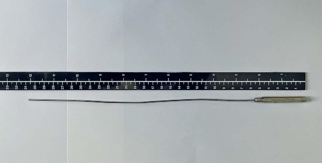
gentle manipulations with the fingers through the vagina to the cervix. The stopwatch was started at the beginning of cervical passage and stopped when the cervix was passed (Figure 2). During the study, operations were carried out by a single operator.
Statistical Analysis
All data obtained from the study were analyzed using SPSS (20.0 for Windows; SPSS, Chicago, IL, USA). Data were represented as mean ± standard error (x±Sx). The Shapiro-Wilk test was used as a normality test. The statistical differences between the groups’ means were determined by an Independent Sample TTest. Differences with values of P<0.05 were considered statistically significant.
RESULTS
In the present study, the mean transit time of the cervix was found to be 83.60±13.63 seconds in the U group, and 168.22±20.83 in the control group (Table 1). The statistical analysis revealed a difference between the groups (P<0.05). In the trial study (before the presented study), we used Allis forceps, Pozzi forceps, and Forester ring forceps. It has been seen that Allis and Pozzi forceps cause a small amount of bleeding. There was no active bleeding when forester ring forceps were used, but redness was observed on the clipped tissue. In the trials, it was also clear that where the forester ring forceps are used to clip is important. This may make it harder for the operator to handle the situation or make the cervical transition process easier.
DISCUSSION
The complex structure of the cervix of sheep and the inability to control the cervical line with rectal manipulations limit
Figure 1 - 1 mm diameter bakes rosebud urethral sound dilator.
the application of non-surgical assisted reproductive techniques, especially embryo production and transfer [5,15,16]. However, the use of different tocolitic agents that will facilitate the passage of the cervical line would increase the applicability of these techniques [17,18,19]. Furthermore, the use of these methods would have positive effects on animal welfare compared to surgical methods [5, 20] .
In sheep, during labor, estrogen and oxytocin levels start to increase; as a result, cervical dilatation and an increase in uterine contractility occur [21]. Due to the cervical dilation effects of these hormones, they have been used in transcervical applications in sheep [4,7,22]. However, Stellflug et al. [23] reported that oxytocin applications before transcervical artificial insemination in sheep decreased the rate of pregnancy-specific protein B and lambing. According to King et al. [24], sheep that received laparoscopic and cervical insemination had a lower lambing rate than those that received oxytocin.
PGE2, which has an anti-inflammatory and relaxing effect on myometrial smooth muscles, is also used for cervical dilation in sheep [9,25]. It is also reported that PGE2 has positive effects on fertility [26]. On the other hand, it has been reported that isoxsuprine provides much better arterial and venous relaxation compared to PGE2 in horses [27,28]. In the studies of Gregory and
Rodrigues (1986) [12] performed by applying isoxsuprine chloride before embryo transfer in cattle, it is seen that the pregnancy rate improved numerically, and the pregnancy rates in the control group and animals treated with isoxsuprine chloride were 39% and 48%, respectively.
Pozzi forceps and Allis forceps have been reported to be used in previous studies to hold and pull the cervix [5, 7, 9, 29]. In our study, in which we used forester ring forceps, it was observed that there was no active bleeding and a slight discoloration of the tissue. For this reason, the use of uterine forceps in transcervical applications seems to be more advantageous for holding and pulling the cervix.
According to the findings of our study, isoxsuprine HCl can be used successfully to provide cervical relaxation in sheep. In this context, we think that the effects of isoxsuprine HCl on pregnancy deserve to be examined in detail in the future.
ACKNOWLEDGEMENTS
None
CONFLICT OF INTEREST
The authors declare that they have no conflict of interest.
AUTHOR CONTRIBUTION SECTION
NTO designed and performed the experiment. NTO, YO, MBT wrote the manuscript. YO provided the isoxsuprine HCL for the experiment. TG, MCK, OS, SY provided assistance during the experiment. NTO and MBT performed the statistical analysis.
DATA AVAILABILITY
The data used to support the findings of this study are available from the corresponding author upon request.
References
1.Armstrong D.T., Evans G. (1983). Factors influencing success of embryo transfer in sheep and goats. Theriogenology. 19(1): 31-42.
2.Bruno-Galarraga M.M., Cueto M., Gibbons A.E., Pereyra-Bonnet F., Catalano R., Gonzalez-Bulnes A. (2014). Repeatability of superovulatory response to successive FSH treatments in Merino sheep. Small Rumin Res. 120(1): 84-89.
3.Santos J.D.R., Ungerfeld R., Balaro M.F.A., Souza-Fabjan J.M.G., Cosentino I.O., Brair V.L., Souza C.V., Pinto P.H.N., Bade A.L.C., Fonseca J.F., Brandão F.Z. (2020). Transcervical vs. laparotomy embryo collection in ewes: the effectiveness and welfare implications of each technique. Theriogenology. 153: 112-121.
4.Wulster-Radcliffe M.C., Costine B.A., Lewis G.S. (1999). Estradiol-17 βoxytocin-induced cervical dilation in sheep: application to transcervical embryo transfer. J Anim Sci. 77(10): 2587-2593.
5.Fonseca J.F., Souza-Fabjan J.M.G., Oliveira M.E.F., Leite C.R., NascimentoPenido P.M.P., Brandão F.Z., Lehloenya K.C. (2016). Nonsurgical embryo recovery and transfer in sheep and goats, Theriogenology. 86(1): 144-151.
6.Candappa I.B.R., Bartlewski P.M. (2014). Induction of cervical dilation for transcervical embryo transfer in ewes. Reprod Biol Endocrinol. 12(1): 1-9.
7.Fonseca J.F., Oliveira M.E.F., Brandão F.Z., Batista R.I., Garcia A.R., Bartlewski P.M., Souza-Fabjan J.M. (2019). Non-surgical embryo transfer in goats and sheep: the Brazilian experience. Reprod Fertil Dev. 31(1): 17-26.
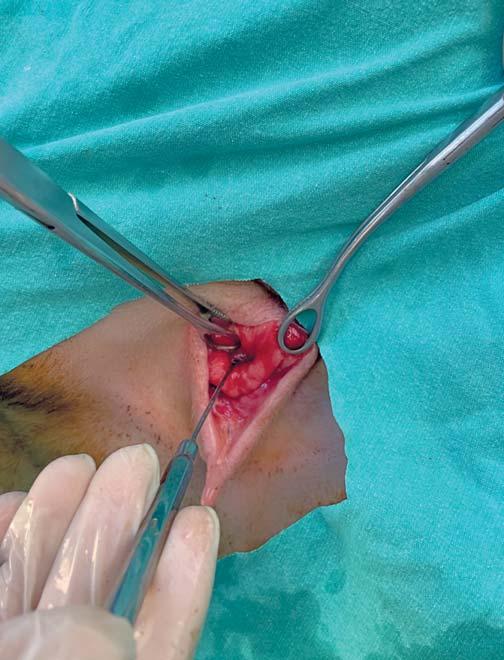
8.Bhoi D.B. (2022) Tocolysis in farm animal reproduction. Livest Sci. 13, 41-47.
9.Pereira R.J.T.A., Sohnrey B., Holtz W. (1998). Nonsurgical embryo collection in goats treated with prostaglandin F2α and oxytocin. J Anim Sci. 76(2): 360-363.
10.Landesman R., Wilson K., Zlatnik F.J. (1966). The Myometrial relaxant properties of isoxsuprine and 2 methanesulfonamido derivatives. Obstet
N.T. Onder et al. Large Animal Review 2023; 29: 89-9291
U83.60±13.63a 19 140 C168.22±20.83b 30238 Table 1 -
GroupsTransit Time (TT)Minimum TTMaximum TT a and b: Values with different superscripts in the same column for each times are significantly different (P < 0.05).
Figure 2 - Cervical passage of the cervix in the sheep.
Cervical transit time in seconds.
92Does isoxsuprine HCl facilitate the passage of the cervix in sheep?: a case series
Gynecol. 28(6): 775-783.
11.Menard L. (1984). Tocolytic drugs for use in veterinary obstetrics. Can Vet J, 25 (10): 389-393.
12.Gregory R.M., Rodrigues J.L. (1986). Pregnancy rates after embryo transfer using drugs that produce relaxation of the uterus. Theriogenology. 25(1): 157.
13.Vural H.A., Koenhemsi L., Gonul R., Yardibi H.D., Gulyasar T., Or E., Hosturk G.T., Barutcu B., Balci H. (2016). Effects of isoxsuprine hydrochloride on electrocardiographic and trace element status in sheeps. J Pak Med Assoc. 66(8): 938-942.
14.Kurtdede A., Börkü M.K., Gürkan M. (1988). Xylazine hydrochloride (rompun) ve ketamine hydrochloride (ketalar) uygulanan koyunlarda bazi klinik gözlemler. Lalahan Hay Araşt Enst Derg. 28(1): 42-50.
15.Kershaw C.M., Khalid M., McGowan M.R., Ingram K., Leethongdee S., Wax G., Scaramuzzi R.J. (2005). The anatomy of the sheep cervix and its influence on the transcervical passage of an inseminating pipette into the uterine lumen. Theriogenology. 64(5): 1225-1235.
16.Robinson J.J., McKelvey W.A.C., King M.E., Mitchell S.E., Mylne M.J.A., McEvoy T.G., Dingwall W.S., Williams L.M. (2011). Traversing the ovine cervix-a challenge for cryopreserved semen and creative science. Animal. 5(11): 1791-1804.
17.Prellwitz L., Zambrini F.N., Guimarães J.D., de Sousa M.A.P., Oliveira
M.E.F., Garcia A.R., Esteves S.N., Bartlewski P.M. Souza-Fabjan J.M.G., Fonseca J.F. (2019). Comparison of the intravenous and intravaginal route of oxytocin administration for cervical dilation protocol and non-surgical embryo recovery in oestrous-induced Santa Inês ewes. Reprod Domest Anim. 54(9): 1230-1235.
18.Figueira L.M., Alves N.G., Batista R.I.T.P., Brair V.L., Lima R.R., Oliveira M.E.F., Fonseca J.F., Souza-Fabjan J.M.G. (2019). Pregnancy rate after fixed-time transfer of cryopreserved embryos collected by non-surgical route in Lacaune sheep. Reprod Domest Anim. 54(11): 1493-1496.
19.Leite C.R., Fonseca J.F., Fernandes D.A.M., Souza-Fabjan J.M.G., Ascoli F.O., Brandão F.Z. (2018). Cervical relaxation for non-surgical uterus access in Santa Inês ewes. Arq Bras Med Vet Zootec. 70: 1671-1679.
20.Morais M.C.C., Esteves L.V., Souza-Fabjan J.M., Oliveira M.E.F., Silva M.R., Brandão F.Z., Fonseca J.F. (2020). Factors affecting pregnancy rates for
goat embryos recovered and transferred by transcervical route. Small Rumin Res. 192: 106215.
21.Leethongdee S. (2010). Development of trans-cervical artificial ınsemination ın sheep with special reference to anatomy of cervix. Suranaree J Sci Technol. 17(1): 57-69.
22.Figueira L.M., Alves N.G., Souza-Fabjan J.M.G., Oliveira M.E.F., Lima R.R., Souza G.N., Fonseca J.F. (2020). Preovulatory follicular dynamics, ovulatory response and embryo yield in Lacaune ewes subjected to synchronous estrus induction protocols and non-surgical embryo recovery. Theriogenology. 145: 238-246.
23.Stellflug J.N., Wulster-Radcliffe M.C., Hensley E.L., Cowardin E.A., Seals R.C., Lewis G.S. (2001). Oxytocin-induced cervical dilation and cervical manipulation in sheep: effects on laparoscopic artificial insemination. J Anim Sci. 79(3): 568-573.
24.King M.E., McKelvey W.A., Dingwall W.S., Matthews K.P., Gebbie F.E., Mylne M.J., Stewart E., Robinson J.J. (2004). Lamb in gratesand litter sizes following intrauterine or cervical insemination of frozen/thawed semen with or without oxytocin administration. Theriogenology. 62(7): 12361244.
25.Slater D.M., Astle S., Woodcock N., Chivers J.E., de Wit N.C., Thornton S., Newton, R. (2006). Anti-inflammatory and relaxatory effects of prostaglandin E2 in myometrial smoothmuscle. Mol Hum Reprod. 12(2): 89-97.
26.Niringiyumukiza J.D., Cai H., Xiang W. (2018). Prostaglandin E2 involvement in mammalian femalefertility: ovulation, fertilization, embryo developmentand early implantation. Reprod Biol Endocrinol. 16(1): 1-10.
27.Baxter G.M., Tackett R.L., Moore J.N. (1989). Reactivity of equine palmar digital arteries and veins to vasodilating agents. Vet Surg. 18(3): 221226.
28.Erkert R.S., Macallister C.G. (2002). Isoxsuprine hydrochloride in the horse: a review. J Vet Pharmacol Ther. 25(2): 81-87.
29.Figueira L.M., Alves N.G., de Souza-Fabjan J.M.G., Batista R.I.T.P., da Cruz Morais M.C., de Lima R.R., Oliveira M.E.F., Fonseca J.F. (2020). Embryo yield and quality are associated with progestogen treatment during superovulation protocol in lactating Lacaune ewes. Theriogenology. 155, 132-138.
ALESSIA DE LUCIA, FILIPPO CALABRESE, STEFANO GRASSIGLI, PARIDE MARCHIORI, TAMARA ORTOLAN, CARLO RAFFAELLI, NICOLA SACCAROLA, STEFANIA TURI, STEFANO ZELCO*
Regione Veneto, Azienda ULSS 3 Serenissima, Dipartimento di Prevenzione, U.O.C Sanità Animale
AFRICAN SWINE FEVER: RISK FACTORS AND BIOSECURITY MEASURES IN BACKYARD HOLDINGS
ABSTRACT
Since 2007, most of the African swine fever (ASF) outbreaks in Europe have involved smallholdings. The aim of this study was to investigate the risk factors that could potentially affect the introduction and spread of ASFV in small pig farms located in ULSS3’ territories.
According to the number of risk factors and the level of biosecurity measures, each farm was ranked into three risk classes. Risk factors were evaluated via questionnaire; in particular farms’ infrastructure, management and feed practice were assessed as well as the risk activities of the pig’s owner or people in charge of pigs. The data collection on the field allowed the updating of the national database (BDN) with geographic coordinates and the actual number of pigs in herds at the time of inspection.
At the beginning of the study, out of the 780 smallholdings registered in BDN almost 50% of backyard holdings were not active anymore as they no longer kept pigs, mitigating the risk of maintenance and spread of ASF often linked to this pig farming system and its poor biosecurity measures.
Greater attention to the internal biosecurity measures compared with the external measure was observed. In particular, the absence of dedicated footwear for visitors (88.1%; 364), lack of evaluation of cleaning and disinfection for trucks (76.3%; 315) and the evidence of risky activities performed by the farmer (69.7%, 288) were the most common risk factors detected.
Swill feeding is not a common practice among our farmers. On the contrary, human forest activities that can represent sources of ASF introduction in pigs’ practices were very common. Despite the interaction between wild boar and domestic pigs being unlikely around Venice’s district, risk activities such as hunting, hiking and mushroom picking represent the main risk factors detected among our farmers especially if performed in the neighboring territorial areas where the wild boar is present.
Most of the premises were isolated with fences and animal feed was kept in properly to avoid wild animal attractions, as expectable considering that for backyard and small farm pigs’ stable is usually part of the house. Results from the analysis showed that the outdoor keeping practices provided with double fences which do not allow animals to come into contact with other animals outside the farm were classified as low risk (6 out of 29). The remaining farms (22 of 29) were at medium risk. These farms were not surrounded by wild boar-proof fences; however, pigs were kept confined in stables to prevent direct contact between production pigs and wild boar.
Only one outdoor farm resulted at high risk and was found with no fence and the stable was not built in a way that prevents wild boar from entering or having contact with the pigs of their farm. Although more than 70% of the farms involved (302 of 413) were classified as medium risk, results from this study highlight the need to organize health education programs to sensitize pig farmers regarding the importance of biosecurity measures and improving good hygiene practices.
KEY WORDS
African swine fever; ASF; biosecurity measures; risk factors; pigs.
INTRODUZIONE
Il territorio dell’Azienda ULSS 3 Serenissima si estende per 1406,09 kmq su buona parte della provincia di Venezia e comprende una popolazione residente di circa 625.235 abitanti. Il patrimonio suinicolo zootecnico è caratterizzato da un’ampia diffusione di allevamenti orientati all’autoconsumo che alle-
Corresponding Author: Stefano Zelco (stefano.zelco@aulss3.veneto.it).
vano al massimo 4 suini in fase di ingrasso.
L’allevamento per uso familiare del suino, finalizzato alla produzione di carne e insaccati destinati alle necessità del nucleo familiare, rappresenta una tradizione ancora radicata e tuttora diffusa nel territorio della Regione Veneto. Questa attività, un tempo legata al contesto economico e sociale del mondo rurale, oggi viene valorizzata come simbolo di tradizione e di ritorno al prodotto “fatto in casa”, ancora molto diffuso in diverse zone del Paese.
Pertanto, l’eventuale diffusione della Peste Suina Africana (PSA) nella popolazione di suini domestici, oltre ad incidere nega-
A. De Lucia et al. Large Animal Review 2023; 29: 93-9893
Peste Suina Africana: fattori di rischio e valutazione delle misure di biosicurezza
O
nelle aziende suinicole a carattere familiare e di piccole dimensioni
tivamente sull’intero settore produttivo, potrebbe innescare dinamiche di drastica riduzione del numero degli allevamenti familiari, con ripercussioni negative nel lungo periodo, come già accaduto in altri paesi dell’Unione Europea. In Estonia, ad esempio, a seguito dei focolai di PSA e a causa delle severe misure di biosicurezza il numero di allevamenti di suini allevati per uso privato è sceso da 696 nel 2014 a 25 nel 2017.
L’evoluzione del ciclo biologico del virus della PSA, che ha contribuito alla sua diffusione geografica in Europa settentrionale e orientale, è rappresentato dal cosiddetto “ciclo cinghialehabitat” che vede come serbatoio del virus i cinghiali e il loro habitat. La circolazione del virus all’interno della popolazione dei cinghiali e la possibilità di contatto tra suini selvatici e domestici rappresentano un rischio costante di introduzione della malattia negli allevamenti.
L’ingresso della PSA in un territorio indenne, caratterizzato dalla presenza di suini domestici, si verifica spesso attraverso l’introduzione di prodotti a base di carne suina contaminata o di animali vivi infetti, mentre la diffusione della malattia tra allevamenti avviene molto probabilmente a causa delle basse misure di biosicurezza degli allevamenti stessi.
L’elevata densità di piccoli allevamenti è da considerarsi un importante fattore di rischio per la diffusione della PSA nelle popolazioni dei suidi domestici, a causa delle scarse condizioni di biosicurezza e di altre caratteristiche comuni a questa tipologia di allevamento, come l’alimentazione dei suini con scarti di cucina, i movimenti non registrati di animali, la macellazione familiare, la possibile vendita di suini infetti e l’accesso all’aperto
degli animali con possibili contatti con altri suini domestici o selvatici. L’analisi spazio-temporale dei dati inerenti ai focolai di PSA nella Federazione Russa nel periodo dal 2007 al 2012 ha identificato gli allevamenti familiari come principale forza trainante per la diffusione del virus, in particolare laddove era diffusa la tecnica dello “swill feeding” ovvero l’alimentazione dei suini con scarti e rifiuti di cucina contaminati. Tuttavia, una recente analisi retrospettiva di 26 focolai in allevamenti di suini domestici in Estonia, ha evidenziato un rischio maggiore di introduzione e diffusione di PSA negli allevamenti commerciali, mostrando una maggior esposizione virale degli allevamenti multi sito e da riproduzione a causa del contatto più frequente e intenso con l’ambiente esterno attraverso le movimentazioni e gli ingressi di veicoli e di persone. Gli Autori segnalano inoltre l’esistenza di una correlazione tra il verificarsi dei focolai nei suini domestici in Estonia e l’intensità dell’infezione nella popolazione di cinghiali identificando quest’ultima come il principale rischio di infezione degli allevamenti di suini domestici.
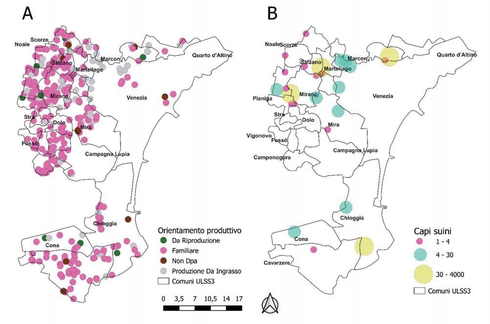
Nel corso di questo studio si è provveduto all’implementazione e all’aggiornamento dei dati anagrafici presenti in BDN relativi al patrimonio suinicolo presente sul territorio di competenza dell’Azienda ULSS 3.
L’obiettivo principale è stato quello di identificare la presenza e la frequenza dei fattori di rischio che possono incidere sulla diffusione della PSA negli allevamenti di piccole dimensioni del territorio di competenza dell’ULSS 3. Successivamente, si è proceduto alla classificazione del rischio di ciascun alle-
A rappresentazione grafica degli allevamenti suddivisi per orientamento riduttivo.
B rappresentazione degli allevamenti suddivisi per capacità produttiva
94Peste
suinicole
Suina Africana: fattori di rischio e valutazione delle misure di biosicurezza nelle aziende
Figure 1 - Distribuzione allevamenti suinicoli nel territorio dell’Azienda ULSS 3 Serenissima.
vamento sulla base della numerosità dei fattori di rischio riscontrati e dei relativi livelli di biosicurezza.
MATERIALI E METODI
I dati relativi alla popolazione suina del territorio dell’Azienda ULSS 3, analizzati di seguito, sono stati estratti dalla BDN. Tutti gli allevamenti registrati in BDN sono stati sottoposti a controllo dai veterinari della UOC di Sanità Animale, che in tale contesto hanno provveduto a raccogliere una serie di informazioni aziendali: effettiva esistenza delle strutture adibite alla stabulazione degli animali e relativa geolocalizzazione, il tipo di stabulazione (stabulati o semibradi), l’orientamento produttivo e il numero di animali presenti, ove non ancora macellati. In merito ai requisiti di biosicurezza, tutti gli allevamenti da riproduzione e quelli da ingrasso con capacità ≥30 capi sono stati ritenuti valutabili con le check list del sistema Classyfarm e, di conseguenza, i dati relativi a questi controlli non verranno presentati in questo studio. L’applicazione di tale check list è pensata per categorizzazione del rischio degli allevamenti industriali ed è difficilmente applicabile in aziende per autoconsumo o a piccole realtà produttive. Per questo motivo, al fine di valutare i livelli di biosicurezza nelle restanti aziende del territorio, nello specifico aziende di piccole dimensioni (autoconsumo o ingrasso <30 capi), la raccolta dati in campo è avvenuta con l’ausilio di un questionario appositamente predisposto (disponibile su richiesta), costituito da 9 domande a risposta multipla e altre 5 di carattere generale. Nel questionario è stata indagata la presenza di fattori di rischio per PSA inerenti alle caratteristiche strutturali dell’allevamento, alla gestione, all’alimentazione degli animali e alle attività del personale addetto alla gestione degli animali considerate a rischio per introduzione in azienda di PSA, quali ad esempio: l’attività di cacciatori, cercatori di funghi o escursionisti. La rilevazione della presenza di ciascun fattore di rischio è stata espressa come percentuale sul totale delle risposte ottenute.
In base alla conformità ai requisiti di biosicurezza imposti dalla legislazione nazionale (DM 28 giugno 2022), pubblicata successivamente alla fase di rilevazione dei dati ed alle domande aggiunte relative alle misure mirate ad impedire il contatto tra suini domestici e selvatici, le aziende sono state divise in tre categorie di rischio.
Per ogni allevamento, ciascun fattore di rischio è stato valutato mediante l’assegnazione dei valori 1 (presenza) o 0 (assenza). Alle domande a cui non è stato possibile rispondere in quanto non applicabili, è stato assegnato un punteggio 0, al contrario di quelle non compilate a cui è stato assegnato il valore 1. Tale decisione è stata presa valutando precauzionalmente come “presente” un
fattore di rischio “sconosciuto”, al fine di non sovrastimare il livello di biosicurezza degli allevamenti soggetti a controllo. Il punteggio di ciascun allevamento (9 è il valore massimo attribuibile) è tanto più elevato quanto peggiore era il livello di biosicurezza. Sulla base della somma dei punteggi ottenuti, le aziende sono state quindi classificate a basso rischio per valori <3, tra 3 e 6 rischio medio e a rischio alto per valori>6.
RISULTATI
A Gennaio 2022, nel territorio di competenza dell’Azienda ULSS 3, gli allevamenti suinicoli registrati in BDN risultavano essere 860, di cui 780 (90,7%) allevamenti familiari, 68 (7,9%) da ingrasso e 12 (1,4%) da riproduzione. Durante il primo semestre del 2022 è stato effettuato un sopralluogo in tutte le aziende, al fine di verificare e aggiornare la BDN sulla base delle informazioni anagrafiche dell’allevamento. Al momento del sopralluogo, sul totale di 780 allevamenti familiari censiti ad inizio anno, 420 hanno sottoscritto la dichiarazione di cessazione attività e 10 sono stati trasferiti in categoria diversa: 8 allevamenti sono entrati nella categoria non destinato alla produzione di alimenti (NON DPA) e due allevamenti familiari sono stati ri-classificati in allevamenti da ingrasso (Tabella 1). Nel mese di agosto 2022, al termine dei sopralluoghi, risultavano censiti nel territorio dell’Azienda ULSS 3 un totale di 440 allevamenti di suini, di cui poco meno dell’80% a carattere familiare (350), 72 da ingrasso, 10 da riproduzione e 8 non DPA. (Tabella 1 e Figura 1).
Per la valutazione della biosicurezza nelle aziende da riproduzione e quelle da ingrasso con capacità superiore ai 30 capi sono state utilizzate le check list Classyfarm. I dati sulla biosicurezza dei piccoli allevamenti con meno di 30 capi sono stati raccolti mediante un apposito questionario. In totale sono state visitati 413 piccoli allevamenti per la valutazione della biosicurezza, e nello specifico 350 familiari, 52 allevamenti da ingrasso, 8 non DPA e 3 allevamenti da riproduzione. Dalla valutazione della frequenza di distribuzione dei fattori di rischio considerati è emerso che l’88,1% (364 su 413) degli allevamenti del territorio non aveva a disposizione calzature specifiche per gli ospiti, il 76,3% (315) non verificava la pulizia e disinfezione dei mezzi in ingresso ed il 69,7% (288) degli allevatori praticava attività considerate potenzialmente a rischio per l’introduzione di PSA. Per i restanti fattori di rischio non soddisfavano i requisiti meno del 50% degli allevamenti (Tabella 2), inclusi quelli relativi alle misure volte ad evitare il contatto con suini selvatici, quali l’assenza della recinzione (36,8%; 152) e l’assenza di protezioni per i mangimi e gli altri alimenti (14,3%;59). Inoltre, è interessante notare che la pratica di integrare la dieta dei suini allevati con scarti di cucina, verdure e prodotti vegetali di autoproduzionenon è risultata essere una pratica diffusa (12,3%; 51 su 413), sebbene percentuali maggiori si riscontrano negli allevamenti semibradi (34,5% di 29) rispetto agli allevamenti stabulati (10,7% di 384). Per entrambe le tipologie produttive considerate (stabulati e semibradi), l’assenza delle calzature dedicate, della mancata verifica della pulizia e disinfezione dei mezzi in ingresso e l’esecuzione di attività considerate a rischio per l’introduzione di PSA da parte del personale sono risultati essere nuovamente i fattori maggiormente rilevati. Relativamente agli allevamenti semibradi, sebbene il 93,1% (27 di 29) fosse in grado di detenere gli animali in locali chiudibili, evitando in tal modo il con-
A. De Lucia et al. Large Animal Review 2023; 29: 93-9895
Familiari780 (90,7%)350 (79,5%) Ingrasso68 (7,9%)72 (16,4%) Riproduzione12 (1,4%)10 (2,3%) Non DPA08 (1,8%) Totale860440
Tabella 1 - Aggiornamento del numero degli allevamenti suddivisi per orientamento produttivo presenti nel territorio di competenza dell’Azienda ULSS 3
Orientamento
N. allevamenti registrati in BDN ProduttivoGennaio 2022Agosto 2022
tatto con i selvatici, circa la metà delle aziende indagate (15 su 29) non era provvista di idonea recinzione. Nello specifico un solo allevamento semibrado presentava entrambi i fattori di rischio, quali l’assenza di recinzione e di locali chiudibili; in base alla classificazione del rischio è per tale motivo risultato essere ad alto rischio (Tabella 2).
In Tabella 3 sono riportati i dati relativi alla classificazione del rischio e alla categorizzazione delle aziende in funzione delle 3 classi di rischio (basso, medio e alto). In generale il 23% delle aziende (95 di 413) è stato classificato come rischio basso; di queste, solo un allevamento stabulato non presentava nessuno dei fattori di rischio indagati.
La maggior parte degli allevamenti è stata posizionata all’interno della categoria di rischio medio (302 di 413, il 73,1%); solo il 3,9% degli allevamenti (16 di 413) ha ottenuto un punteggio superiore a 6 (alto rischio). Di quest’ultimi allevamenti, 15 erano stabulati ed uno solo era semibrado.
DISCUSSIONE
La progressiva espansione della distribuzione geografica della PSA sta minacciando il settore suinicolo in tutto il mondo. Per attivare un efficace controllo, necessario sia in fase di prevenzione che in caso di emergenza, è di fondamentale importanza conoscere la distribuzione e le caratteristiche delle aziende suinicole attraverso l’aggiornamento della BDN, sulla base di informazioni anagrafiche verificate. Durante questo studio, mediante la raccolta dei dati in campo, tramite l’utilizzo di check list appositamente predisposte, sono state verificate tutte le aziende suinicole ed aggiornati i dati anagrafici presenti in BDN. A tal proposito è interessante notare che circa il 50% degli allevamenti familiari hanno deciso di sottoscrivere la dichiarazione di cessazione di attività, piuttosto che aggiornare le pur minime misure di biosicurezza richieste e collegate all’attuale emergenza sanitaria relativa alla PSA.
Sebbene in letteratura la numerosità di piccoli allevamenti è stata descritta come un importante indicatore della potenziale diffusione della PSA in un territorio a causa delle scarse condizioni di biosicurezza che caratterizzano questi allevamenti,
l’aggiornamento dei dati relativi agli allevamenti familiari è un fattore che spesso nelle attività ufficiali è trascurato.
La verifica dell’effettiva presenza degli allevamenti e la conseguente georeferenziazione, insieme all’acquisizione dei dati relativi all’orientamento produttivo ed al numero di capi allevati, ha permesso di mappare tutti gli allevamenti suinicoli presenti. Tale “mappatura” è un requisito preliminare ai fini di una valutazione del rischio di tipo spaziale e della programmazione di misure di emergenza in caso di focolaio.
La maggior parte dei focolai verificatisi nell’ultima epidemia europea ha coinvolto prevalentemente allevamenti familiari. Le aziende di piccole dimensioni sono spesso oggetto di scarsi se non nulli investimenti in infrastrutture e management e dunque ritenuti a maggior rischio di introduzione e diffusione di PSA, assumendo che in questa tipologia di allevamento il livello di biosicurezza sia più carente rispetto alla categoria degli allevamenti commerciali. L’individuazione del livello di biosicurezza adottato da ogni singola struttura registrata in BDN che detenga, anche temporaneamente e/o a qualsiasi titolo, cinghiali, ibridi e suini, anche se non destinati alla produzione di alimenti, rappresenta un pilastro fondamentale delle misure di controllo e prevenzione della diffusione della PSA (DGSAF del 18/01/2022 prot. n. 1195). L’obiettivo principale di questo lavoro è stato quello di acquisire le conoscenze relative ai livelli di biosicurezza di quelle realtà che solitamente sfuggono al controllo ufficiale, in particolare le aziende di piccole dimensioni (familiari e allevamenti con meno di 30 capi), al fine di migliorare l’efficacia del controllo del territorio, sia in fase di prevenzione che in caso di emergenza.
A differenza di quanto generalmente osservato in allevamenti commerciali in cui le misure e le pratiche di biosicurezza esterna sono migliori rispetto a quelle interne, i fattori di rischio più frequentemente riscontrati erano in relazione alle misure adottate per prevenire l’introduzione di malattie trasmissibili. Più del 50% delle aziende prese in esame era carente in almeno una delle seguenti misure di biosicurezza esterna: assenza di calzature specifiche per gli ospiti, omessa applicazione di prassi di verifica di pulizia e disinfezione dei mezzi in ingresso ed esecuzione di attività a rischio da parte dell’allevatore. Al contrario, si è notata una maggiore attenzione per le misure
Tabella 2 - Criteri di base per la valutazione del livello di biosicurezza degli allevamenti di piccole dimensioni nei territori dell’Azienda ULSS 3. Fattori di rischio Presenza fattore di rischio
(N. 413)Stabulati (N. 384)Semibradi (N. 29)
di autoproduzionea
dei locali di stabulazionea
a Requisiti di biosicurezza previsti per gli allevamenti suinicoli familiari secondo il disposto del DM 28/06/2022.
b Requisiti di biosicurezza volti ad impedire il contatto tra suino domestico e selvatico
96Peste
Suina Africana: fattori di rischio e valutazione delle misure di biosicurezza nelle aziende suinicole
Assenza di calzature per ospitia 36488,1%33988,3%2586,2% Mancata verifica lavaggio e disinfezione31576,3%29276,0%2379,3% degli automezzi all’ingressob Attività a rischio del personalea 28869,7%26468,8%2482,8% Assenza recinzioneb 15236,8%13735,7%1551,7% Assenza disinfettanti di provata efficaciaa 14334,6%13936,2%413,8% Assenza protezione degli alimentib 5914,3%5213,5%724,1% Scarti di cucina/verdure/prodotti vegetali 5112,3%4110,7%1034,5%
Assenza locali chiudibilia 399,4%379,6%26,9% Carente manutenzione di pareti e
348,2%328,3%26,9%
pavimenti
Tot
Tabella 3 - Categorizzazione degli allevamenti in funzione della classe di rischio. Classe di rischioFattori di rischio (N.9)Totale (N. 413)Stabulati (N. 384)Semibradi (N. 29)
di biosicurezza interna, probabilmente legata alla natura stessa delle aziende esaminate (prevalentemente familiari); infatti, nella maggior parte degli allevamenti gli animali erano stabulati in locali chiudibili e facilmente sanificabili con prodotti di provata efficacia nei confronti della PSA.
La somministrazione agli animali di scarti di cucina contaminati come integrazione alimentare, ha spesso causato l’insorgenza della malattia nelle popolazioni di suidi domestici di molti Paesi europei. Tuttavia, questo rischio è mitigato se viene rispettato il divieto di somministrare mangimi contenenti scarti di cucina ai maiali come previsto dalla normativa comunitaria. Dalle interviste agli allevatori è emerso che l’alimentazione dei suini con avanzi di cucina non è una pratica diffusa nei Comuni dell’Azienda ULSS 3. Le risposte ottenute riguardo l’integrazione di mangimi erano per di più legate all’autoproduzione ed alla coltivazione di cereali, granaglie e ortaggi. Indagini epidemiologiche condotte in Lituania e Lettonia hanno suggerito che foraggi freschi e semi contaminati da escreti e secreti di cinghiali infetti, possano rappresentare una possibile fonte di infezione per i suini domestici. Ciononostante, il rischio di trasmissione della PSA mediante mangimi autoprodotti, rispetto all’alimentazione dei suini con prodotti a base di carne suina contaminata, risulta essere notevolmente ridotto a maggior ragione nel territorio della dell’Azienda ULSS, caratterizzato da una bassa densità di cinghiali.
In Veneto sono presenti aree territoriali con alta densità di cinghiali; tuttavia, secondo quanto riportato dall’analisi preliminare del rischio svolta a livello regionale (Allegato B DGR 712 del 14 giugno 2022) il territorio della provincia di Venezia è interessato da questo fenomeno solo in minima parte. Data l’importanza del ruolo del cinghiale nella diffusione della PSA, nel questionario somministrato agli allevatori si è ugualmente deciso di indagare alcuni dei fattori di rischio relativi al possibile contatto tra suini domestici e selvatici. Trattandosi per di più di allevamenti familiari o di piccole dimensioni, in cui le strutture di stabulazione degli animali erano vicino alle abitazioni, la maggior parte degli allevamenti era circondato da idonea recinzione e conservava i mangimi in locali adeguati.
Grazie all’elevata capacità del virus di sopravvivere per lunghi periodi nell’ambiente, gli esseri umani possono fungere involontariamente da vettori se esposti ad ambienti contaminati. Il virus della PSA può contaminare e persistere per lunghi periodi sulla superficie di fomiti come vestiti, calzature ed attrezzature. Di conseguenza il rischio legato alla possibilità per i fre-
quentatori dei boschi di agire come fonte di introduzione di PSA in allevamento mediante contatto accidentale con materiale infetto rappresentato ad esempio dalle carcasse di cinghiali, dalle secrezioni ed escrezioni (diarrea, sangue, urine ecc.) eliminate dagli animali ed in generale dall’ambiente boschivo contaminato, non dovrebbe essere sottovalutato.
Nel territorio di competenza dell’Azienda ULSS, in cui le interazioni tra suidi domestici e selvatici sono ancora da considerarsi poco probabili, le attività a rischio di introduzione di PSA nel comparto dei domestici, quali l’attività venatoria o la frequentazione dei boschi da parte di escursionisti o fungaioli, sono risulte essere molto diffuse tra gli allevatori, rappresentando senz’altro il principale rischio in questo contesto, soprattutto se praticate nelle aree territoriali limitrofe dove è presente il cinghiale.
In merito alla tipologia produttiva, gli esperti concordano sul fatto che gli allevamenti all’aperto comportino un rischio notevole di introduzione e diffusione della PSA. Un recente parere dell’Autorità Europea per la Sicurezza Alimentare (EFSA) in cui è stato valutato il rischio di diffusione della malattia negli allevamenti suini all’aperto situati nelle zone dell’UE già interessate dalla presenza dalla PSA, ha concluso che la sola installazione di robuste recinzioni singole o doppie, senza altre misure di biosicurezza, potrebbe di per sé ridurre tale rischio almeno del 50%. Inoltre, il panel di esperti sostiene che una valutazione completa e obiettiva dei parametri di biosicurezza degli allevamenti semibradi, sulla base del rischio di introduzione del virus della PSA, ridurrebbe ulteriormente il rischio di introduzione e diffusione della malattia. In quest’ottica, sulla base della numerosità dei fattori di rischio riscontrati e dei relativi livelli di biosicurezza assegnati, tutte le aziende di piccole dimensioni presenti sul territorio dell’Azienda ULSS 3 sono state suddivise in tre classi di rischio (alto, medio e basso).
Relativamente agli allevamenti semibradi, tutte le aziende provviste di recinzione sono risultate essere a basso rischio; nei restanti allevamenti (circa la metà) classificati come a rischio medio e non delimitati da una recinzione, l’efficace isolamento degli animali era garantito dalla presenza di locali di stabulazione chiudibili. Un solo allevamento semibrado privo di recinzione e locali chiudibili è stato valutato ad alto rischio.
A fronte dei dati raccolti e della classificazione del rischio in funzione del livello di biosicurezza degli allevamenti, appare cruciale l’implementazione dei requisiti di biosicurezza nei 16 allevamenti risultati ad alto rischio. Sebbene il 70% degli alleva-
A. De Lucia et al. Large Animal Review 2023; 29: 93-9897
Basso 0110 132284 262602 <395 (23,0%)89 (23,2%)6 (20,7%) Medio 31371325 491847 556497 618153 Tra 3-6302 (73,1%)280 (72,9%)22 (75,9%) Alto 7220 814131 9000 >616 (3,9%)15 (3,9%)1 (3,4%)
menti siano classificabili a “medio rischio”, i risultati relativi alle frequenze dei fattori di rischio hanno evidenziato la necessità di promuovere iniziative di educazione sanitaria, per spiegare agli allevatori di suini i sistemi di prevenzione dell’introduzione di malattie infettive, anche aumentando le misure di biosicurezza esterna, in particolare quelle di natura igienico sanitarie. Come suggerito dal gruppo di esperti scientifici EFSA , rimane fondamentale il monitoraggio degli allevamenti semibradi, tarato sulla base del rispettivo rischio di biosicurezza. Infine, in ottemperanza al DM del 28 Giugno 2022, a tutti gli allevamenti semibradi verrà imposto l’obbligo di dotarsi di adeguate recinzioni volte ad impedire il contatto tra suidi domestici e selvatici riducendo ulteriormente il rischio di introduzione e diffusione della PSA.
RIASSUNTO
Dal 2007, la maggior parte dei focolai di Peste Suina Africana (PSA) in Europa ha coinvolto prevalentemente gli allevamenti familiari. L’obiettivo di questo studio è stato quello di identificare, attraverso l’utilizzo di un questionario la presenza e la relativa frequenza di distribuzione dei fattori di rischio che incidono sulla diffusione della PSA negli allevamenti di piccole dimensioni presenti sul territorio dell’ULSS 3 Serenissima della regione Veneto. Sulla base di numerosi fattori di rischio riscontrati e dei relativi livelli di biosicurezza, le aziende controllate sono state poi suddivise in tre classi di rischio. Mediante la check list utilizzata sono state valutate le caratteristiche strutturali degli allevamenti, la gestione, l’alimentazione degli animali e le attività del personale addetto alla gestione degli animali considerate a rischio per introduzione in azienda di PSA. La sistematica raccolta dati in campo ha permesso l’aggiornamento della Banca dati nazionale (BDN) con informazioni anagrafiche verificate. Sul totale di 780 allevamenti familiari censiti ad inizio anno circa il 50% degli allevamenti familiari è risultato non più attivo, riducendo in tal modo il potenziale rischio di diffusione della PSA connesso a questa realtà produttiva spesso caratterizzata da scarse misure di biosicurezza.
A tal riguardo, dai questionari raccolti (n. 413) si è notata una maggiore attenzione per le misure di biosicurezza interna. I fattori di rischio maggiormente riscontrati sono stati: assenza di calzature specifiche per gli ospiti (88,1%, 364), l’omessa applicazione di prassi di verifica di pulizia e disinfezione dei mezzi in ingresso (76,3%; 315) ed esecuzione di attività a rischio da parte dell’allevatore (69,7%, 288).
La somministrazione di scarti di cucina contaminati come mangime supplementare degli animali è risultata non essere una pratica diffusa nei nostri comuni. Al contrario, nella provincia di Venezia in cui le interazioni tra suidi domestici e selvatici sono ancora da considerarsi poco probabili, le attività a rischio di introduzione PSA quali ad esempio l’attività venatoria, la raccolta di funghi o l’escursionismo, sono risultate essere molto diffuse tra gli allevatori e rappresentano senz’altro uno dei principali fattori di rischio, soprattutto se praticate nelle aree territoriali limitrofe dove è presente il cinghiale.
Trattandosi di piccole realtà, in cui le strutture di stabulazione degli animali sono in prossimità delle abitazioni, la maggior parte degli allevamenti era circondato da idonea recinzione e conservava i mangimi in locali adeguati.
Relativamente ai semibradi, sulla base della categorizzazione del rischio effettuata, tutti gli allevamenti provvisti di recinzione sono stati classificati a basso rischio (6 di 29), nei restanti allevamenti (22) classificati come a rischio medio e non delimitati da una re-
cinzione, l’efficace isolamento degli animali era garantito dai locali di stabulazione chiudibili. Un solo allevamento semibrado privo di recinzione e locali chiudibili è stato valutato ad alto rischio. Sebbene il 70% degli allevamenti siano stati classificati come “medio rischio”, i risultati emersi dal presente studio hanno evidenziato la necessità di organizzare programmi di educazione sanitaria per sensibilizzare gli allevatori di suini su come prevenire l’introduzione di malattie infettive ed aumentare le misure di biosicurezza esterna in particolare quelle di natura igienico sanitaria.
PAROLE CHIAVE
Peste suina Africana; PSA; biosicurezza; fattori di rischio; suino.
Bibliografia
1.Niro A.,and Sosto G. (2017). Evidenze di efficacia della macellazione a domicilio di suini. veterinariapreventiva.it.
2.Nurmoja I.,Mõtus K.,Kristian M.,Niine T.,Schulz K.,Depner K.,and Viltrop A. (2020). Epidemiological analysis of the 2015-2017 African swine fever outbreaks in Estonia. Prev Vet Med 181 , 104556. 10.1016/J.PREVETMED.2018.10.001.
3.Guberti V.,Khomenko S.,Masiulis M.,and Kerba S. (2022). African swine fever in wild boar: Ecology and biosecurity. Food & Agriculture Org. 28
4.Bellini S.,Rutili D.,and Guberti V. (2016). Preventive measures aimed at minimizing the risk of African swine fever virus spread in pig farming systems. Acta Vet Scand 58, 1-10. 10.1186/S13028-016-0264-X/METRICS.
5.EFSA (2014). Panel on Animal Health and Welfare (AHAW). Scientific opinion on African swine fever. EFSA Journal 12, 3628.
6.EFSA (2019). Panel on Animal Health and Welfare (AHAW), Nielsen, S. S., Alvarez, J., Bicout, D. J., Calistri, P., Canali, E.,... & Gortázar Schmidt, C. (2021). African swine fever and outdoor farming of pigs. EFSA Journal, 19(6), e06639. EFSA Journal 17, e05861. 10.2903/J.EFSA.2019.5861.
7.Oganesyan A.S.,Petrova O.N.,Korennoy F.I.,Bardina N.S.,Gogin A.E.,and Dudnikov S.A. (2013). African swine fever in the Russian Federation: Spatio-temporal analysis and epidemiological overview. Virus Res 173, 204211. 10.1016/J.VIRUSRES.2012.12.009.
8.DM 28 giugno 2022 (2022). Requisiti di biosicurezza degli stabilimenti che detengono suini. (22A04210) (GU Serie Generale n.173 del 26-07-2022). Gazzetta Ufficiale. https://www.gazzettaufficiale.it/showNewsDetail?id=5173&backTo=archivio&anno=2022&provenienza=archivio.
9.EFSA (2019). Panel on Animal Health and Welfare (AHAW), Nielsen, S. S., Alvarez, J., Bicout, D., Calistri, P., Depner, K.,... & Gortázar Schmidt, C. (2019). Risk assessment of African swine fever in the southeastern countries of Europe. Efsa Journal, 17(11), e05861. EFSA Journal 17 . 10.2903/j.efsa.2019.5861.
10.Laanen M.,Persoons D.,Ribbens S.,de Jong E.,Callens B.,Strubbe M.,Maes D.,and Dewulf J. (2013). Relationship between biosecurity and production/antimicrobial treatment characteristics in pig herds. The Veterinary Journal 198, 508-512. 10.1016/J.TVJL.2013.08.029.
11.Postma M.,Backhans A.,Collineau L.,Loesken S.,Sjölund M.,Belloc C.,Emanuelson U.,Grosse Beilage E.,Stärk K.D.C.,and Dewulf J. (2016). The biosecurity status and its associations with production and management characteristics in farrow-to-finish pig herds. animal 10 , 478-489. 10.1017/S1751731115002487.
12.Guinat C.,Gogin A.,Blome S.,Keil G.,Pollin R.,Pfeiffer D.U.,and Dixon L. (2016). Transmission routes of African swine fever virus to domestic pigs: current knowledge and future research directions. Veterinary Record 178, 262-267. 10.1136/VR.103593.
13.Bur n. 73 del 21 giugno 2022 (2002). DGRV n. 712 del 14 giugno 2022“Approvazione del Piano Regionale di Interventi Urgenti (PRIU) per la gestione, il controllo e l’eradicazione della Peste Suina Africana (PSA) nei suini dell’allevamento e nei cinghiali a vita libera.” https://bur.regione.veneto.it/BurvServices/pubblica/DettaglioDgr.aspx?id=479392.
14.Gervasi V.,Marcon A.,and Guberti V. (2022). Estimating the risk of environmental contamination by forest users in African Swine Fever endemic areas. Acta Vet Scand 64. 10.1186/s13028-022-00636-z.
15.EFSA (2021). African swine fever and outdoor farming of pigs Panel on Animal Health and Welfare (AHAW), Nielsen, S. S., Alvarez, J., Bicout, D., Calistri, P., Depner, K., & Gortázar Schmidt, C. EFSA Journal 19 10.2903/j.efsa.2021.6639.
98Peste
aziende suinicole
Suina Africana: fattori di rischio e valutazione delle misure di biosicurezza nelle
Salmonella enterica serovar Dublin infection in dairy cattle: a case study on the management of an outbreak in Italy N
CHIARA TOMMASONI1*, ELIANA SCHIAVON2, ANASTASIA LISUZZO1, MATTEO GIANESELLA1, MARIANNA MERENDA3, PATRIZIO COIN4, TOMMASO PATREGNANI5, SEBASTIANA TOLA6, SALVATORE CATANIA3, ANTONIO BARBERIO2
1 Department of Animal Medicine, Production and Health, University of Padua, Viale dell’Università 16, 35020 Legnaro, Italy
2 Istituto Zooprofilattico Sperimentale delle Venezie, Sezione Diagnostica Padova, Viale dell’Università 10, 35020 Legnaro (PD), Italy
3 Istituto Zooprofilattico Sperimentale delle Venezie, Sezione Diagnostica di Verona, Via Bovolino 1/C, 37060 Buttapietra (VR), Italy
4 Dipartimento veterinario e sicurezza degli alimenti di origine animale “ATS Brescia”, 25124 Brescia, BS, Italy
5 Veterinary Services, Local Health Unit “AULSS 9 Scaligera”, 37057 Verona, VR, Italy
6 Istituto Zooprofilattico Sperimentale della Sardegna, Via Vienna 2, 07100 Sassari, Italy
SUMMARY
Salmonella enterica subsp. enterica serovar Dublin (S. Dublin), is a serovar adapted to cattle, causing both intestinal and systemic infections. The introduction of the bacterium leads to serious economic losses due to abortions, high mortality in calves and persistent infections, also representing a major health problem as zoonotic agent. The aim of this study was to describe an outbreak of S. Dublin on an Italian dairy cattle farm and to assess the effectiveness of the management protocol prepared by the Istituto Zooprofilattico Sperimentale delle Venezie (IZSVe).
S. Dublin was diagnosed on a cattle farm in Northeastern Italy following the conferral at the IZSVe of a newborn calf that died from enteric syndrome. At the autoptic exam pathological findings were observed in gut, liver, pericardium, lungs, joints, lymph nodes and abomasum. Considering the pathogenesis of S. Dublin, authors decided to apply a protocol prepared by the IZSVe based both on direct and indirect prophylaxis. Particularly, an autogenous vaccine against S. Dublin prepared by the Istituto Zooprofilattico Sperimentale della Sardegna (IZS Sardegna) was administered.
Screening tests were performed on fecal and milk samples (bulk tank milk) and on environmental swabs from lactating and dry cows’ boxes.
A pre and post-vaccination screening in 3-times (T0, T1, T2) was performed on serum, feces and milk to assess the immunization of cows and the effectiveness of the protocol itself. The first sampling took place 1-day prior immunization, the second and the third 2 and 11 months later respectively.
Serological examination identified 25%, 100% and 73% positive animals at T0, T1 and T2 respectively. No fecal sample in all time-points was found positive. After vaccination only 1 milk sample turned out positive.
Considering the pathogenesis of S. Dublin, the negativity of the bacteriological exams suggests a positive effect of the protocol in the reduction of clinical cases, circulation of the etiological agent and biocontainment of the infection.
KEY WORDS
Salmonella; Dublin; Cattle; Autogenous vaccine.
INTRODUCTION
Salmonella enterica subsp. enterica serovar Dublin (S. Dublin) is a serovar with high zoonotic potential, adapted to cattle, which causes both intestinal and systemic infections in the host1. Infections have significant impact on productivity and welfare in cattle herds, thus resulting in serious economic losses for the
Corresponding Author:
Chiara Tommasoni (chiara.tommasoni@phd.unipd.it)
farmers2. Bacteria dissemination occurs mainly through feces, although spreading via aerosol, oculo-conjunctival route and milk secretion has been proved in cattle. Thus, contagion is possible either directly between animals or indirectly, due to its ability to survive for long time in the environment3. Ingestion of S. Dublin does not necessarily induce disease. In fact infection may result in chronic and subclinical carriers that may spread organisms continuously or intermittently in the environment not only through feces but also through milk and colostrum, thus constituting an important maintenance factor of the infection within the herd 4,5. The pathogenesis of S. Dublin depends on several factors, such as virulence factors,
C. Tommasoni et al. Large Animal Review 2023; 29: 99-10399
infectious dose, passive transfer of specific immunoglobulins, immunity developed during previous infections, age, and physiological status of the host6,7. In particular, the development of a successful immune response is thus fundamental8, even if several studies highlighted that metabolic pathologies, such as ketosis, might negatively impact on its stimulation9,10. Consequently, also the clinical presentation is affected, distinguishing an hyperacute, acute or chronic form11,12. Adults are generally affected by asymptomatic infection or subacute enteric form, and abortion could be the only clinical sign of infection. Calves may be affected more frequently than adults by the hyperacute form, characterized by sepsis and respiratory signs, with highest incidence between 4 and 28 days13. The severity of the disease is strictly connected both with rearing condition and with management14. Many calves often suddenly succumb 1 to 2 days after the onset of symptoms due to dehydration and generalized systemic distress, especially in case of no or inadequate drug treatment15
Generally, most recurrent clinical signs are apathy, anorexia, hyperthermia, reduced milk production (adults), respiratory distress (calves), mucosal pallor. These are followed by diarrhea, varying from greenish watery to fetid and yellowish, containing blood, mucus, fibrin, and necrotic shreds of the intestinal lining15. Meningoencephalitis, septicemic arthritis16, dry gangrene in the extremities17, urocystitis and ureteritis18 have also been described.
S. Dublin is also an important serovar from human health perspective, as it demonstrates a high level of invasiveness in humans that could lead to severe disease or death19. Therefore, the occurrence of S. Dublin in dairy herds should be faced applying all the biosecurity measures needed to avoid the contamination of milk, the development of chronical infection in cattle and the spread of the disease inside and outside the farm. The aim of this study is to describe the application of a biosecurity protocol to control S. Dublin spread in a dairy farm of the Northeastern Italy.
CASE DESCRIPTION
One Italian herd of 385 Holstein-Friesian dairy cows, of which 210 in milk, was affected by an outbreak of S. Dublin. Only calves younger than 6 months showed clinical signs, in particular enteric syndrome, while deaths occurred only in animals younger than 30 days. In November 2020, a newborn calf died from enteric syndrome and a post-mortem examination was performed by the Istituto Zooprofilattico Sperimentale delle Venezie (IZSVe). Necropsy finding included serious enteritis, peritonitis, and involvement of other organs such as liver, joints, lungs and pericardium. Specifically, icterus, pericarditis, fibrinous polyarthritis and foci of pulmonary congestion and hepatization were detected. Abomasitis, severe hepatosplenomegaly with hepatic degeneration and impressive meseraic lymphadenomegaly were also found. To investigate the etiology of the gross findings, intestine, joints, lymph nodes, spleen, pericardium, lungs and kidney were examined by standard bacteriological method and Minimum inhibitory concentration (MIC), revealing S. Dublin colistin-resistant as causative agent.
The case was reported to the Local Official Veterinary Service (LOVS), and a biosecurity management protocol in compliance with IZSVe was then applied.
PROTOCOL DESCRIPTION
The IZSVe protocol consisted in a farm analysis and epidemiological investigation and the collection and testing of screening samples to assess the spread of the disease and the absence of milk contamination.
Screening tests were performed both on fecal and milk samples. Feces were collected from 80 animals (dry-off cows and calves), in 7 times-points, from November 2020 to February 2021. 25 milk samples were collected from bulk tank milk (BTM) from November 2020 to April 2021. Furthermore, two environmental swabs were also collected from lactating and dry cows’ boxes in November and December 2020.
Moreover, all lactating cows were tested by mean of a fecal swab 2 times, in February and March 2021: a total of 220 and 223 dairy cows were tested.
Due to the specific patterns of S. Dublin, the use of vaccination was included in the protocol.
Considering the difficulty to find an effective commercial vaccine against S. Dublin, an autogenous vaccine was prepared from the S. Dublin strain isolated from the farm by the IZS of Sardegna. The vaccine, consisting of a washed culture inactivated with 0.3% formalin and adjuvanted with 10% aluminum hydroxide, was administered to all the cattle of the farm with a first dose in February and a booster dose one month later.
A pre and post-vaccination screening was performed to assess the immunization of cows and the effectiveness of the protocol itself.
A total of 52 cattle randomly selected among cows and heifers were enrolled for the 3-time effectiveness sampling (T0, T1, T2), the first one day prior immunization, the second and the third 2 and 11 months later respectively, both on feces and serum, collecting blood samples from the coccygeal vein using a vacutainer system20,10. Due to animal culling only 44 animals completed the screening. To monitor the risk of milk contamination, weekly bacteriological control of BTM was performed from November 2020 to April 2021, for a total amount of 25 samples collected and tested.
ANALYTICAL METHODS
Salmonella spp. detection and typing: Salmonella spp. detection on fecal samples, milk, animal tissue and environmental swab was performed according to ISO 6579:2017.
All the Salmonella spp. isolates were delivered to the National Reference Center and subtyped according to Kauffmann-White-Le Minor21.
Antibodies detection:
To identify Salmonella serological antibodies an indirect ELISA commercial kit (PrioCHECK® Salmonella Ab bovine) was performed. Results were analyzed through the Percent positivity (PP). Cut off positivity was established with values of PP≥35%.
RESULTS
The epidemiological investigation highlighted as main biosecurity risk factors the purchase of adult animals, the practice of mountain grazing using promiscuous pastures and the absence
100Salmonella enterica
in dairy cattle
serovar Dublin infection
of regular pests control plans. Factors that facilitated the spread of infection were overcrowding and poor environmental hygienic areas where both lactating and dry cows were housed, just as poor hygiene condition of the milking room and its equipment. Consequently, corrective measures were adopted, such as milking facility and routine hygiene improvement, implementation of an appropriate pest’s control system, systematic cleaning, and disinfection of newborn calves’ cages and of the equipment used to prepare and feed milk to calves. To reduce the risk of contamination, milk after collection was heat treated to be suitable for human consumption; to avoid the spread of infection to veal calves’ farms, the control of all newborn calves was improved at least twice within 7 days, and the movement of positives calves was forbidden.
The screening test performed in the farms highlighted the presence of 6 positive calves with 2 calves that remained positive to 2 consecutive test, and 1 dry-off cow (Figure 1). All the fecal samples collected from the lactating cows in February and March tested negative for S. Dublin. All the environmental swabs collected in lactating and dry-off cows’ pens tested positive for S. Dublin. Eventually 2 BTM samples out of 25 were contaminated with S. Dublin; both the samples were collected in November.
From the effectiveness evaluation sampling, serological examination identified 13/52 (25%), 49/49 (100%) and 32/44 (73%) positive animals at T0, T1 and T2 respectively. No fecal sample in all time-points was found positive. After vaccination only 1 milk sample turned out positive. Considering serological response of cows after vaccination, an initial strong increase in antibody titer at T1 and a subsequent decrease after 11 months (T2) were shown (Figure 2).
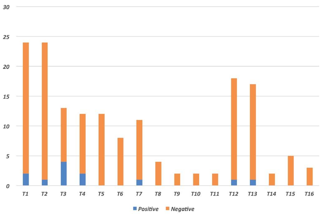
Serological analysis also showed that the presence of seropositivity before the first vaccination (T0) significantly affected the antibody response of cows (Figure 3).
DISCUSSION
The management protocol applied in this outbreak of S. Dublin reflects those commonly applied in case of positivity to S. Typhimurium, including the monophasic variant, S. Dublin and S. Enteritidis in dairy cows’ herds. These serotypes were selected based on 3 factors: isolation rate in samples of bovine origin, public health relevance and zoonotic aspect.
Findings highlighted by the epidemiological investigation agree with several other studies, according to which the purchase of infected carrier animals could be one of the most frequent access ways of S. Dublin into farms3,22,23. Another key point is keeping small and stable groups of calves, avoiding mixing individuals with different immune and infectious status. Boxes of 2 or 4 calves would be optimal; on the contrary, groups of more than 8 calves would greatly increase the risk of S. Dublin outbreaks in young animals2. Subsequently, it has been shown that poorly clean calving boxes and overcrowding of calving animals have a negative impact on the spread of the disease. Proper management of calving boxes is therefore essential in the control of Salmonella spp. infection. Preventive actions should include removing calves from their mothers immediately after calving, setting up a separate box for each cow, maintaining high levels of cleanliness and hygiene in the environment and bedding, and allocating areas for the exclusive use of parturient cows24,25. An-
C. Tommasoni et al. Large Animal Review 2023; 29: 99-103101
Figure 1 - Results of screening test on calves and cows.
other important aspect is that chronically infected cows can eliminate S. Dublin through colostrum and milk. Several studies also showed that contamination of milk and colostrum, tends to increase through collection, storage and feeding processes, thus making cleanliness and hygiene of all the tools used for feeding calves and of barn staff crucial24,26. For this reason, it is essential to clean and sanitize feed and water distribution tools, environments and milking parlors with their respective equipment, as reported by numerous studies4,24
Analysis of the clinical and laboratory data showed that the infection occurred in both cows and calves, although probably with different prevalence between these groups.
The initial contamination of milk may be due to simultaneous presence of adult bovine animals excreting Salmonella and the hygienic deficiencies highlighted during milking and cleaning
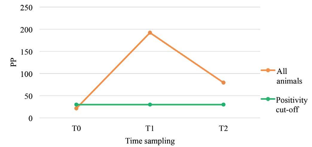
of the equipment used for this activity. Considering the positivity encountered in the housing areas of lactating cows, observed during the first sampling, we should hypothesize that the lack of detection of excretory cows is related to the sporadic excretion, which was probably present before the sampling was carried out. The effectiveness evaluation highlighted the discrepancy between the serological test and the detection of S. Dublin in feces. In fact, at T0 some subjects tested positive to the serological test, without excretion of S. Dublin in the feces. The antibody positive outcome showed that animals had previous contact with the etiologic agent but without pathogen’s spread. Considering the pathogenesis of S. Dublin infection, it cannot be excluded that some positive animals were chronic carriers27. The use of vaccination may improve the immune response and reduce the duration of fecal excretion, thereby helping to limit the spread of infection
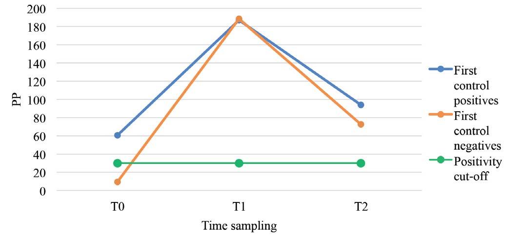
102Salmonella enterica serovar Dublin infection in dairy cattle
Figure 3 - Average Positivity Percentage (PP) trend in animals serologically positive or negative at first sample.
Figure 2 - Average serological Positivity Percentage (PP) trend in animals sampled with positivity cut-off of 30.
within the herd.
The initial strong increase in antibody titer at T1 and the subsequent decrease after 11 months (T2) highlight how the use of vaccination cannot be extemporaneous but must include annual booster to ensure adequate antibody titer to limit excretion phases in chronically infected animals.
Moreover, concerning serological positivity at T0, analysis showed that, after 11 months, these animals had higher values at the T2 than the initially seronegative animals. This difference may be related to a preexisting natural infection, which may induce a more sustained immune response in individuals. This event does not occur in all animals because, due to a malfunction of the cell-mediated immune response, chronic infections may develop4. Previous studies highlighted that animals with the highest risk of becoming active carriers are heifers aged between the first year of life and first calving, and cows infected close to the calving date itself. An increase in incidence was also noted in farms with low eliminator’s prevalence28
Considering the negativity of all fecal bacteriological exams, this would suggest a possible positive effect of the vaccine in reducing the excretion of S. Dublin, considering that the direct prophylaxis measures applied also contributed to the biocontainment of the infection. However, it’s not possible to quantify the vaccination’s effectiveness, as the management protocol applied included both direct and indirect prophylaxis. On the other hand, based on the exams performed, control measures yielded good results in terms of reduction of clinical cases and circulation of the etiological agent.
CONCLUSIONS
Biosecurity and biocontainment measures adopted were effective in identifying eliminators and reducing environmental contamination. In addition, the adoption of a vaccination protocol has been a key factor in the management of the infection. Results of post-vaccination sampling, demonstrated how the integrated application of direct and indirect prophylaxis measures drastically reduced the presence of eliminator animals, leading to the extinction of the infection outbreak, thus constituting a possible model for the management of S. Dublin outbreaks in dairy herds even in complex farm situations.
References
1.Egle Kudirkiene, Gitte Sørensen, Mia Torpdahl, Leonardo V. de Knegt, Liza R. Nielsen ER, Shahana Ahmed JEO. 2020. Epidemiology of Salmonella enterica Serovar Dublin in Cattle. 86(3):1-12.
2.Henderson K, Mason C. 2017. Diagnosis and control of Salmonella Dublin in dairy Herds. In Pract. 39(4):158-168. doi:10.1136/inp.j1160
3.Nielsen LR, Kudahl AB, Østergaard S. 2012. Age-structured dynamic, stochastic and mechanistic simulation model of Salmonella Dublin infection within dairy herds. Prev Vet Med. 105(1-2):59-74. doi:10.1016/j.prevetmed.2012.02.005
4.Holschbach CL, Peek SF. 2018. Salmonella in Dairy Cattle. Vet Clin North Am - Food Anim Pract. 34(1):133-154. doi:10.1016/j.cvfa.2017.10.005
5.Wallis TS, Barrow PA. 2005. Salmonella Epidemiology and Pathogenesis in Food-Producing Animals. EcoSal Plus. doi:10.1128/ecosalplus.8.6.2.1
6.Nielsen LR. 2013. Review of pathogenesis and diagnostic methods of immediate relevance for epidemiology and control of Salmonella Dublin in cattle. Vet Microbiol. 162(1):1-9. doi:10.1016/j.vetmic.2012.08.003
7.Bortolami A, Fiore E, Gianesella M, Corrò M, Catania S, Morgante M. 2015. Evaluation of the udder health status in subclinical mastitis affected dairy cows through bacteriological culture, Somatic Cell Count and thermographic imaging. Pol J Vet Sci. 18(4):799-805. doi:10.1515/pjvs-2015-0104
8.Bobbo T, Ruegg PL, Fiore E, Gianesella M, Morgante M, Pasotto D, Gallo L, Bittante G, Cecchinato A. 2017. Short communication: Association between udder health status and blood serum proteins in dairy cows. J Dairy Sci. 100(12):9775-9780. doi:10.3168/JDS.2017-13111
9.Fiore E, Lisuzzo A, Tessari R, Spissu N, Moscati L, Morgante M, Gianesella M, Badon T, Mazzotta E, Berlanda M, et al. 2021. Milk Fatty Acids Composition Changes According to -Hydroxybutyrate Concentrations in Ewes during Early Lactation. Anim 2021, Vol 11, Page 1371. 11(5):1371. doi:10.3390/ANI11051371
10.Tessari R, Berlanda M, Morgante M, Badon T, Gianesella M, Mazzotta E, Contiero B, Fiore E. 2020. Changes of Plasma Fatty Acids in Four Lipid Classes to Understand Energy Metabolism at Different Levels of Non-Esterified Fatty Acid (NEFA) in Dairy Cows. Anim 2020, Vol 10, Page 1410. 10(8):1410. doi:10.3390/ANI10081410
11.Richardson A, Watson WA. 1971. A Contribution to the Epidemiology of Salmonella Dublin Infection in Cattle. Br Vet J. 127(4):173-183. doi:10.1016/S0007-1935(17)37634-0
12.Wray C, Davies RH. 2000. Salmonella infection in cattle. In: Salmonella in Domestic Animals. 1 st Ed., 169-190, Wray C, Wray A, editors. New York: CABI Publishing.
13.Guizelini CC, Tutija JF, Morais DR, Bacha FB, Ramos CAN, Leal CRB, Zaquetti ME, Lemos RAA. 2020. Outbreak investigation of septicemic salmonellosis in calves. J Infect Dev Ctries. 14(1):104-108. doi:10.3855/jidc.12087
14.Barrow PA, Methner U. 2013. Salmonella Infections in Cattle. In: Salmonella in Domestic Animals, 2nd Ed., 233-262, CABI.
15.Hall GA, Jones PW. 1977. A study of the pathogenesis of experimental Salmonella dublin abortion in cattle. J Comp Pathol. 87(1):53-65. doi:10.1016/0021-9975(77)90079-2
16.Uribe JAZ, Coura FM, Nunes PP, Silva MVP, Carvalho AU de, Moreira MVL, Mendonca FL de M, Salvato LA, Meneses RM, Guedes RMC, et al. 2015. Septicemic Salmonellosis in Pre Weaned Calves Caused by Salmonella dublin. Res J Vet Pract. 3(3):69-75. doi:10.14737/journal.rjvp/2015/3.3.69.75
17.Loeb E, Toussaint MJM, Rutten VPMG, Koeman JP. 2006. Dry Gangrene of the Extremities in Calves Associated with Salmonella dublin Infection; a Possible Immune-mediated Reaction. J Comp Pathol. 134(4):366-369. doi:10.1016/j.jcpa.2006.01.005
18.Costa RA, Casaux ML, Caffarena RD, Macías-Rioseco M, Schild CO, Fraga M, Riet-Correa F, Giannitti F. 2018. Urocystitis and Ureteritis in Holstein Calves with Septicaemia Caused by Salmonella enterica Serotype Dublin. J Comp Pathol. 164:32-36. doi:10.1016/j.jcpa.2018.08.005
19.Lynne AM, Dorsey LL, David DE, Foley SL. 2009. Characterisation of antibiotic resistance in host-adapted Salmonella enterica. Int J Antimicrob Agents. 34(2):169-172. doi:10.1016/j.ijantimicag.2009.02.018
20.Tessari R, Mazzotta E, Blasi F, Morgante M, Badon T, Bedin S, Fabbri G, Lisuzzo A, Contiero B, Fiore E, Berlanda M. 2021. Milk fatty acids as biomarkers of metabolic diseases in dairy cows identified through thin layer chromatography and gas chromatographic techniques (Tlc-gc). Large Anim Rev. 27(4):187-193.
21.Issenhuth-Jeanjean S, Roggentin P, Mikoleit M, Guibourdenche M, De Pinna E, Nair S, Fields PI, Weill FX. 2014. Supplement 2008-2010 (no. 48) to the White-Kauffmann-Le Minor scheme. Res Microbiol 165(7):526-530. doi:10.1016/J.RESMIC.2014.07.004
22.Tablante N, Lane V. 1989. Wild mice as potential reservoirs of Salmonella dublin in a closed dairy herd. Can Vet J. 30(7):590-592. https://www.ncbi.nlm.nih.gov/pmc/articles/PMC1681103/
23.Vaessen MA, Frankena K, Graat EAM, Veling J, Klunder T. 1998. Risk factors for salmonella dublin infection on dairy farms. Vet Q. 20(3):97-99. doi:10.1080/01652176.1998.9694848
24.House JK, Smith BP. 2004. Profitable Strategies to Control Salmonellosis in Dairy Cattle. In: 23rd World Buiatrics Congr Quebec City, Canada.
25.Nielsen TD, Vesterbæk IL, Kudahl AB, Borup KJ, Nielsen LR. 2012. Effect of management on prevention of Salmonella Dublin exposure of calves during a one-year control programme in 84 Danish dairy herds. Prev Vet Med. 105(1-2):101-109. doi:10.1016/j.prevetmed.2012.01.012
26.Mohler VL, Izzo MM, House JK. 2009. Salmonella in Calves. Vet Clin North Am - Food Anim Pract. 25(1):37-54. doi:10.1016/j.cvfa.2008.10.009
27.Veling J, Barkema HW, Van Der Schans J, Van Zijderveld F, Verhoeff J. 2002. Herd-level diagnosis for Salmonella enterica subsp. enterica serovar Dublin infection in bovine dairy herds. Prev Vet Med. 53(1-2):31-42. doi:10.1016/S0167-5877(01)00276-8
28.Nielsen LR, Schukken YH, Gröhn YT, Ersbøll AK. 2004. Salmonella Dublin infection in dairy cattle: Risk factors for becoming a carrier. Prev Vet Med. 65(1-2):47-62. doi:10.1016/j.prevetmed.2004.06.010
C. Tommasoni et al. Large Animal Review 2023; 29: 99-103103
GUIDELINES FOR AUTHORS LAR
Large Animal Review
Large Animal Review is a bimonthly magazine published by SIVAR (Italian Society of Farm Animals Veterinary Practitioners) for scientific updating of veterinarians who deal with animals in livestock production and the supply chain control in the production of food industry. The topics of main interest for the journal are those of internal medicine, surgery, obstetrics, animal nutrition, zootechnics, infectious and parasitic diseases, food safety and security, animal welfare, prevention and management.
■ MANUSCRIPTS
Large Animal Review publishes manuscripts in the form of reviews, original articles and case reports; manuscripts must comply with the guidelines below.
Review - This is a complete coverage of a specific topic accompanied by a detailed and updated bibliography. Authors interested in writing a review should contact the editor of Large Animal Review. The text should not have more than 48.000 characters (including spaces) and not be accompanied by more than 15 figures or tables.
Original Article - The papers published in Large Animal Review are short or full-length research articles related to the topic of the journal. The full text article should not exceed 32.000 characters (including spaces) and should not be accompanied by more than 10 figures or tables. Manuscripts in the form of short articles should not exceed 16.000 characters and no more than 4 figures or tables.
Case Report - Single clinical or herd case report should be presented in Large Animal Review. The manuscript must not exceed 10.000 characters (including spaces) and no more than 4 figures or tables.
■ FORMAT
All manuscripts (review, original articles and case reports) must have the following structure.
Language - English or Italian.
Title - The title of the manuscript has to be short and explicative and written on the front of the first page. Under the title, names of authors should be given indicating the surname and the name (e.g., Smith Tom). Institutional addresses are displayed below the author names; footnotes referring from author names to displayed addresses should be numbered. The full name, mailing address, phone number, and e-mail address of the corresponding author should appear directly below the affiliation lines on the title page. The corresponding author will be identified by a symbol footnote (e.g., Smith Tom*) and e-mail address below the affiliation lines on the first page of the published article (e.g., *Corresponding author: Smith Tom, University of …).
Abstract - Abstract has to be placed on the second page of the manuscript and written in English. Abstracts should be limited from 300 to 500 words. The abstract disseminates scientific information through abstracting journals and is a convenience for readers. Exclude references, figure or table.
Official scientific journal of SIVAR
Key Words - After the abstract, list 3 to 5 key words have to be placed. In case of manuscripts written in Italian, the key words have also be translated into English.
Body of the Paper - The manuscript of the original articles must show the following outline: introduction, materials and methods, results, discussion, conclusions, acknowledgments and bibliography. Regarding the reviews, the outline is not expected, but the topic must be clearly argued and divided in chapters.
The text should be typed in Microsoft Word preferably, or OpenOffice or Rich Text Format, with lines and pages numbered consecutively, using Times New Roman font at 12 points. All margins should be at least 2 centimeters, single spacing and cannot exceed the number of characters (including spaces) indicated in the previous section for each type of manuscript.
Tables and Figures - Tables, graphs and images must be included in the manuscript text and numbered (Arabic numerals). The titles or captions should describe concisely the data shown, sufficiently detailed and comprehensible to the reader. Tables and figures should be placed in separate sections at the end of the manuscript.
References - The references must be selected by the authors (not more than 30, except in a review) and should be cited in the text with a serial number in round brackets and listed in the same numerical order in the bibliography.
For articles from journals you should indicate: surname and first initial of the author and co-author/s names, year of publication, article title, abbreviated indication of the magazine (Index Medicus), volume number, number of first and last pages. For citations to articles or chapters contained in textbooks, you should indicate: surname and first initial of the Author and co-author/s names, year of publication, chapter’s title, book title, volume number (if more than one volume) editors, edition, first and last page of the chapter, publishing house and its location.
Examples:
– Journals - Galey F.D., Terra R., Walker R., Adaska J., Etchebarne M.A., Puschner B., Fisher E., Whitlock R.H., Rocke T., Willoughby D., Tor E. (2000). Type C botulism in dairy cattle from feed contamined with a dead cat. J Vet Diagn Invest, 12: 204-209.
– Books - Gustafson D.P. (1986). Pseudorabies. In: Diseases of swine, Ed. Dunn H.W., 5th ed., 274-289, Iowa State University Press, Ames, IA.
– Conferences - Barbano D. M. (1996). Mozzarella cheese yield: Factors to consider. Page 29 in Proc. Wisconsin Cheese Makers Mtg., Madison. Ctr. Dairy Res., Univ. Wisconsin, Madison.
■ SUBMISSION OF MANUSCRIPTS
The manuscripts have to be submitted exclusively on the following link: www.largeanimalreview.com
Informations: Dr. Enrico Fiore - Technical Editor largeanimalreview@sivarnet.it
- I TALIAN A SSOCIATIONOF F ARM A NIMAL P RACTITIONERS Centro Studi EV - Cremona (I) - Tel. +39 0372 403539 - info@sivarnet.it - www.sivarnet.it
