28 minute read
A Review of the Literature: How Intestinal Microbiota Shape the Immune System and the Related Impact on Human Health and Autoimmune Disease
Shane P. Bross1†
1Geisinger Commonwealth School of Medicine, Scranton, PA 18509 †Doctor of Medicine Program Correspondence: sbross@som.geisinger.edu
Abstract
The symbiotic relationship between gut microbiota and surrounding tissue significantly influences the functioning of our immune system. This review examines the current understanding of how gut microbiota contribute to shaping the immune system by influencing immune system related cells in the surrounding environment. Additionally, it examines how gut microbiota can impact human health by contributing to both intestinal and non-intestinal autoimmune diseases. The microbiota-host interaction is explored by emphasizing the evolutionary and ecological mechanisms which established their relationship; further, how gut microbiota shape the immune system and contribute to autoimmune disease generation via dysbiosis and generation of metabolic byproducts is discussed. To summarize the current understanding of the research topic we searched the PubMed database using combinations of relevant key words (e.g., “microbiome”, “immune system”, “autoimmune”), time limiters of 5 and 10 years, and ancestry technique. A comprehensive understanding of gut microbiota and their impact on the immune system elucidates disease process, mechanism, and associated symptoms. In conclusion, this understanding translates to practicing improved investigative technique, which limits waste and maximizes utility of resources. Judicious experimental design and execution fosters production of safer, more efficacious medical therapies and treatments for relevant autoimmune diseases and symptom management.
Introduction
There are roughly 7.5 billion people on Earth, and each one of us is unique in one way or another. When one thinks of the word unique as it applies to people, one often thinks of a person’s hair color, eye color, personality, DNA, fingerprints, and the like. However, every person is also unique in the way that they house a different population of nearly 100 trillion microorganisms in their gut that make up their individual gut microbiomes (1). These microbes occupy niches within the human host gut, presenting potential pathogens with a survival challenge: They make it difficult for the pathogen to survive and thrive in the already occupied environment (1). As such, microbial community structure is considered a factor that can influence disease presentation, occurrence, and severity depending on the host. Perhaps the most focused area of study between the human host, its microscopic occupants, and disease is how individual microbiota populations play a role in shaping the immune system. The effects that intestinal microbiota have on the immune system paves the path that leads to discussion of how these bacteria have the potential to elicit autoimmune diseases. Inflammatory bowel disease (IBD) is a result of autoimmunogenic mechanism. Crohn’s disease and ulcerative colitis are the two primary forms of IBD; both are relapsing disorders characterized by chronic inflammation in regions of the intestinal tract (2). IBD is found globally, with the highest reported prevalence in Europe (Norway and Germany) and North America (USA and Canada). Additionally, since 1990, the incidence rates for Crohn’s disease and ulcerative colitis have been rising in newly industrialized countries in Africa, Asia, and South America (3). Furthermore, gut microbiota can influence devastating non-intestinal autoimmune diseases. One of the most profound examples is multiple sclerosis. Multiple sclerosis is the most frequently seen demyelinating disease in which the immune system attacks the protective coverings of nerve cells. It has a prevalence rate of nearly 100 per 100,000 people in North America and Europe (4). In addition to addressing the connection between gut microbiota and intestinal related autoimmune diseases (IRADs), this review will explore the less-investigated connection between gut microbiota and their potential impact on nonIRADs. Exploring these relationships allows for better understanding of the potential mechanisms that contribute to disease presentation and severity. Utilizing this knowledge, the following review will also discuss potential therapies and areas of study that could be further investigated to aid in improved control and management of multiple autoimmune diseases. By first taking an ecological and evolutionary look into the relationship between human and microbe interaction, one can explore how diversity of these bacterial populations came to be by interacting with the host. Building off this foundational framework, this review investigates how this interaction shapes the immune system of the host, contributing to autoimmune disease manifestation. The ultimate purpose of this literature review is to help gain insight and understanding on how gut bacterial populations shape the immune system and contribute to disease by elucidating connections between human host and microbe relationships.
Methods
A comprehensive literature review was conducted. This extensive literature search was carried out by utilizing the PubMed database, allowing for the review and collection of relevant subject content. The content analyzed includes
foundational knowledge of the ecological and evolutionary forces that shape microbial diversity and host interaction, epidemiological data and background information concerning IRADs and non-IRADs, interactions between gut microbiome and host immunity, and experimental findings that support how gut microbiota influence IRADs and non-IRADs. The specific search techniques used to locate each information source are listed below, in respective order. A literature search for information pertaining to microbial diversity in humans was conducted using the keywords ecological, evolution, microbial, and diversity in the PubMed database. This initial search produced 733 articles. To narrow the search, the search limiter “humans” was used as a species subtopic selection. A second search was conducted with the same limiter; the key words “human” and “disease” were added to the search, producing 42 articles. After examining titles and abstracts of the articles produced from these searches, two articles were selected to be used in this literature review based on their relevance to the topic. A search for epidemiological literature discussing IRADs was conducted using PubMed database and entering combinations of the keywords “worldwide,” “incidence,” “prevalence,” “inflammatory,” “bowel,” and “disease”. Publication date of 5 years was used as a limiter to increase accuracy and relevance. This search produced 83 articles; the abstracts of multiple articles were examined based off title, with two articles being selected for use in this literature review. To examine epidemiology of non IRADs, the keywords “epidemiology,” “multiple,” and “sclerosis” were used as search terms in the PubMed database, again using publication date of 5 years as a limiter. This search produced 2,367 articles, ultimately leading to the selection of one article being used for this literature review based off applicability via article abstract examination. A literature search focused on targeting the influential effects that the gut microbiome has on shaping immune response was conducted using multiple combinations of the keywords “gut,” “microbiome,” “bacteria,” “shape,” “immune,” and “system” in the PubMed database. Two articles were selected for use in this literature review after confirming their relevancy to the topic by examining their abstracts. An additional search was carried out, yielding 554 articles. A search limiter of 10 years was selected for publication date to narrow the search based on recent findings, which produced 537 articles. One article was selected from this search after examining the abstract and confirming its relevance to the review topic. An extensive literature search was conducted to highlight experimental results that may serve as additional evidence to support the contention that gut microbiota play a role in influencing the development of IRADs and non-IRADs via their impact on host immune system modification. Searches of the PubMed database were conducted using a combination of terms and keywords. The terms used included “intestinal microbiota,” “gut homeostasis,” “human disease,” “autoimmune,” and “immune system.” Key words such as “metabolites” and “imbalance” were added to the searches to determine whether the scopes of certain studies were applicable. The search term “intestinal microbiota” produced 15,462 articles; the addition of “autoimmune diseases” narrowed the results down to 500 articles. Using a search limiter of 5 years further narrowed down the results to 426 articles, of which one was chosen based off title and abstract. Searching the term “intestinal microbiota metabolites” with 5 years as a search limiter yielded 1,111 articles; adding the term “gut homeostasis” narrowed results to 121 articles, leading to the selection of one article to be used in the literature review. Searching the keywords “intestinal,” “microbiota,” “immune,” and “system” produced 2,883 articles. Investigating titles and abstracts for relevancy resulted in the selection of two articles for use in the literature review. A final search of the key terms “microbial,” “community,” and “imbalances” in the PubMed database produced 32 articles; after examining the title of each article for relevancy and investigating the abstracts, one article was chosen to be used in this literature review. Lastly, to better understand the topic and provide additional support, the ancestry technique was used and proved to be very beneficial. Using the ancestry technique, 17 articles were examined based on relevance and were eventually chosen to be included in this literature review.
Discussion
Evolutionary and ecological perspective: the microbehost relationship Since the beginning of time, the coevolution of man and microbe has resulted in a relationship between the two that is as intimate as it is complex. The coevolution between microbial communities and their hosts, enforced by natural selection operating at multiple levels, resulted in diverse populations of microbes that are specific to an individual. Dethlefsen et al. provide ample support for these statements in their discussion about the evolution of mutualism and the potential impact a changing microbial environment might have on the occupying symbionts. It is understood that natural selection favors traits that contribute to the fitness of a species. However, in more recent times, there has been an increase in the number of studies that analyzed the evolution of traits that benefit others in addition to the trait bearer. No-cost mutualism is the result of an instance when an organism’s traits, which contribute to the fitness of that organism, coincidentally also prove to be beneficial to the members of another species. Such a case describes one’s relationship with their gut microbiota: these intestinal symbionts, as Dethlefsen et al. describe, are selected upon due to their effectiveness as consumers through direct effects on their fitness. However, this selection also benefits the host in the form of protection against pathogens. The niche occupied by these selected microbiota serves as competition to potential pathogens, effectively creating a barrier that prevents pathogens from prospering and colonizing (5). If these mutualistic interactions are possible, it would stand to reason that those traits which improve — or at a minimum, stabilize — this mutualistic relationship might evolve in one or both symbionts. Additionally, a species might benefit in increasing its own fitness by subsequently increasing the fitness of its mutualistic associate. In either case, the selection to increase mutualistic benefits between partners can be strengthened with the interaction of alike lineages of partners that spans multiple generations (5). Thus, the evolution of
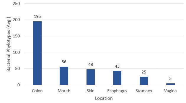
Figure 1. Diversity and distribution of bacterial phylotypes in female human. The distribution of bacterial phylotypes is broad and extensive, with the colon housing the greatest number of distinctive bacteria. These data are not all encompassing and provide a rough estimation of phylotype diversity. Data used for figure construction was obtained from reference (5). mutualism can support why humans have such a prominent population of gut microbiota. It also supports how and why our gut microbiota have stayed with us as humans as a species evolved. Evolution is one way in which to look at the relationship between host and microbe. Evolution requires selection of traits, which are passed on via genes. As it is understood that genes can be affected by environment, so too can a host’s gut microbiota. This environmental impact introduces the utility of an ecological perspective to examine the relationship between host microbiota populations and their respective environments inside the human body. When a human infant is born, they acquire their microbiota from the environment. As the first weeks of life turn into months, multiple species of fauna and flora colonize, diversify, and begin to occupy their specific microbial habitats within the infant body. With time, the ingestion of solid foods facilitates the creation of an individualized microbiome like that of an adult (5). Thus, the bacteria that compose an individualized microbiome are diverse and broadly distributed throughout the human body (Figure 1).

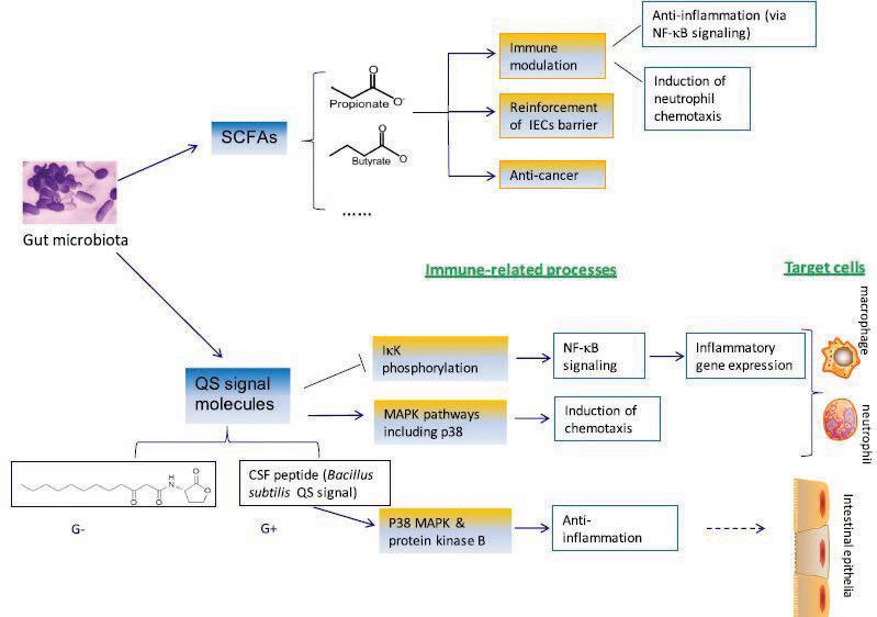
Figure 2. Gut microbial metabolites and effect on host immune responses. Two major avenues eliciting host immune response via microbial metabolites involve short chain fatty acids (SCFAs) and quorum sensing (QS) signaling molecules. This image has been reproduced using original source image from reference (7). CSF: Competence and sporulation factor; G– : Gram-negative bacteria; G+ : Gram-positive bacteria; IECs: Intestinal epithelial cells; IkK: Kinase complex; MAPK: Mitogen-activated protein kinase; NF-kB: Nuclear factor-kappa beta complex.
Microbiota can undergo very drastic changes in response to a change in their environment. For example, a change in the environment of a non-pathogenic microbe can result in host tissue invasion. An immune response is then mounted to eliminate the infection, resulting in the mass death of numerous microbial occupants. These immune defenses introduce selective pressure. To combat death by the immune system in response to a change in environment, the microbe needs to adapt in such a way as to avoid detection from the host immune system. In this case, the microorganism begins adapting toward selection that favors its conversion to a pathogen to conserve fitness (5). These changes highlight the extreme sensitivity and response between the host microbiome and their environment with the human body. Appreciating the importance of evolutionary mechanisms and human ecology regarding the host-microbiota relationship is influential in understand why a disruption in either of these areas could contribute to ailments. An ecological or genetic change that disconnects the human-microbe relationship has the potential to result in serious diseased states.
A closer look: gut microbiota metabolites and dysbiosis As introduced, the community structure of gut bacteria can be intimately related with proper immune system function. Important components of modulation and shaping of the immune system by gut microbiota involve microbial byproducts and dysbiosis, or an unnatural shift in the composition of microbiota. The metabolic byproducts of certain bacterial species can serve to prevent inflammatory autoimmune disease such as IBD, specifically Crohn’s disease and ulcerative colitis. These conditions are classically characterized by increases in pro-inflammatory cytokines, particularly tumor necrosis factor-alpha (TNF-α) and interferon-gamma. More recently, a population of inflammatory T helper lymphocytes has been noted as a factor that could contribute to pathogenesis of human and experimental colitis. These T helper lymphocytes are characterized by the expression of pro inflammatory cytokine interleukin-17 (IL-17) (6). The immunomodulatory effects of products from intestinal bacteria metabolism increases colonic IL-17. Additionally, other types of bacterial-released metabolites, including intermediates or end products of commensal gut bacteria dietary metabolism, are proposed to function in shaping immunity. Lin et al. thoroughly discusses two such metabolites: short chain fatty acid (SCFA) and quorum sensing (QS) signal molecules. There are multiple proposed mechanisms by which these metabolites exert their immunologic effects (Figure 2). The SCFA metabolites include propionate and butyrate. The latter is well known for its anti-inflammatory activities, in which accumulating evidence has shown that butyrate can diminish bacterial translocation across epithelia under stress, as well as improve the gut barrier by reinforcing intestinal epithelial cells. Both SCFAs have demonstrated binding capability to specific G-protein coupled receptors that mediate signal transduction involved in certain inflammatory pathways. Thus, these SCFAs serve to repress inflammation by binding these receptors (7). Quorum sensing—reliant on cell density—is a regulatory mechanism that bacteria use to promote synchronized behavior between and among the bacterial populations. The QS signaling molecules allow for smooth operation of this process, which is utilized by pathogens to facilitate colonization and invasion into a host (7). Thus, it is proposed that QS signals may act as an important weaponry fallback to combat pathogenic encroachment via multiple immunomodulation pathways controlling anti-inflammatory processes, chemotaxis, and inflammatory gene expression.
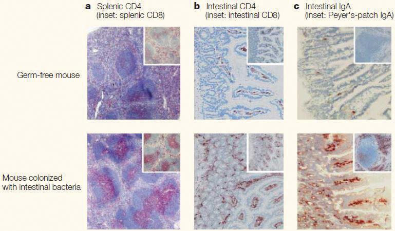
These findings suggest that a dysbiotic event leading to a shift in the proportions of certain bacteria would result in a change in the number and/or frequency of metabolites produced. Theoretically, this change in metabolite production due to dysbiosis could then lead to an immunocompromised state. Alternatively, discussion of how these metabolites hold potential to effect intestinal epithelia cells warrants deeper investigation into their effects on the gut mucosa and mucosal immune system. The degree of development of the mucosal immune system, and the lymphoid structures it contains, differ depending on the absence or presence of commensal bacteria in the gut. Within the mucosa of the small intestine lie Peyer’s patches. These lymphoid tissues are sites in which B and T lymphocytes are activated (8). They are integral members of the mucosal immune system. Histological comparisons of mouse model tissues show evidence that the mucosal immune system is underdeveloped in germ-free (GF) mice: the formation of Peyer’s patches is greatly affected (Figure 3). Additionally, comparisons show a greatly reduced number of immunoglobulin A (IgA)- producing plasma cells and a reduction in CD4+ T lymphocytes in the GF mice. This is a significant observation; IgA functions in protecting mucosal surfaces against pathogenic and non-pathogenic microorganisms (9). However, these immunologic abnormalities are not solely limited to the mucosal immune system. Splenic tissue in association with the systemic immune system also shows considerable changes upon comparison between the two mouse models. The tissue sample from the GF mouse shows poorly formed T and B lymphocytes zones. These observations provide a more concrete understanding of how dysbiosis in commensal microbiota can shape the immune system.
Genetic blemishes and regulatory anomalies Furthering the discussion about bacterial impact on the mucosal immune system is the relationship between bacteria, genetics, and mucosal function. Activation of immune and inflammatory responses can be initiated by stimulating the luminal flora and/ or their products. It has also been suggested that genetically determined variations in key mucosal functions, including cell activation by bacterial pattern molecules, leads to differing immune function and susceptibility to IBDs (10). Pattern recognition receptors (PRRs) play a pivotal role in mediating host immune processes by initiating inflammatory responses upon recognition of a microbial molecule during infection. However, an issue exists in this detection-altering system: PRRs are not discriminatory between ligands produced from pathogenic bacteria and ligands produced from a resident microbiota upon colonization (11). Accordingly, and perhaps helping to clarify the histological abnormalities discussed above, recent studies reveal that these gut bacterial ligands from resident microbiota signal through PRRs to promote host tissue and immune development, thereby facilitating protection from disease (11). There are additional findings
that affirm the relevance of how gut microbiota shape the immune system by exploiting host genetic defects. One such theory contends that Crohn’s disease and ulcerative colitis are the result of continuous microbial antigenic stimulation of pathogenic immune responses because of host genetic mutations in mucosal barrier function, innate bacterial killing, or immunoregulation (12). Furthermore, polymorphisms in the host genome are said to likely interact with functional bacterial changes — perhaps utilizing PRR signaling — to stimulate aggressive immune responses leading to chronic tissue injury (12). Thus, even when gut microbiota are themselves functioning as they should in an environment devoid of dysbiosis, they can still have devastating impacts on host immune function by interacting with the genetic shortcomings that affect pattern recognition, mucosal barrier, killing of innate bacteria, and perhaps least surprisingly, genetic imperfections associated with immunoregulation. Having an established genetic program in the immune system unquestionably aids in proper immune function. To reiterate how an immune system piloted by an ill-functioning genetic program can produce disease driven by gut microbiota, Garrett et. al. examined how communicable ulcerative colitis was induced by an immune cell-specific T-box transcription factor (Tbet) deficiency. Tbet orchestrates inflammatory genetic programs in both adaptive and innate immunity by regulating TNF-α, which drives tissue injury in experimental mouse model colitis. Tbet also fosters harmony between eukaryotes and prokaryotes by affecting the behavior of the colonic epithelium (13). Their study demonstrated that mice deficient of Tbet within the innate immune system developed spontaneous ulcerative colitis, inferring the possibility that episodes of spontaneous ulcerative colitis can be significantly influenced by a compromised immune system because of a malfunctioning colonic environment (and the bacteria that are affected by such
an environment). Moreover, the transfer of the gut microbiota from the affected mice into wild-type recipients induced the disease, providing additional support for this assertion. These results spurred investigation into how the microbiota have effects on the functioning and/or production of other regulatory mechanisms associated with immune response and function. Using a mouse model, Ishikawa et al. provide results that help clarify thoughts of how intestinal microbiota effect the production of cells that function to contribute to immune response, thus shaping the immune system. Oral tolerance is a local and systemic immune unresponsiveness to orally ingested antigens (14). It plays a critical Figure 3. Impact of intestinal bacterial colonization on lymphoid structures in intestinal and role in immune defense, specifically in the systemic tissues from mouse model. Histological comparison of splenic and intestinal tissue gastrointestinal mucosa. Failure of oral sections taken from germ-free wild-type mouse and a mouse of the same strain colonized tolerance induction leads to susceptibility with intestinal bacteria; a: Germ-free mice have poorly formed T cell (pink) and B cell zones in to indigenous bacterial antigens in the splenic tissue; b: Low number of CD4+ cells (brown) from lamina propria are seen in the germ- gastrointestinal tract, potentially leading free mouse tissue; c: Intestinal tissue from the germ-free mouse demonstrates a conspicuous to IBD. It is reported that oral tolerance decrease in the number of IgA producing cells (brown) compared to colonized mouse. This image was reproduced using its original source from reference (8). IgA: Immunoglobulin A. can be maintained by regulatory T (Treg) lymphocytes by facilitating the characteristic hypo-responsiveness to the orally ingested antigen. Specifically, CD25+ CD4+ Treg lymphocytes have been associated with this phenomenon due to their production of the inflammatory suppressive cytokine, transforming growth factor-beta (15). Comparing specific pathogen free (SPF) mice to GF mice, Ishikawa et. al. observed that experimental GF mice seemed to be resistant to oral tolerance. It was concluded that both the frequency and total number of CD25+ CD4+ Treg lymphocytes in the GF mice were significantly lower than those in the SPF mice. Furthermore, cells from the mesenteric lymph node — the reported site where CD25+ CD4+ Treg are generated — in SPF mice suppressed proliferation of certain T lymphocytes that produced pro-inflammatory cytokines in comparison of those from GF mice (15). Thus, the study provides evidence that the microbiota play an important role in generating fully functional CD25+ CD4+ Treg lymphocytes. The absence or incomplete functioning of these regulatory cells, due in part to the absence of a microbial population, has potential to lead to increased susceptibility and altered immune response against indigenous intestinal bacterial antigens, contributing to inflammatory autoimmune diseases such as IBD. Resident gut-microbiota and non-intestinal related autoimmune disease Much of this review thus far has revolved around IRADs (Crohn’s disease and ulcerative colitis), which can be facilitated by an altered immune system due to gut microbiota. However, to more fully appreciate the far-reaching impact that gut commensals have on the immune system, it is necessary to investigate how they can affect systemic immunity. Information previously referenced demonstrates how gut microbiota hold potential to contribute to non-intestinal autoimmune diseases, specifically Hashimoto’s thyroiditis, Type 1 diabetes mellitus (T1D), and multiple sclerosis (16).

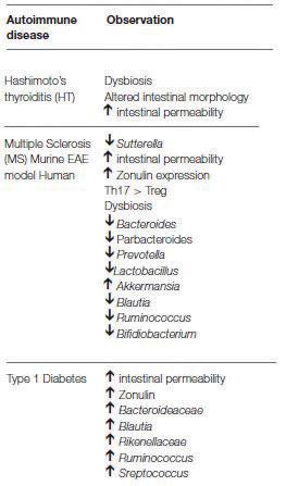
Hashimoto’s thyroiditis is characteristically identified by the infiltration of mononuclear cells in the thyroid along with production of autoantibodies against thyroglobulin and thyroid peroxidase (17). It has been documented that the transfer of microbiota from conventional rats into SPF rats increased the susceptibility of the SPF rats to experimental autoimmune thyroiditis (18). This finding provides evidence that supports the influence of the microbiota on the pathogenesis of Hashimoto’s thyroiditis. Furthermore, to reiterate the effects of dysbiosis and epithelial tissue barriers in the gut, it has been demonstrated that dysbiosis within the microbial environment can manifest as a condition known as “leaky gut” (19). From a histological perspective, leaky gut is characterized by changes in the epithelial cells of the gut, as well as lymphocyte infiltration. Notably, similar observations have been made upon examination of tissues from patients with Hashimoto’s disease (20). T1D is one of the best known and most researched non IRADs. It is characterized by CD4+ and CD8+ T lymphocyteguided destruction, targeting the insulin-storing and releasing beta cells of the pancreas (21). While there is a hereditary component associated with the pathogenesis of T1D, environmental factors — such as cesarean or vaginal birth — can also contribute to the induction of the disease (22). As mentioned, the resident microbiota population is established by the host environment after birth. Results from Endesfelder et al. provided evidence that the induction of T1D can be attributed by intestinal microbiota by their priming of the immune system at an early postnatal period (23). Additionally, a study of Italian children affected by T1D showed an increase in their intestinal permeability that was correlated with modifications in their resident gut microbiota population. Moreover, the same study found three very highly represented microbial biomarkers in the affected children when compared to healthy control children (24). As a final point, and perhaps most striking, is the observation that the entire Bacteroidaceae family of bacteria, as well as two other prominent species, were significantly disproportionately represented in children with T1D (Table 1) (25). Lastly, multiple sclerosis is the most frequently seen demyelinating disease in which the immune system attacks the protective coverings of nerve cells. Like the findings in children with T1D, it was found that multiple sclerosis patients could have a specific type of microbiome due in part to dysbiosis. Studies found abnormally low proportions of some gut specific species while other species were present in abnormally high amounts (26, 27). Importantly, samples of microbiome from patients affected by multiple sclerosis impaired the ability of certain T lymphocytes to properly differentiate into Treg cells (28). As evidenced, this is considerably impactful to the immune system since the proper functioning of these cells are critical in moderating effective and controlled immune responses. In relation to Tregs and the prior discussion of the effect of certain microbial metabolites on immune function, important evidence was derived using an autoimmune encephalomyelitis (AE) model. This model, mediated by T lymphocytes, is an animal model used to study multiple sclerosis due in part to its ability to reproduce most of the symptoms observed in people who have multiple sclerosis (29). Interestingly, orally administered propionate (a SCFA which was one of the bacterial metabolic byproducts discussed earlier) decreased AE clinical scores and increased Tregs at lymph nodes (30). These findings are particularly promising because they demonstrate once more how metabolic products from gut bacteria metabolism could potentially contribute to therapies for multiple sclerosis and other non-intestinal autoimmune diseases.
Conclusion
There have been major strides in research that have focused on identifying specific species of gut microbiota and how these species have evolved to reside in their respective environments within the host. However, clinical practice might benefit from research that focuses more heavily on examining the habitats of the human body and how these environments change over time and vary depending on an individual’s diet and genetic predispositions. This could improve care by allowing health care providers to better anticipate the consequences that a changing

Table 1. Changes observed in microbiota for associated autoimmune diseases. Significant changes in bacterial colonization were observed in multiple non-IRADs. This table was reproduced using material in original source from reference (16). IRADs; Intestinal related autoimmune diseases.
environment might have on our microbiota, and the associated impact on host immunity and severity of disease pathogenesis. By investigating these symbiotic “landscapes” with research focused on examining the ecology of human health, one gains better insight into the relationship between humans and their acquired microorganism populations and the diseases that present when this relationship goes awry. There have also been major advances in related research from a more macroscopic level that focuses on microbial communities and the relationship they have with their host environment, as well as their impact on mucosal and systemic immunity. As discussed, these communities can exploit host genetic defects to alter immune function and contribute to disease. By conducting research that focuses on identifying the polymorphisms that lead to host-microbial alterations, researchers and care providers can work in conjunction to use selective targeted interventions that correct the identified polymorphisms underlying these abnormalities. It is hopeful that these methods would provide improved therapies for treatment of autoimmune diseases via precision medicine. This domain of research requires disciplined and precise methods of examining and obtaining samples of gut microbiota and surrounding tissue. Thus, it is important to consider the technical and ethical limitations that exist when obtaining samples from humans to be used in research. The use and relevance of human studies are clear. However, obtaining samples to be used can be laborious, time consuming, and costly due to the limitations listed above. Focused experimental model systems could be used to obtain preliminary results and findings, guiding researchers towards practicality of methods in humans. Experimental model systems, such as the mice and rats mentioned above (which have experimentally induced and specific autoimmune diseases or malfunction), can fill this role by conserving evolutionary features that are likely to be crucial for function. Model systems would also allow for improved understanding of multiple microbial mechanisms that lead to the same immunologic outcome without compromising precious human samples. Additionally, it could give insight into the evolutionary relationship of host-microbe interaction by stratifying experimentation based on species complexity. Abiding by the ethical regulations set forth to protect animals and their utility in biomedical research is critically essential and must be vigilantly monitored. Nonetheless, results could provide a clearer understanding of microbial interactions with the immune system that could then be explored using human tissue in a more focused, precise, and efficacious manner.
Acknowledgments
I would like to thank Jennifer Boardman, PhD, for her advice and guidance provided throughout the writing process.
Disclosures
The author has no financial affiliations to disclose from sponsors or organizations/associations.
References
1. Ley RE, Peterson DA, Gordon JI. Ecological and evolutionary forces shaping microbial diversity in the human intestine. Cell. 2006;124(4):837-48. 2. Frank DN, St Amand AL, Feldman RA, Boedeker EC, Harpaz
N, Pace NR. Molecular-phylogenetic characterization of microbial community imbalances in human inflammatory bowel diseases. Proc Natl Acad Sci USA. 2007;104(34):13780-5. 3. Ng SC, Shi HY, Hamidi N, Underwood FE, Tang W,
Benchimol EI, et al. Worldwide incidence and prevalence of inflammatory bowel disease in the 21st century: a systemic review of population-based studies. Lancet. 2018;390(10114):2769-78. 4. Leray E, Moreau T, Fromont A, Edan G. Epidemiology of multiple sclerosis. Revue neurologique. 2016;172(1):3-13. 5. Dethlefsen L, McFall-Ngai M, Relman DA. An ecological and evolutionary perspective on human-microbe mutualism and disease. Nature. 2007;449(7164):811-8. 6. Round JL, Mazmanian SK. The gut microbiome shapes intestinal immune responses during health and disease.
Nature reviews Immunology. 2009;9(5):313-23. 7. Lin L, Zhang J. Role of intestinal microbiota and metabolites on gut homeostasis and human diseases. BMC immunology. 2017;18(1):2. 8. Macpherson AJ, Harris NL. Interactions between commensal intestinal bacteria and the immune system.
Nature reviews Immunology. 2004;4(6):478-85. 9. Macpherson AJ, Hunziker L, McCoy K, Lamarre A. IgA responses in the intestinal mucosa against pathogenic and non-pathogenic microorganisms. Microbes and infection. 2001;3(12):1021-35. 10. Podolsky DK. The current future understanding of inflammatory bowel disease. Best practice & research clinical gastroenterology. 2002;16(6):933-43. 11. Chu H, Mazmanian SK. Innate immune recognition of the microbiota promotes host-microbial symbiosis. Nature immunology. 2013;14(7):668-75. 12. Sartor RB. Microbial influences in inflammatory bowel diseases. Gastroenterology. 2008;134(2):577-94. 13. Garrett WS, Lord GM, Punit S, Lugo-Villarino G,
Mazmanian SK, Ito S, et al. Communicable ulcerative colitis induced by T-bet deficiency in the innate immune system.
Cell. 2007;131(1):33-45. 14. Commins SP. Mechanisms of oral tolerance. Pediatric clinics of North America. 2015;62(6):1523-9. 15. Ishikawa H, Tanaka K, Maeda Y, Aiba Y, Hata A, Tsuji NM, et al. Effect of intestinal microbiota on the induction of regulatory CD25+ CD4+ T cells. Clinical experimental immunology. 2008;153(1):127-35. 16. Opazo MC, Ortega-Rocha EM, Coronado-Arrazola
I, Bonifaz LC, Boudin H, Neunlist M, et al. Intestinal microbiota influences non-intestinal related autoimmune diseases. Frontiers in microbiology. 2018;9:432.
17. Antonelli A, Ferrari SM, Corrado A, Di Domenicantonio
A, Fallahi P. Autoimmune thyroid disorders. Autoimmune reviews. 2015;14(2):174-80. 18. Penhale WJ, Young PR. The influence of the normal microbial flora on the susceptibility of rats to experimental autoimmune thyroiditis. Clinical and experimental immunology. 1988;72(2):288-92. 19. Vaarala O, Atkinson MA, Neu J. The “perfect storm” for type 1 diabetes: the complex interplay between intestinal microbiota, gut permeability, and mucosal immunity.
Diabetes. 2008;57(10):2555-62. 20. Sasso FC, Carbonara O, Torella R, Mezzogiorno A,
Esposito V, deMagistris L, et al. Ultrastructural changes in enterocytes in subjects with Hashimoto’s thyroiditis. Gut. 2004;53(12):1878-80. 21. Atkinson MA, Eisenbarth GS, Michels AW. Type 1 diabetes.
Lancet. 2014;383(9911):69-82. 22. Rewers M, Ludvigsson J. Environmental risk factors for type 1 diabetes. Lancet. 2016;387(10035):2340-8. 23. Endesfelder D, Engel M, Zu Castell W. Gut immunity and type 1 diabetes: a mélange of microbes, diet, and host interactions? Current diabetes reports. 2016;16(7):60. 24. Maffeis C, Martina A, Corradi M, Quarella S, Nori N,
Torriani S, et al. Association between intestinal permeability and faecal microbiota composition in Italian children with beta cell autoimmunity at risk for type 1 diabetes. Diabetes/ metabolism research and reviews. 2016;32(7):700-9. 25. De Goffau MC, Luopajarvi K, Knip M, Ilonen J, Ruohtula
T, Harkonen T, et al. Fecal microbiota composition differs between children with beta-cell autoimmunity and those without. Diabetes. 2013;62(4):1238-44. 26. Jangi S, Gandhi R, Cox LM, Li N, von Glehn F, Yan R, et al. Alterations of the human gut microbiome in multiple sclerosis. Nature communications. 2016;7:12015. 27. Freedman SN, Shahi SK, Mangalam AK. The “gut feeling”: breaking down the role of gut microbiome in multiple sclerosis. Neurotherapeutics. The journal of the
American Society for Experimental Neurotherapeutics. 2018;15(1):109-25. 28. Cekanaviciute E, Yoo BB, Runia TF, Debelius JW, Singh
S, Nelson CA, et al. Gut bacteria from multiple sclerosis patients modulate human T cells and exacerbate symptoms in mouse models. Proc Natl Acad Sci USA. 2017;114(40):10713-8. 29. Stromnes IM, Goverman JM. Active induction of experimental allergic encephalomyelitis. Nature protocols. 2006;1(4):1810-9. 30. Mizuno M, Noto D, Kaga N, Chiba A, Miyake S. The dual role of short acid chains in the pathogenesis of autoimmune disease models. PLoS One. 2017;12(2):e0173032.


