4 minute read
M.Ortolani Development of a 0.6 THz Reflection Microscope for Dermatology
Development of a 0.6 THz Reflection Microscope for Dermatology.
A. A. Tanga1,2, V. Giliberti3, M. Ortolani2, and F. Vitucci1 1Crisel Instruments srl, 00167 Rome, Italy 2Dipartimento di Fisica, Sapienza Università di Roma, 00185 Rome, Italy 3Center for Life NanoSciences, Istituto Italiano di Tecnologia, 00166 Rome, Italy
Advertisement
Abstract—A terahertz microscope working at specular oblique reflection angle between 30° and 70° has been developed. The operation frequency of 0.6 THz has been selected for anomaly detection under the skin surface of dermatology patients in vivo.
I. INTRODUCTION
HERE is a long-standing interest in the development of terahertz microscopy techniques for the in-vivo T observation of anomalies below the skin surface. Existing optical techniques, such as epiluminescence and optical coherence tomography are based on visible and near-infrared light and, as such, can penetrate one hundred microns at most. Non-optical techniques based e.g. on ultrasounds can certainly penetrate in-depth, but a clear density-contrast mechanism for the identification of anomalies in the derma and/or in the deep layers of epidermis by ultrasound echoes is still lacking.
Terahertz radiation, in particular in the band 0.4-0.7 THz [1], can penetrate several hundreds of m under the skin surface (depending on local hydration and structure of the skin) mainly because it is not prone to scattering by skin surface roughness [2], due to its long wavelength around 0.5 mm.The future implementation of super-resolution schemes [3]may provide terahertz microscopes with a lateral resolution of the order of 10 m, but, in confocal arrangements [4], the image would be taken in the deep layers of the skin.
In this work we present the design, development and test of a variable-angle oblique-incidence reflection microscope operating at 0.6 THz. First results are obtained on skin simulants made of 3D-printed plastic structures embedded in a collagen gel matrix.
II. MICROSCOPE
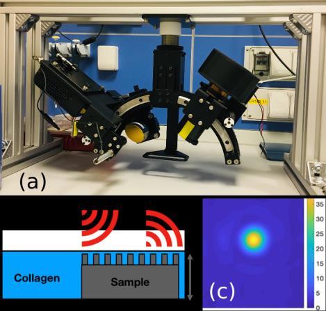
The microscope has been assembled by buying commercial Terahertz components (source, camera and optics) and by designing and then constructing the external mechanical structure. A frequency-multiplied microwave generator (Teraschottky600 by Lytid SaS, France) has been used as the Terahertz source, tunable in the 570-630 GHz range, and capable of emitting a continuous-wave output power around 2 mW. A microbolometer camera with a bandbass filter centered at 600 GHz is employed (TZcam by i2s, France) is used in conjunction with an objective formed by two TPX lenses (by Tydex, Russia) mounted in a tube. Gold-coated parabolic mirrors (Gestione SILO, Italy) are used to focus the radiation produced by the source and emitted by a diagonal horn antenna, onto the sample. The system works at specular reflection angle, variable between 30° and 70°, by displacing both the camera and the source along arc-shaped rails (Fig. 1a). The sample is fixed and the focus is scanned via motors controlling the parabolic mirror position and orientation, as the final aim is to use the system in-vivo.
III. EXPERIMENT
Pure collagen from pork skin is first prepared in liquid form by dissolving solids in water. Skin simulant structures made of different hard plastic disks, with their surface patterned with a laser writer machine, are embedded into the collagen matrix up to a precisely measure height (see Fig. 1b). the collagen matrix is then let to dry for approximately 1 hour, and a gel is formed, embedding the 3D structured plastic disc.
In the experiment, the image of the reflected spot is recorded by the THz camera (Fig. 1c). The eccentricity of the spot is calculated by standard image analysis algorithms (MatLab Region Props) in order to retrieve preferential orientation of the 3D-printed structures. The thickness of the collagen film on top of the plastic disk has been kept between 100 and 600 m, beyond the reach of epiluminescence.
Fig. 1. (a) photograph of the assembled microscope (source with parabolic mirrors on the left arc rail, camera with lens tube on the right arc rail). (b) schematic of the skin simulant sample. (c) image of the reflected spot. IV.SUMMARY
A reflection microscope operating around 600 GHz at specular oblique angle has been developed. Skin simulant samples made of collagen gel matrix and plastic are under test for future dermatology applications.
REFERENCES
[1]. Z. Taylor IEEE trans on Thz Sci Technol. [2]. J. Wang et al., TH in i o mea emen : he effec of e e on kin eflec i i Biomedical Optics Express [3]. M. Flammini, E. Pontecorvo, V. Giliberti, C. Rizza, A. Ciattoni, M. Ortolani, and E. DelRe, Evanescent-Wave Filtering in Images Using Remote Terahertz Structured Illumination Phys. Rev. Applied vol. 8, 054019, 2017 [4]. C. Ciano, M. Flammini, V. Giliberti, P. Calvani, E. DelRe, F. Talarico, M. Torre, M. Missori and M. Ortolani, Confocal Imaging at 0.3 THz with depth resolution of a painted wood artwork for the identification of buried thin metal foils IEEE Trans. THz Sci. Tehcnol. vol. 8, pp. 390-396, 2018


095


