Case 178
Different than the other case presentations, shown will be 3 cases of the same diagnosis. For many, the diagnosis may not be too much of a dilemma, but the purpose is to show the varying appearances of this entity.


Findings
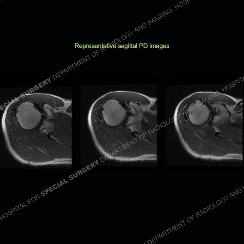
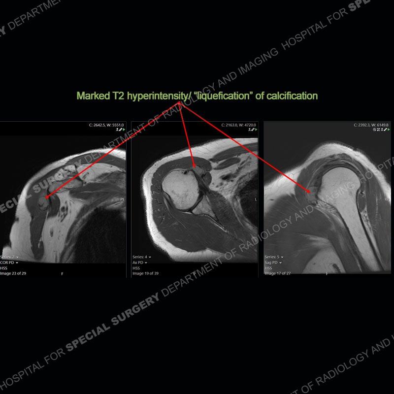
In all cases there are amorphous areas of calcification about the shoulder girdles with varying degrees of density. Varying degrees of edema are seen in the soft tissues on the corresponding MRI examinations. Particularly in case 2, well seen is a change in the density of the calcification from dense to markedly less dense on the radiographs. On the later MRI of case 2, the areas of calcification look almost “liquefied” with surrounding prominent edema in the soft tissue.


Diagnosis: Hydroxyapatite Deposition Disease (HADD)

HADD is of unknown etiology. It frequently deposits in the soft tissue around joints or occasionally in joints. It is most commonly presents in middle aged patients. The shoulder is the most common area of deposition, but other locations are frequently found such as at the pectoralis insertion and of the linea aspera of the femur. Although perhaps not a diagnostic dilemma for many, these cases show the varying architecture of HADD. The deposition typically progresses through stages. A silent phase produces well demarcated and dense calcification with little surrounding edema. These patients may be asymptomatic or have little symptoms. A mechanical or resorptive phase may follow and this produces the greatest degree of symptoms. Pain can be excruciating and patients may not want to move the extremity at all.
On imaging, the calcification becomes less dense and may have a “liquefied” appearance on MRI. On MRI, there is often a marked degree of edema accounting for the patient’s symptoms. Following this, some patients go into a late stage or post calcific stage where there may be some or no pain. Most situations can be treated with oral medication, injection, and aspiration. Occasionally, surgical intervention becomes warranted.
References

Hydroxyapatite crystal deposition disease. Glenn M Garcia, Gary C McCord, Rajendra Kumar. Semin Musculoskelet Radiol. 2003 Sep;7(3):187-93. doi: 10.1055/s-2003-43229.
Calcium hydroxyapatite deposition disease: Imaging features and presentations mimicking other pathologies. Patcharee Hongsmatip, Karen Y Cheng, Christopher Kim, David A Lawrence, Robert Rivera, Edward Smitaman. Eur J Radiol. 2019 Nov;120:108653. doi: 10.1016/j.ejrad.2019.108653. Epub 2019 Sep 8.
