

Findings
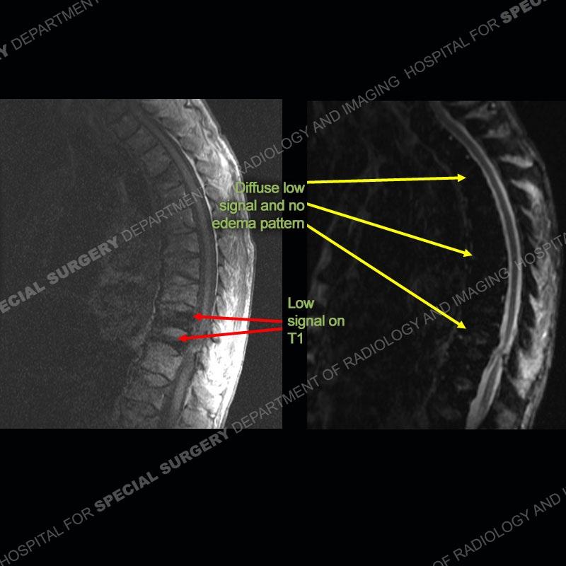
Radiographs of the thoracic and later lumbar spine demonstrate osteoporosis and a wedge compression deformity of at least one lower thoracic vertebrae. On the MRI, the T1 images demonstrate marked low signal of two lower thoracic vertebrae. On the IR sequence, there is diffuse low signal of the bone without any areas of marrow edema pattern. The subsequent bone scan demonstrates increased radiotracer uptake of the two lower thoracic vertebrae. Of important note on the coronal T2 images, there is markedly low signal of the liver and spleen.


Diagnosis: Hemochromatosis (HCT)

This case was utilized to demonstrate underlying hemochromatosis. The patient presented with thoracic wedge compression fractures which may relate to underlying senile osteoporosis or hemochromatosis. HCT can be primary where it is caused by a genetic disorder precipitating abnormal iron deposition in parenchymal cells of preferentially the liver, pancreas, heart, pituitary, thyroid, and synovium. Alternatively, HCT can be secondary where there is iron deposition in tissue related to parenteral iron administration say in the setting of multiple transfusions. In the secondary situation, the deposition is typically of the liver, spleen, and marrow. This iron deposition accounts for the markedly low T2 signal of the liver and spleen on the MR images.
With iron deposition in the marrow, the normal elements are displaced which can produce generalized osteoporosis or focal cyst like areas of the bone. Classic findings of HCT are chondrocalcinosis and hook-like osteophytes at the metacarpal heads. If the iron is deposited in the synovium, premature arthropathy can ensue.
Diagnosis Continued:

This case demonstrates particularly the effect of the iron on the MR imaging. To a certain degree, the T1 and T2 signal of the bone is somewhat decreased but especially the IR signal of the bone is markedly decreased.
Although not well discussed in the literature, when blood product or iron is deposited in the bone, on IR sequences there is a marked low signal of the bone. It can be so much so that even when a fracture is present, no edema pattern can be seen. In this case, the marked low signal of the lower thoracic vertebrae on T1 corresponds to fractures which show increased uptake on the bone scan but no edema pattern on the IR image. This case highlights that simply utilizing the signal on the MRI is not enough. Both morphometry and signal must be utilized and on all pulse sequences to appreciate the pathology.
References
Marcony Queiroz-Andrade, Roberto Blasbalg, Cinthia D. Ortega, Marco A. M.
Rodstein, Ronaldo H. Baroni, Manoel S. Rocha, Giovanni G. Cerri.
MR Imaging Findings of Iron Overload. RadioGraphics Vol. 29, No. 6. Oct 1
2009 https://doi.org/10.1148/rg.296095511

