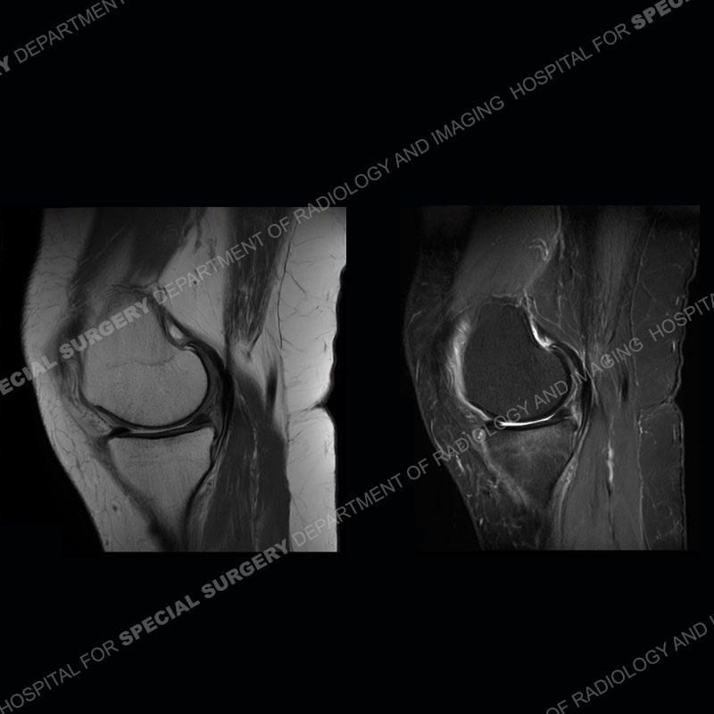


Findings
Unfortunately, no radiographs were available for this case. On the MRI, there is a defect of the posterior horn medial meniscus (PHMM) towards the tibial attachment, the medial meniscus is extruded into the medial gutter (extending 3mm or more into the medial gutter relative to the plateau margin), and there is edema/subchondral sclerosis of the medial tibial plateau. A “ghost” meniscus is seen particularly on the sagittal images where no meniscal tissue is present. In the normal situation, the PHMM can normally be identified on the image adjacent to the PCL on the sagittal images. On these images, there is a defect at this location.


Diagnosis: PHMM Root Tear and Subchondral Fracture
The menisci are attached to the tibia at their root attachment sites. All of these are important, but the most important of these involves the PHMM. When the root attachment is disrupted it renders marked compromise to the function of the medial meniscus. The tear causes a loss of the hoop strength of the meniscus and associated extrusion as seen on this case. The meniscus now also sees markedly decreased loads and the force becomes transmitted to the subchondral bone and the overlying cartilage. Cartilage loss and osteoarthritis frequently develop and often quite rapidly. As seen in this case, subchondral fractures may also be precipitated. In the younger patient population, a meniscal repair via transtibial pullout can be attempted. In the older patient population, these injuries frequently require some type of arthroplasty to be performed.

References

LaPrade RF, Floyd ER, Carlson GB, Moatshe G, Chahla J, Monson JK.
Meniscal root tears: Solving the silent epidemic. J Arthrosc Surg Sports Med 2021;2(1):47-57.
Andrew R. Palisch, Ronald R. Winters, Marc H. Willis, Collin D. Bray, Theodore B. Shybut. Posterior Root Meniscal Tears: Preoperative, Intraoperative, and Postoperative Imaging for Transtibial Pullout Repair.
RadioGraphics 2016 36:6, 1792-1806.
Santiago Pache, MD, Zachary S. Aman, BA, Mitchell Kennedy, BA, Gilberto Y. Nakama, MD, Gilbert Moatshe, MD, Connor Ziegler, MD, Robert F. LaPrade, PhD. Meniscal Root Tears: Current Concepts Review. Arch Bone Jt Surg. 2018 Jul; 6(4): 250–259.
