

Findings
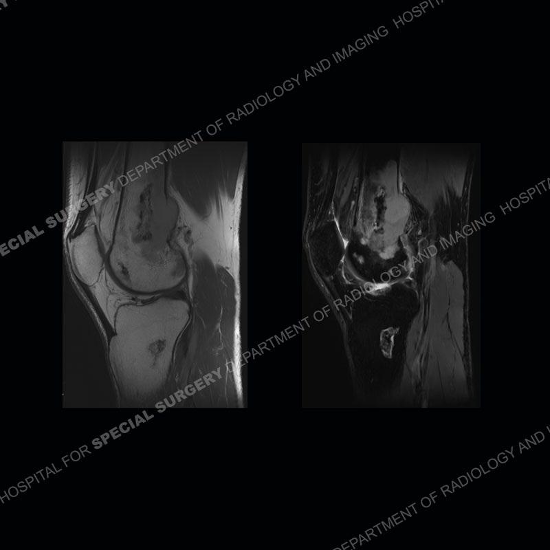
MRI of 2023 demonstrates geographic, serpiginous areas with peripheral double line sign on T2 weighted imaging characteristic of bone infarctions. The central areas mostly demonstrate medullary fat signal. An area of intermediate signal with mass lesion is present along the posterior aspect of the infarct of the distal femur.
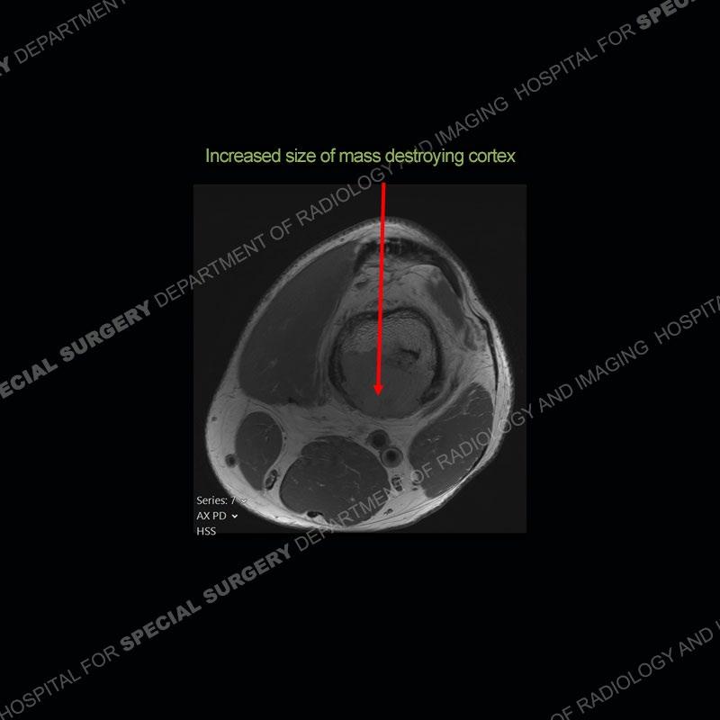
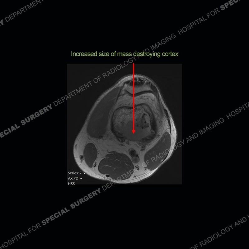
Subsequent imaging of 2024 demonstrates increased size of the mass lesion about the infarct of the distal femur with destruction of the posterior cortex.


Diagnosis: Sarcomatous Transformation of Medullary Infarction
Bone infarcts are fairly common to find and for the most part are asymptomatic. These lesions most typically become symptomatic when they extend to the end of the bone and are associated with subchondral fracture and collapse. Complications of infarct such as infection or sarcomatous transformation are known to occur but because they are extremely rare, exact incidence is not known. Interestingly, most reported cases of sarcomatous transformation occur in middle aged men and about the knee as in this case. Sarcomatous transformation of an infarct can be suggested on radiographs if the infarct goes from being well defined to less well defined. On cross sectional imaging, a destructive mass, as seen in this case, is seen to take the place of the otherwise typical appearance of an infarct. The sarcoma most commonly is an MFH ( more recently delineated as UPS or undifferentiated pleomorphic sarcoma) or osteosarcoma. These sarcomas unfortunately carry a very poor prognosis.

References
Stacy Gregory Scott, Lo Ryan, Montag Anthony. Infarct-Associated Bone Sarcomas: Multimodality Imaging Findings. AJR American Journal of Roentgenology. Volume 205, Issue 4 https://doi.org/10.2214/AJR.14.13871
Alhamdan HA, Alrifai OI, Shaheen MF, Pant R, Altayeb MA. Bone infarct transformation into undifferentiated pleomorphic sarcoma in sickle cell disease: A case report. Int J Surg Case Rep. 2020;77:243-248. doi: 10.1016/j.ijscr.2020.11.004. Epub 2020 Nov 4. PMID: 33189003; PMCID: PMC7672249.
Gaucher, A.A., Regent, D.M., Gillet, P.M. et al. Case report 656. Skeletal Radiol. 20, 137–140 (1991). https://doi.org/10.1007/BF00193829

