
5 minute read
by Julia Furtner and Matthias Preusser


Julia Furtner, MD, PhD, MBA, Associate Professor
Advertisement
Department of Biomedical Imaging and Image-guided Therapy (Division of Neuroradiology and Musculoskeletal Radiology), Medical University of Vienna, Austria Univ.-Prof. Dr. Matthias Preusser, Head - Division of Oncology
Department of Medicine I, Medical University of Vienna, Austria
In the area of personalized therapeutic planning, stratification of glioblastoma patients based on various prognostic factors is essential either for routine patient management decision-making or clinical trial participation . Whereas most of these parameters such as histopathological and molecular characteristics, age, tumor size and localization are objectively assessable, the physical condition of the patient is based mainly on the subjective evaluation of the attending physician . Thus, the use of objective parameters to measure physical performance, such as skeletal muscle mass estimation, has recently become more common .
Methods estimating skeletal muscle mass The reduction of skeletal muscle mass and function is defined as “sarcopenia” and was firstly described by Baumgartner et al . assigned to aging . Moreover, secondarily- induced sarcopenia in various disease entities, such as chronic inflammatory diseases or cancer, has been recognized in the last few years to be an objective parameter indicating a poor prognosis due to patient frailty .
Methods such as dual X-ray absorptiometry, bioelectrical impedance analysis, or the area of lumbar skeletal muscles surveyed by computed tomography or magnetic resonance imaging (MRI) studies of the abdomen are established to estimate the skeletal muscle mass . However, in glioblastoma patients, radiological examinations of the abdomen are generally not routinely available .
In recently published studies the thickness of the temporal muscle, a muscle used for biting on each side of the head, has been shown to have a high association with the area of lumbar skeletal muscles, representing an estimation of the skeletal muscle mass, as well as with the grip strength of a person’s dominant hand, indicating a measure of the muscle function and the nutrition status of patients . Thus, the temporal muscle thickness has been proposed to be a potential marker by which to identify patients at risk for sarcopenia . This is particularly advantageous for patients with neurological or neurooncological diseases, as the thickness of the temporalis muscle can be directly determined on the basis of routine MRI examinations of the brain without prolonging the scanning duration and it helps to avoid additional examinations or a possible increase of the radiation dose for patients, leading to reduced health care costs .
Temporal muscle thickness as a prognostic marker The temporal muscle has previously been proven to have the ability to predict outcome in various disease entities . In trauma patients or children with nonsyndromic craniosynostosis (the premature closure or fusion of the open areas between the skull plates in an infant’s skull, the cause of which is unknown) it was associated with hospital-based clinical outcome markers, such as ventilator days or the
Dr Julia Furtner Dr Matthias Preusser

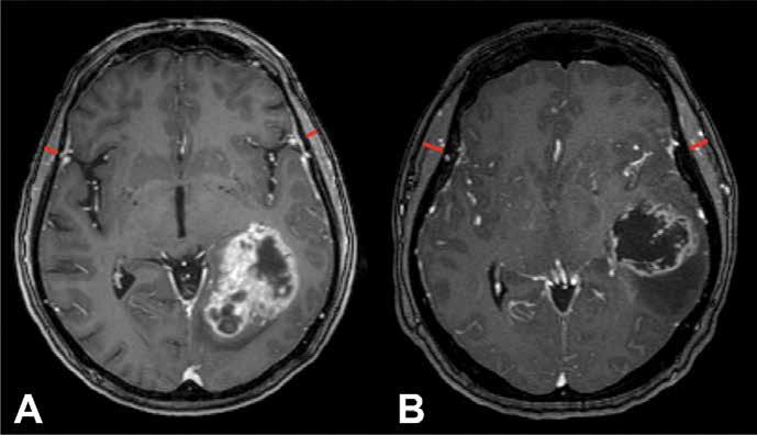
Figure 1A shows a 62-year-old glioblastoma patient at risk for sarcopenia at the time of diagnosis (temporal muscle thickness value below the sex-specific cut-off) with a significantly lower overall survival in comparison to a 63-year-old glioblastoma patient with a normal muscle status presented in Figure 1B.
lengths of the hospital stay . Recently, the thickness of the temporal muscle has been established as an independent outcome prognostication parameter in the field of neuro-oncology by identifying patients at risk for sarcopenia . In this area it showed promising results when associated with the survival of patients with brain metastases of various primary tumor types and primary CNS lymphoma . Moreover, it has also been demonstrated that the temporalis muscle thickness is an independent prognostic marker in patients with newly diagnosed glioblastoma, independent of the MGMT-promoter methylation status in large multicenter international trials (see Figure 1) .
Furthermore, among glioblastoma patients already at risk for sarcopenia at the time of diagnosis, the extent of temporal muscle wasting after chemoradiotherapy was linked inversely with overall survival, but not in patients with normal skeletal muscle status . Interestingly, the temporal muscle thickness showed no correlation with the body-mass-index and only a weak association with age in these patient cohorts . This may be based on the fact that the body mass index focuses on the patient`s weight instead of the body composition and is therefore not able to identify a reduced muscle mass in an obese patient . In terms of the weak association between a patient’s age and the survival prediction, the physical condition of the glioblastoma patients revealed more information than the patient`s chronological age . Similar results have also been shown in large multicenter international trials in recurrent glioblastoma patients .
Impact of implementing the temporal muscle thickness in the clinical workflow When implementing the temporal muscle thickness in routine clinical workflow, it could be used to provide an initial overview of skeletal muscle mass and function . If a patient shows a temporal muscle thickness below the sex-specific cut-off value further testing will be required to confirm the diagnosis of sarcopenia . These measurements should be taken into account at the diagnosis as well as on a regular basis in the follow-up MRI examinations to monitor the muscle status in a longitudinal manner .
If detecting patients with newly developing sarcopenia or patients whose conditions worsen under the course of their disease, the implementation of appropriate strategies for skeletal muscle mass and function preservation, such as nutritional support and resistance training may improve patients' outcomes by reducing skeletal muscle wasting . n
To read more about this topic, please see the paper “Temporal Muscle Thickness as a Prognostic Marker in Patients with Newly Diagnosed Glioblastoma: Translational Imaging Analysis of the CENTRIC EORTC 26071–22072 and CORE Trials” published in Clinical Cancer Research, the journal of the American Association for Cancer Research - Clin Cancer Res (2022) 28 (1): 129–136, https://doi.org/10.1158/10780432.CCR-21-1987


