
56 minute read
A Multi-Systems Approach to Human Movement after ACL Reconstruction The Musculoskeletal System.


REFERENCES
1. National Center for Chronic Disease Prevention and Health Promotion. https://www.cdc.gov/chronicdiseas e/about.
2. Barrett JP, Olivari BS, Price AB, Taylor CA. Cognitive decline and dementia risk reduction: Promoting healthy lifestyles and blood pressure control. Am J Prev Med. 2021;61(3):e157-e160. doi:1 0.1016/j.amepre.2021.03.005
3. Hojman P, Gehl J, Christensen JF, Pedersen BK. Molecular mechanisms linking exercise to cancer prevention and treatment. Cell Metab. 2018;27(1):10-21. doi:10.1016/j.cmet.2017.09.015
4. Stout NL, Baima J, Swisher AK, Winters-Stone KM, Welsh J. A Systematic review of exercise, systematic reviews in the cancer literature (2005-2017). PMR. 2017;9(9S2):S347-S384. doi:10.1016/j.pmrj.2017.07.07 4
5. Lamb SE, Sheehan B, Atherton N, et al. Dementia and physical activity (DAPA) trial of moderate to high intensity exercise training for people with dementia: A randomised controlled trial. BMJ. 2018;361:k1675. doi:10.1136/bmj.k1675
6. Gholamnezhad Z, Boskabady MH, Jahangiri Z. Exercise and dementia. Adv Exp Med Biol. 2020;1228:303-315. doi:10.1007/978-981-15-179 2-1_20
7. The American Physical Therapy Association. APTA Work Force Analysis Published 12/2020.
8. The American Physical Therapy Association. APTA 2021 House of Delegates - 2021 House of Delegates Recap The association’s representative body of the membership addressed racism, DEI, PTA involvement, COVID-19, productivity standards, and more.
9. The American Physical Therapy Association. Educational Degree qualification and nomenclature for physical therapists and physical therapists assistant. HOD P06-18-33-38.
10. The American Physical Therapy Association. Diagnosis by Physical Therapists: HOD P06-2-10-09 Amended HOD P06-08-06-07; HOD P06-97-06-19; HOD 06-95-12-07; HOD 06-94-22-35; Initial HOD 06-84-19-78]. 11. The American Physical Therapy Association. Physical Therapists as Primary Care and Entry-Point Providers. HOD P06-18-28-22.
12. The American Physical Therapy Association. Vision Statement for the Physical Therapy Profession HOD P06-13-18-22.
13. Hislop HJ. Tenth Mary McMillan lecture: the notso-impossible dream. Phys Ther. 1975;55(10):1069-1080. doi:10.1093/ptj/55.10.1069
14. Rothstein JM. Pathokinesiology: a name for our times? Phys Ther. 1986;66:364-365.
15. Sahrmann S. The human movement system, our identity. Phys Ther. 2014;94(7):1034-1042. doi:10.252 2/ptj.20130319
16. Management of the Movement System HOD P06-15-25-24.
17. Spitznagle T, Cabelka C, Clinton S, Abraham K, Norton B. Diagnosis dialog for women’s health conditions: The process and proposed pelvic floor muscle diagnosis. J Womens Health Phys Ther. 2017;41(3):154-162. doi:10.1097/jwh.0000000000000 086
18. Quinn L, Riley N, Tyrell CM, et al. A framework for movement analysis of tasks: Recommendations from the academy of neurologic physical therapy’s movement system task force. Phys Ther. 2021;101(9). doi:10.1093/ptj/pzab154
19. Hedman LD, Quinn L, Gill-Body K, et al. White paper: Movement system diagnoses in neurologic physical therapy. J Neurol Phys Ther. 2018;42(2):110-117. doi:10.1097/npt.00000000000002 15
20. Gill-Body KM, Hedman LD, Plummer L, et al. Movement system diagnoses for balance dysfunction: Recommendations from the academy of neurologic physical therapy’s movement system task force. Phys Ther. 2021;101(9). doi:10.1093/ptj/pzab153
SUPPLEMENTARY MATERIALS
Appendix A
Download: https://ijspt.scholasticahq.com/article/30175-doctors-of-the-movement-system-identity-by-choice-ortherapists-providing-treatment-identity-by-default/attachment/76690.docx?auth_token=eyq6BfCE47iaKMgWsacd
Invited Clinical Commentary
Changing our Diagnostic Paradigm Part II: Movement System Diagnostic Classification
Paula M Ludewig, PT, PhD, FAPTA 1 a , Gaura Saini, PT 2 , Aaron Hellem, PT, OCS, SCS, CSCS 1 , Emily K Kahnert, PT, CCTT 3 , S Cyrus Rezvanifar, PhD 1 , Jonathan P Braman, MD, MHA 4 , Justin L Staker, PT, PhD, OCS, SCS 1
1 Department of Rehabilitation Medicine, University of Minnesota Divisions of Physical Therapy & Rehabilitation Science, 2 Department of Rehabilitation Medicine, University of Minnesota Division of Rehabilitation Science, 3 Department of Rehabilitation Medicine, University of Minnesota Division of Rehabilitation Science; Orofacial Pain & Dental Sleep Medicine Clinic, University of Minnesota School of Dentistry, 4 Department of Orthopaedic Surgery, University of Minnesota Keywords: Movement system, pathokinesiology, pathoanatomy, shoulder https://doi.org/10.26603/001c.30177
International Journal of Sports Physical Therapy
Vol. 17, Issue 1, 2022
Diagnostic classification is a foundational underpinning of providing care of the highest quality and value. Diagnosis is pattern recognition that can result in categories of conditions that ideally direct treatment. While pathoanatomic diagnoses are common and traditional in orthopaedic practice, they often are limited with regard to directing best practice physical therapy intervention. Replacement of pathoanatomic labels with non-specific regional pain labels has been proposed, and occurs frequently in clinical practice. For example non-specific low back pain or shoulder pain of unknown origin. These labels avoid some disadvantages of tissue specific pathoanatomic labels, but are not specific enough to direct treatment. A previously introduced movement system diagnostic framework is proposed and updated with application to shoulder conditions. This framework has potential for broad development and application across musculoskeletal physical therapist practice. Movement system diagnostic classification can advance and streamline practice if considered while recognizing the inherent movement variability across individuals.
INTRODUCTION
In 2013, the American Physical Therapy Association adopted the vision of “Transforming society by optimizing movement to improve the human experience” . 1 Associated guiding principle language (pg. 1) includes “As independent practitioners, doctors of physical therapy in clinical practice will embrace best practice standards in examination, diagnosis/classification, intervention, and outcome measurement.” “The physical therapy profession will demonstrate the value of collaboration with other health care providers, consumers, community organizations, and other disciplines to solve the health-related challenges that society faces”. 1
In this collaborative spirit, we must ask ourselves how do we continue to advance the “best practice standards in examination, diagnosis/classification, intervention, and outcome measurement”? Diagnostic classification is a foundational underpinning of providing care of the highest quality and value. As noted by Zimny in 2004 (pg. 106),2
“the basic advantage of, and therefore rationale for, classifying and diagnosing clinical problems in medicine is to impose order on information from clinical and laboratory findings that otherwise would remain chaotic and unconnected. Classification and labeling allow generalizations to be made that can then be used to identify and treat similar problems so that each new patient need not be treated de novo. Furthermore, diagnostic classification and labeling provide a structure which allows clinicians to better predict and compare outcomes of interventions for given categories of disease.”2
Despite the critical importance of diagnostic classification across all of medicine, many pragmatic challenges exist. Zimny2 succinctly summarized primary concerns to include subjectivity in classification, the lack of mutually exclusive and jointly exhaustive categorizations as relates to clinical problems, and difficulty determining the appropriate level of specificity at which to classify. Despite our 100-year history as a profession, and extensive existing diagnostic labels in medicine, limited diagnostic consistency
a
Corresponding Author:
Paula M Ludewig, PT, PhD, FAPTA University of Minnesota Divisions of Physical Therapy & Rehabilitation Science 426 Church St SE, Minneapolis, MN 55112 ludew001@umn.edu
3 An ongoing concern with a lack of diagnostic consistency or specificity in the profession, and in fact across medicine itself, is variation in practice.4,5 Practice variation limits our ability to define, educate, and provide best practice.
In a 2017 International Journal of Sports Physical Therapy article, we introduced a broad framework for shoulder movement system diagnostic classification as an alternative to traditional pathoanatomic diagnoses.6 The purpose of this current manuscript is to provide an update and further illustration of the framework.
MOVING AWAY FROM PATHOANATOMIC LABELS
Since 2017, there have been growing calls from varied perspectives to move away from medicine’s reliance on pathoanatomic labels.7,8 Rationale for such a change includes considerations of lack of connection between presence of tissue pathology and symptoms such as pain,9 increased understanding of pain processing,10 the presence of comorbid tissue pathologies,11 the high cost and uncertain value of diagnostic imaging,9 the limited value of clinical “special tests”, 12 and the influence a diagnostic label may have on patient expected outcomes and perceived need for invasive treatments such as surgery. 8,13,14 A recent investigation of over 100 patients with unilateral shoulder pain demonstrated a nearly equivalent prevalence of tissue pathology on the asymptomatic versus the symptomatic side.9 Importantly however, tissue pathology should not be uniformly dismissed either. More advanced pathology such as glenohumeral arthritis or full thickness rotator cuff tears were significantly more prevalent on the symptomatic side as compared to the asymptomatic side.9
In addition to the above mentioned limitations to pathoanatomic diagnostic labels, it is important to keep in mind that tissue pathology is the “end stage” of multifactorial cumulative trauma injuries common to musculoskeletal conditions15 (Figure 1). There is evidence that malalignment16 or specific repetitive movement joint loading patterns17 can be risk factors for development of musculoskeletal disease such as osteoarthritis. If we strive as health care providers to provide risk mitigation interventions aiming to prevent pain and pathology, we need to be able to intervene before excess tissue stress or strain leads to tissue pathology. This approach has been used successfully with programs designed to reduce dynamic knee valgus to prevent anterior cruciate ligament injury, as an example.18
In musculoskeletal health and disease, numerous diagnostic labels exist and are employed in clinical practice guidelines, as well as coding and reimbursement. There are advocates of moving from pathoanatomic labels to nonspecific regional labels as preferred terms.8,19 Examples include diagnostic labels for non-specific low-back pain or shoulder pain of unknown origin.19 We agree with previous advocates8,14,19 that these non-specific labels may reduce unnecessary surgery or over reliance on expensive imaging modalities in cases where specific tissue pathologies are being labeled that do not relate to a patient’s symptoms or function.14 However, the lack of specificity of regional pain labels brings us back to the concern of how do such labels
Figure 1. Progression of cumulative trauma disorders from repetitive loading that exceeds tissue fitness,15 to pain and tissue pathology.
Diagnoses occurring only after the presence of pain or tissue pathology are inherently unable to mitigate early movement related risk factors.
go beyond a restating of the patient’s chief complaint and move toward directing best practice? For example, recent changes in Medicare approved ICD-10 codes occurred in an attempt to require increased specificity regarding low back pain diagnoses.20
MOVEMENT SYSTEM DIAGNOSTIC CLASSIFICATION
Advocacy has occurred for the use and development of movement system diagnostic labels and classifications as well.21,22 Several labels already exist within traditional musculoskeletal diagnoses that are compatible with movement system labels, for example - instability. A movement system diagnostic classification identifies characteristic movement system impairments, activity, or functional limitations that presumably cause, contribute to, or are caused by the patient’s pain or dysfunction. This classification leads directly to movement focused interventions (treating these impairments or functional limitations). Physical therapist practice already focuses on treating movement impairments. Diagnostic classifications within the movement system can subsequently further direct treatment. Figure 2 demonstrates how for the same patient problem, a physical therapist will focus on a movement system classification to maximize functional outcome for a patient, while an orthopaedic surgeon will focus on tissue status. Both professionals need to understand the other’s area of expertise (pathoanatomy versus pathokinesiology), and how these components interact to impact function and dysfunction for the client.6
It is important to recognize that a diagnostic classification within the movement system would not and should not require new physical therapy “profession specific” diagnostic labels used and understood only by physical therapists.23,24 Rather the classification is specific to the health of a system – the movement system, rather than specific to the health of musculoskeletal tissue (e.g. rotator cuff).

Figure 2. Depiction of similarities and differences in how an orthopaedic surgeon and a physical therapist may evaluate and treat the same client.
Each provider’s evaluation will focus on the respective area they are able to treat (surgeon - pathoanatomy; physical therapist – pathokinesiology). Both professions are interested in the presence or absence of various tissue pathologies, but from a differing perspective. Both professions are directed toward assisting the client to obtain the best possible functional outcome.
The American Physical Therapy Association (APTA) has endorsed the following criteria for use with a movement system diagnostic classification25: 1) Use recognized movement-related terms to describe the condition or syndrome of the movement system. 2) Include, if deemed necessary, the name of the pathology, disease, disorder, anatomical or physiological terms, and stage of recovery associated with the diagnosis. 3) Be as succinct and direct as possible to improve clinical usefulness. 4) Strive for movement system diagnoses that span all populations, health conditions, and the lifespan. Whenever possible, use similar movement-related terms to describe similar movements, regardless of pathology or other characteristics of the patient or client.25
Historically for atraumatic shoulder pain, the most common diagnoses have been shoulder instability, frozen shoulder/adhesive capsulitis, and shoulder impingement/ rotator cuff disease.6 These conditions can be easily adapted to a movement system framework (Figure 3) by reframing diagnoses broadly as hypermobility/stability deficit, hypomobility/mobility deficit, or aberrant motion/ movement coordination deficit. This classification is not highly specific, but advances specificity beyond regional pain categorizations such as subacromial pain syndrome or shoulder pain of unknown origin. These general movement categories could be easily understood by other health professionals and patients alike, while also beginning to direct physical therapy interventions, since changing movement patterns can alter loading profiles.26,27 Physical therapists can manipulate environmental, individual, or task constraints to allow the patient to attain desired movement patterns through the principles of motor learning.28
Even at this stage of rethinking a classification (three main groups), there are a number of advantages to the movement system based framework, as noted in our previous manuscript.6 First, “the overall treatment goals are derived directly from the diagnostic category: improve functional stability in clients in the hypermobility category; improve functional mobility in clients in the hypomobility category; and improve functional movement coordination or balance of mobility and stability in clients in the aberrant motion category. We would not apply treatments to gain mobility with a client with hypermobility and so forth. This framework further prioritizes the movement in the classification system, and also in the diagnostic process”.
6(p888) A movement examination assessing both quality and quantity of movement follows directly after the patient history (Appendix A). Special tests to identify tissue pathology are best used more selectively to potentially modify the intervention approach and inform prognosis and/or coordination of care after identifying a movement classification. Because the movement system is the focus of the diagnosis, there are no issues with scope of practice,29,30 and no over reliance on costly medical imaging. There is also not an assumed connection to immediate surgical intervention (e.g. tissue torn and not repairable without surgery), as opposed to an evidence-based consideration of all factors with surgical referral when needed.
INCREASED SPECIFICITY
Moving to a greater level of specificity in shoulder movement classification is illustrated in Figure 4. For shoulder conditions for example, based on the history (Appendix A), a qualitative movement examination is performed that includes alignment and repeated shoulder movement assessment. The serratus anterior inferior and trapezius muscles play a critical role in both moving and stabilizing the scapula, but have differential contributions in flexion versus abduction.31 Therefore, evaluation of arm elevation into both flexion and abduction overhead reaching is recommended, along with an evaluation of the “problem” movement as reported by the client history. The history and movement examination provide the ability to formulate hypotheses regarding what movement impairments are contributing to or resulting from the patient’s symptoms or dysfunction. The remaining examination can subsequently

Figure 3. Three proposed broad classifications of shoulder pain following a movement system diagnostic framework, after ruling out conditions not of shoulder origin.
Figure 4. Proposed classification of primary patterns of movement impairments.

Clients may present with shoulder pain of non-mechanical or non-shoulder origin, requiring alternate classification. Within those with symptoms or dysfunction of mechanical origin, glenohumeral or scapulothoracic subtypes are distinguished. Further specificity is provided for the scapulothoracic subtypes. It is recognized that multiple movement impairments may be present and the classification is based on the movement impairment pattern believed most relevant to the client’s presentation.
be directed to confirming/refuting these hypotheses, and reducing reliance on special tests.
In our proposed framework, non-mechanical or unrelated causes (cervicogenic, cardiac conditions) of shoulder pain are ruled out, and primary glenohumeral impairments are distinguished from scapulothoracic impairments. Subtypes of each primary movement impairment are then considered with the understanding that movement emerges as a result of the interactions between individual, environmental, and task constraints.28 From this primary movement impairment pattern, we proceed with additional tests and measures to determine primary movement system contributors such as tissue flexibility, muscle force production, coordination, etc. (Figures 5 and 6). Finally, we assess for important pathoanatomic contributors, such as a tissue tear or nerve injury. This framework of movement system diagnostic classification is presented for shoulder conditions, however, a similar framework can be applied to an array of musculoskeletal conditions. A proposed diagnostic classification for temporomandibular disorders is presented in Figure 7. This classification integrates movement system and pathoanatomic considerations. The flowchart uses objective exam results to classify a movement dysfunction as mobility or coordination deficits with further refinement/ specificity of muscle vs. joint involvement. Additional test results refine the pathoanatomic diagnosis according to the criteria outlined in the Diagnostic Criteria for TMD. 32 Of note, this classification is inherently multidisciplinary, based on accepted diagnostic classification in dental prac-
tice.32,33
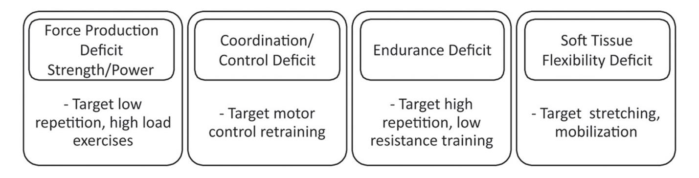
Figure 5. Additional classification of potential movement impairment contributors to a condition, and subsequent targeted treatment approaches that may follow.
Figure 6. Depiction of potential movement system impairments to be assessed following from identification of primary movement pattern abnormalities.

These impairments if present would lead directly to treatment planning decisions.
With regard to the shoulder, Figure 4 presents common movement patterns recognized in a number of previously described classifications.34–37 These patterns are not typically present in isolation. For instance, insufficient scapular upward rotation is often associated with glenohumeral hypermobility, 38,39 and excess scapular internal rotation and insufficient scapular posterior tilt may occur in combination.40 A classification is not determined based on simply the presence of an isolated movement impairment, but instead on the collective history and physical examination, including assistance or symptom relief tests34,41,42 as well as pain provocation tests or movements. Clinical judgement is used to assimilate the collective examination findings in determining which classification is most representative of the client’s movement system dysfunction while incorporating the environmental and personal factors unique to each patient. Figure 5 illustrates that from a movement classification, a clinician can further assess for the associated movement system impairments that would be the focus of a treatment intervention. These representations are not considered all-inclusive or complete, but provide an example of a framework for further investigation. For example, the proposed scapulothoracic patterns represent movement dysfunction in each of the three planes (sagittal - scapular tilting; frontal - clavicle elevation or scapular downward rotation; transverse - scapular internal rotation). Structuring movement patterns in such a way may standardize the clinical evaluation process and the education of new clinicians.37
CASE EXAMPLE
A 22-year-old male presents with a chief complaint of right anterior shoulder joint pain specific to shoulder overhead motions. Pain is easily provoked with unresisted arm elevation, but is of minimal severity (2/10 on a 0-10 pain scale) and does not persist after exacerbating movements are discontinued. Thus he demonstrates a condition with low irritability. He reports aching pain in the joint without numbness, tingling, radiating pain, or substantive weakness. He reports pain began after a feeling of excessive shoulder “strain” while playing volleyball. Arm elevation into flexion is most painful, there is no pain at rest, and arm elevation into abduction is not substantively painful. He is otherwise an active, healthy individual with no confounding demo-

Figure 7. Temporomandibular Disorder Sample Diagnostic Classification.
This classification uses objective exam results to determine movement dysfunction with further delineation of pathoanatomic conditions. MMO = Maximum mouth opening measured in millimeters (mm); TMJ arthralgia = Joint pain; DDwoR = Disc Displacement without Reduction; DDwR = Disc Displacement with Reduction; TMJ OA = TMJ Osteoarthritis (including joint and disc degeneration conditions). Proposed by Kahnert EK integrated with Schiffman diagnostic criteria.32
graphics or co-morbid conditions. No red or yellow flags are identified.
Qualitative and quantitative alignment and movement assessment demonstrates reduced clavicle elevation and reduced scapular upward rotation with his arms relaxed at his side. Cervical and thoracic posture are unremarkable. As he elevates his arm into flexion, his scapula demonstrates increased anterior tilt rather than expected posterior tilt43 (Figure 8). This individual’s posterior tilt first begins at approximately 90 degrees of arm flexion as determined visually, and shoulder pain is present in the mid to end range of shoulder flexion. Flexion and abduction range of motion are within normal limits but demonstrate reduced scapular upward rotation throughout the range. A scapular assistance test44 with manual support to scapular posterior tilt and upward rotation is positive during flexion. Repetitive motion results in slight increases in his aberrant scapular movement patterns.
Incorporation of surface electromyographic (EMG) assessment into his evaluation demonstrates a substantial delay of his serratus anterior muscle activation as compared to activation of the anterior deltoid when raising his arm into flexion (Figure 9, Participant A). This is consistent with the “ reverse action” movement pattern demonstrated whereby unopposed anterior deltoid contraction results in anterior rather than posterior tilt of the scapula as flexion is initiated. Serratus anterior activation begins to noticeably increase above 90 degrees humeral flexion corresponding to the onset of scapular posterior tilt. For comparison, Figure 9 Participant B depicts EMG from another individual who demonstrated typical scapular posterior tilting during shoulder flexion. Serratus anterior muscle activity was sim-
Figure 8. Case example of an individual with excessive scapular anterior tilt during shoulder flexion.

The individual’s scapula anteriorly tilts during the lower half of elevation (A) and begins to posteriorly tilt around 90 degrees of elevation (B).
ilarly increasing along with anterior deltoid muscle activity for the first 70 degrees of flexion producing simultaneous scapular posterior tilt and humeral flexion.
Even without EMG of the muscle activation pattern, the movement examination allows us to streamline our physical examination. We still must assess joint mobility (unremarkable in his case) and overall muscle strength (within normal limits). However, integrating the movement exam and the history allows us to more efficiently complete the physical exam. In this case we need to rule out long thoracic nerve palsy and can do so through basic manual muscle testing of his serratus (within normal limits) which can be further confirmed by surface EMG in this case.
Based on our classification, a movement system diagno-

Figure 9. Root mean square processed EMG (300 ms time constant) collected on two individuals during unresisted shoulder flexion.
Participant A shows delay in full activation of the serratus until later in the range of motion compared to Participant B. EMG is expressed as a percent of maximum voluntary contraction. Participant A was classified with insufficient scapular posterior tilt while Participant B demonstrated a typical scapular posterior tilt pattern.
sis of insufficient scapular posterior tilt associated with coordination/control deficit is provided. Therefore, his treatment follows from his diagnosis and includes movement training exercises to improve serratus anterior activation45 timing including wall slides46 and scapular protraction with flexion movement training. Electromyographic biofeedback could be helpful in accelerating motor learning to improve serratus activation timing. Specific exercise or biofeedback selection based on the individual’s history and physical exam are examples of manipulating task and environmental constraints to attain a desired change in motor behavior of the movement system.
LIMITATIONS
Movement system classification is not without its limitations. First, aberrant movement does not occur in isolation. Rather, movement patterns emerge based on interactions between the individual, environment, and task.28 Thus, it is imperative that clinicians encourage the patient to demonstrate their painful activities in a context similar to that in which symptoms occur. Second, movement is inherently variable occurring on the backdrop of individual biology, anatomy, physiology, and task demands.15,47 Some variability is to be expected and can be assessed as part of the movement system’s ability to adapt to the changing constraints present in daily life, for example eccentric versus concentric loading.48–50 More research is needed to determine how to distinguish expected and potential beneficial movement variation from movement variation that alters tissue loading in detrimental ways. Third, there is potential for misinterpretation of movement systems classification as one “right way” to move. Education should be provided that there is a range of acceptable movement variation. Finally, symptom improvement as a result of interventions may not be related to permanent biomechanical change.27 Effective alterations in movement patterns will redistribute load to reduce symptoms with the goal of allowing a full return to previously aggravating activities. To achieve long-term biomechanical changes, movement system training appears to require task specificity. 27
SUMMARY
Meeting our professional vision requires us to “take a seat at the table” with regard to the development and refinement of diagnostic classifications best able to direct practice, maximize patient outcomes, and determine relative value of services. All of these goals further relate to our ability to produce effective clinical practice guidelines, educate future professionals, and achieve the recognition deserved as advanced practice providers. While effectiveness of physical therapy is well demonstrated for shoulder conditions,51 most outcomes do not demonstrate fully resolved symptoms or positive outcomes for all individuals.52 Continued development and refinement of our diagnostic framework is needed. Movement system diagnostic classification can advance and streamline practice if considered while recognizing the inherent movement variability across individuals. To transform society, we must transform, validate, and translate a movement system diagnostic practice to “solve the health related challenges that society faces”.
Submitted: October 31, 2021 CST, Accepted: November 28, 2021 CST
This is an open-access article distributed under the terms of the Creative Commons Attribution 4.0 International License (CCBY-NC-4.0). View this license’s legal deed at https://creativecommons.org/licenses/by-nc/4.0 and legal code at https://creativecommons.org/licenses/by-nc/4.0/legalcode for more information.
REFERENCES
1. American Physical Therapy Association. Vision Statement for the Physical Therapy Profession. September 2019. https://www.apta.org/siteassets/pdf s/policies/guiding-principles-to-achieve-the-vision.p df. Accessed November 8, 2021.
2. Zimny NJ. Diagnostic classification and orthopaedic physical therapy practice: what we can learn from medicine. J Orthop Sports Phys Ther. 2004;34(3):105-109. doi:10.2519/jospt.2004.34.3.105
3. Miller-Spoto M, Gombatto SP. Diagnostic labels assigned to patients with orthopedic conditions and the influence of the label on selection of interventions: a qualitative study of orthopaedic clinical specialists. Phys Ther. 2014;94(6):776-791. do i:10.2522/ptj.20130244
4. Jewell DV, Moore JD, Goldstein MS. Delivering the physical therapy value proposition: a call to action. Phys Ther. 2013;93(1):104-114. doi:10.2522/ptj.20120 175
5. Committee on Quality of Health Care in America. The Institute of Medicine. Crossing the Quality Chasm: A New Health System for the 21st Century. Washington, DC: National Academies Press; 2001.
6. Ludewig PM, Kamonseki DH, Staker JL, Lawrence RL, Camargo PR, Braman JP. Changing our diagnostic paradigm: movement system diagnostic classification. Int J Sports Phys Ther. 2017;12(6):884-893. doi:10.26603/ijspt20170884
7. Innocenti T, Ristori D, Miele S, Testa M. The management of shoulder impingement and related disorders: a systematic review on diagnostic accuracy of physical tests and manual therapy efficacy. J Bodyw Mov Ther. 2019;23(3):604-618. doi:10.1016/j.jbmt.201 8.08.002
8. Friedman DJ, Tulloh L, Khan KM. Peeling off musculoskeletal labels: sticks and stones may break my bones, but diagnostic labels can hamstring me forever. Br J Sports Med. 2021;55(21):1184-1185. doi:1 0.1136/bjsports-2021-103998
9. Barreto RPG, Braman JP, Ludewig PM, Ribeiro LP, Camargo PR. Bilateral magnetic resonance imaging findings in individuals with unilateral shoulder pain. J Shoulder Elbow Surg. 2019;28(9):1699-1706. doi:10.10 16/j.jse.2019.04.001
10. Moayedi M, Davis KD. Theories of pain: From specificity to gate control. J Neurophysiol. 2013;109(1):5-12. doi:10.1152/jn.00457.2012 11. Vinson EN, Wittstein J, Garrigues GE, Taylor DC. MRI of selected abnormalities at the anterior superior aspect of the shoulder: potential pitfalls and subtle diagnoses. Am J Roentgenol. 2012;199(3):534-545. do i:10.2214/ajr.12.8789
12. Hegedus EJ, Goode AP, Cook CE, et al. Which physical examination tests provide clinicians with the most value when examining the shoulder? Update of a systematic review with meta-analysis of individual tests. Br J Sports Med. 2012;46(14):964-978. doi:10.11 36/bjsports-2012-091066
13. Nickel B, Barratt A, Copp T, Moynihan R, McCaffery K. Words do matter: a systematic review on how different terminology for the same condition influences management preferences. BMJ Open. 2017;7(7):e014129. doi:10.1136/bmjopen-2016-01412 9
14. Zadro JR, O’Keeffe M, Ferreira GE, et al. Diagnostic Labels for Rotator Cuff Disease Can Increase People’s Perceived Need for Shoulder Surgery: An Online Randomized Controlled Experiment. J Orthop Sports Phys Ther. 2021;51(8):401-411. doi:10.2519/jospt.2021.10375
15. Lawrence RL, Ludewig PM, Ward SR. An Integrated Approach to Musculoskeletal Performance, Disease, and Recovery. Phys Ther. September 2021. do i:10.1093/ptj/pzab225
16. Felson DT, Niu J, Gross KD, et al. Valgus malalignment is a risk factor for lateral knee osteoarthritis incidence and progression: findings from the Multicenter Osteoarthritis Study and the Osteoarthritis Initiative. Arthritis Rheumatism. 2013;65(2):355-362. doi:10.1002/art.37726
17. Bittersohl B, Benedikter C, Franz A, et al. Elite rowers demonstrate consistent patterns of hip cartilage damage compared with matched controls: A T2* mapping study. Clin Orthop Rel Res. 2019;477(5):1007-1018. doi:10.1097/corr.0000000000 000576
18. Webster KE, Hewett TE. Meta-analysis of metaanalyses of anterior cruciate ligament injury reduction training programs. J Orthop Res. 2018;36(10):2696-2708. doi:10.1002/jor.24043
19. Schellingerhout JM, Verhagen AP, Thomas S, Koes BW. Lack of uniformity in diagnostic labeling of shoulder pain: time for a different approach. Manual Therapy. 2008;13(6):478-483. doi:10.1016/j.math.200 8.04.005
20. American Physical Therapy Association. Homepage-News-Beginning Oct. 1: Stop Using this ICD-10 Code for LBP. https://www.apta.org/news/202 1/09/29/icd-10-update. Published September 29, 2021. Accessed November 8, 2021.
21. Sahrmann S. Defining our diagnostic labels will help define our movement expertise and guide our next 100 years. Phys Ther. 2021;101(1):pzaa196. doi:1 0.1093/ptj/pzaa196
22. Cools AM, Michener LA. Shoulder pain: can one label satisfy everyone and everything? Br J Sports Med. 2017;51:416-417.
23. Hedman LD, Gill-Body KM, Quinn L, et al. On “Reflections on the Wisdom of Profession-Specific Diagnostic Labels.” Jette AM. Phys Ther. 2021;101:pzab139. Phys Ther. 2021. doi:10.1093/ptj/p zab237
24. Jette AM. Reflections on the Wisdom of Profession-Specific Diagnostic Labels. Phys Ther. 2021;101(6):pzab139. doi:10.1093/ptj/pzab139
25. American Physical Therapy Association. Board of Directors Meeting Minutes. April 2017 26-29. April 2017. http://www.apta.org/BOD/Meetings/Minutes/20 17/4/26/.
26. Kernozek T, Schiller M, Rutherford D, Smith A, Durall C, Almonroeder TG. Real-time visual feedback reduces patellofemoral joint forces during squatting in individuals with patellofemoral pain. Clin Biomech. 2020;77:105050. doi:10.1016/j.clinbiomech.2020.1050 50
27. Willy RW, Willson JD, Clowers K, Baggaley M, Murray N. The effects of body-borne loads and cadence manipulation on patellofemoral and tibiofemoral joint kinetics during running. J Biomech. 2016;49(16):4028-4033. doi:10.1016/j.jbiomech.201 6.10.043
28. Newell KM. Constraints on the Development of Coordination. In: Wade MG, Whiting HTA, eds. Motor Development in Children: Aspects of Coordination and Control. The Netherlands: Martinus Nijhoff; 1986:341-360.
29. Sahrmann SA. The human movement system: our professional identity. Phys Ther. 2014;94(7):1034-1042. doi:10.2522/ptj.20130319
30. Ludewig PM, Lawrence RL, Braman JP. What’s in a name? Using movement system diagnoses versus pathoanatomic diagnoses. J Orthop Sports Phys Ther. 2013;43(5):280-283. doi:10.2519/jospt.2013.0104 31. Inman VT, Saunders JD, Abbott LC. Observations on the function of the shoulder joint. JBJS. 1944;26(1):1-30.
32. Schiffman E, Ohrbach R, Truelove E, et al. Diagnostic Criteria for Temporomandibular Disorders (DC/TMD) for clinical and research applications: recommendations of the international RDC/TMD consortium network and orofacial pain special interest group. J Oral Facial Pain Headache. 2014;28(1):6-27. doi:10.11607/jop.1151
33. Harrison AL, Thorp JN, Ritzline PD. A Proposed Diagnostic Classification of Patients With Temporomandibular Disorders: Implications for Physical Therapists. J Orthop Sports Phys Ther. 2014;44(3):182-197. doi:10.2519/jospt.2014.4847
34. Caldwell C, Sahrmann S, Van Dillen L. Use of a movement system impairment diagnosis for physical therapy in the management of a patient with shoulder pain. J Orthop Sports Phys Ther. 2007;37(9):551-563. doi:10.2519/jospt.2007.2283
35. Burkhart SS, Morgan CD, Kibler WB. The disabled throwing shoulder: Spectrum of pathology part III: The SICK scapula, scapular dyskinesis, the kinetic chain, and rehabilitation. Arthroscopy. 2003;19(6):641-661. doi:10.1016/s0749-8063(03)0038 9-x
36. Uhl TL, Kibler WB, Gecewich B, Tripp BL. Evaluation of clinical assessment methods for scapular dyskinesis. Arthroscopy. 2009;25(11):1240-1248. doi:10.1016/j.arthro.2009.0 6.007
37. Joe Godges, DPT, OCS personal communication.
38. Paletta GA Jr, Warner JJP, Warren RF, Deutsch A, Altchek DW. Shoulder kinematics with two-plane xray evaluation in patients with anterior instability or rotator cuff tearing. J Shoulder Elbow Surg. 1997;6(6):516-527. doi:10.1016/s1058-2746(97)9008 4-7
39. Ogston JB, Ludewig PM. Differences in 3-dimensional shoulder kinematics between persons with multidirectional instability and asymptomatic controls. Am J Sports Med. 2007;35(8):1361-1370. do i:10.1177/0363546507300820
40. Seitz AL, McClure PW, Finucane S, Boardman ND III, Michener LA. Mechanisms of rotator cuff tendinopathy: Intrinsic, extrinsic, or both? Clin Biomech. 2011;26(1):1-12. doi:10.1016/j.clinbiomec h.2010.08.001
41. Sahrmann SA. Diagnosis by the physical therapist-a prerequisite for treatment. A special communication. Phys Ther. 1988;68(11):1703-1706. d oi:10.1093/ptj/68.11.1703
42. Spoto MM, Collins J. Physiotherapy diagnosis in clinical practice: A survey of orthopaedic certified specialists in the USA. Physiother Res Int. 2008;13(1):31-41. doi:10.1002/pri.390
43. Ludewig PM, Phadke V, Braman JP, Hassett DR, Cieminski CJ, LaPrade RF. Motion of the shoulder complex during multiplanar humeral elevation. J Bone Joint Surg Am. 2009;91(2):378-389. doi:10.2106/jbj s.g.01483
44. Rabin A, Chechik O, Dolkart O, Goldstein Y, Maman E. A positive scapular assistance test is equally present in various shoulder disorders but more commonly found among patients with scapular dyskinesis. Phys Ther Sport. 2018;34:129-135. doi:1 0.1016/j.ptsp.2018.09.008
45. Neumann DA, Camargo PR. Kinesiologic considerations for targeting activation of scapulothoracic muscles - part 1: serratus anterior. Braz J Phys Ther. 2019;23(6):459-466. doi:10.1016/j.bj pt.2019.01.008
46. Hardwick DH, Beebe JA, McDonnell MK, Lang CE. A comparison of serratus anterior muscle activation during a wall slide exercise and other traditional exercises. J Orthop Sports Phys Ther. 2006;36(12):903-910. doi:10.2519/jospt.2006.2306 47. Kalkhoven JT, Watsford ML, Impellizzeri FM. A conceptual model and detailed framework for stressrelated, strain-related, and overuse athletic injury. J Sci Med Sport. 2020;23(8):726-734. doi:10.1016/j.jsam s.2020.02.002
48. Bartlett R, Wheat J, Robins M. Is movement variability important for sports biomechanists? Sports Biomechanics. 2007;6(2):224-243. doi:10.1080/147631 40701322994
49. Wade MG, Smith TJ. Variability in human motor and sport performance. In: Variability in Human Performance. CRC Press; 2014:31-46. https://doi.org/1 0.1201/b17319-3.
50. Harbourne RT, Stergiou N. Movement variability and the use of nonlinear tools: principles to guide physical therapist practice. Phys Ther. 2009;89(3):267-282. doi:10.2522/ptj.20080130
51. Pieters L, Lewis J, Kuppens K, et al. An Update of Systematic Reviews Examining the Effectiveness of Conservative Physical Therapy Interventions for Subacromial Shoulder Pain. J Orthop Sports Phys Ther. 2020;50(3):131-141. doi:10.2519/jospt.2020.8498
52. Ludewig PM, Borstad JD. Effects of a home exercise programme on shoulder pain and functional status in construction workers. Occup Environ Med. 2003;60(11):841-849. doi:10.1136/oem.60.11.841
53. Lentz TA, Beneciuk JM, Bialosky JE, et al. Development of a Yellow Flag Assessment Tool for Orthopaedic Physical Therapists: Results From the Optimal Screening for Prediction of Referral and Outcome (OSPRO) Cohort. J Orthop Sports Phys Ther. 2016;46(5):327-343. doi:10.2519/jospt.2016.6487
Proposed elements of a basic diagnostic process for atraumatic shoulder pain: 1. Subjective key questions a. What is the patient’s chief complaint? i. Pain (constant or with movement) ii. Mobility deficit iii. Stability deficit iv. Weakness with or without pain, mobility or stability deficits b. What is the level of condition irritability (provocation required, severity, pain persistence)? c. What type of symptoms are present (pain, numbness, tingling, weakness, stiffness, etc.)? d. What is the location of the symptoms (glenohumeral joint pain rarely radiates past the elbow)? e. Mechanism of “injury” – specific injury, cumulative trauma, insidious onset? f. What if any movements exacerbate/relieve symptoms? g. Demographics, confounding factors (e.g. smoking)? h. History/co-morbid conditions (e.g. diabetes)? i. Red flags? j. Yellow flags (e.g. pain catastrophizing)53 2. Objective a. Focused posture/movement exam
i. Thoracic posture/scoliosis ii. Cervical posture/ROM iii. Shoulder complex initial alignment iv. Bilateral shoulder flexion with and without resistance, repeated movements v. Bilateral shoulder abduction with and without resistance, repeated movements vi. “Problem movement”/hand behind back etc. vii. Symptom relief tests (scapular assistance test etc.) b. Seated follow up exam i. Cervical symptom provocation/relief tests as warranted ii. Shoulder mobility/stability tests (sulcus/AP load and shift) iii. Symptom provocation/strength tests as warranted iv. Select special tests c. Supine/prone follow-up exam i. Select length/strength/stability/nerve entrapment tests as warranted ii. Select special tests (e.g. apprehension, labral tests) iii. Shoulder internal/external rotation in 90 degrees abduction, active and passive 3. Assessment and Movement System Diagnostic Label
Invited Clinical Commentary
Application of the 4-Element Movement System Model to Sports Physical Therapy Practice and Education
Ryan Zarzycki, PhD, PT 1 , Philip Malloy, PhD, PT 1 , Brian J Eckenrode, PT, DPT, OCS 1 , Jane Fagan, PT, DPT, OCS 1 , Molly Malloy, PT, DPT, OCS 1 , Kathleen K Mangione 1 a
1 Physical Therapy, Arcadia University Keywords: education, movement system, clinical practice, post-graduate residency https://doi.org/10.26603/001c.30173
International Journal of Sports Physical Therapy
Vol. 17, Issue 1, 2022
The 4-Element Movement System Model describes primary elements (motion, force, motor control, and energy) essential to the performance of all movements. The model provides a framework or scaffolding which allows for consistent processes to be used in examination and intervention decisions. The process starts with task identification followed by a systematic observation of control, amount, speed, symmetry, and symptoms during movement. Testable hypotheses are generated from the observations which inform the examination and the interventions. This commentary describes the use of the 4-Element Movement System Model in entry level and post-graduate residency educational programs and in clinical care with three common sports-related diagnoses.
Level of Evidence
5
INTRODUCTION
The analysis of human movement is central to the practice of physical therapy. Physical therapists analyze movement in order to identify impairments that contribute to activity limitations and participation restrictions in their patients. Movement analysis and interpretation is a hallmark skill of expert practitioners.1
The movement system was adopted by the American Physical Therapy Association in 2015 and has been defined as the integration of body systems that generate and maintain movement at all levels of bodily function. 2 There have been several approaches to defining the movement system,3–5 but the 4-Element Movement System Model (4-Element Model) has the advantage in that it captures a wide variety of disorders, can meaningfully guide practice and education, can readily be incorporated into entry level and residency training, and is consistent with existing professional models such as the International Classification of Functioning (ICF)6 and the Patient-Client Management Model.7
The 4-Element Model describes the primary elements essential to all movement: motion, force, motor control, and energy (Figure 1). Motion refers specifically to the ability of a joint or tissue to be moved passively. Force refers to the ability of the contractile (i.e. muscles) and non-contractile structures (i.e. tendons) to produce movement, and provide dynamic stability around joints during static and dynamic tasks. Motor control refers to the ability to plan, execute and adapt goal-directed movements such that they are accurate, coordinated and efficient. Lastly, energy refers to the ability to perform sustained or repeated movements, and is dependent on the integrated functioning of the cardiovascular, pulmonary, and neuromuscular systems. The elements overlap in many patient conditions, but can be examined and tested separately. Since all movement occurs within an environmental context and is affected by personal factors specific to the individual, the model depicts how the environment and personal factors surround the 4 elements. Environmental factors are external items that can influence movement such as terrain, support surface, and external distractions. Personal factors include age, gender, comorbidities, self-efficacy, confidence, fear of movement and motivation.5 The comprehensive description of movement in the 4-Element Model is consistent with theories of human movement being a dynamic system involving complex interactions between the task, the person, and the en-
a
Corresponding author:
Kathleen K Mangione, PT, PhD, FAPTA Arcadia University, Dept of Physical Therapy 450 S Easton Rd. Glenside, PA 18901 215-572-2861 mangione@arcadia.edu
vironment.8
Application of the model begins with the identification of an activity or task that the patient will perform, and consists of qualitative observation leading to hypothesis generation. The 4-Element Model encourages a systematic approach to the observation by using five observation targets abbreviated as CASSS (control, amount, speed, symmetry, symptoms). Briefly, control refers to the smoothness, coordination, and timing of movement; amount refers to the amplitude of movement at each joint; symmetry is observed in bilateral tasks or comparing unilateral performance between limbs; speed is the length of time; and symptoms most commonly refer to pain but also can include mechanical symptoms, reports of instability, or fatigue. After using the CASSS observation targets to describe the task, hypotheses are generated about the possible movement system elements that may contribute to the movements observed. Potential impairments are identified and tested which lead the clinician to implement treatment strategies.5 This commentary will address the application of the 4-Element Model to sports physical therapist practice, and its incorporation as part of the education of students and residents.
USE OF THE 4-ELEMENT MODEL IN EDUCATION
Case-based learning with the 4-Element Model is foundational in both the Arcadia University entry-level Doctor of Physical Therapy (DPT) and Orthopedic Residency programs. The model can be incorporated by academic faculty, clinical instructors (CIs), and residency mentors to assess and foster development of clinical reasoning of the learner. The focus of introducing the model in the entry-level DPT program is to help build decision making strategies before entering the clinical education curriculum. The model, terminology, and process are introduced in the first course of the curriculum and applied throughout the rest of the courses in the program. The repetition of 1) activity selection based on patient goals and safety, 2) use of observational targets, and 3) hypothesis driven exam and intervention design is a scaffold by which the student develops clinical reasoning in the didactic curriculum. It is this repetition that helps support the student when they enter the clinical environment and fosters the use of a common examination process and common language to explain the movement impairments with their CI.
CLINICAL EDUCATION
In order to facilitate successful clinical education experiences, the department’s clinical education team has provided clinical faculty with the published paper and a narrated video, as resources to educate them on the 4-Element Model.5,9 The video describes the 4-Element Model and applies it to a sample case of a young athlete with a knee injury. The paper and video help to share insight into the student’s foundational clinical reasoning strategies. We believe this knowledge may allow the CI to more accurately meet the student where they are and provide concrete tools for development. We encourage clinical sites to use the model as a conceptual and visual cue for clinical teaching and reflection in all levels of clinical education experiences, from
Figure 1. The 4-Element Movement System Model.

CASSS is a pneumonic for the five observation targets of the model: C-Control, A-Amount, S-Speed, S-Symmetry, S-Symptoms
beginner to terminal clinical affiliation. A CI may utilize specific components of the model in patient care preparation activities such as helping the student to identify meaningful goals that then drive observational targets. It is also beneficial to use the model as a cue to identify personal or environmental factors which may impact care, and therefore encourages an awareness of safety early on in any clinical experience. The CI may assist the student in streamlining the physical examination by discussing the element(s) contributing to the observed deficits and how each may be tested. To help develop basic clinical reasoning skills a CI may provide detailed prompting questions using common language such as “if this were a force problem, what is the relevant anatomy to test?” or “how do the elements of force and motor control interact during this jumping sequence?” Plan of care development can be facilitated by revisiting the outcomes of hypothesis testing with the student to develop targeted interventions, and using these outcomes in reflection activities. CI’s may also use the 4- Element Model to advance non-linear thinking, the interaction between elements and systems, which is imperative when treating more specialized populations such as the athlete.
POST-GRADUATE TRAINING
A primary focus of a post graduate training program is to facilitate a partnership relationship with mentor and resident/fellow and to help the learner delve deeper into specialty practice for optimal patient care. A challenge with mentoring relationships in residency and fellowship education is that we do not use a common language or examination framework across physical therapy programs and practice. Frequently mentors and learners may have different backgrounds of education and training. The 4-Element Model provides a common framework for a systematic movement assessment of the athlete.
To implement the model, the resident and mentor start the examination by choosing and analyzing a specific functional task. The task is selected by what the patient reports having difficulty performing in their daily activities or sport specific tasks. We have found it beneficial to film the patient performing the challenging task such as a stair climbing, gait assessment, squatting mechanics, overhead throwing or jumping. Filming allows the mentor and resident to slow
the patient’s performance to fully assess movement abnormalities. While looking at the specific functional task, the mentor will ask the resident to assess the observation targets listed in the CASSS. For example, limitations in control would prompt a discussion of what could be affecting the lack of control including poor balance, specific muscle weakness or a patient’s decreased awareness of how to perform the movement. If the resident observes a limited amount of motion this would then prompt discussion of which joints specifically to look at further in the examination of mobility. Speed impairments would prompt a discussion of incorporating a timed functional test or discussion of the “normal” parameters for someone to complete the task (30 second sit to stand test, timed up and go). Symmetry issues would prompt discussions such as “why might a weight shift occur?” Symptom provocation during movement such as pain, clicking, or stiffness and when in the movement sequence these symptoms occur would prompt a mentor and resident to perform a more specific biomechanical assessment. After hypotheses are generated, the novice clinician would continue with the examination delving deeper into the areas of deficit. The mentor and clinician would then use the examination findings to decide which specialty interventions best help that area of deficit. In a post graduate mentoring relationship, the cases that are often discussed have complex clinical presentations, and likely more than one element needs to be addressed to optimize function. It is recommended to return to the specific functional task frequently to assess if the physical therapy interventions are translating into improved functional movement with the limited task.
It is our belief that use of the 4-Element Model in education can be beneficial to both the learner and CI/mentor to help assess and solidify clinical reasoning. This may be demonstrated in entry-level DPT programs, as we look to scaffold learning and build initial reasoning processes, and in post graduate education as we guide novice clinicians to more effective and specialized care.
The 4-Element Model provides a systematic approach to the understanding and management of movement dysfunction for common musculoskeletal conditions that are encountered regularly in sports medicine and orthopedic physical therapist practice. The four elements of the movement system model are essential to all athletic type functional tasks. In the following sections, three clinical case examples will be presented to show how the 4-Element Model and CASSS framework can be used to establish testable clinical hypotheses for observed movement impairments. The findings can then be applied to help develop patient specific treatment plans aimed at improving functional task performance, and can also be used to guide overall clinical decision making.
CASE EXAMPLE 1: FEMALE SOCCER PLAYER POSTOPERATIVE ACL RECONSTRUCTION (ACLR) Marie is a 23-year-old female college student. She ruptured her left anterior cruciate ligament (ACL) while playing soccer. She reported a non-contact injury in which her knee buckled upon attempting to change directions while playing defense. She underwent ACL reconstruction (ACLR) with a quadriceps tendon autograft approximately three weeks after the injury. Acute post-operative effects of effusion and pain, which contribute to limitations in motion and force, are readily observed as the patient enters the treatment room. These findings guide initial treatment. However, once post-operative sequelae are resolved, functional tasks with increased physiologic demand are appropriate to examine. After six weeks of post-operative treatment, motion was restored and strength training was progressed.
Task selection: As closed chain exercises progressed from bilateral to unilateral movements, a lateral step down (8-inch step) was chosen as the task to examine as it was challenging to the patient (see links in reference for video).10,11
CASSS & Key observations: In the sagittal plane, Marie demonstrated good control during the task; however, in the frontal plane, she demonstrated slightly less control of the lower extremity on the right (nonsurgical) side. When examining motion, she demonstrated the appropriate amount of motion through the lower extremity and trunk in the sagittal plane; but in the frontal plane, she demonstrated slightly greater amount of hip adduction on the right (nonsurgical) side during the task which was most apparent towards peak knee flexion. Marie demonstrated normal speed during the task. She exhibited symmetry in the sagittal plane, but in the frontal plane she appeared asymmetric in the control and amount of motion, as described prior. Marie did not report any symptoms of pain, stiffness, or instability during the task. Based on the observational targets of the CASSS, Marie demonstrated slightly greater hip adduction, or greater dynamic lower extremity valgus, in the right (non-surgical) limb compared to the left limb.
Hypotheses and Exam: Hypotheses regarding this movement alteration included: 1) force impairment, weakness in the muscles controlling hip adduction; 2) motor control impairment, perhaps lower extremity valgus decreased in the left (surgical) limb due to extensive unilateral training and feedback; and 3) motion impairment, limited ankle dorsiflexion (DF) in the right limb.12 Based on the observational targets and hypotheses our evaluation included strength testing of the hip abductors and ROM evaluation of ankle DF. Our evaluation revealed symmetrical hip abduction strength and symmetrical DF ROM. Based on the evaluation of Marie’s strength and ROM, we concluded that Marie’s asymmetric performance of the lateral step was a result of learned behavior from extensive unilateral training. Discussion with Marie confirmed this as she was provided with cues to minimize lower extremity valgus throughout post-operative rehabilitation and did not perform substantial exercises with the nonsurgical limb.
Task selection: We chose to observe Marie performing a 32 cm drop vertical jump (DVJ) (see links in reference for the
video).13 The DVJ task was chosen as it may help identify athletes with a higher risk of knee re-injury or second ACL injury.
14–16
CASSS & Key observations: Marie’s control of the movement was smooth and coordinated. In the sagittal plane, she demonstrated a significant amount of hip and knee flexion and a forward trunk position. In the frontal/transverse planes, the amount of motion at the hip was greater on the left (surgical) side. Marie’s speed during the task was normal. From a symmetry perspective, Marie demonstrated asymmetrical loading between the limbs. Specifically, she shifted her weight away from the left (surgical) limb. Marie did not report symptoms during the DVJ. Based on the observational targets, the most significant movement alteration was asymmetrical loading, characterized by greater weight acceptance in the right (non-surgical) lower extremity.
Hypotheses and Exam: Hypotheses regarding this movement alteration included: 1) force impairment, weakness in the surgical limb’s quadriceps muscle may be present; 2) motor control impairment, learned behavior resulting offloading the surgical limb since the injury, after ACLR, and/ or during the rehabilitation process, and 3) psychological factors including readiness to return to sport, confidence, and fear of re-injury. Our evaluation with an isokinetic dynamometer revealed a quadriceps strength index of 85% (L peak torque/body weight = .82 Nm/kg; R peak torque/body weight=.96 Nm/kg). To determine whether motor control alterations were contributing to the asymmetrical loading pattern, Marie performed the drop vertical jump after being provided with external feedback. The external feedback was reaching (with her left hand) for a cone placed to her left. Marie demonstrated more symmetrical loading with the provided external feedback. Also, Marie completed the anterior cruciate ligament return to sport after injury questionnaire (ACL-RSI), scoring a 4/100. Lower scores indicate less psychological readiness (i.e. more fear, less confidence, more concerned about future risks of knee injury). Based on the findings from the evaluation, Marie’s altered loading strategy seemed to be driven primarily by altered motor control and personal factors (i.e. poor psychological readiness). A force impairment (i.e. quadriceps weakness) was likely also contributing to the altered movement. A single assessment of the DVJ, or any other single assessment task, did not allow us to determine if there was potentially an impairment with the energy element. Subsequent testing of Marie’s movement during the DVJ or lateral step down could be evaluated after a fatigue protocol or with repetitive tests/ movements. Repetitive tests appropriate for Marie would be the two-minute lateral step down test or the Tuck Jump
Test.17,18
Targeted intervention: Treatment focused on motor control strategies, such as providing external cueing to promote symmetrical loading during various movement patterns following a graded exposure paradigm. Graded exposure was utilized to address low psychological readiness. Quadriceps strengthening exercises were also continued.
CASE EXAMPLE 2: ACHILLES TENDINOPATHY IN A RUNNER
JP is a 37-year-old male accountant who presented with complaints of an insidious onset of right Achilles tendon pain for the past six months. He denied any significant past medical history, but reported prior right distal iliotibial band pain approximately two years ago which resolved without formal medical attention. JP denied any other symptoms other than Achilles tendon pain, with noticeable pain in the morning upon waking which increases with walking and running. His pre-injury running weekly mileage was 15 to 20 miles per week, but now currently running five to seven miles per week. JP reported pain of 0/10 at rest, and 6/10 at worst on the numeric pain rating scale; he scored a 67/80 on the Lower Extremity Functional Scale and 25/36 on the University of Wisconsin Running Injury and Recovery Index. No diagnostic imaging of the right Achilles tendon was reported. The patient reported taking ibuprofen as needed for pain management.
Task selection: His movement assessment began with examining basic bilateral (ie. squats, lunges) and unilateral lower extremity tasks (ie. lateral step down) to obtain a baseline perspective on the patient’s willingness to move and quality of movement. No major deviations were observed with these movement tasks, so a more complex activity, running, was selected. A running video analysis was conducted, given that this was the primary activity which caused the patient’s pain and for which his participation was limited.
CASSS & Key observations: We used video to record running mechanics on the treadmill in the frontal and sagittal view. Running speed was self-selected by the patient. Using the CASSS framework, for control, we visually observed relatively good trunk and lower extremity control in the sagittal and frontal plane, but cadence was low. Observing amount, we noted excessive vertical displacement in the frontal plane during running. JP demonstrated a self-selected running speed of a 7:45 min/mile running pace. Numerous deviations in symmetry were observed from the frontal plane including increased trunk lean to the right when in mid stance on the right, excessive pelvic drop when in mid stance on the right, and the right lower extremity was in an externally rotated position in stance. (Figure 2) The patient was noted to have a symmetrical rearfoot strike pattern bilaterally from the sagittal plane, but increased right hip flexion in swing. JP reported pain symptoms of 4/10 to the right Achilles tendon during the task.
Hypotheses and Exam: The hypotheses regarding the movement alterations of this patient included: 1) force impairment may be present in the proximal hip/core musculature and right ankle plantar flexors; 2) energy deficit, a possible lack of ankle plantar flexion muscular endurance may contribute to excessive right hip flexion; 3) motor control impairment, the low running cadence rate observed may be contributory to excessive strain on the musculoskeletal system19; 4) motion deficit, hip and ankle ROM could be limited contributing to the excessive external rotation of the right lower extremity in stance. Clinical examination revealed weakness of the right hip abductors (4-/5 right; 5/5 left) and hip extensors (4-/5 right; 5/5 left). In single leg an-
kle plantar flexion, JP could only complete eight repetitions of a heel raise on the right before not being able to continue, compared to completing 25 repetitions on the left. Achilles tendon pain was also reported during the plantar flexion strength test on the right. His cadence was 152 steps per minute which is lower than what is considered optimal (180 steps per minute),20 and a lower running cadence has been associated with increased vertical load rate,21 which in turn has been associated with lower extremity running related injuries.22,23 Hip ROM and Craig’s test did not reveal any meaningful differences between lower limbs, nor were differences found between left and right ankle ROM, therefore a motion deficit was not considered contributory to this patient case.
JP demonstrated significant tenderness and mild swelling at the mid-portion of the right Achilles tendon. A positive arc sign and Royal London Hospital test were noted on the right. While the pathoanatomic diagnosis of Achilles tendinopathy was confirmed through the clinical examination, the movement analysis findings guided the treatment beyond symptom management.
Targeted intervention: Treatment included core, hip abduction, and hip extension strength training, along with eccentric ankle plantar flexion exercises. The eccentric ankle plantar flexion strengthening is specifically designed to painfully load the Achilles tendon with the knee in an extended and flexed position24 and is supported by strong evidence.25 For the energy and motor control impairments, gait retraining to increase step rate with running were included in the treatment, as transitioning to a higher cadence has been shown to result in lower vertical loading rates during running26 which may benefit this patient.
The prognosis for this patient to achieve his goal of pain free running is good with the treatment plan noted. However, in cases which are not responding to traditional management clinicians may need to consider central sensitization27–29 as an explanation for chronic musculoskeletal pain. Although not specifically outlined in the 4-Element Model, the chronic pain symptoms were addressed directly through pain neuroscience education and via the inclusion of noxious electric stimulation in an attempt to modulate the nervous system through decreasing pain sensitiv-
ity. 30,31
CASE EXAMPLE 3: FEMOROACETABULAR IMPINGEMENT SYNDROME IN A YOUNG ACTIVE ADULT
A.M. is a 30-year-old female graphic designer who presented with a two-year history of unilateral left hip pain. She reported that she was extremely active and participated in some form of exercise six days a week. Her regular forms of exercise were running, yoga, and high intensity strength training. AM reported her pain had progressively worsened over the last six months when she began training for a marathon. AM reported that she had difficulty walking a mile without hip pain, therefore, sought a consult from a primary care sports medicine physician. Radiographic evaluation of AM’s hip revealed an alpha angle of 75 degrees on the modified Dunn view, and a lateral center edge angle of 32 degrees on the anterior posterior pelvis (AP) view. AM was diagnosed with femoroacetabular impingement syn-
Figure 2. Mid-stance comparison of frontal plane view of running movement analysis for case example of a patient with chronic right Achilles tendinopathy.
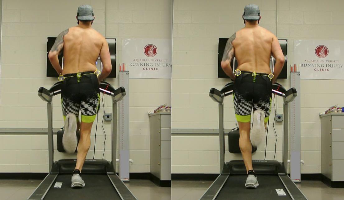
drome (FAIS) due to cam morphology and was referred to physical therapy.
32
Task selection: A single-leg squat has been shown as a useful task to evaluate performance in people with FAIS.33–35 Recently, hip biomechanics and muscle strength were also found to be predictors of impaired performance of a single-leg squat in people with FAIS.36 Since AM desired to return to a high level of activity a single-leg squat task was selected to assess her movement (see link in references for the video).37,38
CASSS & Key observations: Using the CASSS, a visual observation was conducted during a single leg squat with her involved and uninvolved limbs. During the single-leg squat task AM demonstrated good control of the movement in the sagittal plane with both the involved and involved lower extremity. The movement was smooth and coordinated between the segments; however, there was a noticeable reduction in the amount of hip and knee flexion observed between the right and left hip, and slightly more contralateral pelvic drop during the left compared to right single leg squat. The speed of the squat movements between sides were similar. There was a clear asymmetry in the depth of the single leg squat between the left and right sides. She also demonstrated less forward trunk flexion when squatting on the left compared to the right. Symptoms- she reported a level of 3/10 pain when performing a left single-leg squat whereas she reported a 0/10 when performing this task on her right lower extremity.
Hypothesis and Exam: The clinical hypothesis for the movement deviations observed during the single leg squat task included: 1) a hip motion impairment that may be related to the reproduction of symptoms secondary to bony impingement at the hip; 2) knee flexion motion impairment that may be a potentially learned compensation as part of a strategy to limit the overall depth of the squat to avoid moving the hip to near end ranges; 3) motor control impairment as demonstrated by the greater amount of contralateral pelvic drop and hip adduction on the left compared to the right. Additionally, the patient may exhibit 4) force impairments of the hip abductors, extensor, and external rotator muscles that could also limit the ability to achieve equal single-leg squat depth.36,39–41
The physical therapy examination of AM revealed a Csign pain pattern at the hip described as a deep ache with occasional sharp pain during squatting and pivoting. Range of motion assessment showed reduced hip flexion (left: 90 degrees vs. right: 105 degrees) and hip internal rotation at 90 degrees hip flexion (left: 8 degrees vs. right: 20 degrees) with pain noted at end range for both motions. Knee range of motion was not limited, and symmetrical to the uninvolved side. Hip muscle strength measured with a handheld dynamometer revealed reduced hip strength of the left compared to right on the order of: 20% for hip flexion strength, 19% for hip external rotation, and 16% for hip abduction. Reduced hip muscle strength is a common clinical finding in patients with FAIS.39,42,43 AM also exhibited a positive anterior impingement sign, and a positive flexion abduction and external rotation test.
TARGETED INTERVENTIONS
The initial physical therapy treatment interventions for this patient focused on reducing symptoms and restoring pain free hip motion. Treatments in this initial phase included soft tissue mobilization techniques, dry needling, and manual joint mobilizations to help reduce pain, muscle guarding, and restore hip range of motion. Closed and open chain strengthening exercises were performed. Specific movement retraining exercises were also performed to help improve biomechanical faults such as greater contralateral pelvic drop during weight bearing function.
The patient completed 16 visits of physical therapy over 12 weeks. However, limited improvement in hip pain and functional activity occurred with PT management. The CASSS framework was used to evaluate the patient’s singleleg squat prior to discharge and there was little to no change in the observed movement deviations during the task. The patient was referred to an orthopedic hip surgeon for further evaluation, which included magnetic resonance imaging (MRI) to evaluate for additional hip soft tissue injury. In addition to confirming cam type morphology, the patient’s MRI also revealed an acetabular labral tear. The patient underwent hip arthroscopy to address the cam morphology and repair the acetabular labral tear.
In the applied case examples, the 4-Element Model was used to evaluate movement during different tasks in patients with three common clinical diagnoses. The tasks used to assess the patients’ movements were selected based on the functional demands of the patient, but also considered the type of injury and phase of healing in order to ensure safe performance of the task. The movement deviations identified were then used to guide the examination and treatment for each patient. These case examples demonstrate that the 4-Element Model can be applied to clinical conditions commonly seen in orthopedic and sports medicine practice.
SUMMARY
The process underpinning the 4-Element Model can be applied by clinicians, students, and residents to a wide variety of patients. The simplicity of the model is intuitive for master clinicians and provides the scaffolding needed for developing independent clinical reasoning in novice clinicians. Using a common framework and language across settings, patient types, and specialty programs will enhance communication between practitioners.
CONFLICTS OF INTEREST
The authors report no conflicts of interest.
Submitted: November 22, 2021 CST, Accepted: November 22, 2021 CST
This is an open-access article distributed under the terms of the Creative Commons Attribution 4.0 International License (CCBY-NC-4.0). View this license’s legal deed at https://creativecommons.org/licenses/by-nc/4.0 and legal code at https://creativecommons.org/licenses/by-nc/4.0/legalcode for more information.
REFERENCES
1. Jensen GM, Gwyer J, Shepard KF. Expert practice in physical therapy. Phys Ther. 2000;80(1):28-43; discussion 44-52.
2. American Physical Therapy Association. An American Physical Therapy Association white paper. Physical therapist practice and the human movement system. 2015. https://www.apta.org/patient-care/inte rventions/movement-system-management/movemen t-system-white-paper. Accessed October 10, 2021.
3. Sahrmann S, Azevedo DC, Dillen LV. Diagnosis and treatment of movement system impairment syndromes. Braz J Phys Ther. 2017;21(6):391-399. do i:10.1016/j.bjpt.2017.08.001
4. Hedman LD, Quinn L, Gill-Body K, et al. White paper: Movement system diagnoses in neurologic physical therapy. J Neurol Phys Ther. 2018;42(2):110-117. doi:10.1097/npt.00000000000002 15
5. McClure P, Tevald M, Zarzycki R, et al. The 4-element movement system model to guide physical therapist education, practice, and movement-related research. Phys Ther. 2021;101(3).
6. International classification of functioning, disability, and health : ICF [computer program]. Version 1.0. Geneva: World Health Organization; 2001.
7. American Physical Therapy Association. Guide to physical therapist practice. Second edition. Phys Ther. 2001;81(1):9-746.
8. Guccione AA, Neville BT, George SZ. Optimization of movement: A dynamical systems approach to movement systems as emergent phenomena. Phys Ther. 2019;99(1):3-9. doi:10.1093/ptj/pzy116
9. Zarzycki R, Tevald M, Kantak S. 4-element movement system. YouTube; 2021. https://www.youtub e.com/watch?v=O_vVsqpBddA. Accessed November 17, 2021.
10. Zarzycki R. IJSPT ACL case lateral step down right. YouTube; 2021. https://youtu.be/Gk-OhpC02_E. Accessed November 19, 2021.
11. Zarzycki R. IJSPT ACL case lateral step down left. YouTube; 2021. https://youtu.be/P8GKhxZsnQA. Accessed November 19, 2021. 12. Lima YL, Ferreira VMLM, de Paula Lima PO, Bezerra MA, de Oliveira RR, Almeida GPL. The association of ankle dorsiflexion and dynamic knee valgus: A systematic review and meta-analysis. Phys Ther Sport. 2018;29:61-69. doi:10.1016/j.ptsp.2017.0 7.003
13. Zarzycki R. IJSPT ACL case drop vertical jump. YouTube; 2021. https://youtu.be/TYdPqCqrZ6A. Accessed November 19, 2021.
14. King E, Richter C, Daniels KAJ, et al. Biomechanical but not strength or performance measures differentiate male athletes who experience ACL reinjury on return to level 1 sports. Am J Sports Med. 2021;49(4):918-927. doi:10.1177/0363546520988 018
15. Paterno MV, Schmitt LC, Ford KR, et al. Biomechanical measures during landing and postural stability predict second anterior cruciate ligament injury after anterior cruciate ligament reconstruction and return to sport. Am J Sports Med. 2010;38(10):1968-1978. doi:10.1177/03635465103760 53
16. Fältström A, Kvist J, Bittencourt NFN, Mendonça LD, Hägglund M. Clinical risk profile for a second anterior cruciate ligament injury in female soccer players after anterior cruciate ligament reconstruction. Am J Sports Med. 2021;49(6):1421-1430. doi:10.1177/036354652199910 9
17. Read PJ, Oliver JL, de Ste Croix MBA, Myer GD, Lloyd RS. Reliability of the tuck jump injury risk screening assessment in elite male youth soccer players. J Strength Cond Res. 2016;30(6):1510-1516. do i:10.1519/jsc.0000000000001260
18. White AK, Klemetson CJ, Farmer B, Katsavelis D, Bagwell JJ, Grindstaff TL. Comparison of clinical fatigue protocols to decrease single-leg forward hop performance in healthy individuals. Int J Sports Phys Ther. 2018;13(2):143-151. doi:10.26603/ijspt2018014 3
19. Schubert AG, Kempf J, Heiderscheit BC. Influence of stride frequency and length on running mechanics: a systematic review. Sports Health. 2014;6(3):210-217. doi:10.1177/1941738113508544
20. Daniels J. Daniels’ Running Forumla. Champaign, IL: Human Kinetics; 1998.


