38 minute read
Movement System Dysfunction Applied to Youth and Young Adult Throwing Athletes


REFERENCES
1. American Physical Therapy Association. Guiding Principles to Achieve the Vision. https://www.apta.or g/siteassets/pdfs/policies/guiding-principles-to-achie ve-vision.pdf. Accessed September 21, 2021.
2. Saladin L, Voight M. Introduction to the movement system as the foundation for physical therapist practice education and research. Int J Sports Phys Ther. 2017;12(6):858-861. doi:10.16603/ijspt2017085 8
3. Hunter SJ, Norton BJ, Powers CM, Saladin LK, Delitto A. Rothstein roundtable podcast—“Putting all of our eggs in one basket: human movement system.” Phys Ther. 2015;95(11):1466-1466. doi:10.2522/ptj.20 15.95.11.1466
4. McClure P, Tevald M, Zarzycki R, et al. The 4-element movement system model to guide physical therapist education, practice, and movement-related research. Phys Ther. 2021;101(3). doi:10.1093/ptj/pza b024
5. Davids K, Glazier P, Araújo D, Bartlett R. Movement systems as dynamical systems. Sports Medicine. 2003;33(4):245-260. doi:10.2165/00007256-20033304 0-00001
6. Gerrett N, Griggs K, Redortier B, Voelcker T, Kondo N, Havenith G. Sweat from gland to skin surface: production, transport, and skin absorption. J Appl Physiol. 2018;125(2):459-469. doi:10.1152/japplphysi ol.00872.2017
7. Haff GG, Triplett NT, eds. Essentials of Strength Training and Conditioning: National Strength and Conditioning Association. 4th ed. Champaign, IL: Human Kinetics; 2016.
8. Song D, Yu DSF. Effects of a moderate-intensity aerobic exercise programme on the cognitive function and quality of life of community-dwelling elderly people with mild cognitive impairment: A randomised controlled trial. Int J Nur Stud. 2019;93:97-105. doi:10.1016/j.ijnurstu.2019.02.019
9. Stern Y, MacKay-Brandt A, Lee S, et al. Effect of aerobic exercise on cognition in younger adults: A randomized clinical trial. Neurology. 2019;92(9):e905-e916. doi:10.1212/wnl.00000000000 07003 10. Gornitzky AL, Lott A, Yellin JL, Fabricant PD, Lawrence JT, Ganley TJ. Sport-specific yearly risk and incidence of anterior cruciate ligament tears in high school athletes: a systematic review and metaanalysis. Am J Sports Med. 2016;44(10):2716-2723. do i:10.1177/0363546515617742
11. Dawson B. Repeated-sprint ability: where are we? Int J Sports Physiol Perform. 2012;7(3):285-289. doi:1 0.1123/ijspp.7.3.285
12. Girard O, Mendez-Villanueva A, Bishop D. Repeated-sprint ability - part I: factors contributing to fatigue. Sports Med. 2011;41(8):673-694. doi:10.21 65/11590550-000000000-00000
13. Almeida AM, Santos Silva PR, Pedrinelli A, Hernandez AJ. Aerobic fitness in professional soccer players after anterior cruciate ligament reconstruction. PLoS One. 2018;13(3):e0194432. doi:1 0.1371/journal.pone.0194432
14. Scanlan AT, Dascombe BJ, Reaburn P, Dalbo VJ. The physiological and activity demands experienced by Australian female basketball players during competition. J Sci Med Sport. 2012;15(4):341-347. do i:10.1016/j.jsams.2011.12.008
15. Stojanović E, Stojiljković N, Scanlan AT, Dalbo VJ, Berkelmans DM, Milanović Z. The activity demands and physiological responses encountered during basketball match-play: a systematic review. Sports Med. 2018;48(1):111-135. doi:10.1007/s40279-017-07 94-z
16. Joseph AM, Collins CL, Henke NM, Yard EE, Fields SK, Comstock RD. A multisport epidemiologic comparison of anterior cruciate ligament injuries in high school athletics. J Athl Training. 2013;48(6):810-817. doi:10.4085/1062-6050-48.6.03
17. Dye SF. Therapeutic implications of a tissue homeostasis approach to patellofemoral pain. Sports Med Arthrosc Rev. 2001;9:306-311.
18. Morrison S, Ward P, duManoir GR. Energy system development and load management through the rehabilitation and return to play process. Int J Sports Phys Ther. 2017;12(4):697-710.
19. Logerstedt DS, Scalzitti D, Risberg MA, et al. Knee stability and movement coordination impairments: knee ligament sprain revision 2017. J Orthop Sports Phys Ther. 2017;47(11):A1-A47. doi:10.2519/jospt.201 7.0303
20. Stølen T, Chamari K, Castagna C, Wisløff U. Physiology of soccer: an update. Sports Med. 2005;35(6):501-536. doi:10.2165/00007256-20053506 0-00004
21. Zinner C, Sperlich B, Wahl P, Mester J. Classification of selected cardiopulmonary variables of elite athletes of different age, gender, and disciplines during incremental exercise testing. Springerplus. 2015;4:544. doi:10.1186/s40064-015-13 41-8
22. Bloomfield J, Polman R, O’Donoghue P. Physical demands of different positions in FA Premier League soccer. J Sport Sci Med. 2007;6(1):63-70.
23. Ward PA, Ramsden S, Coutts AJ, Hulton AT, Drust B. Positional differences in running and nonrunning activities during elite American football training. J Strength Cond Res. 2018;32(7):2072-2084. doi:10.151 9/jsc.0000000000002294
24. Wellman AD, Coad SC, Goulet GC, McLellan CP. Quantification of accelerometer derived impacts associated with competitive games in National Collegiate Athletic Association division I college football players. J Strength Cond Res. 2017;31(2):330-338. doi:10.1519/jsc.00000000000015 06
25. Wellman AD, Coad SC, Flynn PJ, Siam TK, McLellan CP. Comparison of preseason and in-season practice and game loads in National Collegiate Athletic Association division I football players. J Strength Cond Res. 2019;33(4):1020-1027. doi:10.151 9/jsc.0000000000002173
26. Petway AJ, Freitas TT, Calleja-González J, Medina Leal D, Alcaraz PE. Training load and match-play demands in basketball based on competition level: A systematic review. PLoS One. 2020;15(3):e0229212. do i:10.1371/journal.pone.0229212
27. Bangsbo J, Iaia FM, Krustrup P. Metabolic response and fatigue in soccer. Int J Sports Physiol Perform. 2007;2(2):111-127. doi:10.1123/ijspp.2.2.111
28. Krustrup P, Mohr M, Steensberg A, Bencke J, Kjaer M, Bangsbo J. Muscle and blood metabolites during a soccer game: implications for sprint performance. Med Sci Sports Exerc. 2006;38(6):1165-1174. doi:10.12 49/01.mss.0000222845.89262.cd
29. Söderlund K, Hultman E. ATP and phosphocreatine changes in single human muscle fibers after intense electrical stimulation. Am J Physiol. 1991;261(6 Pt 1):E737-41. doi:10.1152/ajpend o.1991.261.6.E737
30. MacDougall D, Sale D. The Physiology of Training for High Performance. Oxford University Press; 2014. 31. Nalbandian M, Takeda M. Lactate as a signaling molecule that regulates exercise-induced adaptations. Biology (Basel). 2016;5(4). doi:10.3390/bi ology5040038
32. Philp A, Macdonald AL, Watt PW. Lactate--a signal coordinating cell and systemic function. J Exp Biol. 2005;208(Pt 24):4561-4575. doi:10.1242/jeb.019 61
33. Ali A, Farrally M. Recording soccer players’ heart rates during matches. J Sports Sci. 1991;9(2):183-189. doi:10.1080/02640419108729879
34. Bangsbo J, Nørregaard L, Thorsø F. Activity profile of competition soccer. Can J Sport Sci. 1991;16(2):110-116.
35. Capranica L, Tessitore A, Guidetti L, Figura F. Heart rate and match analysis in pre-pubescent soccer players. J Sports Sci. 2001;19(6):379-384. doi:1 0.1080/026404101300149339
36. Carli G, Bonifazi M, Lodi L, Lupo C, Martelli G, Viti A. Hormonal and metabolic effects following a football match. Int J Sports Med. 1986;7(1):36-38. do i:10.1055/s-2008-1025732
37. Tessitore A, Meeusen R, Tiberi M, Cortis C, Pagano R, Capranica L. Aerobic and anaerobic profiles, heart rate and match analysis in older soccer players. Ergonomics. 2005;48(11-14):1365-1377. doi:10.1080/0 0140130500101569
38. Berg JM, Tymoczko JL, Stryer L. Glycogen Metabolism. In: Biochemistry. New York: W H Freeman; 2002.
39. Bogdanis GC, Nevill ME, Boobis LH, Lakomy HK. Contribution of phosphocreatine and aerobic metabolism to energy supply during repeated sprint exercise. J Appl Physiol (1985). 1996;80(3):876-884. do i:10.1152/jappl.1996.80.3.876
40. Mujika I, Padilla S. Detraining: loss of traininginduced physiological and performance adaptations. Part I: short term insufficient training stimulus. Sports Med. 2000;30(2):79-87. doi:10.2165/0000725 6-200030020-00002
41. Mujika I, Padilla S. Detraining: loss of traininginduced physiological and performance adaptations. Part II: Long term insufficient training stimulus. Sports Med. 2000;30(3):145-154. doi:10.2165/0000725 6-200030030-00001
42. Girardi M, Casolo A, Nuccio S, Gattoni C, Capelli C. Detraining effects prevention: a new rising challenge for athletes. Front Physiol. 2020;11(1234). d oi:10.3389/fphys.2020.588784
43. Bishop D, Spencer M. Determinants of repeatedsprint ability in well-trained team-sport athletes and endurance-trained athletes. J Sports Med Phys Fitness. 2004;44(1):1-7.
44. Steding-Ehrenborg K, Hedén B, Herbertsson P, Arheden H. A longitudinal study on cardiac effects of deconditioning and physical reconditioning using the anterior cruciate ligament injury as a model. Clin Physiol Funct Imaging. 2013;33(6):423-430. doi:10.111 1/cpf.12048
45. Burnsed-Torres ML, Wichmann TK, Clayton ZS, Hahn ME. Comparison of the Gauntlet Test with standard laboratory measures of aerobic fitness. J Strength Cond Res. 2019. doi:10.1519/JSC.0000000000 003452
46. Ness BM, Zimney K, Schweinle WE. Analysis of Gauntlet Test performance and injury risk in intercollegiate division I female soccer (football) players: a retrospective study. J Sport Rehabil. 2017;26:536-543. doi:10.1123/jsr.2016-0097
47. Heidt RS Jr, Sweeterman LM, Carlonas RL, Traub JA, Tekulve FX. Avoidance of soccer injuries with preseason conditioning. Am J Sports Med. 2000;28(5):659-662. doi:10.1177/03635465000280050 601
48. Pedlar CR, Brown MG, Shave RE, et al. Cardiovascular response to prescribed detraining among recreational athletes. J Appl Physiol. 2018;124(4):813-820. doi:10.1152/japplphysiol.0091 1.2017
49. Cullinane EM, Sady SP, Vadeboncoeur L, Burke M, Thompson PD. Cardiac size and VO2max do not decrease after short-term exercise cessation. Med Sci Sports Exerc. 1986;18(4):420-424.
50. Coyle EF, Hemmert MK, Coggan AR. Effects of detraining on cardiovascular responses to exercise: role of blood volume. J Appl Physiol. 1986;60(1):95-99. doi:10.1152/jappl.1986.60.1.95
51. Hughson RL, Shoemaker JK. Autonomic responses to exercise: deconditioning/inactivity. Auton Neurosci. 2015;188:32-35. doi:10.1016/j.autneu.2014.10.012
52. Beaver WL, Wasserman K, Whipp BJ. A new method for detecting anaerobic threshold by gas exchange. J Appl Physiol. 1986;60(6):2020-2027. doi:1 0.1152/jappl.1986.60.6.2020
53. Santos-Silva PR, Fonseca AJ, Castro AW, Greve JM, Hernandez AJ. Reproducibility of maximum aerobic power (VO2max) among soccer players using a modified heck protocol. Clinics (Sao Paulo). 2007;62(4):391-396. doi:10.1590/s1807-59322007000 400004 54. Olivier N, Legrand R, Rogez J, Berthoin S, Weissland T. Effects of knee surgery on cardiac function in soccer players. Am J Phys Med Rehabil. 2007;86(1):45-49. doi:10.1097/phm.0b013e31802b833 a
55. Neufer PD. The effect of detraining and reduced training on the physiological adaptations to aerobic exercise training. Sports Med. 1989;8(5):302-320. do i:10.2165/00007256-198908050-00004
56. Stebbings GK, Morse CI, McMahon GE, Onambele GL. Resting arterial diameter and blood flow changes with resistance training and detraining in healthy young individuals. J Athl Train. 2013;48(2):209-219. d oi:10.4085/1062-6050-48.1.17
57. Palmieri-Smith RM, Lepley LK. Quadriceps strength asymmetry after anterior cruciate ligament reconstruction alters knee joint biomechanics and functional performance at time of return to activity. Am J Sports Med. 2015;43(7):1662-1669. doi:10.1177/0 363546515578252
58. Zwolski C, Schmitt LC, Quatman-Yates C, Thomas S, Hewett TE, Paterno MV. The influence of quadriceps strength asymmetry on patient-reported function at time of return to sport after anterior cruciate ligament reconstruction. Am J Sports Med. 2015;43(9):2242-2249. doi:10.1177/036354651559125 8
59. Ishida K, Katayama K, Akima H, et al. Effects of deconditioning on the initial ventilatory and circulatory responses at the onset of exercise in man. Adv Exp Med Biol. 2010;669:319-322. doi:10.1007/97 8-1-4419-5692-7_65
60. Alvero-Cruz JR, Ronconi M, Garcia Romero J, Naranjo Orellana J. Effects of detraining on breathing pattern and ventilatory efficiency in young soccer players. J Sports Med Phys Fitness. 2019;59(1):71-75. d oi:10.23736/s0022-4707.17.07619-8
61. Impellizzeri FM, Marcora SM, Coutts AJ. Internal and external training load: 15 years on. Int J Sports Physiol Perform. 2019;14(2):270-273. doi:10.1123/ijsp p.2018-0935
62. Kaminsky LA, Arena R, Myers J. Reference standards for cardiorespiratory fitness measured with cardiopulmonary exercise festing: data from the fitness registry and the importance of exercise national database. Mayo Clin Proc. 2015;90(11):1515-1523. doi:10.1016/j.mayocp.2015.0 7.026
63. Anderson L, Orme P, Naughton RJ, et al. Energy intake and expenditure of professional soccer players of the English Premier League: evidence of carbohydrate periodization. Int J Sport Nutr Exerc Metab. 2017;27(3):228-238. doi:10.1123/ijsnem.201 6-0259
64. Taylor M, Nagle EF, Goss FL, Rubinstein EN, Simonson A. Evaluating energy expenditure estimated by wearable technology during variable intensity activity on female collegiate athletes. Int J Exerc Sci. 2018;11(7):598-608.
65. Kenney WL, Wilmore JH, Costill DL. Physiology of Sport and Exercise. 7th ed. Human Kinetics Publishers; 2019.
66. Shephard RJ, Bailey DA, Mirwald RL. Development of the Canadian Home Fitness Test. CMAJ. 1976;114(8):675-679.
67. Wang R, Blackburn G, Desai M, et al. Accuracy of wrist-worn heart rate monitors. JAMA Cardiology. 2017;2(1):104-106. doi:10.1001/jamacardio.2016.3340
68. Gillinov S, Etiwy M, Wang R, et al. Variable accuracy of wearable heart rate monitors during aerobic exercise. Med Sci Sports Exerc. 2017;49(8):1697-1703. doi:10.1249/mss.00000000000 01284
69. Stahl SE, An HS, Dinkel DM, Noble JM, Lee JM. How accurate are the wrist-based heart rate monitors during walking and running activities? Are they accurate enough? BMJ Open Sport & Exercise Medicine. 2016;2(1):e000106. doi:10.1136/bmjsem-20 15-000106
70. Terbizan DJ, Dolezal BA, Albano C. Validity of seven commercially available heart rate monitors. Meas Phys Educ Exerc Sci. 2002;6(4):243-247. doi:10.1 207/S15327841MPEE0604_3
71. Scherr J, Wolfarth B, Christle JW, Pressler A, Wagenpfeil S, Halle M. Associations between Borg’s rating of perceived exertion and physiological measures of exercise intensity. Eur J Appl Physiol. 2013;113(1):147-155. doi:10.1007/s00421-012-2421-x
72. Borg GA. Psychophysical bases of perceived exertion. Med Sci Sports Exerc. 1982;14(5):377-381.
73. Banerjee D, Kamuren J, Baird GL, et al. The Modified Borg Dyspnea Scale does not predict hospitalization in pulmonary arterial hypertension. Pulm Circ. 2017;7(2):384-390. doi:10.1177/204589321 7695568 74. Borg E, Borg G, Larsson K, Letzter M, Sundblad BM. An index for breathlessness and leg fatigue. Scand J Med Sci Sports. 2010;20(4):644-650. doi:10.11 11/j.1600-0838.2009.00985.x
75. Balady GJ, Arena R, Sietsema K, et al. Clinician’s Guide to cardiopulmonary exercise testing in adults: a scientific statement from the American Heart Association. Circulation. 2010;122(2):191-225. doi:1 0.1161/CIR.0b013e3181e52e69
76. Fletcher GF, Ades PA, Kligfield P, et al. Exercise standards for testing and training: a scientific statement from the American Heart Association. Circulation. 2013;128(8):873-934. doi:10.1161/CIR.0b 013e31829b5b44
77. Dreger RW, Quinney HA. Development of a hockey-specific, skate-treadmill VO2 max protocol. Can J Appl Physiol. 1999;24(6):559-569. doi:10.1139/h 99-037
78. Edwards AM, Clark N, Macfadyen AM. Lactate and ventilatory thresholds reflect the training status of professional soccer players where maximum aerobic power is unchanged. J Sports Sci Med. 2003;2(1):23-29.
79. Watson A, Brindle J, Brickson S, Allee T, Sanfilippo J. Preseason aerobic capacity is an independent predictor of in-season injury in collegiate soccer players. Clin J Sport Med. 2017;27(3):302-307. doi:10.1097/jsm.00000000000003 31
80. Clark NA, Edwards AM, Morton RH, Butterly RJ. Season-to-season variations of physiological fitness within a squad of professional male soccer players. J Sports Sci Med. 2008;7(1):157-165.
81. Bellenger CR, Fuller JT, Nelson MJ, Hartland M, Buckley JD, Debenedictis TA. Predicting maximal aerobic speed through set distance time-trials. Eur J Appl Physiol. 2015;115(12):2593-2598. doi:10.1007/s0 0421-015-3233-6
82. Berthoin S, Gerbeaux M, Turpin E, Guerrin F, Lensel-Corbeil G, Vandendorpe F. Comparison of two field tests to estimate maximum aerobic speed. J Sports Sci. 1994;12(4):355-362. doi:10.1080/02640419 408732181
83. Bediz CS, Gökbel H, Kara M, Uçok K, Cikrikçi E, Ergene N. Comparison of the aerobic contributions to Wingate anaerobic tests performed with two different loads. J Sports Med Phys Fitness. 1998;38(1):30-34.
84. Jaafar H, Rouis M, Coudrat L, Attiogbé E, Vandewalle H, Driss T. Effects of load on wingate test performances and reliability. J Strength Cond Res. 2014;28(12):3462-3468. doi:10.1519/jsc.00000000000 00575
85. Bar-Or O. The Wingate anaerobic test. An update on methodology, reliability and validity. Sports Med. 1987;4(6):381-394. doi:10.2165/00007256-19870406 0-00001
86. Nikolaidis PT, Matos B, Clemente FM, et al. Normative data of the Wingate anaerobic test in 1 year age groups of male soccer players. Front Physiol. 2018;9(1619). doi:10.3389/fphys.2018.01619
87. Baker UC, Heath EM, Smith DR, Oden GL. Development of Wingate Anaerobic Test norms for highly-trained women. J Exerc Physiol Online. 2011;14:68+.
88. Schmitz B, Pfeifer C, Kreitz K, Borowski M, Faldum A, Brand SM. The Yo-Yo intermittent tests: a systematic review and structured compendium of test results. Front Physiol. 2018;9:870. doi:10.3389/fphys.2 018.00870
89. Martinez-Lagunas V, Hartmann U. Validity of the Yo-Yo Intermittent Recovery Test Level 1 for direct measurement or indirect estimation of maximal oxygen uptake in female soccer players. Int J Sports Physiol Perform. 2014;9(5):825-831. doi:10.1123/ijsp p.2013-0313
90. Buckthorpe M, Tamisari A, Villa FD. A ten taskbased progression in rehabilitation after ACL reconstruction: from post-surgery to return to play - a clinical commentary. Int J Sports Phys Ther. 2020;15(4):611-623.
91. Della Villa S, Boldrini L, Ricci M, et al. Clinical outcomes and return-to-sports participation of 50 soccer players after anterior cruciate ligament reconstruction through a sport-specific rehabilitation protocol. Sports Health. 2012;4(1):17-24. doi:10.1177/ 1941738111417564
92. Fleming BC, Beynnon BD, Renstrom PA, Peura GD, Nichols CE, Johnson RJ. The strain behavior of the anterior cruciate ligament during bicycling. An in vivo study. Am J Sports Med. 1998;26(1):109-118. do i:10.1177/03635465980260010301
93. Beynnon BD, Fleming BC. Anterior cruciate ligament strain in-vivo: a review of previous work. J Biomech. 1998;31(6):519-525. doi:10.1016/s0021-929 0(98)00044-x 94. Beynnon BD, Johnson RJ, Fleming BC, Stankewich CJ, Renström PA, Nichols CE. The strain behavior of the anterior cruciate ligament during squatting and active flexion-extension. A comparison of an open and a closed kinetic chain exercise. Am J Sports Med. 1997;25(6):823-829. doi:10.1177/03635465970250061 6
95. Rodeo SA. Knee pain in competitive swimming. Clin Sports Med. 1999;18(2):379-387, viii. doi:10.1016/ s0278-5919(05)70152-6
96. Schaal CM, Collins LH, Ashley C. Cardiorespiratory responses to underwater treadmill running versus land-based treadmill running. Int J Aquatic Res Educ. 2012;6:6.
97. Bukowski EL. Aquatic exercise. In: Kisner C, Colby LA, Borstad J, eds. Therapeutic Exercise: Foundations and Techniques. 7th ed. Philadephia: F.A. Davis; 2018:295-319.
98. Batté AL, Darling J, Evans J, Lance LM, Olson EI, Pincivero DM. Physiologic response to a prescribed rating of perceived exertion on an elliptical fitness cross-trainer. J Sports Med Phys Fitness. 2003;43(3):300-305.
99. Egaña M, Donne B. Physiological changes following a 12 week gym based stair-climbing, elliptical trainer and treadmill running program in females. J Sports Med Phys Fitness. 2004;44(2):141-146.
100. Lu TW, Chien HL, Chen HL. Joint loading in the lower extremities during elliptical exercise. Med Sci Sports Exerc. 2007;39(9):1651-1658. doi:10.1249/ms s.0b013e3180dc9970
101. Bowersock CD, Willy RW, DeVita P, Willson JD. Reduced step length reduces knee joint contact forces during running following anterior cruciate ligament reconstruction but does not alter inter-limb asymmetry. Clin Biomech. 2017;43:79-85. doi:10.101 6/j.clinbiomech.2017.02.004
102. Pietrosimone B, Seeley MK, Johnston C, Pfeiffer SJ, Spang JT, Blackburn JT. Walking ground reaction force post-ACL reconstruction: analysis of time and symptoms. Med Sci Sports Exerc. 2019;51(2):246-254. doi:10.1249/MSS.0000000000001776
103. Zadpoor AA, Nikooyan AA. The effects of lowerextremity muscle fatigue on the vertical ground reaction force: a meta-analysis. Proc Inst Mech Eng H. 2012;226(8):579-588. doi:10.1177/0954411912447021
104. Grabowski AM, Kram R. Effects of velocity and weight support on ground reaction forces and metabolic power during running. J Appl Biomech. 2008;24(3):288-297. doi:10.1123/jab.24.3.288
105. Rambaud AJM, Ardern CL, Thoreux P, Regnaux JP, Edouard P. Criteria for return to running after anterior cruciate ligament reconstruction: a scoping review. Br J Sports Med. 2018;52(22):1437-1444. doi:1 0.1136/bjsports-2017-098602
106. Nigg BM, De Boer RW, Fisher V. A kinematic comparison of overground and treadmill running. Med Sci Sports Exerc. 1995;27(1):98-105.
107. Riley PO, Dicharry J, Franz J, Della Croce U, Wilder RP, Kerrigan DC. A kinematics and kinetic comparison of overground and treadmill running. Med Sci Sports Exerc. 2008;40(6):1093-1100. doi:10.12 49/MSS.0b013e3181677530
108. Laursen PB, Shing CM, Peake JM, Coombes JS, Jenkins DG. Interval training program optimization in highly trained endurance cyclists. Med Sci Sports Exerc. 2002;34(11):1801-1807. doi:10.1097/0000576 8-200211000-00017
109. Menz V, Marterer N, Amin SB, Faulhaber M, Hansen AB, Lawley JS. Functional vs. running lowvolume high-intensity interval training: effects on VO2max and muscular endurance. J Sports Sci Med. 2019;18(3):497-504.
110. García-Pinillos F, Soto-Hermoso VM, LatorreRomán PA. How does high-intensity intermittent training affect recreational endurance runners? Acute and chronic adaptations: a systematic review. J Sport Health Sci. 2017;6(1):54-67. doi:10.1016/j.jshs.2016.0 8.010 111. Esfarjani F, Laursen PB. Manipulating highintensity interval training: effects on VO2max, the lactate threshold and 3000 m running performance in moderately trained males. J Sci Med Sport. 2007;10(1):27-35. doi:10.1016/j.jsams.2006.05.014
112. Gibala MJ, Little JP, Macdonald MJ, Hawley JA. Physiological adaptations to low-volume, highintensity interval training in health and disease. J Physiol. 2012;590(5):1077-1084. doi:10.1113/jphysio l.2011.224725
113. Wong PL, Chaouachi A, Chamari K, Dellal A, Wisloff U. Effect of preseason concurrent muscular strength and high-intensity interval training in professional soccer players. J Strength Cond Res. 2010;24(3):653-660. doi:10.1519/JSC.0b013e3181aa36 a2
114. Sperlich B, De Marées M, Koehler K, Linville J, Holmberg HC, Mester J. Effects of 5 weeks of highintensity interval training vs. volume training in 14-year-old soccer players. J Strength Cond Res. 2011;25(5):1271-1278. doi:10.1519/JSC.0b013e3181d 67c38
115. Aschendorf PF, Zinner C, Delextrat A, Engelmeyer E, Mester J. Effects of basketball-specific high-intensity interval training on aerobic performance and physical capacities in youth female basketball players. Phys Sportsmed. 2019;47(1):65-70. doi:10.1080/00913847.2018.1520054
Clinical Commentary/Current Concept Review
A Multi-Systems Approach to Human Movement after ACL Reconstruction: The Integumentary System
Kathryn Lucas, PT, DPT, PhD, SCS, OCS 1 , Patricia Todd, MD 2 , Brandon M Ness, PT, DPT, PhD, SCS 3 a
1 Kosair Charities Center for Pediatric NeuroRecovery, University of Louisville; Kentucky Spinal Cord Injury Research Center; Department of Neurological Surgery, University of Louisville, 2 Department of Pediatrics, University of Louisville School of Medicine, Norton Children's Pediatric Dermatology, 3 Doctor of Physical Therapy Program, Tufts University School of Medicine Keywords: anterior cruciate ligament, anterior cruciate ligament reconstruction, integumentary system, movement system, rehabilitation https://doi.org/10.26603/001c.29454
International Journal of Sports Physical Therapy
Vol. 17, Issue 1, 2022
Postoperative management of anterior cruciate ligament (ACL) reconstruction has traditionally focused on the evaluation and intervention of musculoskeletal components such as range of motion and patients’ reports of function. The integumentary system can provide early indications that rehabilitation may be prolonged due to protracted or poor healing of the incision sites. Full evaluation of the reconstruction over time, including direction of the incisions, appearance of surgical sites, level of residual innervation, and health of the individual should be considered when determining time-based goals and plans for returning an athlete to activity. Skin care techniques should be used to minimize strain and promote wound healing at the surgical sites, which in turn allows for implementation of other interventions that target other body systems such as locomotion, strength training, and cardiopulmonary conditioning. The integration of the integumentary system with cardiovascular, neurological, and muscular systems is required for a successful return to activity. A multi-physiologic systems approach may provide a unique viewpoint when aiming to attain a greater appreciation of the integumentary system and its integration with other body systems following ACL reconstruction. The purpose of this clinical commentary is to discuss integumentary considerations within a multi-physiologic systems approach to human movement after ACL reconstruction, including an anatomical review, key elements of assessment, and integrated intervention strategies.
Level of Evidence
5
INTRODUCTION
In 2013, the American Physical Therapy Association (APTA) adopted a new vision statement that called for physical therapists to ‘transform society by optimizing movement to improve the human experience.’1 A product of this vision was the development of the movement system, where the APTA described the movement system as a term to represent the interaction of a collection of systems that ultimately contribute to human movement.2 Several physical therapy publications (position papers, editorials, commentaries) have voiced support for using the movement system as a foundation for physical therapist practice, education, and research.3–6 One of the most critical elements of the proposed movement system model is the integration of physiological systems and the multi-system contributions to purposeful, efficient human movement.
The integumentary system (IS) is rarely the focus of rehabilitation after anterior cruciate ligament reconstruction (ACLR); however, impairments of the IS can contribute to deficits in other physiologic systems that lead to movement limitations. The IS contributes to thermoregulation during cardiovascular training,7 sensation for neuromuscular control,8 and fascial mobility for range of motion of the
a
Correspondence:
Brandon Ness, PT, DPT, PhD Doctor of Physical Therapy Program Tufts University School of Medicine 136 Harrison Ave Boston, MA, 02111 Email: Brandon.Ness@tufts.edu
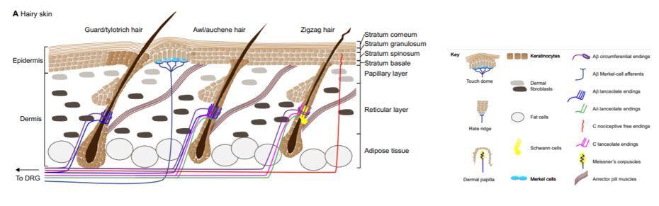
Figure 1. Anatomy of the Skin
Used with permission from Development. Jenkins BA, Lumpkin EA. Developing a sense of touch. Development. 2017;144(22):4078-4090. doi:10.1242/dev.120402
joint.9,10 Despite traditional rehabilitation protocols that focus on incision site healing for approximately two weeks,10,11 the IS continues to contribute to an athlete’s rehabilitation for months after surgery. These contributions illustrate the crucial role of the IS in supporting optimal movement strategies in the long term. The purpose of this clinical commentary is to discuss integumentary considerations within a multi-physiologic systems approach to human movement after ACLR, including an anatomical review, key elements of assessment, and integrated intervention strategies. Additionally, this commentary will identify potential warning signs indicative of impaired IS healing after ACLR.
REVIEW OF ANATOMY
The IS is one of the largest organs of the body. It functions as a structural barrier as well as providing thermoregulation, sensation, and balancing of hydration and electrolytes.12 Age, fitness, nutrition, and genetics all play a large role in the health of the skin.13 The epidermis is the outermost layer, followed by the dermis, the most critical layer in wound healing. The dermis gives the skin its strength and elasticity while containing superficial vessels and nerves. Beneath the dermis lies the third portion of the IS: adipose tissue. Within the subcutaneous fat, there are larger nerves and vascular structures.12 (Figure 1)
Skin thickness, innervation, adnexal structures, and elasticity vary depending on the location in the body. 13 Areas of the knee with prominent and superficial bony structures, such as the patella, tibial tuberosity, and anterior crest of the tibia have a mobile dermal layer that is relatively inelastic with minimal fat composition.13 Subcutaneous bursae reduce friction between the dermis and fascia while Havers’ glands (“fat pads”) surround the patellar tendon.
SURGICAL RECONSTRUCTION OF THE ACL
There are currently four common surgical reconstruction techniques used for the ACL. The bone-patellar-tendonbone (BPTB), hamstring (HS), quadriceps tendon (QT), and allograft are the most frequently used ACLR grafts.9,14 The organization of the IS and the impact scarring has on the body varies depending on the incision site’s location, the depth and direction of the incision, the underlying tissues (muscle, fascia, tendon, bone, etc.), and the overall health (including blood flow and mobility) of the soft tissues being incised. Skin tension lines run vertically along the shin and horizontally at the knee.13 Scars that run contrary to the skin tension lines are more likely to require revision due to increased strain and risk of dehiscence or spread.15 A visual of surgical site locations and skin tension lines can be seen in Figure 2.
Each of the autografts (BPTB, HS, QT) requires healing of the harvest site and the healing of the ACL graft including bone, cartilage, and soft tissue remodeling.9,11 Not all graft sites heal at the same rate; BPTB and QT grafts recover slower than HS and allografts in muscle and tendon strength.14,16 Despite a quicker recovery of strength, HS grafts and allografts have been associated with higher retear rates, especially in younger athletes.17 While not interdependent, the healing of the fascia, harvest sites, and skin progress concurrently. Gradually increasing weightbearing and progressively increasing the resistance of exercises throughout the range of motion can simultaneously facilitate remodeling of the integumentary and other body systems, including the neuromuscular and musculoskeletal systems.11 Weight-bearing progression may have specific implications for the musculoskeletal system, given decreased bone mineral density has been reported across several joints of the lower extremity. 18–20 Protracted healing and adverse scarring can slow the athletes’ progression in multiple facets of rehabilitation (range of motion, locomotion, strength training) due to limited range of motion and pain.
Several methods exist to approximate the skin in order to promote healing.15 The most frequent technique after ACLR is closure of the dermis and subcutaneous tissue with absorbable sutures and reinforcement with superficial epidermal sutures, steri-strips or cyanoacrylate adhesive (such as Dermabond; Ethicon Inc, Somerville, NJ). Surgical sutures that are not absorbable are typically removed within two weeks to prevent hash marks (a series of parallel scars connecting suture sites perpendicular to the incision) due to re-epithelialization at the suture puncture sites. Steri-
strips or similar devices can be used to support healing once sutures are removed.
ASSESSMENT OF THE INTEGUMENTARY SYSTEM
Assessment of the IS should be performed in a clean, well lit room. The patient’s history, including nutrition, smoking history, history of previous scarring or infections, and systemic conditions such as immunosuppression or diabetes should be noted.21 Surgical history, including length of surgical procedure, should also be documented, with procedures lasting over two hours at higher risk of infection.22 Skin should be observed for incision closure and skin integrity, including areas of discoloration, edema, scarring, nodules, and/or scaly skin.23 Any discharge from the incision site should be noted with location, color, and smell. Areas that feel warm should be documented.22 Skin mobility should be assessed around the incision and any areas with edema or altered skin integrity should be noted. Signs of infection should be recognized as localized warmth, erythema (redness), localized pain and/or drainage within 30 days of the surgery, most commonly in the first 4-10 days post-operatively.
22
As the athlete progresses, the incision site should continue to be monitored. Scars can take 1 year or longer to mature. Gentle massage and treatments described below can aid in the remodeling process.24 Many athletes report sensation changes around the incision site.17,25,26 Sensation after ACLR can be evaluated using pain reported during kneeling,17 monofilaments to determine the cutaneous sensation detection thresholds,25 and vibratory perception thresholds for vibration sense.26 Proper management and treatment of the IS will aid in the athlete’s ability to recover after ACLR.
TREATING THE INCISION SITES
Cutaneous wounds heal in three phases: inflammatory (two to three days), proliferative (two to three weeks) and remodeling (up to 12 months). Creating an optimized environment for wound healing allows these phases to progress appropriately. After surgery, the tensile strength of the skin progresses from 3% of normal tissue at week one to 20% at week three and roughly 80% at week twelve.27 Increased age, smoking status and other comorbidities (diabetes, nutritional deficiencies, etc.) that that hinder nutrient and oxygen delivery to wound tissue will prolong healing.28,29 If the healing of the incision site is protracted due to bleeding, pain, infection or dehiscence, it could contribute to limitations in achieving range-of-motion goals and adversely affect multiple body systems.29
The first 12 days post-operatively are the most important for management of the incision site and prevention of wound dehiscence.28,29 Wound care for the incision site is essential for efficient healing and prevention of infection. Surgical sites should be gently cleaned daily with soap and water followed by application of semi-occlusive hydrating emollients, such as petroleum jelly, and a dressing. Emollients can provide a protective barrier as well as hydration to the healing skin.28 These interventions promote mobility of the IS and decrease stress on the newly developing
Figure 2. Surgical site locations for the five most common graft types and how they relate to the skin tension lines
1) fat pad portal sites for all grafts, including the allograft, 2) horizontal and vertical quadriceps tendon grafts above the patella with a potential small secondary site by the pes anserine, 3&4) superior-lateral portal sites for the hamstring grafts with incisions by the pes anserine for the hamstring grafts, 5) large anterior medial incision site for the bone patellar tendon bone graft.
skin.29
Patients should avoid submersion into a public water source such as a pool until the incision is completely closed. Hot tubs and other environments that pose a high risk of infection (lakes, rivers) should be avoided until the remodeling phase which starts approximately one month postoperatively. If an infection occurs, systemic antibiotics prescribed by the referring physician are considered the first-line treatment with subsequent operative debridement if the antibiotics are ineffective.30 Silicone sheets and gels, as well as scar massage, have shown efficacy in minimizing excess scar tissue formation.31 Athletes should also be encouraged to use sunscreen the first year post-operatively to protect the new skin around the incision site.
Providers should be aware that many topical products used during wound care can lead to contact sensitization. Allergy to adhesives (as found in postoperative bandaging), antimicrobial products, and other agents may produce an inflammatory rash that can be confused with infection. It is important to recognize that a geometric distribution of an erythematous, scaly rash (typically very pruritic) with a lack of other infectious signs and symptoms are suggestive of an allergic reaction and can be treated with topical steroids and removal of the offending agent.
Range of motion goals are critical to achieve during this time as well. However, because wound strength is lowest in the early period after surgery, a conservative mobility routine is advised the first weeks. Keeping the incision clean
and hydrated while maintaining skin mobility and promoting neural regeneration of cutaneous receptors will provide the best results of managing the rehabilitation of the IS following an ACLR. Addressing underlying IS impairments contributes to optimizing the recovery of other physiologic systems after ACLR, which in turn impacts the patent’s ability to participate in movement-related interventions.
TREATING SENSORY LOSS
Due to the knee’s use as a kneeling structure and the somatosensory changes throughout the limb after ACLR, superficial nerves of the knee, foot, and ankle, should be evaluated pre- and post-operatively.
Locations around and below the knee may feel numb due to sensory nerve severance during the surgical procedure. These areas can remain numb for weeks, months, or years25 after ACLR. The greatest risk for sensation impairments is found along the infrapatellar branch of the saphenous nerve. The graft’s incision site is also likely to sever superficial nerves.25,32,33 Zones of the leg with decreased sensation should be identified as potential risks of other sensory impairments such as: burns, razor burns (or shaving injuries), poor thermoregulation, hypersensitivity, or altered proprioceptive feedback.34 The loss of the native ACL diminishes proprioceptive input and likely affects movement during functional tasks, requiring rehabilitation interventions that target the neuromuscular system.
Sensory integration training should attempt to promote neural regeneration by providing a gradual progression from non-noxious stimuli to more noxious (painful) stimuli, utilizing electrical stimulation and manual therapy interventions to decrease pain and promote healing and mobility of the skin.34 Skin care promotes an optimal environment for neural regeneration, starting with electrical stimulation, scar massage, and regular use of emollients.28,34 An easy way to progress and promote normal sensation, and potentially help identify neurogenic pain,35 is for athletes to wear clothes and coverings on the injured leg as soon as possible. Gentle massage can also provide the skin sensory feedback and improve skin mobility. Concerns of chronic pain or hypersensitivity should be communicated with the physician. Conditions such as nerve entrapment, complex regional pain syndrome (CRPS), or chronic pain may require additional surgical corrections or methods of analgesia (nerve block or medication changes) and pain reduction (such as TENS) techniques to reduce the neurogenic pain.34,35 Progressive return to less than comfortable activities should be integrated into rehabilitation for sensory normalization as appropriate. Kneeling is an early example of a method to identify abnormal sensations (i.e.: pain) and modulating the intensity (such as moving from foam to hard surfaces and from quadruped to tall kneeling) in order to diminish the painful response. As an athlete progresses, sport-specific activities may be painful, requiring progressive loading or sensory integration to reduce fearavoidance movement patterns secondary to altered afferent feedback related to altered sensation and chronic pain.36 The two most common scars that concern patients are those that are stretched and atrophic and those that are thickened (i.e. hypertrophic or keloidal scars). Scars that stretch occur when tension on the wound, created by movement of underlying muscles, overcomes the strength of the skin at the line of closure.12 These changes are most remarkable in the first eight weeks post-operatively and typically do not worsen after the twelfth week.13 Referral to dermatology or plastic surgery for scar revision may be warranted if the patient is displeased with the scar aesthetics.
Hypertrophic scars and keloids are due to excess deposition of collagen at the surgical site. There may be a genetic predisposition for keloid formation thus inquiry into previous wound healing and family history of keloids is important in the preoperative period.37 Keloids and hypertrophic scars often are associated with pain or pruritus and are managed with scar massage and silicone sheets. More aggressive therapy includes intralesional steroid injections, intralesional 5-fluorouracil injections, radiation treatments, and cryotherapy, among others.37,38 These are typically performed by a dermatologist or plastic surgeon. Frequent conversations should be had with the surgeon with a potential referral warranted if symptoms interfere with the knee’s functional movement. As a collaborative practitioner, physical therapists are able to offer a unique perspective on movement-related impairments and interventions to optimize functional capacity and recovery after ACLR.2
CONSIDERING HEALTH OF THE PATIENT
It is important to assess patients’ underlying medical conditions and behaviors as pre-existing conditions could prolong or complicate the healing of the incision or scar. 28,29,34 The skin of younger individuals tends to heal better than older adults after surgery given healthy cardiovascular system and lack of actinic damage (damage from the sun) to the skin. However, these patients are more at risk for hypertrophic or keloidal scars, especially if there is a family or personal history. 39 Genetic conditions such as hypermobility syndromes (i.e., Ehlers Danlos Syndrome) that alter the normal function of connective tissue elements (i.e. collagen, elastin, etc.) predispose affected patients to the potential for poor wound healing and protracted rehabilitation39 and should be considered. Being prepared to address each patient’s potential obstacles to rehabilitation will allow providers to tailor programs appropriately and positively impact the recovery of multiple body systems throughout rehabilitation.
CONCLUSION
The IS contributes to success after ACLR and can provide an early indication of a prolonged rehabilitation process in certain instances, which may ultimately impact other physiologic systems and the patient’s ability to participate in movement-related interventions. Creating incisions along skin tension lines, keeping the incision clean and hydrated with appropriate dressings, and regular close assessment
of wound and scar progression will improve the aesthetic of the scar and limit the potential for poor scar mobility. Furthermore, proper care of the IS can decrease the risk of an infection that could delay or severely dismantle the progress of an individual’s ACL rehabilitation. Despite being overlooked at times, the IS plays a crucial role in the functioning of other physiologic systems after ACLR.
FINANCIAL DISCLOSURES
The authors have declared no conflict of interest. None declared.
ACKNOWLEDGEMENTS
None.
Submitted: October 01, 2021 CST, Accepted: November 01, 2021 CST
This is an open-access article distributed under the terms of the Creative Commons Attribution 4.0 International License (CCBY-NC-SA-4.0). View this license’s legal deed at https://creativecommons.org/licenses/by-nc-sa/4.0 and legal code at https://creativecommons.org/licenses/by-nc-sa/4.0/legalcode for more information.
REFERENCES
1. Mission, Vision, and Strategic Plan. American Physical Therapy Association Website. https://www.a pta.org/apta-and-you/leadership-and-governance/vis ion-mission-and-strategic-plan. Accessed June 17, 2021.
2. American Physical Therapy Association. An American Physical Therapy Association white paper. Physical therapist practice and the human movement system. 2015:1-4.
3. Saladin L, Voight M. Introduction to the movement system as the foundation for physical therapist practice education and research. Int J Sports Phys Ther. 2017;12(6):858-861. doi:10.16603/ijspt2017085 8
4. Hedman LD, Quinn L, Gill-Body K, et al. White paper: movement system diagnoses in neurologic physical therapy. J Neurol Phys Ther. 2018;42(2):110-117. doi:10.1097/npt.00000000000002 15
5. Ludewig PM, Lawrence RL, Braman JP. What’s in a name? Using movement system diagnoses versus pathoanatomic diagnoses. J Orthop Sports Phys Ther. 2013;43(5):280-283. doi:10.2519/jospt.2013.0104
6. Quinn L, Riley N, Tyrell CM, et al. A framework for movement analysis of tasks: recommendations from the Academy of Neurologic Physical Therapy’s Movement System Task Force. Phys Ther. 2021;101(9). doi:10.1093/ptj/pzab154
7. Gerrett N, Griggs K, Redortier B, Voelcker T, Kondo N, Havenith G. Sweat from gland to skin surface: production, transport, and skin absorption. J Appl Physiol. 2018;125(2):459-469. doi:10.1152/japplphysi ol.00872.2017
8. Jenkins BA, Lumpkin EA. Developing a sense of touch. Development. 2017;144(22):4078-4090. doi:1 0.1242/dev.120402
9. Beynnon BD, Johnson RJ, Abate JA, Fleming BC, Nichols CE. Treatment of anterior cruciate ligament injuries, part I. Am J Sports Med. 2005;33(10):1579-1602. doi:10.1177/03635465052799 13
10. van Grinsven S, van Cingel RE, Holla CJ, van Loon CJ. Evidence-based rehabilitation following anterior cruciate ligament reconstruction. Knee Surg Sports Traumatol Arthrosc. 2010;18(8):1128-1144. doi:10.100 7/s00167-009-1027-2 11. Wright RW, Haas AK, Anderson J, et al. Anterior cruciate ligament reconstruction rehabilitation: MOON guidelines. Sports Health. 2015;7(3):239-243. doi:10.1177/1941738113517855
12. Bolognia JL, Jorizzo JL, Schaffer JV. Dermatology. 3rd ed. London: Elsevier; 2012.
13. Robinson JK. Anatomy for procedural dermatology. In: Robinson JK, Hanke CW, Siegel DM, Fratila A, Bhatia AC, Rohrer TE, eds. Surgery of the Skin. 3rd ed. St. Louis: Elsevier; 2015.
14. Mouarbes D, Menetrey J, Marot V, Courtot L, Berard E, Cavaignac E. Anterior cruciate ligament reconstruction: a systematic review and metaanalysis of outcomes for quadriceps tendon autograft versus bone-patellar tendon-bone and hamstringtendon autografts. Am J Sports Med. 2019;47(14):3531-3540. doi:10.1177/03635465188253 40
15. Weitzul S, Taylor RS. Suturing technique and other closure materials. In: Robinson JK, Hanke CW, Sengelmann R, Siegel DM, eds. Surgery of the Skin. 1st ed. St. Louis: Mosby; 2005:225-244.
16. Smith AH, Capin JJ, Zarzycki R, Snyder-Mackler L. Athletes with bone-patellar tendon-bone autograft for anterior cruciate ligament reconstruction were slower to meet rehabilitation milestones and returnto-sport criteria than athletes with hamstring tendon autograft or soft tissue allograft: secondary analysis from the ACL-SPORTS trial. J Orthop Sports Phys Ther. 2020;50(5):259-266. doi:10.2519/jospt.2020.9111
17. Widner M, Dunleavy M, Lynch S. Outcomes following ACL reconstruction based on graft type: are all grafts equivalent? Curr Rev Musculoskelet Med. 2019;12(4):460-465. doi:10.1007/s12178-019-0958 8-w
18. Nyland J, Fisher B, Brand E, Krupp R, Caborn DN. Osseous deficits after anterior cruciate ligament injury and reconstruction: a systematic literature review with suggestions to improve osseous homeostasis. Arthroscopy. 2010;26(9):1248-1257. do i:10.1016/j.arthro.2010.03.017
19. van Meer BL, Waarsing JH, van Eijsden WA, et al. Bone mineral density changes in the knee following anterior cruciate ligament rupture. Osteoarthritis Cartilage. 2014;22(1):154-161. doi:10.1016/j.joca.201 3.11.005
20. Reiman MP, Rogers ME, Manske RC. Interlimb differences in lower extremity bone mineral density following anterior cruciate ligament reconstruction. J Orthop Sports Phys Ther. 2006;36(11):837-844. doi:1 0.2519/jospt.2006.2278
21. Weinstein MC, Vidimos AT. Patient evaluation, informed consent, preoperative assessment, and care. In: Robinson JK, Hanke CW, Siegel DM, Fratila A, Bhatia AC, Rohrer TE, eds. Surgery of the Skin. 3rd ed. St. Louis: Elsevier; 2015.
22. Marlwalla K. Antibiotics. In: Robinson JK, Hanke CW, Siegel DM, Fratila A, Bhatia AC, Rohrer TE, eds. Surgery of the Skin. 3rd ed. St. Louis: Elsevier; 2015.
23. High WA, Tomasini CF, Argenziano G, Zalaudek I. Basic principles of dermatology. In: Bolognia JL, Schaffer JV, Cerroni L, eds. Dermatology. 4th ed. China: Elsevier; 2018.
24. Leitenberger JJ, Isenhath SN, Swason NA, Lee KK. Revision of surgical scars. In: Robinson JK, Hanke CW, Siegel DM, Fratila A, Bhatia AC, Rohrer TE, eds. Surgery of the Skin. 3rd ed. St. Louis: Elsevier; 2015.
25. Hoch JM, Perkins WO, Hartman JR, Hoch MC. Somatosensory deficits in post-ACL reconstruction patients: a case-control study. Muscle Nerve. 2017;55(1):5-8. doi:10.1002/mus.25167
26. Cronström A, Ageberg E. Association between sensory function and medio-lateral knee position during functional tasks in patients with anterior cruciate ligament injury. BMC Musculoskelet Disord. 2014;15:430. doi:10.1186/1471-2474-15-430
27. Son D, Harijan A. Overview of surgical scar prevention and management. J Korean Med Sci. 2014;29(6):751-757. doi:10.3346/jkms.2014.29.6.751
28. Jourdan M, Madfes DC, Lima E, Tian Y, Seité S. Skin care management for medical and aesthetic procedures to prevent scarring. Clin Cosmet Investig Dermatol. 2019;12:799-804. doi:10.2147/ccid.S218134
29. Agha R, Ogawa R, Pietramaggiori G, Orgill DP. A review of the role of mechanical forces in cutaneous wound healing. J Surg Res. 2011;171(2):700-708. doi:1 0.1016/j.jss.2011.07.007
30. Judd D, Bottoni C, Kim D, Burke M, Hooker S. Infections following arthroscopic anterior cruciate ligament reconstruction. Arthroscopy. 2006;22(4):375-384. doi:10.1016/j.arthro.2005.12.002 31. Marini L, Odendaal D, Smirnyi S. Importance of scar prevention and treatment-an approach from wound care principles. Dermatol Surg. 2017;43 Suppl 1:S85-s90. doi:10.1097/dss.0000000000001001
32. Grassi A, Perdisa F, Samuelsson K, et al. Association between incision technique for hamstring tendon harvest in anterior cruciate ligament reconstruction and the risk of injury to the infrapatellar branch of the saphenous nerve: a metaanalysis. Knee Surg Sports Traumatol Arthrosc. 2018;26(8):2410-2423. doi:10.1007/s00167-018-485 8-x
33. Sharaby MMF, Alfikey A, Alhabsi IS, Al-Ghannami S. No difference in sensory outcome between vertical and oblique incisions for hamstring graft harvest during ACL reconstruction. Knee Surg Sports Traumatol Arthrosc. 2019;27(1):146-152. doi:10.1007/ s00167-018-5057-5
34. Lebonvallet N, Laverdet B, Misery L, Desmoulière A, Girard D. New insights into the roles of myofibroblasts and innervation during skin healing and innovative therapies to improve scar innervation. Exp Dermatol. 2018;27(9):950-958. doi:10.1111/exd.1 3681
35. Hong AJ, Agarwalla A, Liu JN, et al. Neurological structures and mediators of pain sensation in anterior cruciate ligament reconstruction. Ann Anat. 2019;225:28-32. doi:10.1016/j.aanat.2019.05.010
36. Burland JP, Lepley AS, Cormier M, DiStefano LJ, Arciero R, Lepley LK. Learned helplessness after anterior cruciate ligament reconstruction: an altered neurocognitive state? Sports Med. 2019;49(5):647-657. doi:10.1007/s40279-019-01054-4
37. Ud-Din S, Bayat A. Strategic management of keloid disease in ethnic skin: a structured approach supported by the emerging literature. Br J Dermatol. 2013;169 Suppl 3:71-81. doi:10.1111/bjd.12588
38. Berman B, Maderal A, Raphael B. Keloids and hypertrophic scars: pathophysiology, classification, and treatment. Dermatol Surg. 2017;43 Suppl 1:S3-s18. doi:10.1097/dss.0000000000000819
39. Bolognia JL, Schaffer JV, Cerroni L. Dermatology. 4th ed. China: Elsevier; 2018.
Clinical Commentary/Current Concept Review
Forefoot Injuries in Athletes: Integration of the Movement System
Lindsay A Carroll 1 , Stephen Paulseth 2 , RobRoy L Martin 1 a
1 Physical Therapy, Duquesne University, 2 Paulseth & Associates Physical Therapy, Inc Keywords: taping, foot, joint mobilization, sports injuries, movement system https://doi.org/10.26603/001c.30021
International Journal of Sports Physical Therapy
Vol. 17, Issue 1, 2022
Despite the prevalence of forefoot related problems in athletes, there are few comprehensive summaries on examination and intervention strategies for those with forefoot related symptoms. While many factors may contribute to pathology and injury, the presence of abnormal foot alignment can negatively affect lower extremity biomechanics and be associated with injuries. Physical therapists may use the characteristics associated abnormal pronation or abnormal supination to describe the movement system disorder and serve as a guide for evaluating and managing athletes with forefoot pathologies. Athletes with an abnormal pronation movement system diagnosis typically demonstrate foot hypermobility, have decreased strength of the tibialis posterior muscle, and present with a medially rotated lower extremity position. Athletes with abnormal supination movement system diagnosis typically demonstrate foot hypomobility, decreased strength of the fibularis muscles, and a laterally rotated lower extremity position. Interventions of manual therapy, taping, strengthening exercises, and neuromuscular reeducation can be directed at the identified impairments and abnormal movements. The purpose of this clinical commentary is to integrate a movement system approach in pathoanatomical, evaluation, and intervention considerations for athletes with common forefoot pathologies, including stress fractures, metatarsalgia, neuroma, turf toe, and sesamoiditis. By applying a prioritized, objective problem list and movement system diagnosis, emphasis is shifted from a pathoanatomical diagnosis-based treatment plan to a more impairment and movement focused treatment.
Level of Evidence
5
INTRODUCTION
It is well known that athletes are at risk for foot and ankle injuries. These include injuries in the forefoot, defined as the region of the foot distal to the tarsometatarsal joints. The forefoot, unlike the mid- and hindfoot, is unconstrained with movement occurring freely in all three planes. Because the forefoot is the most distal weight bearing segment of the lower extremity, it can undergo a substantial amount of stress and strain, and can be affected by footwear, terrain, and biomechanical factors in the entire lower kinetic chain. Despite the prevalence of forefoot related problems in athletes, there are few comprehensive summaries on examination and intervention strategies for those with forefoot related symptoms. The purpose of the clinical commentary is to integrate a movement system approach in pathoanatomical, evaluation, and intervention considerations for athletes with common forefoot pathologies, including stress fractures, metatarsalgia, neuroma, and sesamoiditis.
ANATOMY OF THE FOREFOOT
The forefoot is composed of five rays that are functionally
a
Corresponding Author:
RobRoy L. Martin, PT, PhD, CSCS Duquesne University Department of Physical Therapy 111A RSHS Pittsburgh, PA 15282 martinr280@duq.edu Phone: 412-396-1811 Fax: 412-396-4399


