IJSRD - International Journal for Scientific Research & Development| Vol. 3, Issue 08, 2015 | ISSN (online): 2321-0613
Medical Image De-Noising using Wavelet Transform Jagdeep Kaur1 Ruchika Manchanda2 1,2 Department of Electronics & Communication Engineering 1,2 Guru Nanak Institute of Technology, Mullana, India Abstract— The goal of de-noising method in Wavelet denoising removes the noise present in the image while preserving the image characteristics regardless of its frequency content. Wavelets preserve visual quality and also maintain the diagnostically significant details of medical images. The purpose of de-noising is to remove the noise while retaining the edges and other detailed features as much as possible. In this paper a wavelet based denoising technique is evaluated by implementing the improved algorithm on medical images with simulated noise. To conclude standard parameters like MSE and PSNR are used for image evaluation. Key words: Medical Image De-Noising, Wavelet Transform
Noise Ratio[15]. Wavelet based de-noising has opened up other fields and important techniques such as denoising and non-linear approximation, smoothing and restoration of images. The goal of image de-noising is to remove noise by differentiating it from the signal. The wavelet transform’s energy compactness helps greatly in de-noising. The Discrete Wavelet Transform (DWT) is a powerful tool for multi resolution decomposition of image.
I. INTRODUCTION Technological developments in X-Rays, ultrasound, magnetic resonance imaging and other imaging techniques have produced a non-invasive method for diagnosis. A number of new techniques have also flourished, making use of latest technology for better imaging. Imaging can provide uniquely valuable information about physical and structural properties of organs, as well as quantitative descriptions of many fundamental biological processes. There are many advanced methods ofimage processing involving techniques that include the traditional Fourier transform and the wavelet transform [1]. Recently there exist a variety of wavelet transform based methods developed with added advantages over classical methods, these include shift invariance [2] and improved texture conservation. The denoising algorithms apply a chosen method of wavelet decomposition for the reconstruction of medical images [4] The image-processing process amplifies part of the noise and adds its own rounding noise. Rounding noise occurs because there are only a finite number of bits to represent the intermediate floating point results during computations. Most de-noising algorithms assume zero mean additive white Gaussian noise (AWGN) because it is symmetric, continuous, and has a smooth density distribution [11]. Researchers published different ways to compute the parameters forthe thresholding of wavelet coefficients. Data adaptive thresholds [6] were introduced to achieve optimumvalue of threshold. Later efforts found that substantial improvements in perceptual quality could be obtained by translation invariant methods based on Thresholding of an Un-decimated Wavelet Transform [ 3] These thresholding techniques were applied to the nonorthogonal wavelet coefficients to reduce artifacts. Multiwavelets were also used to achieve similar results. Probabilistic models using the statistical properties of the wavelet coefficient seemed to out perform the thresholding techniques and gained ground. Recently, much effort has been devoted to Bayesian de-noising in Wavelet domain [13]. The performance of the de-noising algorithms is quantitatively assessed using different criteria namely the PSNR, MSE and the visual appearance. Two commonly used measures are Mean-Squared Error and Peak Signal-to-
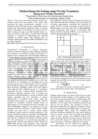
Fig. 1: Wavelet Decomposition Tree The wavelet decomposition of an image is done as follows: In the first level of decomposition, the image is split into four sub bands, namely HH1, HL1, LH1, and LL1, as illustrated in Figure 1. The HH1 sub-band gives the diagonal details of the image; the HL1 sub band gives the horizontal features, while the LH1 represents the vertical structures. The LL1subband is the low resolution residual consisting of low frequency components and it is this sub band which is further split at higher levels of decomposition. It has been shown that the noise standard deviation žw can be accurately estimated from the first decomposition level diagonal sub band HH1 by the robust and accurate median estimator [4], as given by (|
|)
(1) The choice of wavelet is dictated by the signal or image characteristics and the nature of the application. II. METHODOLOGY Standard medical images are taken for the process to study the performance of the algorithm. A noise addition code is first written to add noise artificially in original image. The parameter standard deviation for noise is controlled in algorithm. A set of images with varying SD is produced and kept for the test purpose. The simulation of noise is performed to mimic the real conditions of a medical system. Here, we used ultrasound and MRI images for testing of denoising algorithm. These images are corrupted with uniformly distributed multiplicative noise having different levels of variance of the noise. By taking four different values of noise variances, four different medical noisy images are being obtained. The range of noise variance are [0.1 1], where the variance 0.1 represents low level noisy image and variance 1 represents a high level noisy image. De-noising can be achieved by applying a thresholding operator to the wavelet coefficients (in the
All rights reserved by www.ijsrd.com
320