EXPRESS RADIOLOGY | 2025

Viveka Roychowdhury, Editor viveka.r@expressindia.com

EXPRESS RADIOLOGY | 2025
Viveka Roychowdhury, Editor viveka.r@expressindia.com
As Chennai prepares to host the 23rd Asian Oceanian Congress of Radiology (AOCR 2025) and the 77th Annual Conference of Indian Radiological & Imaging Association, this becomes the third AOCR conference hosted by IRIA ,after the 6th AOCR in New Delhi (1991) and the 17th AOCR in Mumbai (2018).
Tagged as the largest international radiological conference in India,the largest in Asia and the third biggest in the world, as per the AOCR 2025 website, radiologists can look forward to a lot of learning crammed into the four day-event.
The theme chosen for the conference, Clinical Radiology Decoded - See like a Surgeon: Think Like a Physician, is indeed timely and reflects the urgent need to integrate, train and prepare radiologists for working as part of multidisciplinary medical teams.
As Noriyuki Tomiyama, President, Asian Oceanian Society of Radiology (AOSR) mentions in his message on the AOCR 2025 site, “The theme … sets a clear direction for radiologists. As diagnostic modalities such as CT and MR continue to advance and new scanning techniques emerge, radiologists play an increasingly crucial role, making significant contributions to healthcare.”
Treating a patient with a serious illness is nothing short of a relay race. While new age tools like AI, ML, augmented
intelligence et al,provide the means to analyse more reports, in less time and across international boundaries, it is human intelligence and compassion which must interpret the image, convert the data into a coherent diagnosis and then convey the same to the next set of treating medical specialists.
This could be a surgeon who needs specific information before she/he operates and /or a physician who needs to decide how to manage pre and post operative care. Or help the patient and caregivers manage the patient’s daily routine.
Unless there is a smooth hand over of the baton of care between each medical expert, there could be tragic consequences. And in today’s social media-charged society, tragic cases translate into physical violence endangering the lives of medical staff. If such incidents are not attended to, it could result in loss of reputation and prolonged medico-legal cases.
This edition of Express Radiology, an
AOCR 2025 special edition, has practicing radiologists weigh in on topics like the use of AI tools to augment radiologists’ ability to interpret complex cases, as well as the need for collaboration between radiologists, surgeons, and physicians. And most importantly, how the community is training the next generation of radiologists to develop a surgeon’s eye.
Writing for Express Radiology, Dr Sneha Kini, Consultant Imaging, P. D. Hinduja Hospital & MRC, Mahim, Mumbai sums up the situation best: Preoperative imaging forms a ‘Google map’ for our new age Abhimanyu surgeons to skillfully navigate through the ‘Chakravyuh’ or ‘labyrinth’ of human body.
But the same tools that are being celebrated today could disrupt the sector beyond imagination. While AI tools can help radiology clinics process more scans in less time, at lower prices, will radiologists become too expensive and obsolete? Is this the commoditisation of a valuable skill set?
So how do radiologists stay relevant midst the onslaught of artificial intelligence driven diagnosis? How far are we from the day that an AI driven diagnosis is preferred over a human-made diagnosis?
These are existential questions that need to be asked, even if they are uncomfortable. Because asking the right questions is the first step in the search for the right answer.


P14: Dr (Lt Col )Priscilla Joshi Prof and Head-Department of Radiodiagnosis,Vice Principal PG Academics,Bharati Vidyapeeth (DU) Medical College Pune
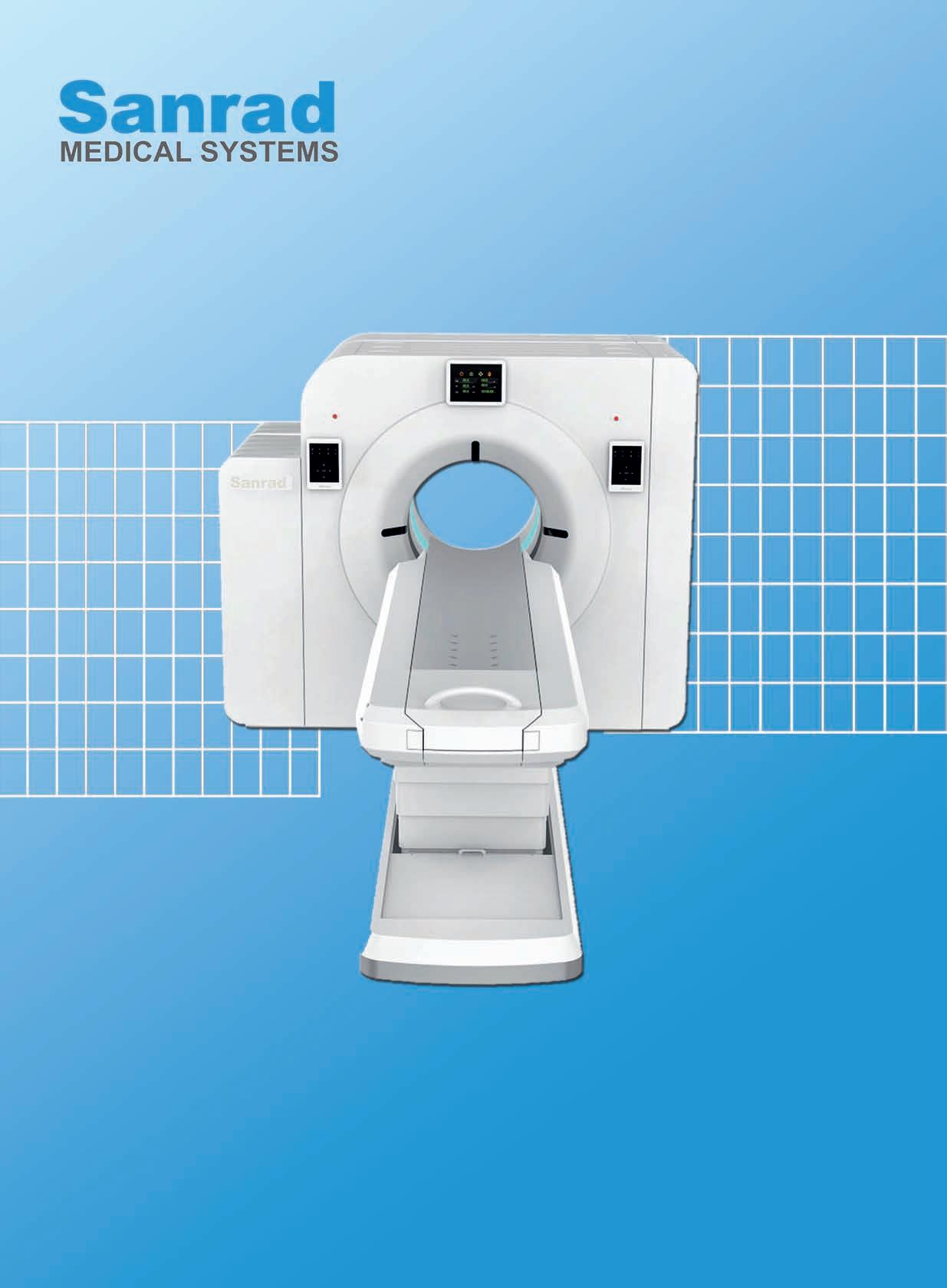
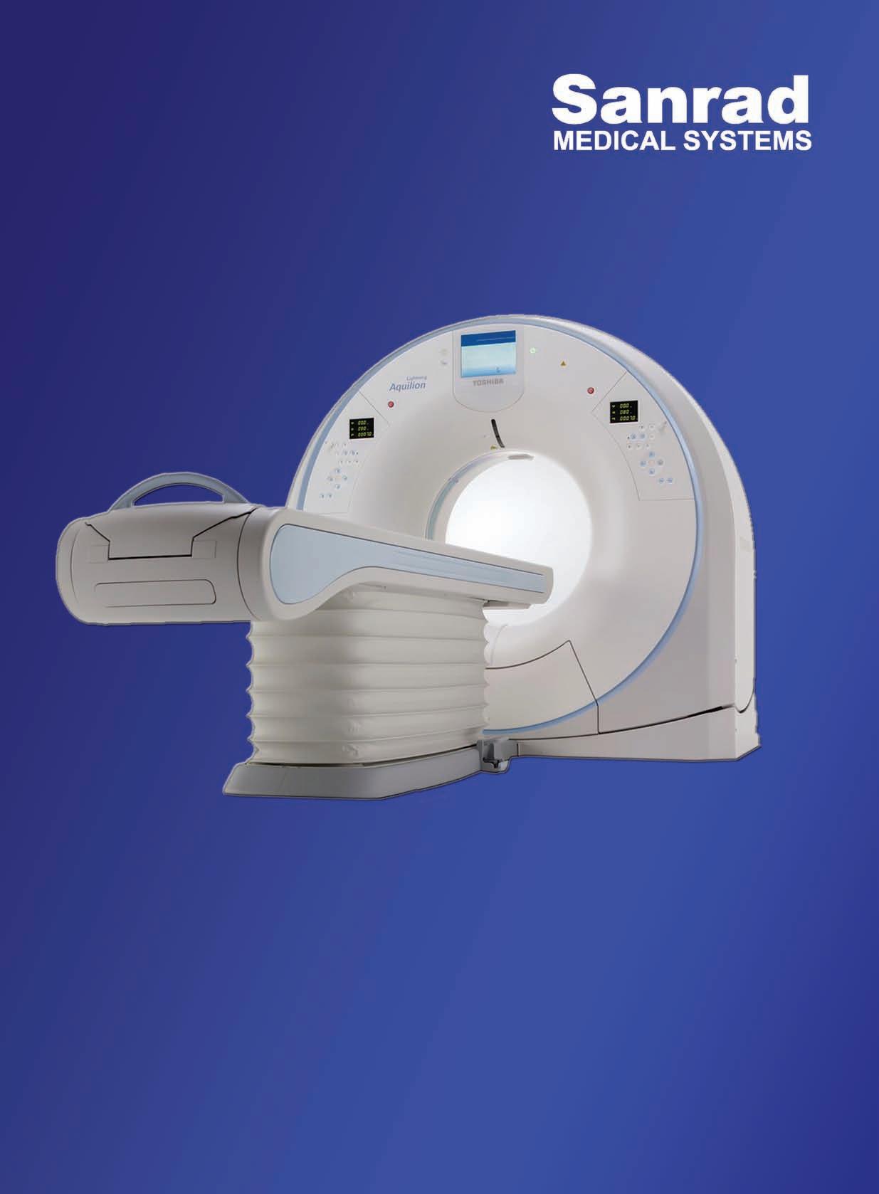
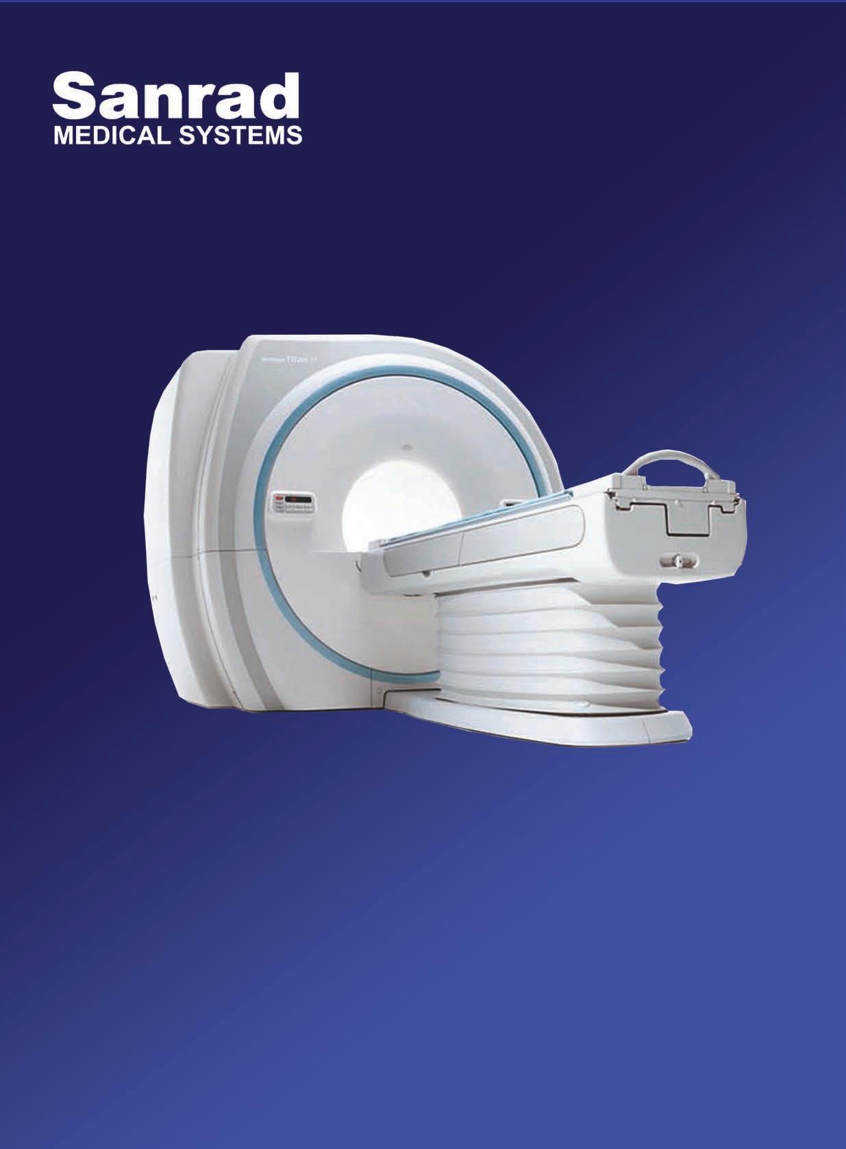
P18: Ratish Nair CEO, Sanrad Medical Systems
P21: Brijesh Suneja Director, Phantom Healthcare
P24: Praveen Rajgopal Global product marketing Manager, Carestream Health
P30: Prakash Padmalwar Managing Director, Evertech
P32: S Viswanathan Managing Director, Sequoia Healthcare


P41:

Chairman of the Board
ViveckGoenka
Sr.Vice President-BPD
Neil Viegas
Vice President-BPD
Harit Mohanty
Editor
Viveka Roychowdhury*
Editorial Team
Lakshmipriya Nair
Kalyani Sharma
Kavita Jani
Neha Aathavale
Design
Art Director
Pravin Temble
Senior Designer
Rekha Bisht
Senior Artist
Rakesh Sharma
Marketing Team
Rajesh Bhatkal
Douglas Menezes
Ashish Rampure
Debnarayan Dutta
Production Co-ordinator
DhananjayNidre
Scheduling & Coordination
Pushkar Waralikar
CIRCULATION
Mohan Varadkar

Iam Professor Noriyuki Tomiyama from Osaka University, and I have had the privilege of serving as the President of the Asian Oceanian Society of Radiology (AOSR) since 2023. It is my distinct pleasure and honor to extend a warm welcome to everyone attending the 23rd Asian Oceanian Congress of Radiology (AOCR 2025) in Chennai, hosted by the Indian Radiological and Imaging Association (IRIA). Notably, IRIA previously hosted the 6th AOCR in New Delhi in 1991 and the 17th AOCR in Mumbai in 2018.
On behalf of AOSR, we extend our deepest gratitude to IRIA for hosting AOCR 2025. We anticipate another successful event and an enriching experience. Congratulations to Dr V.N. Varaprasad, President of IRIA, and Dr T.S. Swaminathan and Dr N. Kulasekaran, Patrons of AOCR 2025. Additionally, we express our appreciation to Dr C. Amarnath, the Organising Chairman, Dr L. Murali Krishna, the Organising Secretary, and all staff members contributing
Noriyuki Tomiyama President
to this outstanding congress.
The theme for AOCR 2025, 'Clinical Radiology Decoded - See like a Surgeon: Think Like a Physician,' sets a clear direction for radiologists. As diagnostic modalities such as CT and MR continue to advance and new scanning techniques emerge,
radiologists play an increasingly crucial role, making significant contributions to healthcare.
Chennai is the gateway to South Indian art, culture, and traditions, encompassing resplendent classical dance, the rich flavors of its iconic cuisine, and a wealth of ancient temples and natural wonders. I am sure we would all enjoy our time there during AOCR 2025.
See you in Chennai!
We are delighted to welcome you all to the 23rd Asian Oceanian Congress of Radiologyand the 77th Annual Conference of Indian
Dr C.Amarnath Organising Chairman
Radiological & Imaging Association
AOCR 2025set to take place in the
vibrant city of Chennai in January 2025. With its rich cultural heritage and technological advancements, Chennai promises to be an ideal backdrop for our upcoming
attendees. The delegates will also have a platform to present their cutting edge scientific work. Chennai, known for its bustling energy and hospitable culture, will
OUR SCIENTIFIC COMMITTEE HAS DILIGENTLYCRAFTED A COMPREHENSIVE PROGRAM THATWILLCATER TO THE DIVERSE NEEDS OFBOTH SEASONED RADIOLOGISTS AND ENTHUSIASTIC RADIOLOGYRESIDENTS
conference.
Our scientific committee is diligently crafting a comprehensive program that will cater to the diverse needs of both seasoned radiologists and enthusiastic radiology residents. The themeClinical Radiology Decoded - See like a Surgeon: Think Like a Physicianis relevant in today's evolving radiology practice. Renowned experts and thought leaders in the field will share their insights through engaging lectures, innovative demonstrations, hands on workshops ensuring an enriching experience for all
provide the perfect setting for a conference that seamlessly blends academic excellence with opportunities for networking and camaraderie. Join us for an unforgettable experience that combines the best of academic enrichment and the vibrant spirit of Chennai.
We eagerly anticipate the opportunity to welcome you and your esteemed colleagues to the 77th Annual Radiology Conference. Let's come together to learn, share, and forge new connections in the ever-evolving field of radiology.
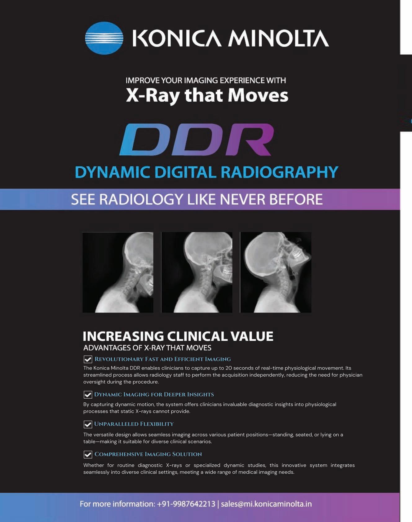
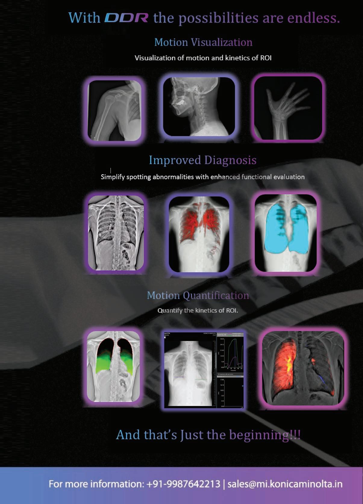
What motivated you to adopt Dynamic Digital Radiology (DDR) in your practice?
My first exposure to DDR was at the IRIA 2020 in Gandhinagar – just pre COVID when I visited the Konica Minolta’s stall and saw a video film on it . It seemed to be a very promising and exciting development, so when we were looking at acquiring a new DR (our previous DR is also Konica Minolta’s first full room DR installation in India) we were offered this machine as the first DDR installation in India. I was quite excited about it.
None of us really knew all its applications and capabilities and we are still discovering several new applications with the help of the applications team from Konica Minolta.
Evaluation of lung perfusion with a breath hold of 7 secs seems very promising and we have compared DDR with pulmonary angiography for detection of Pulmonary embolism. We had a paper presentation on this from our institute at the RSNA 2023.
What specific features of DDR do you find most beneficial in your daily practice?
In addition to evaluation of lung perfusion, DDR offers several features that I find highly beneficial in daily practice, especially in areas

requiring detailed functional assessment of moving structures. One of the most impactful aspects is its ability to provide real-time, motion-based imaging. In respiratory studies, DDR enables dynamic visualisation of lung and diaphragm motion, which is invaluable in diagnosing conditions like COPD or restrictive lung disease as well as in the evaluation of the pre and post operative thoracic region.
Another key feature is its ability to capture multiple frames over time, which facilitates more comprehensive joint evaluations, such as assessing biomechanics in weight-bearing studies of the knee or spine. This level of detail is difficult to achieve with
conventional radiography. Additionally, DDR's reduced radiation dose compared to fluoroscopy which makes it a safer option for patients, especially those requiring repeated imaging. Overall, the combination of dynamic imaging capability, diagnostic accuracy, and patient safety makes DDR an essential tool in my practice
Have you noticed any improvements in patient outcomes since using DDR? Yes, I have noticed significant improvements in patient outcomes since incorporating DDR into practice. One of the most notable benefits is its ability to provide dynamic, functional imaging, which enables earlier and more accurate diagnoses.
For example, DDR has improved outcomes is in musculoskeletal evaluations. By visualising joint movement dynamically, we can identify subtle biomechanical abnormalities that might be missed with static imaging. This has been particularly helpful in guiding physical therapy or surgical interventions, ensuring patients receive the most appropriate care. Another area where DDR has improved outcomes is in cases of respiratory conditions like COPD or diaphragm dysfunction, DDR allows us to assess lung and diaphragm
motion in real time, which leads to more targeted treatment plans. This has resulted in better symptom management and improved quality of life for patients.
How do patients react or respond when you use this technology during their treatment?
Patients generally respond very positively when we use DDR to facilitate diagnosis and management. One of the most common reactions is a sense of reassurance and trust, as the dynamic imaging allows us to show them a real-time visual of how their body is functioning—for example, lung motion or joint movement. Patients also appreciate the efficiency of DDR. Since the technology provides both dynamic and static information in one session, they don’t have to undergo multiple tests, which reduces their time spent in the clinic and minimises discomfort. Additionally, they are often relieved to hear that DDR involves lower radiation exposure compared to other imaging techniques, particularly for conditions that require ongoing monitoring.
Overall, I’ve noticed that the interactive nature of DDR improves patient engagement. They feel more involved in their care and are more likely to adhere to treatment plans because they understand their condition better. This has a direct impact on patient satisfaction and outcomes.
Has DDR helped streamline your workflow or save time in any specific areas?
Yes, DDR has significantly
ONE OFTHE KEY ADVANTAGES IS ITS ABILITYTO PROVIDE DYNAMIC FUNCTIONAL IMAGING ALONGSIDE STATIC RADIOGRAPHS IN A SINGLE SESSION
While integrating DDR into routine practice has brought many benefits, there were some initial challenges. One of the primary challenges was the learning curve associated with interpreting dynamic images. Unlike static radiography, DDR provides motion-based data, so it required additional training to analyse the functional aspects of joint movement, lung function and other dynamic studies accurately.
streamlined workflow and saved time in several ways. One of the key advantages is its ability to provide dynamic functional imaging alongside static radiographs in a single session. For example, in respiratory assessments or musculoskeletal evaluations, DDR eliminates the need for separate tests like fluoroscopy or repeated imaging sessions, which speeds up the diagnostic process.
Another benefit is the immediate availability of high-quality, detailed images. The dynamic data DDR provides often eliminates the need for follow-up imaging because it gives a more comprehensive picture of the patient’s condition upfront. This reduces repeat appointments and shortens the overall timeline for diagnosis and treatment planning.
Overall, DDR has allowed us to work more efficiently while improving diagnostic accuracy, which ultimately benefits both the clinical team and the patient.
Are there any challenges you've faced while integrating DDR into your routine?
Another challenge was optimising workflow during the implementation phase. Incorporating new technology meant adjusting scheduling and ensuring the radiology team was familiar with both the operation of the equipment and the interpretation of results.
Educating referring physicians about the added value of DDR was also essential to ensure they understood when to order this modality and how it could improve patient management.
Lastly, like with any advanced technology, there were initial concerns about cost and resource allocation. H owever, as the team became more adept at using DDR and its clinical value became evident, these challenges were mitigated. Today, DDR has become a seamless part of the workflow and has prov en to be an invaluable tool in improving patient outcomes.
How user-friendly do you find the interface and functionality of DDR?
I find the interface and functionality of DDR to be highly user-friendly, especially once the initial learning curve is overcome. The software is intuitive, with clearly organized
tools for adjusting image parameters, reviewing dynamic sequences and one click measurement summary.
One aspect I particularly appreciate is how easily I can switch between static and dynamic views. The ability to analyse motion frame by frame or as a continuous sequence is both efficient and straightforward. Additionally, the integration of automated tools, like motion analysis or quantitative measurements, enhances the functionality without being overly complex.
Overall, DDR’s interface is designed to complement clinical workflows, and the system's responsiveness helps ensure a smooth and efficient experience during imaging sessions. This usability is critical in a busy clinical environment, as it allows both radiologists and technologists to adopt the technology with minimal disruption.
How do you perceive the overall value of DDR compared to its cost?
I perceive the overall value of DDR as being well worth the investment, especially when considering the clinical benefit it offers. While the initial cost of DDR may be higher than traditional radiography systems, the long-term value it provides in terms of improved diagnostic accuracy, enhanced patient outcomes, and streamlined workflows outweighs the expense.
From a clinical perspective, DDR enables dynamic, real-time imaging that can reveal functional abnormalities—such as joint mechanics or lung movement—that
static imaging cannot capture. This leads to earlier and more precise diagnoses, reducing the need for additional imaging studies or invasive procedures. In turn, this not only improves patient care but also lowers costs associated with repeat tests or delayed diagnosis.
Moreover, DDR’s ability to integrate seamlessly into existing workflows, with lower radiation dose compared to traditional fluoroscopy, adds significant value from both a safety and operational standpoint. While the upfront cost maybe higher, the return on investment becomes evident as the technology enhances efficiency, reduces patient risks, and supports better decision-making for complex cases.
Overall, I think DDR would prove to be a valuable tool and would justify the cost in the long run.
Would you recommend DDR to your colleagues?
Yes, I would absolutely recommend DDR to my colleagues. The primary reason is the significant clinical value it offers in enhancing diagnostic accuracy and patient care. For example, DDR’s ability to capture real-time motion—whether it’s lung function in respiratory studies or joint mechanics in musculoskeletal assessments— provides insights that static imaging simply cannot. This often leads to earlier diagnoses, more targeted treatments, and better patient outcomes.
Another reason is the efficiency DDR brings to workflow. It consolidates multiple imaging steps into one session, saving time for both patients and clinicians.
Additionally, its lower radiation dose compared to traditional fluoroscopy makes it a safer option, which is particularly valuable for patients requiring frequent followup imaging.
That said, I would also advise my colleagues to ensure their teams are adequately trained to interpret dynamic images and integrate DDR into their workflows. While there’s a learning curve, the benefits far outweigh the challenges, and once implemented, DDR becomes a game-changer in routine and specialised imaging.
Would you be willing to share your experience as a case study or testimonial?
Definitely. I would be more than happy to share our experience with the equipment and would invite those interested to visit our hospital .
I was also very happy to see the portable DDR at the RSNA 2023 and subsequently in Dec 2024 when it was brought to our hospital for a demonstration. I think it would be a valuable addition to any intensive care unit.
Do you have any data or stories that highlight the product's success in your practice?
Our previous pilot study of comparing DDR with CT pulmonary angiography for suspected pulmonary embolism is now being continued and we are comparing DDR, 2D echo and CT pulmonary angiography findings and performing a follow up DDR and 2D echo after one week in these patients. This is showing promising results.


Ratish Nair
insights into the products that will be featured by the company at AOCR 2025
What innovative solutions is Sanrad showcasing at AOCR 25 ?
As a logical addition to our existing range of preowned diagnostic imaging products like CT Scanners & MRI Systems, SANRAD will introduce NEW PETCT Scanners at AOCR 2025. Our ongoing partnership with the likes of Fujifilm will continue to move ahead strongly for state- ofthe-art High End NEW CT Scanners and MRI systems.
We're also proud to introduce our new medical imaging equipment leasing services to our existing customers, which represents a significant innovation in making advanced technology accessible to healthcare providers across India. This initiative allows healthcare facilities to expand their horizons and have access to cutting-edge equipment without the burden of substantial upfront capital investment.
How has the landscape of medical imaging technology evolved in recent years, particularly regarding accessibility and affordability?
The medical imaging landscape has transformed significantly. While there have been remarkable technological advancements, the key challenge has been making these innovations accessible to healthcare providers across different geographical scales in our country. At Sanrad, we've addressed this through a dual approach - offering both new
and pre-owned equipment solutions. We've seen significant growth in the pre-owned equipment market, particularly in tier 2 and tier 3 cities where cost-effectiveness and low maintenance is crucial.
What role does quality assurance play in your pre-owned equipment offerings?
Quality is paramount in our preowned equipment division. Every piece of equipment undergoes rigorous inspections, upgrades, and quality assurance processes to meet or exceed original manufacturer specifications. We ensure that all imported preowned equipment meets stringent quality standards as per regulatory requirements. Our commitment extends to providing spare parts and technical support for at least 7 - 8 years post-installation, ensuring long-term reliability and performance.
What role does advanced imaging play in enhancing minimally invasive surgeries, and what new
modalities do you see impacting surgical planning?
Our advanced imaging solutions play a crucial role in revolutionising surgical planning and minimally invasive procedures. For instance, our new systems offer deep learning reconstruction capabilities that provide exceptionally clear and detailed images, essential for precise surgical navigation. Their ability to scan multiple anatomical areas within a single breath hold makes it particularly valuable for complex surgical planning.
Our high end systems bring additional precision to surgical planning through features like automated bone removal capabilities. The system's (Metal Artifact Reduction) technology is particularly valuable for post-operative imaging and follow-up care, especially in cases involving metallic implants.
Looking toward the future, our fully Digital PET/CT system represents a significant advancement in surgical planning capabilities. Its expanded axial field of view and digital gating technology provide detailed molecular imaging that can be crucial for tumour localisation and surgical approach planning. The system's free-breathing scanning capability without auxiliary devices is particularly beneficial for respiratory-gated procedures and thoracic surgeries.
These imaging modalities collectively enhance surgical precision by providing detailed
anatomical information and functional imaging data, enabling surgeons to plan and execute minimally invasive procedures with greater confidence and accuracy.
Looking ahead, what developments in medical imaging technology are you most excited about?
We're particularly excited about our latest addition to our portfolio - the fully DIGITAL PET/CT system. This comprehensive digital PET/CT scanner represents a significant advancement in molecular imaging solutions, combining precision scanning with proficient dose management algorithms. What makes this system particularly innovative is its Digital SiPM-based PET technology integrated with ScintCare Blue Series CT. The system offers several cutting-edge features, passive calibration, and digital gating technology. These features collectively enhance both the patient experience and diagnostic accuracy.
In addition to this, we're seeing great potential in AI-integrated imaging systems that enhances diagnostic capabilities, ease of reporting and streamline workflows.
How does Sanrad support healthcare facilities in their digital transformation journey?
Beyond equipment provision, we offer comprehensive support through our expert technical team, regular maintenance services, and specialised training programs. Our new leasing services include customisable plans that allow facilities to upgrade their technology as advancements occur. We understand that digital
transformation is an ongoing journey, and we position ourselves as longterm partners in this evolution.
Finally, what are your thoughts on the future of medical imaging accessibility in India?
We envision a future where no healthcare provider is limited by budget constraints in accessing cutting-edge imaging technology. Through our multi-faceted approachcombining new equipment sales, preowned equipment solutions, and flexible leasing options - we're working to democratise access to advanced diagnostic capabilities. The growing acceptance of pre-owned equipment and the positive response to our leasing programs suggest that we're moving in the right direction. Our commitment remains focused on being an integral part of India's healthcare ecosystem, making advanced diagnostics accessible to all.
What key takeaways do you hope to gain from AOCR 2025?
AOCR 2025 presents a valuable platform for us to showcase our significantly expanded portfolio, particularly our newest offering - the fully DIGITAL PET/CT system, which represents our entry into advanced molecular imaging solutions. We aim to demonstrate how our comprehensive solutions - from high-end new equipment to quality pre-owned systems and innovative leasing options - are making advanced imaging technology accessible to healthcare providers across different geographical scales. This expanded portfolio, including PET/CT technology, strengthens our position as a complete imaging
solutions provider. The AOCR 2025 conference also provides an opportunity to demonstrate our commitment to environmental sustainability through low cost maintenance programs and to highlight how our solutions are helping bridge the healthcare accessibility gap in India.
With a rich experience of more than 30 years in the medical imaging industry, our company is embarking on an ambitious and transformative journey into the healthcare sector by localising the production of Low-cost 4-, 8-, and16-Slice CT Scanners. This venture aligns with the growing demand for affordable and accessible diagnostic imaging solutions in India and other emerging markets. By leveraging local resources and global expertise, we aim to produce highquality equipment tailored to meet the unique needs of the country’s growing population and different regions while ensuring affordability and reliability.
1. Affordability: By localising production and leveraging costeffective sourcing strategies, we aim to provide CT scanners at a significantly lower price than our competitors.
2. Customisation: Our solutions will be tailored to meet the specific needs of the Indian healthcare sector, including compact designs, userfriendly interfaces, and support for local languages.
3. Job creation: This initiative will generate employment opportunities across the supply chain, from manufacturing and assembly to distribution and after-sales service.
4. Self-reliance: This project aligns

with the "Make in India" initiative, contributing to India's goal of reducing dependence on imports for advanced medical equipment.
India’s healthcare sector is expanding rapidly, driven by a growing population, increasing awareness of early disease
detection, and a push toward universal healthcare. Yet, access to advanced diagnostic tools like CT scanners remains limited, particularly in rural and semi- urban areas. Our low-cost CT scanners will address this gap, opening up opportunities to serve hospitals, diagnostic centres, and government healthcare initiatives.
Distribution: A dedicated sales and service network will ensure that our products reach all parts of the country, with a focus on tier-2 and tier-3 cities.
Support: Comprehensive after-sales service, including maintenance and training programs, will ensure customer satisfaction and product longevity.
Introducing the latest breakthrough in medical imaging technology, the ScintCare 720E Digital PET-CT, unveiled to the world at RSNA 2023 in USA. This cutting-edge technology system represents a significant advancement in diagnostic capabilities, combining state-of- the-art features to deliver unprecedented precision and efficiency in medical imaging.
The ScintCare 720E boasts Time-ofFlight PET technology with 1:1 SiPM coupling, ensuring unparalleled sensitivity and accuracy in detecting minute anatomical details and metabolic activity within the body. With its lightning-fast scan capabilities, this system reduces examination times, minimises patient discomfort, and optimises clinical workflow.
But that's not all – the ScintCare 720E sets new standards for patient
care and safety by delivering exceptionally low radiation doses, making it a preferred choice for both physicians and patients. Its precise imaging capabilities allow for early and
accurate diagnosis, aiding in the timely treatment of various medical conditions.
Furthermore, the system offers an array of intelligent features, such as Smart Data analytics and Intelligent Scan protocols, enhancing the overall diagnostic process. With an impressive axial field of view (FOV) measuring 20.2 cm, it covers a broad range of applications and ensures comprehensive patient evaluations.
The ScintCare 720E Digital PET-CT represents the future of medical imaging, promising to revolutionize healthcare by providing clinicians with the tools they need to make more informed decisions, while prioritising patient comfort and safety. Witness the future of diagnostics at ARAB Health 2025 – it's a game-changer you won't want to miss !!
Brijesh Suneja,Director Phantom Healthcare shares an overview of the innovative products his company will showcase at AOCR 2025
What innovative solutions or technologies is your company showcasing at AOCR 2025?
At AOCR 2025, Phantom Healthcare is excited to showcase our advanced refurbished radiology solutions, including MRI Machines, CT Scanners, PET-CT systems, Cath Labs, and Bone Densitometers. We will be displaying a prototype of the MRI machine which will be educative for students, Also we will be displaying PET-CT without covers which will show all its internal components including crystals on an Open-IN display. This will be very interesting and educative for students and interested buyers. Displaying workstations for different modalities showcasing diagnostic images acquired on our systems. In addition to offering state-of-the-art refurbished equipment, we present our customised service packages, such as AMC (Annual Maintenance Contract) and CMC (Comprehensive Maintenance Contract), ensuring seamless operational efficiency. We’re also introducing AI-powered upgrades and software enhancements that enhance imaging accuracy and streamline workflow for healthcare providers. Our emphasis on sustainability through refurbishment and cuttingedge technology integration demonstrates our commitment to delivering innovative, cost-effective, and eco-friendly healthcare solutions.
AOCR 2025 will focus on "Clinical Radiology Decoded - See Like a
Surgeon: Think Like a Physician."
How have advancements in real-time imaging transformed the precision and planning of surgical procedures? Real-time imaging has revolutionised the way surgeries are planned and executed. With advancements in modalities like intraoperative MRI, Hybrid Combination of CT and Cath Lab technologies(Miyabi Solutions), and high-resolution CT scans, radiologists, surgeons, and cardiologists now have unparalleled visualisation of anatomical structures. These technologies provide dynamic, three-dimensional views, allowing for precise mapping and minimising intraoperative surprises. This level of precision enhances the accuracy of interventions, reduces operative times, and improves patient outcomes. By making detailed imaging accessible even during surgeries, radiology has truly bridged the gap between diagnostic insight and surgical precision, enabling clinicians to "see like a surgeon and think like a physician."
What role does advanced imaging play in enhancing minimally invasive or robotic surgeries? Are there any new imaging modalities that you believe will significantly impact surgical planning in the near future? Advanced imaging serves as a cornerstone of minimally invasive and robotic surgeries, delivering the clarity and precision essential for highly intricate procedures. Cutting-edge modalities, including 3D CT, highdefinition ultrasound, PET-CT, Hybrid Combination of CT and Cath Lab technologies(Miyabi Solutions), biopsy-guided CT, PET-MRI, and intraoperative MRI, ensure precise targeting of lesions while minimizing damage to healthy tissues.
In robotic surgeries, advanced imaging tools play a pivotal role in enhancing the surgeon's control and decision-making. Systems like the Da Vinci and Hugo robotic platforms utilise these imaging modalities to guide surgeons with unparalleled precision, improving patient outcomes.
Looking ahead, emerging technologies such as functional MRI (fMRI) and spectral imaging are set to transform surgical planning and execution. By providing not only structural but also functional and molecular insights, these innovations will greatly enhance preoperative planning and intraoperative decisionmaking, paving the way for more personalised and effective treatments.
What future developments in
radiology are you most excited about?
The future of radiology is incredibly promising, In the last decade and so radiology has seen remarkable advancement and will grow at a much faster pace in times to come. Today advancements like artificial intelligence (AI), hybrid imaging modalities, and molecular imaging are leading the charge. AI-powered tools are set to revolutionise diagnostics by providing faster, more accurate readings and predictive insights. Hybrid systems like PET-MRI, Hybrid Combination of CT and Cath Lab technologies(Miyabi Solutions) and spectral CT are breaking new ground by combining anatomical and functional imaging, paving the way for earlier and more precise disease detection. We’re also excited about the increasing role of portable imaging devices, which will bring diagnostic capabilities to remote and underserved areas. At Phantom Healthcare, we are committed to staying at the forefront of these developments by integrating them into our refurbished systems to
REAL-TIME IMAGING HAS REVOLUTIONISED THE WAY SURGERIES ARE PLANNED AND EXECUTED.WITH ADVANCEMENTS IN MODALITIES LIKE INTRAOPERATIVE MRI,HYBRID COMBINATION OFCTAND CATH LAB
TECHNOLOGIES(MIYABI SOLUTIONS),AND HIGHRESOLUTION CTSCANS,RADIOLOGISTS,SURGEONS,AND CARDIOLOGISTS NOWHAVE UNPARALLELED VISUALISATION OFANATOMICALSTRUCTURES
make cutting-edge technology accessible to all.
What are the key takeaways you hope to gain from AOCR 2025?
AOCR 2025 represents a unique opportunity to engage with the global radiology community and gain insights into the latest trends and innovations in the field. We aim to deepen our understanding of how cutting-edge imaging techniques are shaping clinical outcomes and explore potential
collaborations that align with our mission of delivering affordable, highquality diagnostic solutions. Additionally, we look forward to sharing our expertise, learning from industry leaders, and identifying new market opportunities to expand our footprint globally, particularly in emerging markets. This conference serves as a platform to reaffirm our commitment to driving excellence and innovation in refurbished radiology solutions.
Brijesh Suneja,Director and Rochi Nargotra
,CEO & MD of Phantom Healthcare highlights how refurbished medical imaging is bridging the technology gap
In an era where healthcare accessibility and environmental sustainability are paramount concerns, the medical imaging industry stands at a crucial crossroads. The rising demand for advanced diagnostic capabilities, coupled with budget constraints and
environmental considerations, has created a pressing need for innovative solutions. Phantom Healthcare emerges as a pioneer in this space, offering sustainable alternatives that bridge the technology gap while promoting environmental responsibility
The growing demand for advanced imaging in modern healthcare
The healthcare landscape is witnessing an unprecedented surge in the need for sophisticated imaging technologies. From routine diagnostics to complex medical
"Our
vision extends beyond just providing equipment – we're creating sustainable healthcare solutions that make advanced imaging technology accessible to all while minimizing environmental impact. This approach is crucial for the future of healthcare delivery."
-Brijesh Suneja Director
procedures, advanced imaging equipment has become indispensable. However, this growing demand often collides with financial constraints, particularly in developing regions and smaller healthcare facilities. Phantom Healthcare addresses this challenge by providing high-quality refurbished imaging solutions that meet modern healthcare requirements without compromising on technological capabilities.
The medical equipment industry's environmental footprint is a growing concern. Refurbishing existing equipment not only extends its lifecycle but significantly reduces electronic waste and carbon emissions associated with manufacturing new devices. Through
our comprehensive refurbishment process, Phantom Healthcare contributes to environmental sustainability while ensuring healthcare facilities can access the technology they need.
Modernising existing imaging systems presents a sustainable alternative to complete replacement. Phantom Healthcare specialises in upgrading legacy equipment with current technology, ensuring compatibility with modern healthcare requirements. This approach includes software updates, hardware improvements, and integration capabilities that extend equipment lifespan while maintaining optimal performance.
The financial advantages of choosing refurbished equipment extend beyond initial cost savings. Our analysis shows that healthcare facilities can achieve savings of 4060 per cent compared to new equipment while maintaining comparable diagnostic capabilities. These savings enable facilities to allocate resources to other critical areas of patient care, ultimately improving overall healthcare delivery.
At Phantom Healthcare, quality assurance is non-negotiable. Each piece of refurbished equipment undergoes rigorous testing and certification processes to ensure
"We believe in maximising the potential of existing medical imaging systems through strategic upgrades and modernisation. This approach not only makes economic sense but also promotes sustainable healthcare practices."
Rochi Nargotra CEO & Managing Director
compliance with international standards. Our comprehensive quality control measures include:
◆ Detailed technical inspections
◆ Performance testing under clinical conditions
◆ Compliance verification with current safety standards
◆ Documentation of all modifications and upgrades
◆ Post-installation support and maintenance services
The future of sustainable healthcare lies in finding innovative solutions that balance technological advancement with environmental responsibility and economic feasibility. Through our commitment to providing high-quality refurbished medical imaging equipment, Phantom Healthcare continues to lead the way in making advanced healthcare technology accessible while promoting sustainable practices in the medical industry.
Praveen Rajgopal,Global product marketing manager,Carestream Health highlights that his company is proud to showcase its diagnostic imaging solutions that drive improved clinical outcomes,enhance the imaging experience for both users and patients
Can you provide an overview of your company's key differentiators that have contributed to its success in the industry?
Carestream Health is a global leader with over 100 years of expertise in supplying radiology and imaging products and services. Carestream remains at the forefront of numerous technological advancements in imaging, and we are deeply committed to helping our customers succeed by providing high-value technology, products, and services, all driven by engaged employees who foster continuous innovation and operational excellence. As an industry leader, we offer game-changing healthcare solutions backed by exceptional service and support. Our customerfocused approach and life-changing innovations have been key differentiators, helping Carestream grow into a leading global company.
What innovative solutions or technologies your company is showcasing at AOCR 25?
Building on a century-long foundation of research in Radiology & Imaging, Carestream is proud to showcase its diagnostic imaging solutions that drive improved clinical outcomes, enhance the imaging experience for both users
and patients, and strengthen the financial performance of healthcare facilities. At the AOCR 25, our product portfolio highlights
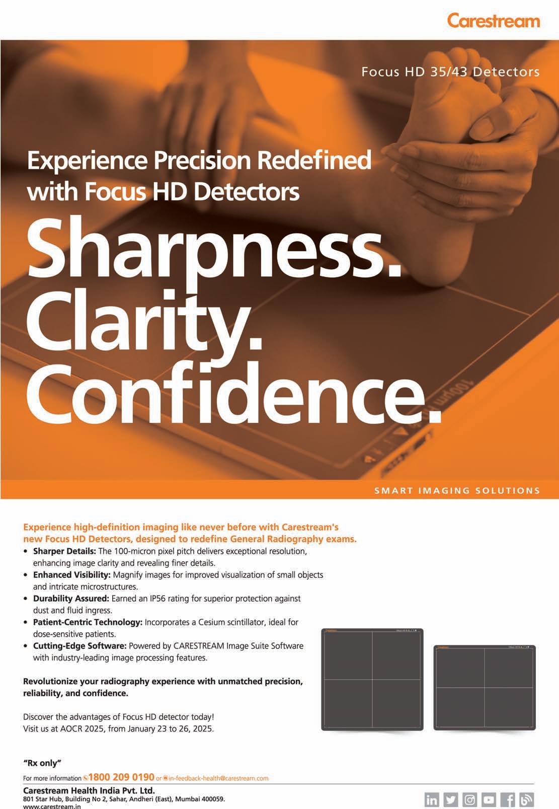
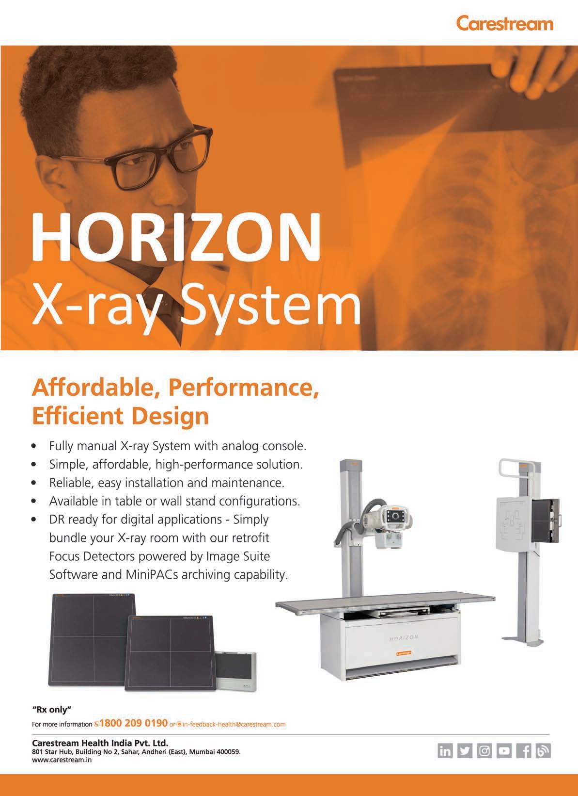
Capture-Print solutions designed to boost productivity and elevate patient care. We're also thrilled to introduce our new FOCUS HD
Detector as part of our expanding Retrofit DR solutions. This cuttingedge technology delivers highdefinition imaging at an affordable price, addressing our customers' evolving needs. Along with promoting our innovative products and solutions, we are excited by the tremendous response our CROP program has received. This program, which aims to support the ongoing education of radiographers, has garnered significant attention, and we are eager to further expand it at this year's show.
AOCR 25 will focus on "Clinical Radiology Decoded - See like a Surgeon: Think Like a Physician". How have advancements in realtime imaging transformed the precision and planning of surgical procedures?
The journey of radiology and imaging has been continuously shaped by groundbreaking technological innovations since its inception. Digital technology has not only facilitated new discoveries and diagnostic modalities but has also expanded healthcare access, reaching a broader population across the country. Hybrid imaging, which combines multiple imaging modalities, has become a gamechanger for medical practitioners, offering faster diagnostic accuracy and better patient management. While past breakthroughs focused on new imaging devices, the emphasis is now shifting towards software developments that enhance integration and improve radiology workflows. Advances in real-time imaging technology have significantly imp roved the accuracy of radiology procedures.
DIGITALTECHNOLOGYHAS
Radiologists can now target even the smallest areas with precision, enhancing success rates for early detection, timely treatment, and cure. These developments are poised to revolutionise surgical procedures, leading to improved patient outcomes, particularly in the management of critical diseases where time is of the essence.
What future developments in radiology are you most excited about?
The role of radiology is pivotal in disease management, patient treatment and saving lives. While conventional technology products will continue to address the current needs of the industry, the digital ecosystem will serve as the foundation for an integrated healthcare infrastructure, bridging gaps between stakeholders and benefiting the population as a whole. This integration will facilitate the convergence of emerging technologies that enhance healthcare efficiency, enable predictive medicine, minimise errors, and reduce costs. Advancements such as artificial intelligence, machine learning, and deep learning are adding significant value to the field of radiology. These innovations are empowering
radiologists by improving workflow efficiency, diagnostic accuracy, and decision-making. Driven by the creativity of numerous start-ups, these technologies are set to transform the future of radiology, ushering in a wave of change that will reshape the industry in unprecedented ways.
What are the key takeaways you hope to gain from AOCR 2025? Exhibitions and tradeshows provide an ideal platform for innovation and collaboration, bringing together customers, business partners, and healthcare providers. With the AOCR 25 theme set by the organisers, this promises to be an enriching event, offering valuable opportunities to engage with international delegates as well. At Carestream, we are excited to gain deeper market insights into the latest trends, challenges, and innovations in healthcare and radiology. This event will allow us to explore new technologies and solutions that are shaping the future of the industry. It’s also an excellent opportunity for us to showcase our innovative products and solutions, gather direct feedback from customers, and better understand their needs and areas for improve ment.
The workload of radiology departments is continuously increasing. This growing demand is putting pressure on staff and equipment, leading to an increasing need which allows radiology department to do more with less.
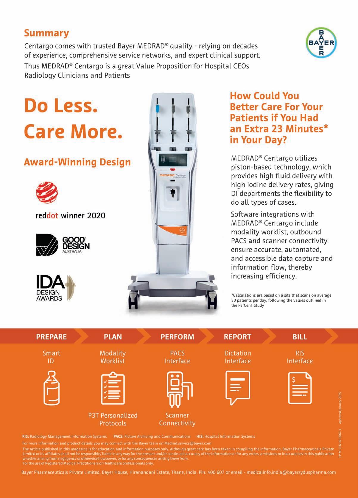
Introducing the next big step in CT injector technology, multiaward winner Medrad Centargo, an innovative CT injection system, that simplifies workflow improving the overall productivity of the radiology department/CT scan suite through automation. Centargo addresses multiple
stakeholder challenges through its wide range of features. Therefore, poising itself as Solution system and not just as simple injector.
Centargo individualises protocols without manual calculations,
Its unique design saves time and manual steps, thus increasing opportunity for scanning more patients or spending more time with each patient.
Centargo offers Automated Documentation thus greater peace of mind when it comes to complying with documentation
these Smart Protocols automate the protocol adjustments in a simple way making it possible to treat every patient as an individual. Protocols are personalised by adjusting contrast protocols for tube voltage, considering patient weight kidney function, contrast media volume, and flow rate.
Centargo maximises efficiency through automating workflow, allowing radiology staff to spend more time with their patients.
requirements. By effortlessly capturing contrast and injection parameters along the workflow, it reduces the number of manual tasks and hence, potential.
It makes the right information available when needed, so more time can be spent focusing on patient care.
Financial challenges/Benefits
Through contrast optimisation, freeing up staff for scanning more patients by digitalising the workflow, It may contribute to improving financial outcomes of a radiology department.
Dr
explains the crucial role of a radiologist in a healthcare system
Radiology is a crucial component of the modern healthcare system and a quickly developing medical subspecialty. It is not only a crucial instrument for verifying the diagnosis of many medical disorders, but it is also often utilised for small and large radiological operations, treatment monitoring, and result prediction. In the area of medical radiography, ongoing research and technical advancements are resulting in the creation of more complex and sophisticated equipment and methods that allow physicians to treat patients more quickly and accurately, thereby improving patient care.
Healthcare and medical radiology are two sectors in India that are expanding quickly. With the increasing healthcare demands of the Indian population, a large number of hospitals, nursing homes, medical labs, and diagnostic centres are being established. Expertise and training are necessary for the proper usage of radiography equipment and picture interpretation. But for now, the field of radiology is lacking in competent and trained personnel. Both in urban and rural India,
radiology specialists are in great demand. Thus, for individuals who are interested in this profession, radiography is a great career choice. Radiologists are physicians with training in imaging techniques such as CT, MRI and X-rays. They are also trained to diagnose and treat diseases.
Because there is a growing need for medical imaging to address a wide range of health disorders, radiologists are in great demand worldwide. To become an MD radiologist in India, an individual must complete a three-year postgraduate degree in Radiology following their MBBS. Moreover, a physician may choose to enrol in a two-year medical radiodiagnosis diploma program.
Cardio-radiology, Neuroradiology, Fetal Medicine and Interventional radiology are among the subspecialties in radiology training. To obtain training in one of these subspecialties, one can undertake one- to two-year fellowship programs.
Medical image interpretation is a speciality of diagnostic radiologists, who help with diagnosis. Within the field of diagnostic radiology, there are several subspecialties, such as neuroradiology, pediatric radiology, breast imaging, chest radiology, and emergency radiology.
The therapy of several illnesses, including uterine fibroids, cancer, aortic disease, and stroke, is handled by interventional radiologists with great skill using image-guided
surgical techniques.
In a hospital, the department of radiology is among the busiest. In most emergency and accident situations, imaging is frequently required; therefore, in most hospital settings, the department must operate around the clock. A thorough understanding of the handling and operation of equipment is also necessary for radiology operations because they are intricate. For that reason, those who want to pursue a successful career in radiology must develop and acquire the following abilities.
◆ A quick and inquisitive mind, familiarity with medicine, meticulousness, a fix-any-problem mindset, and adaptability
◆ Capacity to maintain and calibrate radiographic equipment; autonomously perform and interpret a variety of radiology procedures; sympathetically and empathetically care for patients throughout the procedure; and assure patient compliance, particularly in the case of pediatric patients, pregnant women, and individuals with special needs, among others.
RADIOLOGYIS AMONG THE BUSIEST.IN
MOST
EMERGENCYAND ACCIDENTSITUATIONS,IMAGING IS FREQUENTLYREQUIRED; THEREFORE,IN MOST HOSPITALSETTINGS,THE DEPARTMENTMUST OPERATE AROUND THE CLOCK.ATHOROUGH UNDERSTANDING OFTHE HANDLING AND OPERATION OFEQUIPMENTIS ALSO NECESSARY FOR RADIOLOGYOPERATIONS BECAUSE THEY ARE INTRICATE
◆ Adhere to policies and procedures to guarantee radiation safety for patients and employees.
◆ Possess strong communication abilities to effectively explain complicated imaging information to patients, secure informed consent, soothe anxious patients, and collaborate with other medical experts.
◆ Working in multidisciplinary
teams and as a team with other medical professionals;
◆ Managing stress and burnout in a high-stress atmosphere
◆ Regular upgrading of knowledge and ongoing education about the most recent advancements in radiology by participation in fellowship programs, reading journal articles, attending CMEs, etc.

Prakash Padmalwar,Managing Director,Evertech shares an overview of the innovative products his company will showcase at AOCR 2025
What innovative solutions or technologies your company is showcasing at AOCR 25?
Advance analytical dash board
◆ Platform independent advance diagnostics ZFP viewer with all tools
◆ Seamless work flow
◆ Easily configurable and customisable features and modules
◆ Easy integration and data exchange with other healthcare IT system like LIS ,EMR , AI tools etc
◆ Builtin speech to text and also built in AI tools
What role does advanced imaging play in enhancing minimally invasive or robotic surgeries? Are there any new imaging modalities that you believe will significantly impact surgical planning in the near future?
Advanced imaging plays a pivotal role in enhancing minimally invasive and robotic surgeries by providing detailed anatomical and functional insights that enable precision, reduce risks, and imp rove outcomes. These technologies are integral to preoperative planning, intraoperative guidance, and postoperative assessment.
Preoperative planning: Highresolution imaging modalities such as MRI and CT scans allow surgeons to map out complex anatomical structures, identify pathology, and develop precise surgical plans
tailored to each patient.
Intraoperative guidance: Imaging technologies like fluoroscopy, intraoperative ultrasound, and 3D imaging provide real-time visualisation, aiding in accurate navigation during surgeries. In robotic procedures, integration of imaging with systems like the da Vinci Surgical System enhances precision by overlaying anatomical data onto the surgical field. Reduced invasiveness: Advanced imaging minimises the need for exploratory procedures by offering
detailed insights into target areas, thereby reducing surgical trauma, recovery time, and complications. Emerging imaging modalities impacting surgical planning: Several new imaging modalities and advancements are poised to significantly influence surgical planning in the near future: 4D imaging: Building on 3D imaging, 4D imaging incorporates time as a dimension, enabling dynamic visualisation of moving structures like the heart or lungs. This is particularly beneficial in
procedures requiring motion tracking.
AI-enhanced imaging: Artificial intelligence (AI) is transforming imaging interpretation by offering automated, precise analysis of complex datasets. AI integration into imaging systems can identify anomalies, optimise surgical pathways, and predict outcomes with unprecedented accuracy.
Molecular imaging: Techniques like PET-MRI combine functional and anatomical imaging, allowing surgeons to differentiate between healthy and pathological tissues with greater specificity, crucial in oncological surgeries.
Photoacoustic imaging: This hybrid technology merges laserinduced ultrasound with optical imaging to provide high-resolution vascular and tissue visualisation, enhancing precision in microsurgeries and tumor resections.
Augmented Reality (AR) and Mixed Reality (MR): These technologies overlay imaging data onto the surgical field, offering immersive 3D visualisations for enhanced depth perception and spatial awareness.
Advanced imaging technologies are revolutionising minimally invasive and robotic surgeries by increasing precision, safety, and personalisation. Emerging modalities like 4D imaging, AIenhanced imaging, and molecular imaging hold immense potential to further refine surgical planning and execution, ensuring better outcomes for patients. Continued innovation in imaging will remain essential for advancing the capabilities of modern surgery.
What future developments in radiology are you most excited about?
Automated structured reporting: Seamlessly generating structured, AI-enhanced radiology reports that highlight critical findings and reduce turnaround times
The high speed teleradiology and portable radiology modality will make the radio diagnosis possible from anywhere /any time
Improve the TAT, quality, efficiency of the radiology services and resources by using BI tools enterprise centralised PACS solutions enable faster, scalable, and secure access to imaging data across
What drives Everttech's rapid client acquisition?
Cutting edge technology
Our PACS platform integrates the latest advancements, including Advance diagnostics ZFP viewer &
reporting module , Structured reporting , Enterprise multi hospital centralised solution , high speed image transmission, high quality images, user friendly and highly customisable modules and features and seamless interoperability with other healthcare systems.
Customisation and scalability
We offer highly customisable solutions that cater to healthcare providers of all sizes, from small clinics to large hospital networks. Our scalable architecture grows with the client’s requirements, providing long-term value.
Seamless implementation
Our streamlined implementation process minimizes disruptions and ensures a quick go-live timeline, which is critical for healthcare institutions looking to upgrade or adopt new systems.
Exceptional customer support Everttech prides itself on providing 24/7 support and training to clients. This ensures users maximise the benefits of our platform, fostering long-term relationships. Special thanks to our support and implementation team for building strong personal connections with our clients and ensuring instant availability as and when required
Strong industry reputation
With a proven track record and glowing testimonials from leading healthcare providers, we’ve built a reputation for reliability, innovation, and excellence in the PACS industry
What are the key takeaways you hope to gain from AOCR 2025?
Meet all key customers and vendors. See what’s new technologies coming in radiology healthcare IT.
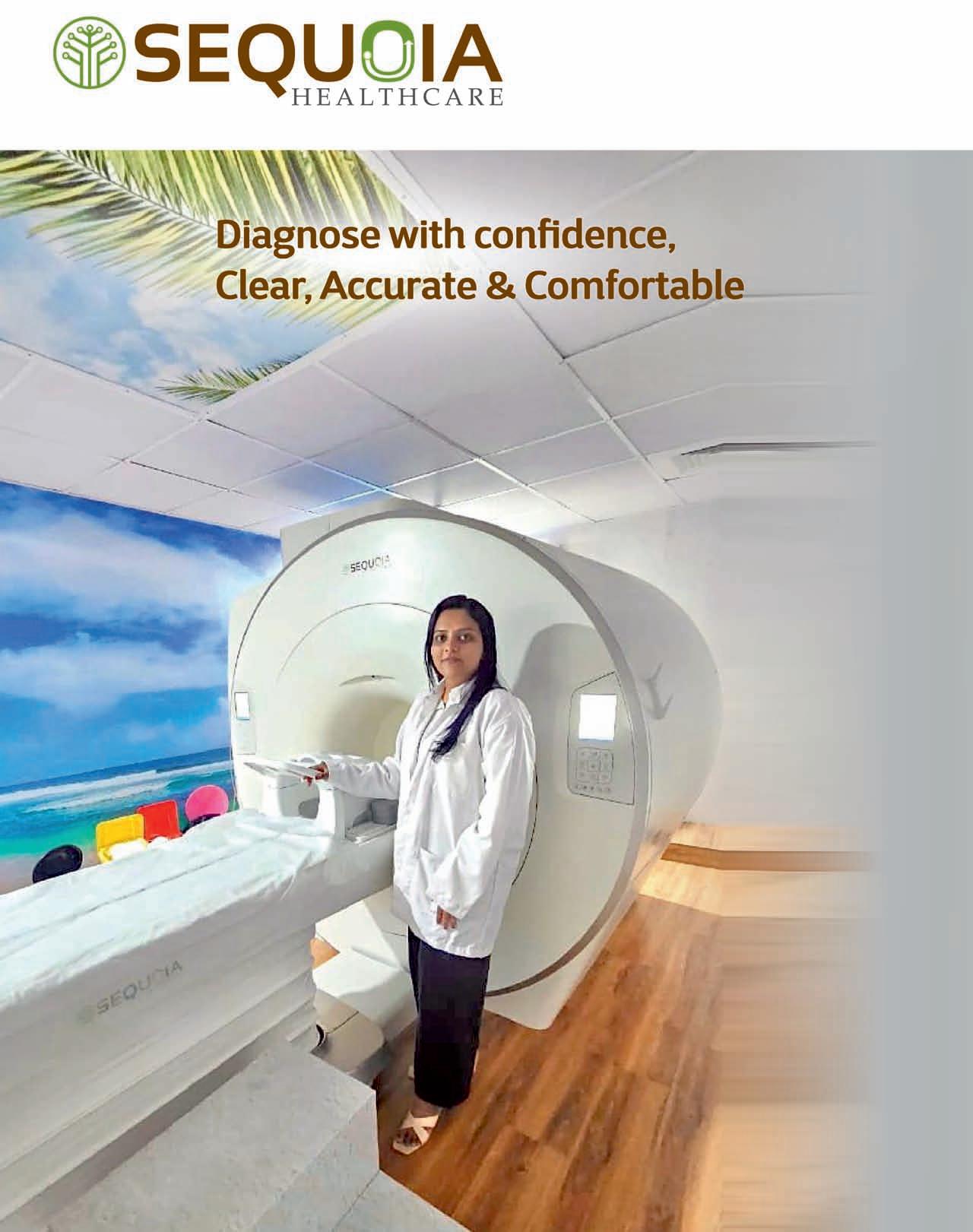
S.Viswanathan,Managing Director,Sequoia Healthcare,discusses the role of advanced imaging and shares an overview of the innovative products his company will showcase at AOCR 2025
What innovative solutions or technologies is Sequoia Healthcare showcasing at AOCR 2025?
At AOCR 2025, Sequoia Healthcare is thrilled to showcase several groundbreaking innovations that align perfectly with this year’s theme, “Clinical Radiology Decoded – See Like a Surgeon, Think Like a Physician.” Our flagship product, the Clarity 1.5 Tesla Helium-Free MRI Scanner, is a testament to our commitment to advancing radiology. It is not only environmentally sustainable but also delivers exceptional imaging clarity, making it an invaluable tool for clinical radiology and pre-surgical planning.
We are also unveiling our Advanced Radiology Workflow and Last-Mile Connectivity solutions. These enable seamless integration with numerous companies working on discrete AI solutions in medical diagnostics. As a consolidator of different AI suites through our collaborators at Innowave Healthcare, we are creating a unified platform that allows healthcare providers to access cutting-edge AI tools for diagnostics. This ensures a streamlined workflow from imaging acquisition to advanced diagnostic insights, empowering radiologists and clinicians to work more efficiently and effectively.
Additionally, Sequoia Healthcare is has expanded its portfolio with the of MiE Gamma Camera also works is in
progress for a super fast DIGITAL PET CT and a low helium 3.0 Tesla sealed MRI. The 3.0 Tesla MRI is designed for enhanced imaging capabilities while maintaining helium efficiency. Furthermore, we are working on a 512Slice Ultrafast CT scanner capable of freezing heart motion and creating multiphase images of the heart in a single beat. These innovations are designed to enhance the diagnostic imaging landscape, offering greater precision and diagnostic capability to healthcare providers.
AOCR will focus on Clinical Radiology Decoded – See Like a Surgeon, Think Like a Physician. How has real-time imaging improved the precision and planning of surgical procedures? Real-time imaging has revolutionised surgical precision and planning. Technologies like real-time MRI and intraoperative CT imaging provide surgeons with immediate feedback during procedures. This ensures that surgical interventions are more precise, reducing the risk of complications and improving patient outcomes.
In our solutions, we’ve integrated 3D reconstruction and live volumetric imaging, which allow surgeons to visualise anatomical structures in incredible detail. This has a profound impact on surgical decision-making, particularly in complex procedures like
neurosurgery and cardiovascular interventions. For example, clarity ng neurosurgeons to navigate critical areas with unparalleled confidence. These advancements also enhance multidisciplinary collaboration. Radiologists, surgeons, and other clinicians can work together seamlessly, leveraging real-time data to make more informed decisions, which ultimately translates into better patient care.
What role does advanced imaging play in enhancing minimally invasive or robotic surgeries? Are there any new imaging modalities that you believe will significantly impact the field in the near future?
Advanced imaging is the backbone of minimally invasive and robotic surgeries. Modalities like highresolution MRI, intraoperative ultrasound, and 512-Slice Ultrafast CT scanner provide real-time, highprecision imaging that guides robotic instruments with exceptional accuracy. These technologies reduce the invasiveness of procedures, shorten recovery times, and improve patient safety.
Looking ahead, Sequoia Healthcare is planning to further develop our
Clarity 1.5 Tesla Helium-Free MRI system into an intraoperative MRI solution. Its helium-free design makes it uniquely suited for operating room environments, as it eliminates many logistical and safety challenges associated with traditional MRI systems. Additionally, Hybrid Imaging systems, including PET-CT and PETMRI, combining modalities like PETCTI, are paving the way for unparalleled diagnostic and therapeutic capabilities.
What future developments in radiology are you the most excited about?
The future of radiology is incredibly exciting, driven by the convergence of advanced imaging modalities, AI, and personalised medicine. One area that excites me is the integration of AIdriven decision support systems, which will empower radiologists to provide faster and more accurate interpretations. This is particularly
critical in addressing the growing demand for diagnostic imaging amidst a shortage of skilled professionals.
Another exciting development is the rise of theranostic imaging—a fusion of therapy and diagnostics. This approach enables imaging-guided, targeted treatment, especially in oncology. Imagine a scenario where a tumour is not only visualised but also treated simultaneously using imaging-guided interventions.
Lastly, I’m deeply fascinated by the potential of cloud-based radiology platforms that leverage telemedicine to bring advanced imaging capabilities to underserved areas. At Sequoia, we are already exploring these avenues to democratise access to world-class radiology services.
What are the key takeaways you hope to gain from AOCR 2025?
AOCR 2025 is a premier platform for innovation and collaboration, and I look forward to engaging with global
Having spent decades as a radiologist, I’ve had the privilege of working with and owning MRI systems from nearly every major global brand. Each system brought its own strengths, but also challenges especially when it came to the high operational costs and maintenance complexities of traditional helium-based MRI machines. It was this very landscape, dominated by established players, that made me skeptical when I first heard
about Sequoia Healthcare, a new entrant in the field of high-field MRI. What changed my perspective and eventually won me over was the commitment and vision of the Sequoia team, coupled with their groundbreaking innovation: the Clarity 1.5T helium-free MRI. This system not only addressed the long-standing challenges of helium dependency but also brought new levels of efficiency and performance to MRI imaging. It was
thought leaders in radiology. One key takeaway I hope to gain is insights into emerging clinical needs and technological trends, which will guide our product development roadmap. I’m also keen on exploring collaborative opportunities with institutions and other industry leaders. At Sequoia, we strongly believe that partnerships are essential to driving meaningful change in healthcare. Finally, I hope to gain feedback from our customers and stakeholders about how our innovations are impacting their clinical workflows. These insights will be invaluable as we continue our mission of delivering precision technology with a human focus.
This year’s AOCR theme aligns closely with our ethos at Sequoia Healthcare. We remain committed to empowering healthcare providers with cutting-edge imaging solutions that not only decode clinical radiology but also inspire a new era of diagnostic and therapeutic excellence.
Dr Alok Singhai,Senior Radiologist and Founder,Pulse Diagnostics
clear that Sequoia was not just another manufacturer but a disruptor with a purpose. The Clarity 1.5T’s helium-free magnet design eliminates the need for
liquid helium, significantly reducing operating costs, environmental impact, and maintenance headaches. Yet, what truly stands out is its performance, which rivals and even surpasses conventional systems. The inclusion of 66-element, 16-channel “Music” coils is a testament to Sequoia’s engineering brilliance. These coils provide an exceptional signal-to-noise ratio and
seamless anatomical coverage, ensuring uniform, high-resolution imaging across the entire body. Whether scanning the brain, spine, or joints, the workflow is smooth, and the results are consistently outstanding.
Sequoia’s Clarity 1.5T isn’t just about innovation—it’s about accessibility. Its reduced installation and operational costs make high-field MRI attainable
for smaller diagnostic centers, democratising advanced imaging.
Choosing to partner with Sequoia was a leap of faith, but it has proven to be one of the most rewarding decisions in my career. The Clarity 1.5T is not just a machine—it’s a revolution, redefining what’s possible in MRI technology and bringing high-field imaging into a new era of sustainability and excellence.
Dr Pungavkar is a celebrated radiologist known for her obsession with accuracy and quality diagnosis. As part of the Disease Management Group for NeuroOncology at Tata Memorial Hospital, a founder member of the Indian Society of Neuro-Oncology, and a teacher for both, the Diplomate of National Board and the College of Physicians and Surgeons, Dr Pungavkar has set a gold standard in radiology. Currently, she is serving a second term as a member of the Radiology Advisory Board for the Blue Books of Pathology for brain tumors, a project by International Agency for Research on Cancer (IARC) and World health organisation (WHO).
For Dr Pungavkar, uncompromising image quality is non-negotiable. Her diagnostic center thrives on trust and precision, earning referrals from hospitals and clinicians who expect nothing less than excellence.
Dr Pungavkar’s adoption of the Inspiration 64 CT scanner from Sequoia Healthcare underscores her
commitment to cutting-edge technology. Installed nearly two years ago, this ultrafast scanner with three times faster coverage and high temporal and spatial resolution has become indispensable, enabling over 20 cases daily with minimal radiation dose to patients. She also performs guided interventions on the scanner with ease.
“I look beyond specifications. The inspiration 64 has exceeded my expectations and has elevated the quality of care I am able to provide,” says Dr Pungavkar.
She acknowledges the support from Sequoia for having been able to meet her expectations, having put to rest her initial skepticism.
The scanner’s speed and precision make it invaluable, especially in neuro-oncology and gastroenterology as well as pediatric work up, where early diagnosis can direct accurate management. Its high-resolution imaging ensures no detail is missed, empowering clinicians with critical insights while safeguarding patients with minimal radiation exposure.
Dr Sona Pungavkar,Founder and Medical Director,Siddhi Diagnostic & Research Center,Mumbai
Dr Pungavkar’s relentless pursuit of perfection inspires radiologists nationwide. Her association with IARC/WHO and her work at Siddhi Diagnostic & Research Center reflect her vision of transforming lives through radiology.
“Every image tells a story. It’s our responsibility to ensure that story leads to the best possible outcome for our patients,” she says. Dr Pungavkar continues to shape the future of radiology with passion and precision.
The theme of AOCR 2025, "See Like a Surgeon, Think Like a Physician," perfectly encapsulates the dual mindset required in modern radiology. It emphasises precision akin to a surgeon’s eye while fostering holistic, patientcentric insights like a physician.
The Sequoia Inspiration 64 CT Scanner has been transformative for AV Multispecialty Hospital, enabling rapid, high-resolution imaging critical for trauma cases and detailed evaluations. Its advanced detector technology ensures precision, mirroring the surgeon’s need for clarity and speed. From identifying subtle lung nodules to intricate vascular anomalies, it minimises diagnostic errors and aids confident surgical planning.
The 64-slice Sequoia Inspiration CT is not only significantly superior to the 16- and 32-slice CT systems available from other vendors but also makes better economic sense. With improved coverage—three times faster than other CT scanners—lower radiation exposure, and more accurate contrast capture, the scanner delivers enhanced clinical outcomes. Additionally, faster scanning times translate to higher throughput, lower electricity bills, and longer tube life due to shorter exam durations. The use of a smaller tube also reduces replacement costs when the tube eventually needs replacement.
Beyond precision, the scanner enables comprehensive assessments, aligning with the physician’s broader
Dr Rajesh Murthy,Founder and Radiologist, AVMultispecialty Hospital,Bangalore
perspective. For instance, it has been instrumental in detecting early-stage diseases in patients with vague symptoms, allowing timely intervention. By combining imaging insights with clinical context, radiologists can deliver comprehensive diagnoses and treatment recommendations.
AOCR 2025 theme inspires us to redefine radiology’s role, leveraging cutting-edge technology to deliver care that is both precise and compassionate. The Sequoia Inspiration 64 CT Scanner exemplifies this vision, empowering radiologists to elevate patient outcomes.
Six months ago, I installed the Clarity 1.5T Superconducting MRI with Zero Boil-Off technology, and it has revolutionised my practice. The high homogeneity magnet and 66-element MUSIC coils deliver exceptional image clarity, while the powerful 35 mT/m SR 150 gradients ensure unmatched precision, especially in complex cases like neuroimaging and musculoskeletal studies. Referring physicians are highly impressed with the crisp, high-resolution images we now provide. Orthopaedic, neurologists, and oncologists have praised the accuracy and detail, strengthening our relationships and boosting our reputation. In one case, the system detected a small ischemic stroke with exceptional clarity, enabling timely diagnosis and treatment.
Patients also appreciate the widebore design, which reduces the anxiety often associated with MRI scans. Claustrophobic and paediatric patients, as well as those requiring bariatric imaging, find the experience far more comfortable and non-intimidating compared to traditional systems.
Operationally, the Zero Boil-Off technology ensures sustainability, eliminating concerns about helium costs
and availability. The advanced gradients and MUSIC coils enable faster scans without compromising quality, enhancing productivity and reducing patient waiting times.The Clarity MRI has been a game-changer, improving diagnostic accuracy, patient comfort, and operational efficiency. It has positioned our centre as a leader in Aurangabad, providing world-class imaging services. For any radiologist considering an upgrade, I wholeheartedly recommend the Clarity 1.5T MRI. It’s a worthy investment, transforming both patient care and diagnostic excellence.
*Dr Shriram Mate is a radiologist in Aurangabad with over a decade of experience, specialising in diagnostic imaging
Agarwal,Managing Director,Innvolution Healthcare gives an overview about his company and its products
Founded in 2010 by a team of experts with over a century of combined experience, Innvolution Healthcare is a leading provider of innovative cardiovascular solutions in India. With a mission to deliver cuttingedge technologies that save lives, the company has transformed cardiac care through its comprehensive portfolio of highquality products, including award winning Cath Labs, drug-eluting stents, balloon catheters, and advanced imaging technologies like LVAD, vFFR, and intravascular ultrasound.
Innvolution pioneered the development of India’s most compact, cost-effective cath labs, designed to enhance accessibility in underserved regions. With over 450 installations across India and growing international recognition, the company addresses critical healthcare gaps by combining affordability with advanced features. Their flagship CE-marked Pinnacle Agile and Premier Elite cath labs exemp lify i nnova tion, offering superior image quality, reduced radiation exposure, and lifetime software upgrades.
Innvolution is dedicated to redefining healthcare with its "Made in India for the World" vision. Its inn ovative a pproach is
INNVOLUTION PIONEERED THE DEVELOPMENTOFINDIA’S MOSTCOMPACT,COST-EFFECTIVE CATH LABS,DESIGNED TO ENHANCE ACCESSIBILITYIN UNDERSERVED REGIONS
strengthened by strategic partnerships, such as with OrbusNeich, and a legacy of excellence marked by 17 prestigious awards. As a leader in medtech, Innvolution is advancing cuttingedge solutions like AI-driven imaging, robotics, AIMAG,
AInstaQCA, and virtual FFR, shaping the future of cardiac care. Recognised as a "Great Place to Work," the company fosters a culture of innova tion, collaboration, and compassion, solidifying its position as a global medtech pioneer.
Fujifilm's comprehensive range of healthcare products is designed to meet the evolving needs of healthcare professionals and patients across India
As a leader in the healthcare industry, Fujifilm India is committed to providing innovative solutions that enhance patient care and outcomes.
Fujifilm's comprehensive range of healthcare products is designed to meet the evolving needs of healthcare professionals and patients across India.
"Diagnostic Imaging Solutions"
Fujifilm India offers a range of diagnostic imaging solutions, including: -
◆ Supria CT: A cutting-edge computed tomography system that delivers exceptional image quality and low dose radiation.
◆ Echelon MRI: A high-field magnetic resonance imaging system that provides advanced clinical applications and patient-centric design.
◆ Lucent Open MRI: An opendesign MRI system that reduces claustrophobia and anxiety, providing a more comfortable experience for patients.
◆ Arietta Series Ultrasound: A range of ultrasound systems that deliver exceptional image quality, ease of use, and advanced clinical applications.
"Medical Informatics Solutions"
Fujifilm India's medical informatics solutions are designed to enhance workflow efficiency, patient care, and outcomes. Solutions include:
India's digital X-ray solutions are designed to provide high-quality images, enhance workflow efficiency, and reduce radiation dose. Solutions include:
◆ FDR Smart X-ray: A digital X-ray system that delivers high-quality images, advanced clinical
"DIGITALX-RAYSOLUTIONS" FUJIFILM INDIA'S DIGITALX-RAYSOLUTIONS ARE DESIGNED TO PROVIDE HIGH-QUALITYIMAGES,ENHANCE WORKFLOW EFFICIENCY,AND REDUCE RADIATION DOSE.A DIGITALX-RAYSYSTEM THATDELIVERS HIGH-QUALITY IMAGES,ADVANCED CLINICALAPPLICATIONS,AND LOW DOSE RADIATION
◆ Synapse PACS: A comprehensive picture archiving and communication system that enables secure storage, retrieval, and sharing of medical images.
◆ Synapse RIS: A radiology information system that streamlines workflow, enhances patient care, and improves outcomes.
"Digital X-ray Solutions" Fujifilm
applications, and low dose radiation. Fujifilm India is committed to providing innovative healthcare solutions that enhance patient care and outcomes. Their comprehensive range of healthcare products is designed to meet the evolving needs of healthcare professionals and patients across India.
Visit the booth to learn more about our healthcare products and solutions.
The integration of Artificial Intelligence (AI) tools in radiology is revolutionising medical imaging, significantly enhancing the ability of radiologists to interpret complex cases. In India, where there is a rapidly growing burden of noncommunicable diseases (NCDs) and an urgent need for improved
Dr Aneeta Bajaj Sr.Consultant Radiology, Apollo Hospitals Navi Mumbai
healthcare delivery, AI is emerging as a pivotal resource. The country faces challenges such as a shortage of specialists, particularly radiologists, and a fragmented healthcare system, but AI’s ability to enhance diagnostic accuracy, foster collaboration among healthcare providers, and support real-time surgical planning is transforming the field.
As the role of AI in radiology expands, it is critical that both healthcare professionals and the government continue to invest in the development and implementation of these technologies, ensuring that India can provide cutting-edge care to its vast and diverse population.
One of the most promising aspects of AI is its potential to bridge gaps in radiology expertise,
Dr Nikhil Kamat Senior Consulting Radiologist and Head- Department of Radiology,Jupiter Hospital,Thane
particularly in underserved areas. In remote regions where access to specialists is limited, AI tools trained on diverse datasets can serve as virtual second opinions. For example, AI systems designed to detect tuberculosis in chest X-rays have been successfully deployed in low-resource settings, enabling early diagnosis and treatment. Similarly, these tools help radiologists in smaller hospitals tackle rare or complex cases by offering reliable, evidence-based insights. The successful adoption of AI requires radiologists to act as both interpreters and gatekeepers, validating AI findings and understanding where its recommendations fit into clinical practice. Transparent algorithms, continuous monitoring, and clear regulations are also essential to ensure reliability and build trust in the technology.
One of the key ways AI enhances workflow productivity is by automating mundane jobs. For example, image sorting and preliminary analysis are time-consuming tasks that AI can complete quickly and accurately. AI algorithms are trained to organize and prioritize imaging studies depending on their urgency and complexity.
This implies that instances that require immediate care, such as possible strokes or fractures, are immediately pushed to the front of the
Dr Ajit Pratap Singh,Director- Imaging & Radiology,Asian Hospital,Faridabad
work list. This prioritising ensures that crucial situations are dealt with as soon as possible, which improves outcomes for patients in need.
Furthermore, AI improves the overall efficiency of picture analysis. Advanced algorithms can first examine the scans, flagging any possible anomalies and annotating significant aspects. This preliminary analysis helps radiologists save time by pinpointing areas of interest throughout each scan.
The collaborative approach between radiologists, surgeons, and other physicians is crucial in modern healthcare, ensuring comprehensive and coordinated patient management. Each specialist brings unique expertise that
Prof.(Dr) Pratiksha Yadav,Professor Radiology,Head-Interventional Radiology, Dr.D.Y.Patil Medical College,Hospital and Research Centre,Pune
contributes to accurate diagnosis, treatment planning, management, and monitoring. It benefits from the collective knowledge and expertise of the various disciples of medicine to develop multidisciplinary treatment plans and provide the patient with comprehensive and personalised treatment. It is necessary to plan a multidisciplinary meeting to discuss and evaluate the best treatment plan for the particular case. The collaborative approach reduces the delays in diagnosis, and teamwork ensures that all the experts are working towards the common goal of providing the best possible care for
the patient. This approach not only addresses the management but also the long-term well-being of the patient.
A collaborative approach among radiologists, surgeons, and other physicians enhances the quality of healthcare, leading to better diagnosis, treatment outcomes, and patient satisfaction. When these professionals work together seamlessly, the patient benefits from a comprehensive, multidisciplinary approach to care that is wellcoordinated, timely, and personalised. This approach ultimately leads to more effective treatments, reduced errors, and improved patient safety, contributing to better long-term health outcomes.
Teamwork in healthcare reduces the time taken in reaching appropriate diagnoses as well as developing individualised treatment regimens for the patients. By coming
Dr Chanabasappa V.Chavadi,Senior Consultant Radiologist,Manipal Hospitals
together, doctors are able to offer more treatment modalities thus giving patients more time to consider treatment and reducing the chances of treatment related risks. This team-centred model also translates to improved patient satisfaction and positive patient care results.
For physicians, especially for primary care or specialised fields like cardiologists, radiologists

Dr Dharav Kheradia,Consultant-Neuro and Vascular Interventional Radiologist, Wockhardt Hospitals,Mumbai Central
are their precious partners. In presenting unexplained symptoms, many times, imaging can provide pieces of the puzzle. In a case of pulmonary embolism, detailed scans by radiologists help physicians diagnose and promptly treat the problem. Regular collaboration between these professionals ensures that no detail is overlooked for patients to have the best chance of recovery. Collaboration isn't just a
good idea—it's the future of medicine. And for patients, that's a win every time.
Radiologists are known to come up with accurate imaging findings by helping the surgeons to know the tumor size, and location and then initiate treatment. Surgeons then can check the risks of operative approaches and suggest the treatment plan accordingly. Oncologists and physicians will design a proper treatment plan that may consist of chemotherapy, radiation therapy, or even surgery. By joining hands, radiologists, surgeons, and physicians are not only improving patient outcomes but also creating a revolution in the healthcare sector and changing the patient outlook for the better.
technology. Knowledge sharing not only enhances individual expertise but also drives improvements in diagnostic accuracy and treatment strategies.
For instance, imaging technologies can be applied more effectively through collaboration with clinical teams on the part of radiologists. Through detailed interpretations, radiologists can help surgeons and physicians incorporate cutting-edge solutions in their practices through the potential applications of these tools.
TThe collaborative model also promotes continuous learning and inn ovation. Regular interactions between radiologists, surgeons, and physicians create opportunities to share insights, discuss complex cases, and stay updated on advancements in medical
he collaborative nature of radiology is crucial, especially in complex medical scenarios such as surgical planning and the management of chronic diseases. Radiologists work closely with other specialists to determine the most accurate diagnosis and the best treatment pathways.
For example, in the context of joint trauma cases, the radiologist
depicts exact nature of injury to various structures in joint including ligaments, meniscus and cartilage. They also help in determining extent of bony injury (eg. Glenoid bone loss in shoulder dislocation). This critical information helps the surgeon in deciding about best surgical option for that patient.
Almost all cancer cases need to be imaged by various radiological modalities so as to determine the extent of disease, spread to other organs and staging, based on which appropriate treatment decisions are taken by onco-physicians and oncosurgeons.
Dr Vardhan Joshi,Radiologist,Sahyadri Super Speciality Hospital
Almost all medical branches including ICU specialists, General Physicians, Nephrologists, Cardiologists, Haematologists, Rheumatologists, Gastroenterologists, Paediatricians, Gynaecologists etc., apart from their clinical judgement also take important inputs from imaging findings provided by the radiologists.
Training the next generation of radiologists to have a surgeon's mindset involves equipping them with skills to be able to anticipate and assess imaging keeping in mind surgical considerations. The radiology training differs across India, and often, the curriculum fails to include adequate interaction between radiologists and surgeons, thus creating a gap between understanding specific needs of surgeons, mainly in surgical cases.
Dr Pranav Devani,Consultant-Radiology, Sterling Hospitals,Gurukul
Many radiologists, especially newly qualified ones, may lack awareness of the surgical aspects they need to address in their reports. Therefore, the reports would either be incomplete or unclear and leave the surgeons to seek review reporting or venture into more discussion in clarifying vital points. So, there is a need for bridging this gap through reorienting the training curriculum for radiology in creating a background of surgery and encouraging interdisciplinary approaches. Interdisciplinary meetings and clinical discussions are crucial for
aligning radiology training with surgical requirements.
In the long run, the radiologists should be educated to think more about the detailed information relevant to surgical planning and beyond diagnosis. This should require a high level of understanding between the radiologists and surgeons, with crucial points in the report and pre-surgical discussions that bring out the nuances of each case. In advanced settings, such as in the United States, inter-specialty fellowships where both surgeons and radiologists undergo training together, will buttress how collaboration can ascertain an improved outcome. When these two work together, especially on the treatment of complex cases, such as cancer surgeries, liver resections, and orthopedic implant procedures, their working will ensure proper planning on a patient's case to lead to better outcomes. Training in radiology should involve exposure to practical surgery, simulation technologies, and interdisciplinary meeting. Such an exposure shall enable the next generation to be trained to deal actively with surgical problems and reduce the gap between radiology and surgery. Radiologists would then prepare reports that are not only diagnostic but also of immense value for arriving at surgical decisions.
Technology cannot be a substitute for good clinical skill and assessment. Clinicians need radiologists who understand the clinical issues, keep updated with the most
Dr Sharad Maheshwari,Consultant, Radiologist (Abdominal Imaging),Kokilaben Dhirubhai Ambani Hospital,Mumbai
recent advances in the disease pathophysiology, and be aware of the relevant newer treatment strategies. For a good diagnostic radiologist, clinical experience is required before embarking on training in imaging. Once Radiology training is complete, it may be appropriate to further training in a specific organ or region-based subspecialty, which will enable the radiologist to interact at the appropriate level with clinicians. The process for basic radiology training should start in Medical school, which can give insight into the anatomy in the context of the clinical problem. Approachability is the keyword. Integrated reading rooms within clinical areas or technology-assisted virtual consultation may provide greater opportunities for direct interactions. Radiologists will have to change many of their attitudes and rethink their professional training to accommodate to the dramatic revolution and evolution of clinical radiology.
Dr Aparna Kulkarni,Consultant-Radiology,Jupiter Hospital,Pune highlights how real time imaging techniques improve surgical planning and navigation
Modern imaging technologies are transforming the way surgeries are planned and performed. Although radiological imaging by CT, MRI, their additional advanced techniques and real time imaging studies remain the base of diagnosis, it is the real time imaging that also give the surgeons a clear, live view of the body, helping them make precise decisions during procedures. These advanced tools, often powered by artificial and augmented intelligence among other trends like nuclear imaging, wearables, etc. make surgeries safer and more effective by reducing complications and improving outcomes. AI has the potential to transform the advanced medical imaging sector in a way that AI-driven and predictive analytics can increase the accuracy and speed of treatment decision-making. While real AI replicates human-like thinking without assistance, augmented intelligence technologies in healthcare seek to improve tedious or difficult medical operations. Augmented intelligence enhances communication between radiologists and oncologists by minimising repetitive data input activities and automatically attaching imaging and diagnostic data to electronic health records (EHRs). It may also be utilised to enhance picture quality.
The true revolutionary impact of virtual reality (VR) is shown in healthcare, where it is utilised to treat patients and assist clinicians in providing treatment more efficiently.
The most prominent of these new uses is 3D medical imaging. As strong as MRIs and CT scans are now, their 2D renderings require clinicians to conceive a spatial dimension they cannot see. New augmented reality technology enables clinicians to produce 3D images of MRIs. They may then analyse the image with a standard VR headset.
Physicians can use peripheral pointing devices and other tools to rotate, zoom in, and create cross sections of the 3D picture. This allows for better visualising and planning before an operation. Some VR platforms even allow doctors to simulate procedures with these visuals or produce real models with 3D printers. AR (augmented reality), like VR, generates three-dimensional visuals and projects them into the actual world utilising spatial computing and a specialised gear. Companies are utilising machine learning and AR to assist surgeons in navigating hurdles and bottlenecks that may delay a highrisk procedure.
A patient ingests radioactive compounds known as radiotracers (or radiopharmaceuticals) before undergoing a medical imaging scan. During the scan, a camera focuses on the areas where radioactive material is concentrated. These scans are very useful for identifying internal illnesses such as thyroid disease, cancer, and Alzheimer's disease.
For example, amyloid PET imaging (one of the recent advances, done at very few places as of now) can aid in predicting Alzheimer's development. This scan detects if individuals with memory problems have an excessive amount of amyloid plaques, which are a sign of Alzheimer's disease. Prior to amyloid PET imaging, these plaques could only be seen at autopsies. Early identification leads to more effective therapy and improved patient outcomes.
Wearable medical gadgets are no longer solely for monitoring heart rates and insulin levels (though those functions remain important). Researchers recently revealed the development of wearable ultrasound scanners for use in bladder and kidney disorders, breast cancer screening, and other applications. These various advancements enhance surgical planning and navigation, offering precise, continuous monitoring, improving accuracy, and reducing risks during procedures. Additionally, they make advanced diagnostic imaging more accessible and affordable, especially in underserved areas.
Dr Sunil Kumar Puri,Director,Radiology,Rajiv
Gandhi
Cancer
Institute and
Research Centre
highlights the significance of emerging technology that enables precise tumour localisation and characterisation,allowing healthcare professionals to develop precision medicine
Radiology encompasses a broad array of techniques essential to disease management.
Optimally used, radiology and imaging can lower medical costs and improve patient outcomes through early disease detection and accurate diagnosis.
The significance of radiology in healthcare is especially pronounced in cancer care, covering all stages from early diagnosis to treatment response, prognostication, and follow-up evaluation. Techniques like mammography, computed tomography (CT), magnetic resonance imaging (MRI), and positron emission tomography (PET) are crucial for the early detection of cancer. These technologies enable precise tumour localisation and characterisation, allowing healthcare professionals to develop tailored treatment plans based on the unique needs of each patient, a concept known as personalised or precision medicine.
Advanced hybrid imaging techniques such as PET CT and PET MRI not only locate tumours but also help in understanding their molecular composition. Additionally, imageguided therapies, including interventional radiology procedures, facilitate targeted treatments that minimise damage to healthy tissues. This level of precision is particularly
ADVANCED HYBRID IMAGING TECHNIQUES SUCH AS PETCTAND PET MRI NOTONLYLOCATE TUMOURS BUTALSO HELP IN UNDERSTANDING THEIR MOLECULAR COMPOSITION
beneficial for treating complex cancers in patients who are too sick for traditional surgical approaches. Artificial intelligence (AI) has moved beyond being a mere buzzword
to becoming a transformative force in healthcare, particularly in radiology and imaging. AI’s ability to analyse vast amounts of data and identify subtle patterns and abnormalities in imaging studies aids radiologists in making more accurate and timely diagnoses. This accelerates the diagnostic process and reduces the risk of oversight. AI algorithms are now employed not just for diagnosis but also for predicting outcomes and treatment responses, often outperforming trained professionals in specific scenarios. During the COVID-19 pandemic, AI played a critical role by assessing lung involvement in patients and addressing the shortage of human resources. AI is also extensively used in screening programs, notably for breast cancer detection.
The field of radiology and imaging faces several challenges, including the need for better interoperability of imaging systems, ensuring data security and patient privacy, managing the growing volume of imaging data, and standardising imaging protocols. Additionally, integrating advanced technologies like AI for image interpretation and diagnosis poses challenges, as does training healthcare
Dr Manas Kumar Sahoo,Associate Director-Radiology & Imaging, Medanta,highlights AI's transformative role in PETimaging,enhancing accuracy,reducing radiation,and expanding clinical applications
Positron emission tomography/computed tomography (PET/CT) is a hybrid medical imaging (MI) modality clinically used for functional imaging predominantly useful in neurology, cardiology, nephron-urology, oncology and endocrinology. PET examinations provide physiological information of organs using radiopharmaceutical products (radiotracers) while CT provides the morphological details of the anatomical region under study.
Artificial intelligence (AI) is a growing field of research that is emerging as a promising adjunct to nuclear medicine. The growing synergy between AI and PET imaging promises to revolutionise diagnosis, improve accuracy, reduction in radiation exposure and expand clinical utility.
AI is defined as the study of “intelligent agents” that can perceive the surrounding environment and take actions to maximise the chances of successfully achieving predefined goals. Machine learning (ML) is a subset of AI that involves computer algorithms1. Deep learning (DL) is a type of ML methodology that is based on artificial neural networks. DL is considered representation learning which includes a set of methods that would enable a machine to be fed with raw data which results in automatic detection of the needed representation for classification. A
convolutional neural network (CNN) is a type of deep learning architecture inspired by neural system that can learn representations of input data2.
With the advent of fast wave form digitisers, DL methods have become fundamental for improving the gamma detection from scintillators, predicting the time-of-flight (TOF) of photons, and improving the coincidence-time-resolution1. The ability to add information about the position of the positron annihilation along the Line-of-response (LOR) is an important parameter in the reconstruction process, leading to an improvement in signal-to-noise ratio and lesion detectability. Deep convolutional neural networks (CNN) have been implemented to estimate the TOF directly from the pair of digitised detector waveforms for a coincident event 2,3. Randomly scattered photons result in
degradation of image quality, in this case Neural Network (NN) approaches showed very good LOR recovery rate (75 per cent), yielding an overall high sensitivity increase of 55 per cent (real scanner conditions) 4 . Image pre-processing techniques using of regions of interest (ROI) segmentation are a critical step for several clinical tasks, including quantitative analysis, treatment planning, response assessment, etc. AI will progressively replace the practice of manual segmentation, which is typically time-consuming, suffers from intra- and inter-reader variability, and low reproducibility. DL-based segmentation algorithms have been successfully applied to PET images with different radiopharmaceuticals and in different clinical settings, from oncology to cardiac and brain imaging5.
With AI, the process of analysing medical images can be accelerated. It can help streamline workflow by automating routine tasks, allowing to focus more on complex cases and providing better patient care. AIdriven diagnostic imaging has the potential to enhance the accuracy of diagnoses by analysing vast amounts of data and identifying subtle patterns.
As the, accuracy of AI-driven radiology heavily relies on the quality and diversity of the data it is trained
on, if the training data is biased or incomplete, it may lead to inaccurate results. There are ethical considerations regarding the use of AI in nuclear medicine and molecular imaging, the ethical, social, and legal issues specifically relate to the data used, the algorithms applied, and how algorithms are used in practice6. Lastly, implementing AI-driven systems can be additional financial burden requiring significant investment in infrastructure, training, and maintenance.
The most profound impact of AI applications in PET/CT with respect to the patient experience arise from the capacity of AI to enable deeper, more meaningful interactions between the patient and the physician. The paradigm shift created by applications of AI in nuclear medicine has the potential to inspire a culture shift where emphasis is increased on the value of the interaction between the patient and the provider. AI will
WITH AI,THE PROCESS OF ANALYSING MEDICAL IMAGES CAN BE ACCELERATED
definitely bring tremendous change in PET/CT not only by playing a major role in the process of clinical diagnosis but also guiding the reduction in radiation exposure to patients with modification of imaging parameters and ultimately improving the image quality. AI offers a powerful toolkit for the rapid and safe automation of tedious or repetitive tasks and deep analysis beyond the capability of the human mind and, in doing so, provides time and energy for the physicians to more expertly review patient data and to communicate with the patient.
References:
1. Choy et al., “Current Applications and Future Impact of Machine Learning in Radiology.”
professionals to effectively use these technologies.
The future of radiology and imaging is filled with exciting possibilities. Advances in AI will further improve diagnostic accuracy, automate routine tasks, and assist in interpreting complex images. The integration of machine learning algorithms will enhance predictive analytics, facilitating early disease detection. Innovations in imaging technology, such as photon-counting CT and functional MRI, promise
2. LeCun, Bengio, and Hinton, “Deep Learning.”
3. Carra et al., “A Neural Network-Based Algorithm for Simultaneous Event Positioning and Timestamping in Monolithic Scintillators.”
4. Berg and Cherry, “Using Convolutional Neural Networks to Estimate Time-of-Flight from PET Detector Waveforms.”
5. Maebe and Vandenberghe, “Simulation Study on 3D Convolutional Neural Networks for Time-of-Flight Prediction in Monolithic PET Detectors Using Digitized Waveforms.”
6. Michaud et al., “Sensitivity Increase Through a Neural Network Method for LOR Recovery of ICS Triple Coincidences in High-Resolution Pixelated- Detect
7. “Artificial Intelligence with Deep Learning in Nuclear Medicine and Radiology | EJNMMI Physics | Full Text.”
8. SFR-IA Group, CERF, and French Radiology Community, “Artificial Intelligence and Medical Imaging 2018.”
The...
higher resolution and improved tissue characterisation. The rise of telemedicine and remote monitoring will broaden access to imaging services globally. Collaborations between radiologists and data scientists will drive innovation, leading to a future where personalised medicine and targeted therapies based on advanced imaging findings become more common, ultimately improving patient outcomes and transforming healthcare.
In conclusion, the evolution of radiology and imaging has profoundly impacted cancer care. From early detection to personalised treatment strategies, radiology is central to improving patient outcomes. While the initial investment in advanced imaging technologies can be significant, the long-term benefits— both in terms of patient health and healthcare system efficiency— highlight the importance of continued advancements in radiology for cancer care.
Dr Radheshyam Naik,Medical Oncologist,HCG,highlights the push for better breast cancer screening technologies while stressing the importance of thorough evaluation before adoption
Breast cancer poses a significant global threat to women, with the Lancet Commission projecting over three million cases and one million deaths annually by 2040. This alarming rise, particularly among younger women, necessitates a shift in focus from treatment to early detection. Early detection is crucial, dramatically improving treatment outcomes. While mammography and ultrasound remain standard screening tools, they present challenges. These include cost, infrastructure limitations, reduced sensitivity for younger women and those with dense breast tissue, and the potential for false positives or negatives. Additionally, mammography involves exposure to low doses of radiation.
The need for better breast cancer screening has spurred the development of new technologies. However, careful evaluation is crucial before widespread adoption. As a leading oncologist, I believe we must critically assess any new screening method. The following key questions are essential in identifying and developing truly innovative screening tools:
◆ Is it scientifically proven?Solid scientific evidence, backed by rigorous research and testing, is paramount.
◆ Is it supported by credible clinical studies?Published studies in reputable journals provide validation and transparency.
◆ Has it been successfully implemented on a large scale?Realworld success demonstrates practicality and effectiveness.
◆ What are the infrastructure requirements?Understanding the resources needed for implementation is crucial for accessibility.
◆ Is it US FDA cleared?FDA clearance signifies that a technology has met rigorous safety and efficacy standards.
One technology that has gained attention is thermography. While marketed as a non-invasive alternative, it falls short of these critical criteria. Thermography lacks the sensitivity and specificity needed for reliable breast cancer screening.
Many factors, including hormonal fluctuations, infections, even everyday activities like applying deodorant or drinking coffee, can influence thermography results, leading to inaccurate readings and false positives. Importantly, the US FDA hasnotcleared thermography for breast cancer screening and has issued warnings about its potential to mislead patients.
Choosing the right screening technology is a critical decision. While mammography remains a cornerstone of breast cancer detection, exciting advancements are emerging. New and improved technologies are harnessing diverse approaches from electrical impedance imaging and contrastenhanced MRI to ultrasound tomography and the insights of cellular biology to offer potentially groundbreaking screening tools. These innovations hold the promise of overcoming the limitations of current methods and improving early detection for all women.
Understanding individual risk factors allows for tailored screening
Dr
Sneha Kini,Consultant-Imaging,P.D.Hinduja Hospital & MRC,Mahim
explains how USG in musculoskeletal applications is a ‘well established’ technique which can assist the surgeons in accurate surgical planning
Medical sciences have shown astronomical advances in past few decades. Diagnosis and treatment of conditions which were hitherto considered untreatable, have been made possible. These advancements have been achieved because of conjoint progress of all the fields of Medicine leading to tremendous improvement in patient care and outcomes.
Radiology, now plays a pivotal role in patient management algorithm and helps in prudent clinical decision making.
Preoperative imaging forms a ‘Google map’ for our new age Abhimanyu surgeons to skillfully navigate through the ‘Chakravyuh’ or ‘labyrinth’ of human body.
Ultrasound (US)/Ultrasonography (USG) technology today is equipped to depict structures ranging from large tumors to smallest nerve fascicles due to the high-resolution images. It is a rapid and real time depiction of structures and dynamic motion with no associated radiation. This potential of USG is fully tapped in its musculoskeletal applications.
‘Dynamic/ real time’ evaluation of
which may vary depending on the duration of the tear. Tendon dislocations, snapping, impingement, friction syndromes are easily demonstrated on dynamic ultrasound since this is the only modality which can visualise soft tissues during actual motion. It can help surgeons to triage patients with acute injuries, like ankle twist injuries by evaluating ankle ligaments and hence decide need for further investigations. Assessments
RADIOLOGY,NOWPLAYS APIVOTALROLE IN PATIENT MANAGEMENTALGORITHM AND HELPS IN PRUDENT CLINICALDECISION MAKING
various joints and adjacent structures, ranging from large joints like shoulder and hip to small joints of the fingers and toes, gives ultrasound the edge over other modalities. Various tendons and superficial ligaments can be explicitly delineated by USG in acute as well as chronic conditions. It is a quick way to detect tendon tears, thickness of tendon involvement and pre-operatively assess the exact retraction of torn tendon (if any),
of muscles ranges from static evaluation in the form of muscle bulk assessment, atrophy/ fatty infiltration, tear or hematomas to dynamic evaluation of muscle contraction and free tendon movement. All ‘swellings’/‘lumps and bumps’ involving/around the joints can be evaluated ranging from joint effusion to masses. Also, in cases of multiple joint involvement
Pg50 ➤
Srinivas
Fetal growth assessment is a critical component of prenatal care, providing valuable insights into fetal development and overall pregnancy health.
Traditionally, this process involves manual measurements of fetal growth parameters such as Biparietal Diameter (BPD), Head Circumference (HC), Abdominal Circumference (AC), and Femur Length (FL). While effective, these methods are often time-intensive and prone to variability among practitioners, leading to inconsistencies and potential inaccuracies. Additionally, manual measurements are subject to human error, as different practitioners may interpret ultrasound images differently, further exacerbating variability. The skill level and experience of the practitioner also significantly influence the accuracy and reliability of these measurements.
To address these challenges, there is a growing need for AI-based algorithms capable of providing fetal growth assessment parameters with high precision. Such algorithms can considerably enhance prenatal care by automating the measurement process, thereby improving both the accuracy and efficiency of fetal
AI into this process, we can enhance precision, efficiency, and reproducibility, ultimately elevating the quality of prenatal care.
fetal growth assessment
The integration of AI in fetal growth assessment offers numerous advantages over traditional methods. AI algorithms streamline and simplify the extraction and analysis of fetal growth data from ultrasound images, ensuring consistent and reliable measurements. These
advancements lead to several improvements in prenatal care:
◆ Enhanced accuracy
AI algorithms reduce the variability associated with manual measurements, providing more precise and consistent biometry data. This accuracy is critical for detecting subtle developmental issues that might be missed due to human error.
◆ Increased efficiency
Automating the measurement process saves time for healthcare practitioners, enabling them to focus on other essential aspects of prenatal care, such as patient consultations, detailed examinations, and personalised care plans.
◆ Improved reproducibility
AI ensures that measurements reproducible measurements across different practitioners and clinical settings, facilitating standardised care. This consistency is vital for tracking fetal development and comparing results across healthcare facilities.
◆ Comprehensive analysis
AI can analyse large volumes of data quickly, identifying patterns and
trends that might not be immediately apparent through manual methods. This ability supports early detection of potential issues and fosters a holistic understanding of fetal health.
◆ Earlydetection of growth abnormalities
Accurate and consistent measurements enable early detection of fetal growth abnormalities, allowing timely interventions. Early detection is essential for managing complications and improving outcomes for both mother and fetus.
◆ Better monitoring of high-risk pregnancies
AI-driven assessments provide detailed insights into fetal development, which are particularly beneficial for high-risk pregnancies. Enhanced monitoring capabilities enable proactive and informed decision-making.
◆ Enhanced patient care
By improving the accuracy and efficiency of fetal growth assessments, AI technology contributes to better overall patient care. Patients benefit from more reliable assessments, reduced wait times, and higher quality of care.
◆ Standardisation of care
The use of AI promotes standardised care protocols, reducing disparities in healthcare delivery. This ensures all patients receive consistent, high quality assessments, regardless of the practitioner or facility.
◆ Reduction in human error
AI algorithms minimise the risk of human error in measurements and
interpretations, leading to more reliable diagnostic outcomes and better clinical decision-making. These multifaceted benefits underscore the transformative potential of AI-based fetal growth assessment in advancing prenatal care and improving maternal and fetal health outcomes.
Future directions in AI-based fetal growth assessment
◆ Point-of-care devices
The future of AI in fetal growth assessment includes portable, userfriendly devices that enable immediate and accurate evaluation. These devices, equipped with AI algorithms, offer real-time analysis and decision support, making highquality prenatal care accessible even in remote areas.
◆ Emerging markets
AI-driven fetal growth assessment holds significant potential in emerging markets, where access to advanced medical technologies is limited. Implementing affordable and accurate AI solutions can bridge gaps in prenatal care, improving outcomes in underserved regions.
◆ Integration with telemedicine
AI integration with telemedicine platforms enhances the reach and efficiency of prenatal care. Remote AI-powered assessments allow healthcare providers to monitor fetal development and address concerns without requiring in-person visits, benefiting patients in rural or underserved areas.
◆ Continuous improvement through data
As AI algorithms analyse increasing volumes of data, for fetal their accuracy and comprehensiveness will improve. Continuous learning capabilities will drive advancements, with collaboration among healthcare providers, researchers, and developers ensuring algorithms remain current with latest medical knowledge.
◆ Population-specific nomograms
Another promising direction for AIdriven fetal growth assessment is the development and implementation of population-specific nomograms. These customised growth charts, designed to reflect the unique characteristics and growth patterns of different populations, greatly enhance the precision and relevance of assessments. Leveraging large datasets and sophisticated AI algorithms, healthcare providers can develop detailed growth charts that consider genetic, environmental, and socio-economic factors unique to specific groups. This personalised approach ensures that assessments are not only accurate but also culturally sensitive, leading to better maternal and fetal health outcomes across diverse populations. These future directions highlight the vast potential of AI to revolutionise prenatal care, making it
more accurate, efficient, and accessible on a global scale.
Conclusion
The development of AI algorithms marks a significant leap forward in prenatal care. By automating the measurement of fetal biometry parameters and enhancing the accuracy and efficiency of fetal growth assessments, AI technology is transforming how we monitor and
approaches. High-risk women may benefit from more frequent screenings, supplemental screening modalities, or specialised imaging techniques, maximising early detection potential while minimising unnecessary procedures for lower-risk individuals. Advanced screening technologies can detect even subtle abnormalities in high-risk women, often before they're detectable by traditional methods. This earlier detection is crucial for timely intervention and improved outcomes.
We must prioritise methods with proven efficacy, regulatory clearance, and successful real-world implementation. As healthcare professionals, our commitment is to evidence-based practice and the well-being of our patients. By carefully evaluating emerging technologies and adhering to established standards, we can ensure accurate and effective breast cancer screening, ultimately saving lives.
manage pregnancy health.
References
Smith, J. A., & Brown, L. M. (2021). Advances in AI for Prenatal Care: A Review. Journal of Medical AI Research, 15(3), 345-359.
Jones, R. T., & White, K. P. (2022). AI Algorithms for Fetal Growth: Challenges and Opportunities. International Journal of AI in Medicine, 27(2), 112-125.
Nguyen, T. H., & Lee, C. J. (2023). Telemedicine and AI: Transforming Prenatal Monitoring. Telehealth and Medicine Today, 18(1), 56-68.
Garcia, M. E., & Patel, S. R. (2020). Bridging the Gap: AI-Driven Prenatal Care in Emerging Markets. Global Health Innovations, 12(4), 401-416. Kim, H. J., & Chen, Y. Y. (2024). Continuous Learning in AI for Fetal Assessment. Journal of Continuous Medical Education, 20(2), 78-90.
like inflammatory arthritis, USG comes in handy while evaluating multiple joints in one sitting.USG provides a real time guidance to perform interventions like aspirations, biopsies, pain management procedures etc which improves procedure results and minimises the complications. It also forms an important tool to assess post operative patients due to presence of orthopedic implants. Tendon re-tears, post operative hematomas/collections, infections can be identified and managed accordingly.
USG can finely delineate peripheral nerves with visualisation of even the nerve fascicles. It forms a robust tool to investigate conditions like nerve injuries, post trauma or post-surgery/intervention. It can diagnose the grade of nerve injury, exact retraction in cases of complete nerve transections, presence of neuromas, scarring around the nerve, nerve compression, if any. This gives a surgeon the required information, if surgical exploration
is planned. Presence of orthopedic metal implants leads to susceptibility artifacts on MRI and hence suboptimal evaluation of postsurgical nerve injuries. In these cases, USG scores over MRI and can delineate the entire nerve. Also, intra-operative USG can be performed to aid the surgeon in delineating the nerves intraoperatively, especially in posttrauma or post-surgical nerve injuries which already have altered anatomy. USG can evaluate compressive neuropathy and the entire course of the nerve can be traced in one sitting, for site, degree and cause of compression. Multiple nerve involvement, cases of leprosy or multiple masses along the nerves like in neurofibromatosis can be easily assessed in single sitting. USG in musculoskeletal applications is a ‘well established’, ‘specialised’ and ‘doer dependent’ technique which can assist the surgeons in accurate surgical planning and aid in better patient management.