
International Research Journal of Engineering and Technology (IRJET) e-ISSN: 2395-0056 p-ISSN: 2395-0072

Volume: 11 Issue:12 | Dec 2024 www.irjet.net


International Research Journal of Engineering and Technology (IRJET) e-ISSN: 2395-0056 p-ISSN: 2395-0072
Volume: 11 Issue:12 | Dec 2024 www.irjet.net
Tanushree T1 , Shreya Kanti M2 , Rashmitha K3,Prathibha H B4
1234Bachelor of Engineering, Information Science and Engineering, Bapuji Institute of Engineering and Technology, Davangere, affiliated to VTU Belagavi, Karnataka, India.
Abstract - : Indevelopingcountries,breastcancer isoneof the main causes of mortality for women, and early detection and treatment are essential. Invasive ductal carcinoma (IDC) and ductal carcinoma in situ (DCIS) are the two primary subtypes of the illness. More precise diagnostic models are the result of developments in artificial intelligence (AI) and machine learning (ML). Magnetic resonance imaging (MRI) and convolutional neural networks (CNNs) are important diagnostic and prophylactic techniques for breast cancer. CNN-basedmodels, suchasCNNI-BCC, help classifysubtypes of thedisease. Thesemodels, however, demandalargeamount of processing power. Using three feature selection techniques low-variance elimination, recursive feature elimination, and univariate feature selection this study suggests an efficient deep learning model for detecting breast cancer from digital mammograms with different densities. 3002 photos from 1501 patients who had digital mammograms between February 2007 and May 2015 are included in the dataset. The following six classification techniques were assessed: logistic regression (LR), support vector classifier (SVC), k-nearest neighbors (KNN), decision trees (DT), random forests (RF), and linear support vector classifier (linear SVC). The findings demonstrate that the suggested approach provides excellent accuracy and efficiency while using less processing resources.
Key Words: Breast Cancer ; Invasive Ductal Carcinoma (IDC);DuctalCarcinomaInSitu(DCIS);ArtificialIntelligence (AI); Convolutional Neural Networks (CNNs);Magnetic ResonanceImaging(MRI);DigitalMammograms.
Cancerisaglobalhealthissuethataffectspeopleofallages andbackgrounds.Amongvariouscancers,breastcanceris particularlyprevalentinwomenandisthesecondwleading causeofcancer-relateddeathsinwomen,afterlungcancer [1].Earlydetectionanddiagnosisarecriticalforimproving survival rates, and machine learning (ML) techniques can significantlyenhancethisprocess.Breastcancerdevelopsin thebreastcellsandcanpresentinseveralforms,including DuctalCarcinomaInSitu(DCIS),whichisnon-invasive,and InvasiveDuctalCarcinoma(IDC),themostcommon

form,accountingfor80%ofcases[2].Earlysymptomsmay include unexplained swelling in the breast or armpit, changes in breast size or appearance, pain or discomfort, nipple discharge, or skin changes. For early detection, mammography remains a vital tool. However, mammographycan beless effectivein women withdense breast tissue, where diagnostic ultrasonography, thermography, or radiography might offer more precise results. Magnetic Resonance Imaging (MRI), particularly Dynamic Contrast-Enhanced MRI (DCE-MRI) [3], is highly sensitive for identifying and characterizing breast cancer lesions.Recentadvancesinartificialintelligence(AI)have furtherimproveddiagnosticaccuracybyenablingAI-based systemstoanalyzemedicalimages,suchasmammograms, andidentifycancerouslesions.Inthiscontext,deeplearning modelscanbeusedtoanalyzecomputerizedmammograms of varying breast tissue densities. This study proposes an effectivedeeplearningapproachtoenhancebreastcancer detection across different breast densities, ultimately improvingtheaccuracyofearlydiagnosis.
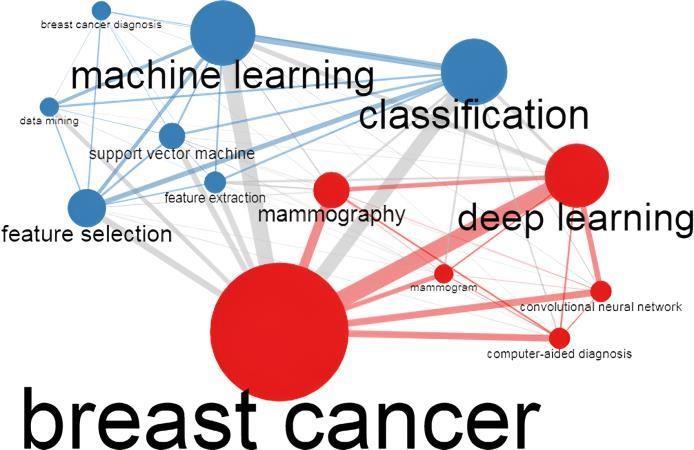
The image highlights the relationship between advanced computationalmethodsandbreastcancerdiagnosis.Atits core,"breastcancer"connectstotechnologieslikemachine learning and deep learning. Machine learning is linked to termssuch as "classification,""supportvectormachine,"and "feature selection," emphasizing its role in predictive modeling , Deep learning focuses on applications like "convolutionalneuralnetworks,""mammography,"and

Volume: 11 Issue:12 | Dec 2024 www.irjet.net
International Research Journal of Engineering and Technology (IRJET) e-ISSN: 2395-0056 p-ISSN: 2395-0072
"computer-aided diagnosis," enhancing imaging and automation. The diagram shows how these technologies work together to advance breast cancer detection and classification.
InordertoidentifyBC,anumberofresearchhaveemployed machine learning (ML) approaches in the healthcare industry in recentyears.Otherscientists haveapplied the algorithms to difficult problems because they yield satisfactory results [4]. In breast cancer photos, invasive ductalcarcinomawaspredictedanddiagnosedusingaCNN algorithm,anditsucceededinanapproximate88%accuracy rate [5,6]. Furthermore, it is frequently employed in the medicalindustrytodiagnoseandforeseeabnormaleventsin ordertogainabetterknowledgeofdiseasesthatcannotbe cured, like cancer [7]. Imaging and genetic-based breast cancer screening methods have been the subject of numerous investigations. Additionally, as far as we are aware,noresearchhasbeendonethatcombinesthetwoof thesemethods. Theauthorsof[8]providedanoverviewof thevariousmethodsforhistologicalimageanalysis(HIA)in thedetectionofbreastcancer.Thesemethodsarebasedon several kindsofconvolutional neural networks(CNN)[9]. Theauthorscategorizedtheirworkaccordingtothetypeof dataset they used. Everything was arranged in reverse chronological order, starting with the most recent event. Accordingtothefindingsofthisstudy,ANNswereinitially appliedinthefieldofHIAsomewherearoundthemiddleof 2012.ANNsandPNNswerethemostwidelyusedalgorithm types [10]. The suggested approach was created to assist medicalprofessionalsindeterminingwhethertodoabreast biopsy on a worrisome lesion following a review of mammography results. Several distinct classification algorithmsareusedintherandomforest(RF)classifier,an ensembleapproach.Usingadecisiontree,eachofthemcan be implemented. Using multiple decision trees improves classificationaccuracy(Dai,Chen,etal.,2018)[11].Toputit simply,theRFisanensembleclassifiercomposedofseveral decision trees that cooperate to increase output and prediction precision. In Nguyen, Wang, et al. (2013) [12], scientistsdevelopedaclassifierbasedonRF.Theresultsof trainingthealgorithm they employed on two datasetsare promising:Itperformedwellandwithgreataccuracy.
Thisstudyexplorestheuseofsixmachinelearningmodels forbreastcancerdiagnosis:RandomForest(RF),Decision Tree(DT),k-NearestNeighbors(KNN),LogisticRegression (LR),SupportVectorClassifier(SVC),andLinearSVC.Breast Cancer Wisconsin(Diagnostic)data wasusedtotrainand assessthemodels.Toguaranteeconsistentanddependable results,thedataunderwentpreprocessingproceduressuch
recursivefeatureeliminationandunivariateselectionwere used. The study emphasizes the value of preprocessing, whichinvolvedseparatingthedataintotraining(70%)and testing(30%)setsandeliminatingduplicatevalues.Inorder to improve interpretability, decision trees were displayed and feature significance analysis was performed for treebasedclassifierssuchasRFandDT.Toincreasethemodels' resilienceanddependability,ensembleapproachesincluding bagginginRFandstackingmethodswereapplied.Toassess theeffectivenessofthemodel,performanceindicatorssuch asaccuracy,precision,recall,sensitivity,andF1-scorewere utilized. Additionally, deep learning architectures such as convolutional neural networks (CNNs) were used to investigatesophisticatedpicturecategorizationapproaches. Fordeepfeatureextraction,pre-trainednetworkslikeVGG andResNetwereemployedinadditiontomoreconventional techniques like Scale-Invariant Feature Transform (SIFT) andHistogramofOrientedGradients(HOG).Byutilizingthe advantages of several feature extraction and classification techniques, this multimodal approach showed a notable improvementintheidentificationofbenignandmalignant tumors. Genetic Programming (GP) was also used in the study to optimize machine learning pipelines. The investigationofvariouspipelinetopologieswasmadeeasier byGP'sevolutionarymethodology,whichincludedmutation, crossover,andselectionoperators.Becauseofitsflexibility, the system was able to improve classification accuracy, optimize hyperparameters, and refine preprocessing procedures. The pipelines that performed the best were chosen for deployment thanks to the iterative approach. Overall, the work shows that feature selection, preprocessing, ensemble learning, and evolutionary algorithms can be used to diagnose breast cancer. The findingshighlighthowwellmachinelearningalgorithmscan classifytumors,openingthedoorformoreadvancementsin clinicaldiagnostics.
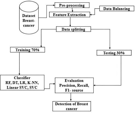
Fig-2: Proposed methodology. as normalization, low-variance feature removal, and data balancing. To preserve the most informative qualities for classification, important feature selection strategies such
Volume: 11 Issue:12 | Dec 2024 www.irjet.net
4. Results and Discussion
Using the assessment measures EDA dataset, we have presenteda systematicapproachinthisstudytocompare thestate-of-the-art.Weinitiallygatheredthematerialfrom kaggle.com, which we accessed on April 15, 2023. For the simulation,weutilizedPythonJupiterNotebook6.4.12.The initialfindingsofexplanatorydataanalysesemployingmany models.PythonextensionslikePandas,Seaborn,Plotly,and BokehareexamplesofEDAresources.Pandasisapowerful dataanalysislibrarythatprovidesDataFramesandSeries. Plotly is a versatile framework for making interactive visualizationsincludingdashboardsand3Dplots,whereas Seaborn is a high-level interface for making statistical visualizations. A library called Bokeh is used to create interactive dashboards that can be used online. The predictionsareappliedtothedataset'ssubsetsinthisgraph. Thirty percent of the test data and seventy percent of the training data make up each batch. The table provided a summary and comparison of various metrics, including radiusmeanandid
Table-1:Exploratorydataanalysis
Withouttheexploratorydataanalysis(EDA)phase,thedata analysisprocessdescribedabovewouldnotbecomplete.In ordertoidentifypatterns,drawinferences,identifyoutliers, andformulatehypotheses,dataanalysisinvolvesdisplaying thedata.Beforeadvancingtomoreintricatestatisticaland machine learning techniques, EDA is helpful for understanding the structure and characteristics of the dataset. Recognizing the dataset's characteristics, finding issueswithdataquality,andspottingearlytrendsthatcould inspireadditionalmodelingandanalysisEDAisanessential stepinthedataanalysisprocesssinceitisusedtomakeall ofthedecisions.It isalsoessential to the reliability of the data-drivenoutcomes.
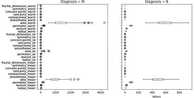
Fig-3: Boxplotsofbreastcancerdataset.
International Research Journal of Engineering and Technology (IRJET) e-ISSN: 2395-0056 p-ISSN: 2395-0072
Volume: 11 Issue:12 | Dec 2024 www.irjet.net
Theboxplotsofthedatasetonbreastcancerareshownin Figure3.Thedistributionofbreastcancerdetectionscoresis seen in these boxplots in this figure. Every graph that we obtained had an abnormality. The median of the data will decreaseiftheyareeliminated,whichcouldmakeitmore difficult to correctly diagnose tumors, especially if a malignanttumorismistakenlyclassifiedasbenign.However, inordertoavoidinterferingwiththeidentificationofserious cancers, we will only eliminate abnormalities in benign tumors. The maximum value of the "worst" area after processingis932.7,comparedto1210.0priortoprocessing. Prior to processing, the maximum area of the mean value was 992.1; now, it is 788.5. This could result in false positives,whereinbenigntumorsareincorrectlyclassified asmalignant.Ontheotherhand,thisapproachisbetterthan the other. By identifying tumors at an early stage, when treatmentismosteffective,thisdiagnosismaysavelives. In order to take an image, a mammographer will place the patient's breast on theplateof themammography machineand gentlycompressitwithasecondplatetospreadoutthebreast tissue.According to standard procedure, each breastisX-rayed twice: oncefrom the side(mediolateral obliqueview) and oncefromthetopdown(cranio-caudalview).Thereisthe opportunitytoaddmoreviewsifnecessary.
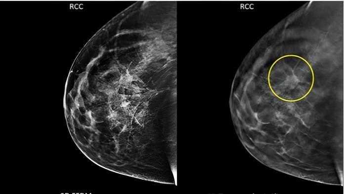
Figure4illustrateshowthelocation,size,andstageofbreast cancermightaffectitsappearanceonmammograms.Tofind breast cancer, radiologists look for anomalies and trends. Masses, microcalcifications, and architectural deformities are typical findings.Nodules, spiculated borders, and asymmetries. Microcalcifications are microscopic calcium deposits in breast tissue, whereas masses, microcalcifications,andarchitecturalabnormalitiesareearly indicatorsofbreastcancer.K-foldcross-validationisabreast cancerscreeningmethodthatdividesadatasetintoKequalsizedsubgroups.TheotherK-1subsetsareusedfortraining, andoneoftheKsubsetsisusedasthetestset.Kevaluations and trainings are performed on the model. Performance metricsarecalculatedonthetestsettoassesshowwellthe model generalizes to new data. After all K iterations, the performancemetricsfromeachfoldareaveragedtoprovide acomprehensiveoverview.
Breast Cancer detection using machine learning: K-fold cross-validation is necessary for robustness, overfitting detection,optimalhyperparametertuning,biasandvariance assessment, and realistic performance estimations. It guaranteesthattheevaluationismoretrustworthyandless dependent on a specific data split, allowing for more accurateandexactpredictionsofthemodel'sperformance onfresh,untesteddata.
The top 10 correlation findings across all variables are shown in e 6. Diagnostic, radius_mean, testure_mean, perimeter_mean,area_mean,smoothness_mean,andother parametersaredisplayedonthey-axis.Whereasapositive ornegativevaluedenotesastrongrelationshipbetweenthe two variables, a correlation coefficient between -1 and 1 impliesnoassociationatall.Itiscrucialtorememberthat correlationcanonlyquantifythelinearrelationshipbetween variables.Eachoftheseindicatorshasacorrelationwiththe patient's prognosis of at least 69%.Diagnostics 2023, 13, 3113 16 of 22 that the correlation can be used to make measurements. Each of these indicators has a correlation withthepatient'sprognosisofatleast69%.
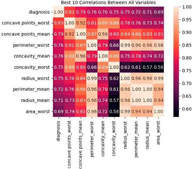
Fig-5: Correlationbetweenthetenbestvariables.
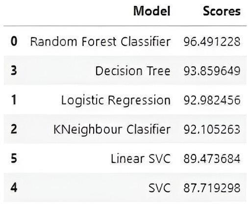
Fig-6:Accuracyresultforclassifiers.
International Research Journal of Engineering and Technology (IRJET) e-ISSN: 2395-0056 p-ISSN: 2395-0072
Volume: 11 Issue:12 | Dec 2024 www.irjet.net
Theaccurateoutcomesofeveryclassifierweemployedare displayed in Figure 6. With an accuracy of 96.49%, the random forest is the best result in this figure. The explanationofothersarelistedbelow.
* Whencompared toothermodels,random forest models performbetteratpredictingthetargetvariable,asseenby theirgreatestaccuracyscores.
* KNN anddecision treesare comparable toothermodels andhavegreatervaluesthanLGandSVC, suggestingthat theymightnotbethebestmodelsforpredictingthetarget variable.
*SVCandlogisticregressionbothperformwellinpredicting thetargetvariable,asevidencedbytheircomparablevalues.
Becauseofitsadaptability,simplicityininterpretation,and capacitytoidentifywhichcharacteristicsarenearlyessential to the categorization decision-making process, random forestmaybeagoodsubstituteforothermethodswhenit comestocancerdetection.
UsingtheBreastCancerWisconsin(diagnostic)dataset,this study assesses six machine learning models for the categorizationofbreastcancer.TheStandardScalermodule was used for data preparation, and scikit-learn in Python wasusedforfeatureselection.RandomForest(RF),Decision Tree(DT),K-NearestNeighbors(KNN),LogisticRegression (LR), and Support Vector Classifier (SVC) are among the modelsthatwereputtothetest.Confusionmatriceswere used to evaluate performance parameters such accuracy, precision,recall,sensitivity,F1-score,andAUC.Correlations between characteristics and patient outcomes were emphasizedbyexploratorydataanalysis,whichshowedthat RFhadthehighestaccuracy,followedbyDTandKNN.SVC andlogisticregressionperformedsimilarly.RFisnotablefor its adaptability, readability, and capacity to recognize important characteristics. Given that breast cancer can be classedaseitherinvasiveductalcarcinoma(IDC)orductal carcinomainsitu(DCIS),thestudyhighlightstheimportance ofearlyidentification.Diagnosticimagingmethodssuchas thermography, ultrasonography, and mammography are essential. Advances in artificial intelligence, namely in the areasofdeeplearningandconvolutionalneuralnetworks, are boosting the accuracy of mammography analysis. An extremelysensitiveimagingtechniqueisstillbreastMRI.To further enhance breast cancer detection and treatment, future studies should investigate the incorporation of cutting-edge AI approaches and promote cooperation between data scientists,medicalexperts,andresearchers. This could revolutionize early diagnosis and patient outcomes.
[1] Bayrak,E.A.;Kırcı,P.;Ensari,T.Comparisonofmachine learningmethodsforbreastcancerdiagnosis.InProceedings of the 2019 Scientific Meeting on Electrical-Electronics &
Biomedical Engineering and Computer Science (EBBT), Istanbul,Turkey,24–26April2019.
[2] Loomans-Kropp,H.A.;Umar, A.IncreasingIncidenceof ColorectalCancerinYoungAdults. J. Cancer Epidemiol. 2019, 2019,9841295.[CrossRef][PubMed].
[3] Din,N.M.U.;Dar,R.A.;Rasool,M.;Assad,A.Breastcancer detection using deep learning: Datasets, methods, and challenges ahead. Comput. Biol. Med. 2022, 149, 106073. [CrossRef][PubMed].
[4] Pang,T.;Wong,J.H.D.;Ng,W.L.;Chan,C.S.Deeplearning radiomics in breast cancer with different modalities: Overviewandfuture. Expert Syst. Appl. 2020, 158,113501. [CrossRef]
[5] Zhou, X.; Li, C.; Rahaman, M.M.; Yao, Y.; Ai, S.; Sun, C.; Wang, Q.; Zhang, Y.; Li, M.; Li, X.; et al., A comprehensive review for breast histopathology image analysis using classical and deep neural networks. IEEE Access 2020, 8, 90931–90956.[CrossRef]
[6] Wisesty, U.N.; Mengko, T.R.; Purwarianti, A. Gene mutationdetectionforbreastcancerdisease:Areview. IOP Conf. Ser. Mater. Sci. Eng. 2020, 830,032051.[CrossRef]
[7] Mahmood,T.;Li,J.;Pei,Y.;Akhtar,F.;Imran,A.;Rehman, K.U.ABriefSurveyonBreastCancerDiagnosticwithDeep LearningSchemesUsingMulti-ImageModalities. IEEEAccess 2020, 8,165779–165809.[CrossRef]
[8] Sutanto,D.H.;Ghani,M.K.A.Abenchmarkofclassification framework for non-communicable disease prediction: A review. ARPN J. Eng. Appl. Sci. 2015, 10,9941–9955.
[9] Gautam,R.;Kaur,P.;Sharma,M.Acomprehensivereview onnatureinspiredcomputingalgorithmsforthediagnosisof chronicdisordersinhumanbeings. Prog. Artif. Intell. 2019, 8,401–424.[CrossRef]
[10] Mahmood,M.;Al-Khateeb,B.;Alwash,W.M.Areviewon neuralnetworksapproachonclassifyingcancers. IAESInt.J. ArtifIntell. 2020, 9,317–326.[CrossRef]
[11] Dai, B.; Chen, R.-C.; Zhu, S.-Z.; Zhang, W.-W. Using Random Forest Algorithm for Breast Cancer Diagnosis. In Proceedings of the 2018 International Symposium on Computer,ConsumerandControl(IS3C),Taichung,Taiwan, 6–8December2018;pp.449–452.[CrossRef].
[12] Nguyen, C.; Wang, Y.; Nguyen, H.N. Random forest classifiercombinedwithfeatureselectionforbreastcancer diagnosisandprognostic. J. Biomed. Sci. Eng. 2013, 06,551–560.[CrossRef].