

Department of Ophthalmology

CONTENTS

Message From Department of Ophthalmology Leadership
Defining the Emerging Field of Oculomics Through Our Advanced Imaging and Technology
• Sounding the Alarm on Signs of Sickle Cell Dis ease
• New Evidence Cites Reduced Ocular Perfusion as a Mechanism for Macular Degeneration
Understanding Papilledema Through the Lens of Machine Learning
Cataract Surgery Is on the Cusp of Seismic Change ... Again
Remote Diagnosis Gives Hope to Eye Stroke Patients
Investing in Our Patients
Virtual Reality Prepares Residents for Realities of Cataract Surgery
Genetic Screening Tilts the Scales for Glaucoma Patients
Microsurgical Training Has a New Look at the Buxton Education Center
Unlocking the Secrets of Dry Eye, One Layer at a Time
Golden Eye: A Patient With an Eye-Threatening Injury Has Sight Saved by the City’s Premier Trauma Center
OCT Angiography Is About to Take a ‘Wide Turn’ With Unprecedented New Reach
‘Waste Not, Want Not’ Drives Study of Eye Drop Usage
Department of Ophthalmology at a Glance
Faculty News
Faculty Listing
Message From Department of Ophthalmology Leadership

Paul A. Sidoti, MD
Chair,
Department of Ophthalmology, New York Eye and Ear Infirmary of Mount Sinai

James C. Tsai, MD, MBA
President,
Infirmary
New York Eye and Ear
of Mount Sinai Chair, Department of Ophthalmology, Icahn School of Medicine at Mount Sinai and Mount Sinai Health System
From artificial intelligence to a new state-of-the-art center for cataract and refractive procedures to simulated surgical training for residents with a techsavvy global partner, New York Eye and Ear Infirmary of Mount Sinai (NYEE) continues its dedication to innovation and new directions in the field of ophthalmology. Backed by the extensive resources of the Icahn School of Medicine at Mount Sinai and the Mount Sinai Health System, we build upon a foundation supported by a culture of mentorship and technological innovation, as well as a 200-year legacy of community service to ensure that patients receive the highest level of ophthalmic care.
The rapidly evolving field of cataract surgery illustrates the wide spectrum of our activities—as well as our commitment to change. As described more fully in the following pages, we opened the Center for Refractive Solutions to provide the best solution for enhancing vision, whether it be premium-lens cataract surgery or refractive procedures for individuals focused on enhancing their vision and/or reducing their need for

Louis R. Pasquale, MD, FARVO
Chair,
Department of Ophthalmology, The Mount Sinai Hospital and Mount Sinai Queens
glasses or contact lenses. With the support of advanced corneal and anterior segment imaging and the latest intraocular lens platforms, our skilled clinicians and surgeons oversee a streamlined process tailored to patient satisfaction and superlative outcomes adapted to their lifestyles.
We are making great strides in enhancing our educational curriculum by adding unique simulation surgical training. This training involves a partnership with the global education organization HelpMeSee and our robust microsurgical training program at the Jorge N. Buxton, MD, and Douglas F. Buxton, MD, Microsurgical Education Center. This hybrid training model is an invaluable tool to help prepare our second-year residents to operate safely and efficiently on patients. Surgical training is further enhanced this year through the installation of 10 highresolution microscopes equipped with video cameras at the Buxton Center, benefiting both ophthalmic and ENT surgeons. And very shortly, we will open a new Education Center on the first floor of NYEE’s Bank Building, across 14th Street from the hospital. A state-of-the-art, dedicated
conference space for the ophthalmology and ENT departments, the Center will host grand rounds and visiting professor lectures and serve as a central educational hub for trainees.
In terms of cutting-edge surgical technology, we are one of a handful of institutions in the country to trial the use of miCOR, a procedural innovation that packs the power and performance of bulky phacoemulsification machines into a safe and effective handheld instrument for cataract surgery. Developed by one of our faculty members, Tsontcho (Sean) Ianchulev, MD, MPH, and now being used in our hospital practice, miCOR is likely to pave the way for a new era of office-based cataract procedures in the United States and globally.
The deployment of artificial intelligence and machine learning to transform ophthalmology remained front and center for us following our 2023 launch of the Center for Ophthalmic Artificial Intelligence and Human Health. An example of our leading-edge work is using an artificial neural network to evaluate, from standard fundus photographs of patients’ eyes, the clinical stage and severity of papilledema. Moreover, we are at the forefront of the emerging science known as oculomics, driven by machine learning and powerful imaging technologies. We are advancing this field with the development of the Intermittent Perfusion Index as an effective biomarker for measuring the efficacy of novel CRISPR-based gene editing for treating sickle cell disease.
Our eye trauma center, a top referral source for the tristate area, continues to provide sight-saving surgical expertise to patients with traumatic eye injuries. It is staffed by a team of dedicated voluntary and full-time faculty members, residents, and fellows.
Together, these ambitious projects paint a vibrant picture of how our enduring strengths in research, education, and uncompromising patient care have enabled us to remain a world-renowned specialty hospital—the first of its kind in the Western Hemisphere—for more than two centuries. We are working more diligently and purposefully than ever to maintain that stature, even as our specialty and medical institutions feel the inevitable winds of change.


Top Awards Our Specialty Reports Garner


The success of our scientists in uncovering breakthrough therapies and of our clinicians in treating patients with the latest advances in ocular medicine and surgery are brought to vivid life each year in the pages of the Department of Ophthalmology Specialty Report, the publication you are now reading. We are incredibly proud that these reports have won a total of 22 top industry honors over the past six years for their creative excellence and commitment to the highest communication and professional standards.
Credit for this repeated industry recognition lies with the Mount Sinai Health System’s Marketing and Communications Department, comprising Creative and Design, Communications and Content, and Print Services teams, along with our freelance writers, photographers, and graphic designers. Each year, this team marshals its collective talents, creative energy, and passion to find imaginative new ways to engage readers with memorable patient stories, visionary leadership pieces, and fresh accounts of our research and clinical accomplishments that embody the spirit of innovation that drives everything we do at NYEE.
Among the coveted honors recognizing outstanding performance in advertising, marketing, and communications we have received are the Aster Award, Hermes Award, Healthcare Advertising Award, and MarCom Award.

Defining the Emerging Field of Oculomics Through Our Advanced Imaging And Technology
The notion of the eye as a revealing window on the systemic health of the entire human body dates back centuries. The advent in recent years of powerful imaging techniques that can examine the eye in microscopic detail and then apply machine learning to identify biomarkers of systemic disease has given rise to a name for this emerging and highly promising science: oculomics.
continued on page 8

Sounding the Alarm on Signs of Sickle Cell Disease
New York Eye and Ear Infirmary of Mount Sinai (NYEE) was active in oculomics well before it became an industry buzzword and area of interest around 2020. We are now poised to be a major player in the field through our groundbreaking work with optical coherence tomography angiography (OCTA) and adaptive optics scanning light ophthalmoscopy (AOSLO) to image the retinal capillary bed and thereby measure the activity and severity of sickle cell disease. These technologies also measure the response of patients to treatment, including the first CRISPRbased gene editing regimen approved by the U.S. Food and Drug Administration (FDA) in December 2023 for sickle cell disease, signaling a huge advance in genome editing technology.
This is welcome news to more than 100,000 Americans, most of them of African descent, who suffer from sickle cell disease, which causes red blood cells to become hard, sticky, and crescent shape, blocking blood flow to the rest of the body. “Many conditions of the eye give us early clues about the presence of other systemic diseases in the body— including diabetes, cardiovascular, hypertension, sickle cell, and Alzheimer’s—that might otherwise
go undetected,” says Richard B. Rosen, MD, Vice Chair and Director of Research at NYEE, and the Belinda Bingham Pierce and Gerald G. Pierce, MD, Distinguished Chair of Ophthalmology at the Icahn School of Medicine at Mount Sinai. “The ability of our advanced imaging technology to study intermittent perfusion events in sickle cell disease clearly shows the potential of oculomics, while giving us an advantage in this important space.”
The eye as a harbinger of a wide range of pathologies gained credence with the work of R. Theodore Smith, MD, PhD, Director of Biomolecular Retinal Imaging at NYEE. His lab identified a strong association between subretinal drusenoid deposits found above the retinal pigment epithelium in age-related macular degeneration (AMD) and systemic vascular disorders impacting the heart, including coronary artery disease, cardiac valvular disease, hypertension, and stroke. That revelation is meaningful for ophthalmologists and cardiologists alike, who now have evidence to justify cross-treating patients who show either AMD or heart disease.
Coupling advanced imaging with machine learning

is now allowing clinicians for the first time to watch sickle red blood cells flow through the capillaries and visualize the mechanisms of vaso-occlusion. “We noticed that capillaries would open and close momentarily, an early sign of vaso-occlusion, which can result in blindness if blood flow blockage is prolonged,” explains Toco Y.P. Chui, PhD, Director of the David E. Marrus Adoptive Optics Imaging Laboratory at NYEE.
That observation led Dr. Chui to develop the Intermittent Perfusion Index, an algorithmic-driven measure of what percentage of capillaries in the retina are transiently blocked over the course of an hour, based on repeat or sequential imaging during that timeframe. In a study published in Case Reports in Hematology, Dr. Chui and research colleague Jeffrey Glassberg, MD, MA, Director of the Mount Sinai Comprehensive Sickle Cell Program, found the OCTA/ Intermittent Perfusion Index approach to be an effective biomarker for measuring the severity of sickle cell
disease, as well as the efficacy of novel CRISPRbased gene editing.
NYEE plans to add to that sophisticated tool set a new AOSLO capability that can measure the blood flow velocity within single capillary segments, giving clinicians a more granular view of disease progression and response to therapy.
“Following the FDA’s recent approval of gene therapy for sickle cell disease, we look forward to seeing more patients who are cured of the disease and to better understand their recovery. There are a wide range of additional vascular, neurologic, and metabolic conditions that may soon be treatable with similar gene therapy approaches,” notes Dr. Rosen. “Clinical cellular imaging will be an important tool for assessing an individual’s response to therapy and will pave the way toward advancing the adoption of gene therapy as a standard clinical solution.”
New Evidence Cites Reduced Ocular Perfusion As a Mechanism for Macular Degeneration
Scientists have sought for 30 years to make the connection between age-related macular degeneration (AMD) and systemic vascular disease, which includes myocardial infarction and ischemic stroke. A group of recently published studies from researchers at New York Eye and Ear Infirmary of Mount Sinai (NYEE) makes that association tighter than ever through strong evidence that vascular disorders compromise the flow of blood (or perfusion) to the retina, in turn serving as a mechanism for the formation of subretinal drusenoid deposits (SDDs), telltale signs of age-related macular degeneration. Just as importantly, the new findings make a strong case for using SDDs as highly sensitive biomarkers for not just AMD, but vascular pathologies that might otherwise go unrecognized.
“The public health message from our studies couldn’t be more clear: if SDDs are detected, especially in patients with cholesterol abnormalities, they should trigger a cardiovascular consult for the patient to detect potential high-risk vascular diseases, and potentially save lives,” says R. Theodore Smith, MD, PhD, Professor
of Ophthalmology at the Icahn School of Medicine at Mount Sinai and senior author of the studies. “Likewise, widespread screening of patients with known vascular disorders for SDDs could lead to the discovery of AMD, and potentially save their sight.”
The research team advanced another mechanism for the SDD phenotype: genetic risk and, more specifically, the ARMS2 gene, which provides instructions for making a protein found in light-sensing tissue in the retina. “We showed that HARM2, as we renamed a double dose of the harmful form of ARMS2, is a second major risk factor for SDDs independent of high-risk vascular disease,” notes Dr. Smith, who is also Director of Biomolecular Retinal Imaging at NYEE. “While more research is required, recognizing this gene’s central role in AMD could position it as a future target for therapeutic interventions.”
Following is a more detailed look at the three NYEE studies that could open an important new chapter in diagnosing and treating macular degeneration by navigating promising cardiovascular pathways.
continued on page 10
The Carotid Artery Connection
In their effort to determine if ocular hypoperfusion from internal carotid artery stenosis is associated with subretinal drusenoid deposits, scientists relied on a perfect experimental system handed to them by nature. They learned from evaluating ischemic stroke patients that moderate or greater blockage of the carotid artery was strongly associated with SDDs and thinning of the choroid—the vascular membrane that lies between the retina and the sclera—on the same or ipsilateral side of the body. Since the unblocked side was unaffected, this pathology offered convincing proof that downstream ophthalmic artery and choroidal hypoperfusion from internal carotid artery stenosis was a mechanism for SDD formation.
As reported in the February issue of Investigative Ophthalmology and Visual Science , that research is the first to demonstrate a significant association between increasing ICA stenosis and ipsilateral SDDs. Investigators concluded that additional research is warranted to determine whether using optical coherence tomography imaging to detect SDDs could serve as an inexpensive mass-screening tool for high-risk vascular disorders, potentially leading to life-saving evaluations for patients.
Linking SDDs to Thinning of the Photoreceptors
Age-related macular degeneration is known to damage the ellipsoid zone of the photoreceptors, the cellular components of the retina that convert light entering the eye into signals that are transmitted to the brain. In this study, published in the February issue of BMJ Open Ophthalmology , NYEE researchers demonstrated in their cohort of patients with AMD a quantitative link between SDDs and decreased ellipsoid zone thickness.
This major finding, they emphasized, is consistent with and offers strong evidence that ellipsoid zone thinning is a critical marker of the reduced and insufficient ophthalmic perfusion that causes SDDs and, therefore, hypoxic photoreceptor damage in cases of high-risk cardiovascular disease and stroke. Clinicians, they concluded, could provide enhanced multidisciplinary care to their retinal and cardiovascular patients by better understanding this emerging link between AMD and systemic vascular disease.
The public health message from our studies couldn’t be more clear: if SDDs are detected, especially in patients with cholesterol abnormalities, they should trigger a cardiovascular consult for the patient to detect potential high-risk vascular diseases, and potentially save lives.
—R. Theodore Smith, MD
Decreased Perfusion From Valvular Heart Disease
This study’s model of SDDs in age-related macular degeneration draws a straightforward line to valvular heart disease, particularly mitral and aortic valve disorders, which affect about 2.5 percent of the U.S. population, rising to 13 percent in those over 80. And as in their companion papers, researchers learned that SDD deposits were significantly more common with valvular heart disease of moderate severity or greater, once again supporting a proposed mechanism of SDD formation through a cardiovascular, then an ocular, perfusion deficit.
In a paper published in the March issue of European Journal of Ophthalmology, scientists said their findings implied that an ophthalmologist detecting SDDs should promptly refer the patient to a cardiologist for a standard workup for vascular disease. The urgency is enhanced in patients who also have low levels of HDL, the “good” cholesterol that protects against vascular disease. Likewise, the presence of mitral or aortic valve disease should trigger a workup by an ophthalmologist for SDDs and AMD as part of a public health effort for early detection of potentially blinding and life-threatening diseases.

Fig. 1 SDDs caused by severe cardiac valve disease. (A): SD-OCT scan, right eye, shows myriad SDDs (yellow arrows) above the RPE and several drusen (blue arrows) beneath the RPE in a patient with severe mitral valve regurgitation and compromised cardiac output. The insufficient ocular perfusion downstream then results in the SDDs. The left eye was similar. (B): A color doppler transthoracic echocardiogram of the left ventricle during systole, showing markedly reduced cardiac index, 1.87 L/min/m2, normal range 2.5-4.0, and severe mitral valve regurgitation. Left atrium shows turbid jet of arterial blood flowing retrograde from the left ventricle.


AFig. 2 Unilateral SDDs caused by a dose response to unilateral internal carotid artery (ICA) stenosis. (A): SD-OCT scan, right eye, downstream of mild right ICA stenosis, shows no SDDs and a few drusen. (B): SD-OCT scan, left eye, downstream of moderate left ICA stenosis, shows confluent SDDs and choroidal thinning are significantly associated (p=0.002, p=0.008) with ipsilateral ICA stenosis in a dose response to moderate (> 50%), not mild, ICA stenosis.
Understanding Papilledema Through the Lens of Machine Learning

Machine learning is no stranger to the field of glaucoma, but its application to the disorders that swell the optic nerve and cause severe vision loss and potentially lifethreatening diseases has been limited.
Researchers at the Mount Sinai/New York Eye and Ear (NYEE) Eye and Vision Research Institute have led a team from several institutions to advance the science, showing how ML can quantify the clinical stage of papilledema and guide the treatment of this optic neuropathy, characterized by swelling of the optic discs due to increased intracranial pressure. The results of the study were reported in the July issue of BMJ Neurology
“For the first time, we’ve tasked an artificial neural network to assign a Frisén grade to fundus photographs of eyes with papilledema,” says senior author Mark Kupersmith, MD, Professor of Neurosurgery, Ophthalmology, and Neurology at the Icahn School of Medicine at Mount
Sinai, and Chief of Neuro-Ophthalmology for the Mount Sinai Health System. “This information could be critical to clinicians in the diagnosis, treatment, and management of not just papilledema, but possibly other non-glaucomatous optic nerve problems such as optic neuritis and nonarteritic anterior ischemic optic neuropathy.”
Inasmuch as prior research has shown that machine learning can distinguish eyes with papilledema from normal eyes, Mount Sinai investigators were determined to learn if those curated data sets could be coupled with the Frisén scale to rate the severity of the disease and, just as importantly, indicate changes over time with a common treatment, acetazolamide, which is a carbonic anhydrase inhibitor.
To that end, the team, which also included members from Harvard Medical School, the University of Iowa, and Cork University Hospital in Ireland, trained a convolutional
The application of machine learning methodologies, such as convolutional neural network, is becoming a valuable diagnostic tool for ophthalmologists for a wide variety of optic nerve disorders.

neural network to assign a Frisén grade to fundus photographs taken from the eyes of 158 participants enrolled in the Idiopathic Intracranial Hypertension Treatment Trial, the first large study on the use of acetazolamide and weight loss for treating idiopathic intracranial-associated vision loss.
They learned that machine learning could indeed build on the Frisén scale to show differential changes over time in the eyes of study participants taking acetazolamide versus a placebo group.
“Our machine learning results correlated well with the expert evaluation by humans—certainly good enough for clinical use to show responses in patients to medications,” notes Dr. Kupersmith. “Given the increasing availability of fundus photography, including handheld cameras, neurologists should be able to use machine learning to quantify papilledema on a
continuous basis that incorporates features of the Frisén grade to monitor interventions.” The technology could also help clinicians who have less experience and training in ophthalmology, he points out, making it a valuable diagnostic tool in emergency departments, intensive care units, and primary care offices.
The current study on papilledema is part of a larger effort by Dr. Kupersmith and his lab to explore nonglaucomatous optic nerve pathologies through the lens of fundus photography, OCT, and visual field, with eight studies under their belt. “New York Eye and Ear Infirmary of Mount Sinai has become a center of excellence for applying artificial intelligence methodologies to optic nerve disorders,” he emphasizes. “And we’re hopeful this work will not only lead to better detection and prediction of a set of diseases that may be associated with lifethreatening illness, but will do so at a stage when they can be effectively treated.”

Cataract Surgery
Is on the Cusp of Seismic Change … Again
continued on page 16
Dr. Ianchulev is the technological founder of the miCOR and miLOOP devices.

In 1962, cataract surgery was revolutionized with the introduction of phacoemulsification, which uses ultrasonic waves to break up and aspirate out cloudy fragments of the eye’s lens. The father of that technology was Charles D. Kelman, MD, a surgeon and attending on the staff of what was then known as The New York Eye and Ear Infirmary (NYEE).
Six decades later, the next wave of innovation has arrived. It promises to reconfigure the cataract landscape through the advent of miCOR and miLOOP, which pack the power and performance of bulky phaco machines into simple handheld devices. Just as cataract surgery’s move into ambulatory surgery centers in the mid-1980s was disruptive at the time, but soon became recognized as a safe, affordable, and familiar choice for surgeons, the movement toward office-based cataract procedures has been gaining ground in the United States.
“Cataract surgery is transitioning to a new era of convenient office-based settings made possible by miniaturized devices like miCOR and miLOOP,” says James C. Tsai, MD, MBA, President, NYEE. “As a major referral center that performs thousands of cataract cases a year, we’re helping to shape this frontier by participating in a trial to refine the technology so that it’s safe and effective when finally rolled out to patients across the country. Equally as far-reaching are the implications it could have for making cataract surgery more affordable and accessible to people in developing nations globally.”
In January 2024, New York Eye and Ear Infirmary of Mount Sinai became the only institution in the Northeast, and one of 10 in the country, to inaugurate use of miCOR for cataract surgery.

“For more than five decades, ophthalmologists have been waiting for new cataract technology that is not just an incremental extension of the conventional phacoemulsification technology, and miCOR puts the power of a 250-pound ‘mainframe’ machine into a miniaturized handheld lens pen, which can fragment and extract the cataract in less than five minutes with less thermal energy, no cavitation power, and less fluidic impact—not to mention the capital expense of the big phaco machines,” says Tsontcho (Sean) Ianchulev, MD, MPH, Director of Ophthalmic Innovation and Technology at NYEE, who is also the technological founder of the miCOR and miLOOP devices, ultimately acquired by ZEISS Medical, a global technology leader in the field of optics and ophthalmology.
For all its benefits, phacoemulsification has always had its share of vulnerabilities. It requires, for example, complex instrument-heavy equipment with significant installation and maintenance costs and a steep learning curve for ophthalmologists, and it presents challenges for treating advanced cataracts. The costs, complexity, and extended learning curve have held back cataract surgery from solving the global blindness epidemic despite a highly effective (95%+) 10-minute procedure that can restore a person’s vision and dramatically improve their life. A recent report by the World Health Organization still lists cataracts as a leading cause of blindness and visual impairment in the world, with almost 100 million people experiencing this curable condition.
To overcome these barriers, more efficient, streamlined, and scalable techniques and instrumentation that could impact cataract surgery are needed not just in the developed world, but in developing countries where

Close-up view of miCOR lens pen with a nucleus removal tip.
Lens fragmentation and extraction using miCOR with irrigation/aspiration removal tip attachment.

cataract-related blindness is a critical and worsening public health problem.
Enter miLOOP, a groundbreaking microinterventional device with the ability to fragment all densities of cataracts using self-expanding nitinol technology to substantially reduce phaco energy and fluid use. Yet miLOOP still leaves the need for phacoemulsification to further fragment and extract dense lens segments. That’s where miCOR serves as the ideal companion for phaco-free nuclear extraction. Approved by the U.S. Food and Drug Administration in 2022, miCOR is a self-contained lens pen that eliminates the need for a console, foot pedal, and other expensive machinery. The mechanism of fragmentation is low-frequency 40 Hz, non-ultrasonic, non-cavitating vibrations of the tip, so heat, and thus the need for fluidic irrigation, is dramatically reduced.
“Use of the device is very intuitive and requires just over a minute for my team to set up, making it much quicker to turn the room over for the next patient,” explains Kira Manusis, MD, Director of the Center for Refractive Solutions at NYEE, who has successfully performed the first procedures with miCOR at the hospital. “Everything
is contained in a handpiece, and I’m able to do surgery just as efficiently as with a much larger phaco machine.”
As for getting up to speed on the device, Dr. Manusis cites “a bit of a learning curve, but not a steep one.”
In addition to the technology figuring in a growing number of successful patient procedures, it is being introduced to residents as part of their surgical training. “We want our trainees to be prepared for the future and the changes in technology that may have a profound impact on how they practice their craft,” points out Dr. Manusis.
Dr. Ianchulev, a faculty member at NYEE, is a founder, board member, and/or equity owner of multiple life-science companies, including the public company Eyenovia, Inc., and private companies Iantrek, Inc., and Aeye, Inc. As a prominent innovator in the field of ophthalmology, he is the holder of multiple issued and pending patents including the technology related to the development of cataract surgery devices discussed in this article. This technology was sold to ZEISS; Dr. Ianchulev has no current financial interests with ZEISS and does not benefit financially from the sale of these devices.
Dr. Manusis performs the first cataract surgery at NYEE using miCOR, a subsonic, non-cavitating iso-thermic lensectomy system.
Remote Diagnosis Gives Hope to Eye Stroke Patients
The COVID-19 pandemic underscored the power and promise of remote diagnosis of patients by clinical experts. A novel program at New York Eye and Ear Infirmary of Mount Sinai (NYEE) has built on that paradigm to transform care for one of the most time-sensitive ophthalmic emergencies—central retinal artery occlusion (CRAO), or “eye stroke.”
In a study published in the June issue of Ophthalmology, NYEE physicians reported encouraging results for the CRAO remote consultation program, a collaboration between ophthalmologists and Mount Sinai’s stroke service. During the first 18 months of the program, visual acuity for most patients diagnosed with CRAO and treated with intra-arterial tissue plasminogen activator (IA-tPA) improved significantly within 24 hours and remained stable at one month. Specifically, six of the nine patients treated saw vision improve from worse than 20/200 to 20/100 or better. Four of the nine improved to 20/40 or better the first day.
“For an ocular emergency in which very few people historically improve at all, most of our patients experienced same-day recovery of their vision,” says first author Gareth M.C. Lema, MD, PhD, Vice Chair for Quality, Safety, and Experience for the Department of Ophthalmology at Mount Sinai, and a retinal specialist who helped develop the program. “It’s a proof of concept for an initiative that effectively combines the latest technology and treatment for CRAO with solid commitment from a wide range of clinical partners.”
In practice, the program integrates remote diagnosis and optical coherence tomography (OCT) in the emergency rooms of three Mount Sinai hospitals. When patients present with painless monocular vision loss, they are evaluated by the stroke neurology service, which uses an onsite automated OCT machine to acquire cross-sectional images of the retina at the resolution of just a few microns. Images are uploaded to retina specialists at NYEE, who are available around the clock to read and expertly interpret them.
Despite the criticality, delays in diagnosing eye stroke have been the norm across the country with patients often waiting hours or days before seeking medical help.
Even after arriving at an emergency room, diagnosis may be delayed by the availability of on-call house staff, timeconsuming referrals to an ophthalmologist, and availability of imaging specialists who can perform fluorescein angiography, traditionally used to diagnose CRAO. In 2021, the American Heart Association called for development of systems of care to prioritize the diagnosis of retinal artery occlusion. Mount Sinai’s unique point-of-care protocol fully responds to that imperative. Among the keys to its success, says Dr. Lema, is getting patients treatment as soon as possible, optimally within six hours or even less. (Improvement has been seen even within 12 hours, but less commonly.) According to the study, the mean time from “last known well” (the last time the patient remembers seeing well) to injection of IA-tPA was around nine hours, and from presentation at a stroke center to injection was around two and a half hours.
Another reason for the program’s impressive results has been the uncompromising buy-in from all participating parties. “If you don’t have an onsite stroke team committed to getting usable images and data to remote retina specialists who are just as committed to making a diagnosis as quickly as possible, the program won’t be effective,” comments Richard B. Rosen, MD, Vice Chair and Director of Ophthalmology Research at NYEE. “We helped ensure its success by choosing OCT machines that are easy to learn and use, have a small footprint for busy emergency departments, and linked our eye stroke protocols to the existing protocols of stroke teams so that they didn’t have to change much.”
Dr. Lema sees vast opportunities for expansion of the initiative to hospitals both within and outside Mount Sinai—particularly in remote regions with a shortage of ophthalmology coverage—and beyond just CRAO. “We’ve shown its effectiveness for retinal artery occlusion, but our remote consultation program could offer tremendous benefits for offsite diagnosis and treatment of many other ophthalmic emergencies.”
With the 12-hour tPA intervention model firmly in place, Dr. Rosen is turning his focus on new therapies targeting patients who can’t get the standard intra-arterial therapy. “Our goal is to be able to restore some vision to patients who come to us 24 and even 48 hours after the eye stroke took place.”

Illustration by TJASART, Tjaša Žurga Žabkar s.p
Investing in Our Patients

Amid all the science and innovation, no investment is more meaningful to New York Eye and Ear Infirmary of Mount Sinai (NYEE) than enhancing interactions with its patients. Three recent physical improvements—the opening of the Center for Refractive Solutions, the reopening of the Optical Shop in prime retail space, and renovations of the Laser Vision Correction Center—drive the point home.
Streamlining the Patient Experience
The new Center for Refractive Solutions caters to individuals determined to improve their vision or reduce their dependence on glasses or contacts through cataract surgery or corneal refractive procedures like LASIK. The dedicated and newly renovated space at NYEE brings together state-of-the-art corneal and ocular imaging, a staff of highly experienced surgeons and specialists, and a streamlined process from check-in to testing to professional evaluation to vastly improve the patient experience.
“We’re now able to offer our patients more complete and efficient access to premium services, including intraocular lens implants, laser-assisted cataract surgery, and custom LASIK,” says Kira Manusis, MD, Director, Center for Refractive Solutions and Co-Director of Cataract Services at NYEE. “We’ve centralized all the necessary equipment and services to deliver an efficient and thorough evaluation for patients in just one day—no small advantage for individuals who lead busy lives.”
Supporting that effort are state-of-the-art technologies that allow our ophthalmic specialists to image and map the multiple layers of the cornea, in addition to precisely measuring the dimensions of the eye. “This thoroughness enables us to design a surgical plan tailored to each patient’s lifestyle and ophthalmic needs,” explains Angie Wen, MD, Director of the Keratorefractive Surgery Division at NYEE. “Our surgeons are skilled at implanting the latest intraocular lens platforms, including trifocal and extended depth-of-focus lenses, as well as the new light-adjustable lens. For patients who don’t yet have cataracts, the best refractive solution may be LASIK, PRK, or the EVO phakic intraocular lens.”
NYEE is grateful to the generous support of the Toni and Martin T. Sosnoff family for helping fund the Center for Refractive Solutions.
Optical Shop Is Now a Convenient Storefront
Upon the initial opening of its doors on the third floor of the South Building at NYEE, the Optical Shop helped fill a crucial void for patients looking to improve the quality of their daily lives. Three years later, the Optical Shop has gone through another patient-friendly makeover and relocation to a convenient new storefront location at the corner of 14th Street and Second Avenue in Manhattan.
“The reopening of the Optical Shop at ground level will ensure greater visibility and ease of access for patients from either within or outside the hospital,” notes Paul Sidoti, MD, Chair of Ophthalmology at NYEE. “This will help solve the problem that many of our patients in need of eyeglasses or low-vision assistive devices have had in accessing our optical dispensary.” Another service enhancement now includes an experienced optometrist who provides comprehensive eye exams and offers helpful advice on eyeglass frames, corrective lenses, and sunglasses ranging from high-end luxury brands to more affordable versions covered by insurance.
The Optical Shop, originally funded as part of a grant from the Lavelle Fund for the Blind, Inc. to enhance low-vison services at NYEE, also provides devices to allow people with limited vision to make the most of the sight they still have. These include traditional aids like handheld and stand magnifiers to enlarge print material, and non-optical equipment like flexible-arm task lamps and absorptive sunglasses that filter out bothersome glare. In addition, patients are apprised of the latest technology-driven devices and applications, such as text-to-speech smartphone apps and screen readers that read emails, web pages, and books aloud.
The improved services and expanded, more accessible location are “our way of improving care for all our patients,” sums up Dr. Sidoti, “and providing more equitable delivery of services to the entire community.”
The Laser Vision Correction Center Gets a Fresh Look
As part of Mount Sinai’s ongoing investment into the Downtown Campus, the Laser Vision Correction Center has been renovated. The redesigned space provides a fresh, contemporary décor with a newly furnished reception area and well-appointed exam/treatment rooms. We look forward to continuing to serve our patients in our beautiful new offices.
Center for Refractive Solutions Clinical Team
Left to right: Angie Wen, MD, Anita Gupta, MD (first row) and Yandong (Yanna) Bian, MD, Sumayya Ahmad, MD, Kira Manusis, MD, Masako Chen, MD (second row)
Virtual Reality Prepares Residents for Realities Of Cataract Surgery
No goal is more critical to surgical training at New York Eye and Ear Infirmary of Mount Sinai (NYEE) than preparing residents for the operating room earlier with the broadest complement of skills and proficiencies. That objective is being met with help from a budding partnership with a nonprofit organization known as HelpMeSee, which is committed to providing stateof-the-art training to ophthalmologists so they can address the leading cause of visual impairment and blindness globally: cataracts.
This year’s class of 10 second-year residents, the largest in the country, is the first at NYEE to benefit from the new alliance. Each resident will attend a weeklong course at HelpMeSee’s instructor-led training center in Jersey City, one of 12 around the world that use high-fidelity, virtual reality simulators to teach the basic skills they’ll need to operate safely and efficiently on patients.
“The program aligns nicely with our plan to move surgical training earlier in the residency,” says Paul A. Sidoti, MD, Chair, Department of Ophthalmology, NYEE. “The HelpMeSee platform closely approximates the actual surgical procedure, taking residents methodically through every step of cataract surgery, which will
Khushali Shah, MD, an NYEE Resident, receives instruction from Nasar Mohammed, MD on manual small-incision cataract surgery using the high-fidelity virtual eye simulator at the HelpMeSee location in Jersey City.


ultimately give them a real advantage when they enter the OR for the first time with a live patient.”
At the core of the program is the Eye Surgery Simulator, developed by HelpMeSee engineers and ophthalmologists over the past 10 years. “The simulator provides trainees in a controlled practice environment with spatial, visual, and tactile realism that’s very comparable to participating in real surgery,”


explains Bonnie An Henderson, MD, President and CEO of HelpMeSee, which trains around 1,000 ophthalmologists a year in locations spanning the globe. “Now, trainees can practice hundreds of times before they begin actual surgery, which increases not only the pace at which they learn, but safety for their patients.”
Making that level of interactive experience possible was
the organization’s creation of a software algorithm that replicates through its simulator the texture, feel, and look of real tissue. Peering through a binocular microscope at workstations with simulated microsurgical instruments in hand, students can navigate with three-dimensional rigor each intricate step of cataract surgery, from the initial corneal incision to emulsification of the nucleus to implantation of a foldable intraocular lens. Other workstations, each staffed by an instructor who provides
continued on page 24

real-time feedback, are focused on suturing, managing surgical complications, and performing manual smallincision cataract surgery, which is practiced in regions of the world where expensive phacoemulsification equipment is not practical.
“The goal of our residency program is to develop the best cataract surgeons in the world, and to that end, we’re adding this program to the excellent wet lab curriculum, courses, and models that already exist at our Jorge N. Buxton, MD, and Douglas F. Buxton, MD, Microsurgical Education Center,” says Harsha S. Reddy, MD, Ophthalmology Residency Director and Site Director for Oculoplastics, Orbital, and Reconstructive Surgery at NYEE. “One important aspect of that training is managing and resolving

complications that can happen intraoperatively, and HelpMeSee’s advanced simulator is well equipped to do that.”
The collaboration of NYEE and HelpMeSee makes eminent sense in the context of their common goal: working to prevent loss of sight by individuals and whole communities by marrying technology with the latest training techniques.
“We’re thrilled to be partnering with NYEE because we know they’re one of the leading institutions in the country for ophthalmic education,” says Dr. Henderson, who is a past president of the American Society of Cataract and Refractive Surgery. “Their use of our advanced simulator along with the intense, hands-on practice it affords will allow them to build on the quality and the reach of their educational commitment.”

Close-up view of the virtual reality console with simulated microsurgical instruments used during training.
Left: Simulation of emulsification of the nucleus using phaco.
Right: Simulation of ophthalmic suturing, passing the needle.
Genetic Screening Tilts the Scales for Glaucoma Patients
Scientists are hard at work developing the nextgeneration screening strategy for glaucoma, and large biorepositories like the Mount Sinai Bio Me BioBank, with its 54,000 participants, are well equipped with the data necessary to support that work. For that resource to be efficiently tapped, however, an advanced system of polygenic risk scores (PRSs) that provides clinicians with individual genetic risk for primary open-angle glaucoma (POAG) must be developed and fine-tuned over time.
A recently published study in the online June issue of American Journal of Ophthalmology , led by researchers from New York Eye and Ear Infirmary of Mount Sinai (NYEE) and Mass General Brigham, moves the field of ophthalmology closer to that important goal. The twocenter investigation showed that using International Classification of Diseases (ICD) codes, which are billing codes residing in electronic health record (EHR)-linked biobank data, are as effective as labor-intensive manual reviews of those records in constructing a system of POAG risk stratification and prognostication.
“The challenge is determining how we can build the next-generation screening strategy so we can pick up more cases of glaucoma earlier and quicker—and genetic testing through polygenic risk scores is clearly the answer,” says Louis R. Pasquale, MD, Deputy Chair for Research and Director, Mount Sinai/NYEE Eye and Vision Research Institute. “First though, we need to test the diagnostic accuracy of the current PRS tool with existing databases. What we found was that even though ICD codes are somewhat imprecise, they are an acceptable surrogate for POAG disease classification leading to polygenic risk score construction and use.”
Many EHR-linked biorepositories employ ICD codes to identify diseases of interest to researchers and clinicians. In the case of POAG, however, the accuracy of these billing codes for disease detection was unknown, posing significant challenges for the translation of genomic tools into clinical practice aimed at screening and detection. In their study, the
The challenge is determing how we can build the next-generation screening strategy so we can pick up more cases of glaucoma earlier and quicker—and genetic testing through polygenic risk scores is clearly the answer.
—Louis R. Pasquale, MD
researchers learned that ICD codes were as effective as manually reviewing test data across ancestries in two EHR-linked biobanks: Mount Sinai’s Bio Me BioBank and Mass General Brigham’s Biobank, which was founded in 2008 and contains 135,000 current participants.
Partnering with Mass General Brigham was particularly helpful to researchers in establishing a disease classification for POAG. The data repositories serve to replicate research findings at each site and serve to enhance their generalizability across ancestral groups.
“We believe that there will be future iterations of the PRS that will be more powerful than the current iteration,” emphasizes Dr. Pasquale. “And by showing that PRS performed similarly when glaucoma is defined by ICD codes or by manually testing medical record data, it allows for rapid testing of future PRS tools.”
A resident physician is performing a capsulorhexis as a part of a simulated cataract surgery on a model eye under an operating microscope. The procedure is captured by a high-definition video camera and displayed on a monitor in real time so the instructor can monitor his technique.


Has a New Look at the Buxton Education Center Microsurgical Training
As one of the country’s leading training sites for a new generation of ophthalmic and ENT surgeons, the Jorge N. Buxton, MD, and Douglas F. Buxton, MD, Microsurgical Education Center is no stranger to advanced technology. In 2006, for example, it installed a virtual reality EyeSi Surgical Simulator to sharpen the intricate microsurgical skills trainees will soon bring to the operating room. Extensive audiovisual improvements soon followed.
The latest upgrade further elevates the educational experience for students and instructors alike. Ten out of 16 training stations within the wet lab environment have been outfitted with ZEISS EXTARO 300
microscopes, each equipped with a high-resolution video camera so that faculty can view practice sessions on a screen in real time, or recorded for later viewing.
“These high-definition microscopes will make training much more realistic for our residents and fellows through exquisite visualization and depth perception,” says Douglas F. Buxton, MD, Clinical Professor of Ophthalmology at the Icahn School of Medicine at Mount Sinai, and son of Jorge N. Buxton, MD, the first chief of the cornea service at New York Eye and Ear Infirmary of Mount Sinai (NYEE) and a champion of modern microsurgery. “In addition, cameras attached to each microscope will allow for co-observation by
Richard D. Najac, MD, a voluntary attending physician and former NYEE resident, provides instruction on cataract surgery to residents using the new microscopes.

instructors and trainees in a way that promotes on-thespot interaction and discussion.”
The replacement of older microscopes with the latest models is consistent with the core mission of NYEE. “The upgrade demonstrates our commitment to not just maintaining but enhancing our educational programs and the investment we continue to make in the future of our residents and fellows,” says Paul A. Sidoti, MD, Deputy Chair for Education, Department of Ophthalmology, Icahn Mount Sinai. “Having a microsurgical training lab with a robust simulation program is also vital to our ability to attract top candidates from across the country who look for such a capability.”
Some 47 ophthalmic residents and fellows and 30 ear, nose, and throat trainees will benefit each year from the enhanced training. In fact, the Department of Otolaryngology at Icahn Mount Sinai is financing half the cost of the lab upgrade, with the remainder being split between the Buxton Microsurgical Education Fund and the Mount Sinai Department of Ophthalmology. Furthermore, the improvements were championed by both James C. Tsai, MD, MBA, President of NYEE, and Eric M. Genden, MD,

Laboratory
FACS, the Isidore Friesner Professor and Chair of Otolaryngology – Head and Neck Surgery at Icahn Mount Sinai.
“By significantly increasing the power of illumination and magnification, the new microscopes will provide students with better and crisper images of small and fine details of structures, especially of the ear and eye,” says George Wanna, MD, Professor and Executive Vice Chair of the Department of Otolaryngology – Head and Neck Surgery. “This is a huge advantage when learning general otolaryngologic procedures such as tympanoplasty, myringotomy, mastoidectomy, stapedectomy, ossicular chain reconstruction, and cochlear implants.”
The training of otolaryngologists has been an important part of NYEE’s rich history since 1864. In 1958, the Temporal Bone Laboratory was established, the first of its kind on the East Coast, to instruct physicians on performing delicate surgery on the inner ear and around the facial nerves. The laboratory became the Jorge N. Buxton, MD, Microsurgical Education Center upon its opening in September 2004.
For Dr. Buxton, the latest lab improvement is a natural fit for the Buxton Microsurgical Education Fund, which he created 15 years ago. “We’re trying to create a microsurgical educational environment that is second to none,” he declares. “And by allowing residents to imitate as closely as possible the real OR surgical environment, these new enhancements are true to our mission.”
Archival image of the Temporal Bone
where ENT residents practiced surgery on the inner ear and around the facial nerves.
Unlocking The Secrets
O f Dry Eye, One Layer
At a Time
The abundance of over-the-counter products available today for dry eye leaves consumers and ophthalmologists alike scratching their heads over choosing the best treatment for their specific condition. New York Eye and Ear Infirmary of Mount Sinai (NYEE) researchers decided to take a scientific approach to their quandary by evaluating the clinical effects of three of the most popular artificial tears (ATs) on the market.
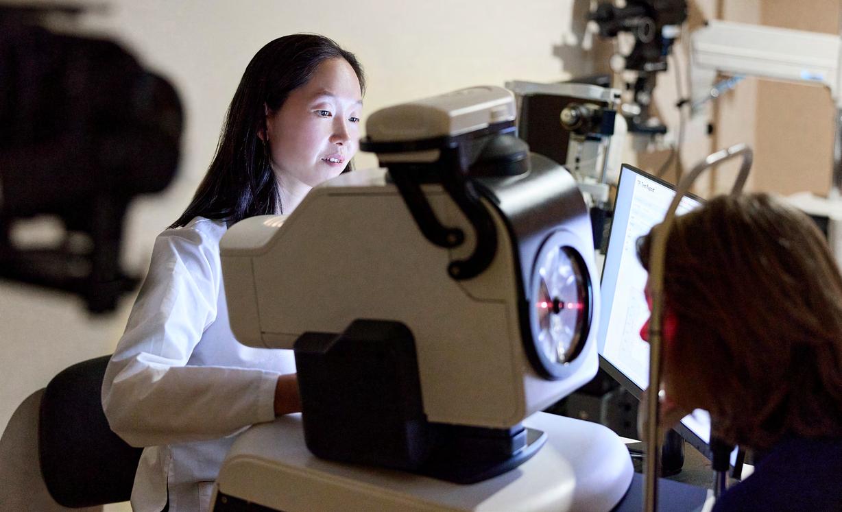
For the investigation, they used a state-of-the-art instrument known as the Tear Film Imager (TFI) to analyze the intricate and ultra-thin layers of the eye’s tear film. The team reported for the first time, in a study published in the February issue of Cornea , how various AT options impact the muco-aqueous and lipid sublayers, suggesting the importance of choosing a product based on the patient’s specific ocular surface pathology.
“There’s an abundance of artificial tear options for the major types of dry eye disease, including evaporative, aqueous deficient, and mixed,” says Masako Chen, MD, Assistant Professor of Ophthalmology at the Icahn School of Medicine at Mount Sinai and senior author of the study. “But with few methods to objectively assess them, confusion regarding the most suitable formulation for a particular individual often results.”
Several diagnostic tests have been developed over the years to examine ocular surface health, such as fluorescein-assisted tear break-up time, which evaluates tear stability; Schirmer strips to assess tear production; and vital dyes to highlight epithelial irregularity. Each of these approaches, however, uses mostly static metrics that fail to assess changes in tear film dynamics over time—a glaring gap.
Dr. Chen uses spectral interference technology to image and map the corneal surface of a patient.
Soaps and creams


Tear film imagery and analysis from a 72-year-old female with dry eye symptoms. Images were taken during and two days after discontinuation of use of facial creams near the eye. Note the difference in the lipid layer and the lipid map uniformity, including oil droplets, with (top image) and without (bottom image) recent application of facial creams.


The TFI is helping to push the science by demonstrating the significant differences in short-term effects on the thickness of the muco-aqueous and lipid layers by the three tear film products researchers studied. The quantification of tear film sublayers, according to Dr. Chen, could help clinicians tailor a therapeutic regimen to the patient’s dry eye disorder.
Combining spectrometry and imaging to analyze tear film layers, the TFI is on the leading edge of an emerging family of sophisticated devices. Developed by Advanced Optical Methods (AdOM), an Israeli company, TFI uses spectral interference technology to image and map the corneal surface with extraordinary precision and clarity thanks to a large field of view (6 mm diameter) and high axial resolution. Measurements are taken as often as 10 times per second over 40 seconds throughout the natural blinking process.
“In ophthalmology, we administer a lot of topical medications for glaucoma, inflammation, dry eye, and postsurgical care, all of which disturb the surface cells of the eye,” says Richard B. Rosen, MD, Vice Chair and Director of Research at NYEE, and the Belinda Bingham Pierce and Gerald G. Pierce, MD, Distinguished Chair of Ophthalmology at Icahn Mount Sinai. “The Tear Film Imager is the first device that measures the individual
layers of the tear film dynamically so we can better assess the efficacy of particular therapies and understand the impact of the surgical choices we make for patients.”
Fueling the field’s interest in tear film analysis is the surge of dry eye among people of all ages. Researchers have responded by exploring the many ways in which the protective layers of the tear film can break down, as well as the most effective therapies to repair the surface damage. That work embraces not just dry eye, but other conditions that result in a lack of tear homeostasis, including glaucoma, Sjogren’s syndrome, refractive and retinal surgery, nerve dysfunction, diabetes, and meibomian gland dysfunction.
To that end, the TFI is proving to be an indispensable research tool. “The uniqueness of the device is its ability to slice and quantify the various layers of the tear film with nanometer resolution, which in turn provides valuable scientific information to better understand surface diseases of the eye and the impact of various therapeutics,” explains Alon Harris, PhD, FARVO, Professor and Vice Chair of Ophthalmology and CoDirector of the Center for Ophthalmic Artificial Intelligence and Human Health at Icahn Mount Sinai, who has helped drive development for the TFI.
A recent NYEE study analyzing the impact of facial creams and lotions on the sublayers of the tear film in patients with meibomian gland dysfunction is further evidence of TFI’s practical capabilities. Compared to a cohort of non-lotion users, significant increases in the lipid layer thickness and liquid map uniformity were found in those using lotion products, demonstrating how cosmetics may play a role in ocular surface disruption.
Work is also underway at NYEE using the TFI to study the effect of topical therapies on the tear film of glaucoma patients by imaging them before and after treatment. “Some patients do very well on one type of compound for controlling intraocular pressure, but not on another,” points out Paul A. Sidoti, MD, Chair of Ophthalmology at NYEE and Glaucoma Service Chief at Mount Sinai Health System. “We could greatly improve the science of treating glaucoma by quantifying the impact of eye drops on the various layers of the tear film and surface epithelium for individual patients. This will help better tailor glaucoma treatments to each patient’s unique physiology.”
Lipid Layer Map (nm)
Lipid Layer Map (nm)

Golden
Eye: A Patient With an Eye-Threatening Injury Has Sight Saved by the City’s Premier Trauma Center

Richard Nuñez


The pair of physicians at New York Eye and Ear Infirmary of Mount Sinai (NYEE) pulled no punches with Richard Nuñez when he arrived early on a Sunday morning in March 2024 with a large, jagged piece of gold-colored metal impaled in his right eye.
“We told him our No. 1 goal was to try and save the eyeball, but there was a good chance he could lose it given the severity of his injury,” recalls Julia Fallon, MD, a retina fellow who, along with Alexander Barash, MD, a member of the NYEE Retina Service teaching faculty and the eye trauma surgeon on call, responded to the emergency.
Through the intense pain that kept building, Mr. Nuñez, 39, was frightfully aware of how his life could be changing in a flash from the freakish fall he had taken just hours before, projecting the metallic object like a missile through the delicate outer membranes of his eye. “I’m a service technician who needs both eyes and both hands,” he says, “and I knew there was a chance I wouldn’t be able to go back to work.”
As ophthalmic emergencies go, this was one of the highest order. An intraocular foreign body lodged in the eye can trigger infection, oxidation, and retinal damage with each passing minute. And if the optic nerve is impacted, the chances of restoring eyesight are extremely low.
Fortunately for Mr. Nuñez, a father of three young boys, he was transported to NYEE by ambulance from a suburban hospital that lacked the resources to treat his
injury. Drs. Fallon and Barash were waiting for him, along with other members of one of the city’s most experienced eye trauma teams, to begin the complex, three-hour-long procedure to salvage his sight.
An initial inspection of the injury revealed how extensive it was. The errant piece of metal, measuring 60 x 40 x 10 mm, started its journey by piercing the upper eyelid, then traveled through the conjunctiva and sclera into the eyeball, where it burrowed into the vitreous gel, and ended up in the inferior portion of the eyeball. It tore the inferior retina—and this required laser treatment to seal the break. “He was remarkably lucky the trauma didn’t damage his optic nerve, or affect the other eye,” notes Dr. Barash, a vitreo-retinal surgeon in private practice who also trains residents and fellows at NYEE.
While removing the metal was the ultimate goal, the surgeons first had to meticulously close the large opening in the eyeball so they could re-establish a closed system that would enable them to proceed with the remainder of the procedure without losing ocular contents through a leak.
“It became obvious to us that the superior part of the sclera was open, so we carefully dissected down to the huge opening in the eyeball, and sutured it shut,” explains Dr. Fallon, who is now in private practice and a member of the NYEE voluntary teaching faculty. “Because some of the muscles of the eye had also been pierced by the object, we had to sew them up as well, and put them back in place. It was a pretty complex job.”


We couldn’t be more happy and grateful for the care and service the hospital provided. The doctors were incredibly helpful, informative, and invested in making sure Richard could see again out of his eye.
—Jessica Muñiz, Richard’s fiancée
Once blood was cleared from the back of the eye—fully exposing the piece of metal—and the shape of the eyeball re-established, the surgeons faced their next challenge: safely removing the object without damaging the fragile superstructure of the eye. They methodically surveyed the two options open to them: passing the object through the same wound by which it entered, or guiding it through the cornea, which would require removing the lens of the eye. “The point of the surgery was to save the eye and hopefully promote visual recovery down the road,” points out Dr. Fallon, “and that often means sacrificing the lens. We deemed that to be the best option for this patient.”
Dr. Barash (left) and Dr. Fallon (right) prepare to perform surgical removal of a gold-colored metallic fragment (center) through the cornea. Measuring 60 x 40 x 10 mm, the metal shard covered over half the diameter of the cornea.
During the months-long recovery and regular visits to the hospital that followed, the wisdom of that surgical decision became clear. Within weeks, the patient’s vision had improved to 20/60. Steady healing of the eye enabled doctors to fit Mr. Nuñez with a special aphakic contact lens to promote visual rehabilitation, and by July, his vision was 20/25, one line away from 20/20. This encouraging progress enabled Mr. Nuñez to proceed with his final surgery in early September to replace his contact with a permanent intraocular lens. Because the surgeons intentionally left scaffolding in place to anatomically secure the lens, the procedure was more akin to a standard cataract surgery instead of a more invasive operation. When Mr. Nuñez returned for his follow-up appointment the next day, his vision was 20/20.
“We know that a lot of people don’t recover from something this traumatic,” acknowledges Mr. Nuñez’s fiancée, Jessica Muñiz, “so we couldn’t be more happy and grateful for the care and service the hospital provided. The doctors were incredibly helpful, informative, and invested in making sure Richard could see again out of his eye.”
For Dr. Barash, the outcome of this case fits a familiar pattern. “It speaks to the strength of our unique training program,” he says, “where fellows and residents get regular exposure to these types of severe injuries. That requires them to make crucial game-time decisions, which turns them into smart and competent surgeons— and NYEE into a center where other institutions feel comfortable sending their most difficult trauma cases.”
OCT Angiography
Is About to Take a ‘Wide Turn’ With Unprecedented New Reach


OCT angiogram of patient with proliferative sickle retinopathy. Left: Shows areas of capillary loss and sea fan neovascularization temporally. Right: OCT cross section shows vitreous attachment to sea fan, which adds risk of hemorrhage.
Using infrared light to visualize cross-sectional details of the retina, known as optical coherence tomography (OCT), has had a major impact on the quality of ophthalmic care since its clinical introduction nearly three decades ago. The technology is now on the cusp of a new breakthrough thanks to the clinical introduction of wide-field OCT angiography (OCTA), which provides details of the retinal blood vessels for detection of diabetes and other peripheral retinal vascular diseases quickly and noninvasively.
New York Eye and Ear Infirmary of Mount Sinai (NYEE) has already installed a clinical prototype of wide-field OCT/OCTA—dubbed the DREAM OCT, from Intalight, based in China—in its Einhorn Clinical Research Center and is studying its clinical application in anticipation of full approval by the U.S. Food and Drug Administration.
For all of their advantages, current commercial OCTA systems allowed imaging of only small 3 x 3 mm to 9 x 9 mm central segments of the retina.
“That was a major revolution for examining blood flow to the macula, but it left clinicians chomping at the bit to view the full retina,” says Richard B. Rosen, MD, Retina Chief at the Mount Sinai Health System,
Vice Chair and Director of Research at NYEE, and a leader over the years in bringing state-of-theart imaging to the field of clinical ophthalmology. “The new wide-field OCTA camera allows us to noninvasively see almost the entire posterior retina in a single scan. And the pictures we get, without dilating the pupil, are spectacular.”
Based on swept-source OCT and a proprietary system, DREAM OCT can image the retina to a depth of 12 mm and the anterior segment to 16.2 mm (from the cornea to the anterior part of the vitreous), while covering an ultra-wide field of 130° , 26 x 21 mm, of the posterior segment in a single scan. What’s more, its scan speed of up to 400 KHz is considerably faster than current commercial OCT devices, with high accuracy, minimal artifact, and a comfortable patient experience.
“Patients who are asymptomatic may gain the most,” points out Dr. Rosen. “If we’re able to pick up early disease in people who may have peripheral disease of the retina, like sickle cell and diabetes, we may be able to begin to adjust their systemic therapy before they notice vision loss and before their condition spirals out of control.”


Main: Diabetic retinopathy with capillary dropout. Inset: Color capillary density map highlighting losses.
‘Waste Not, Want Not’ Drives Study of Eye Drop Usage
A large bin filled with ophthalmic eye drops in an eye clinic storeroom caught the attention of Gareth M.C. Lema, MD, PhD, one day. Asking around, he learned the bin was a collection site for multi-dose ophthalmic drops that were being discarded shortly after they were opened and well before their expiration dates.
“It was clear there were thousands of dollars of unused drops sitting in these bottles,” recalls Dr. Lema, Associate Professor of Ophthalmology at the Icahn School of Medicine at Mount Sinai. “And the reason was selfimposed use cessation dates that are set by the Health System, usually within 28 days of opening or less, which was much shorter than the Food and Drug Administration (FDA)-regulated expiration dates. As a result of this premature disposal, we were experiencing unnecessary and significant waste of medications, increased costs, and disposal of plastic into the environment.”
Dr. Lema, who is also Vice Chair of Quality, Safety, and Experience for the Mount Sinai Health System Department of Ophthalmology, was quick to act on his discovery. He led a study with colleagues to estimate the volume of drugs discarded in hospital-based ophthalmology clinics, along with the financial, clinical, and environmental implications. Collecting and measuring the remaining content in multi-use eyedrop bottles slated for disposal from three New York Eye and Ear Infirmary of Mount Sinai (NYEE) clinics with 61 exam rooms, the researchers unearthed some surprising findings.
Among the most stunning findings described in the study, published in the July issue of Ophthalmology, was that on average nearly 72 percent of the total volume of medication remained at the time the bottles were discarded. Moreover, the researchers estimated that 91 percent of all bottles would have been fully depleted before expiration if the FDA-regulated expiration date—which is based on extensive testing of the drop in its marketed container with appropriate preservatives—was followed.

At issue is the so-called self-imposed use cessation date (SUCD) of 14 to 28 days after opening, an arbitrary timeframe that became widely accepted in hospital-based and academic institutions, versus the longer FDA-regulated expiration date printed on the bottle, known as FRED.
“The fear was that if you exceeded the SUCD, you could transmit infection,” explains Dr. Lema. “But that doesn’t appear to be the case based on concrete evidence. There are no reports of infections from FDA-regulated dropper bottles used in clinical settings. What does prevent colonization of bacteria on the tips of dropper bottles is proper training of medical and technical staff in the eye clinic.”
In actual practice, the research team calculated that the number of eye drop bottles could be reduced by 72 percent at the three NYEE clinics studied if they were discarded according to the FRED schedule. That economization translates into an annual cost savings of 73 percent, or more than $80,000, across the clinics. Less easily measured but still meaningful are the environmental benefits of cutting back on plastic bottle disposal, and the clinical importance of reducing wastage of drops like phenylephrine and tropicamide, which have faced recent shortages.
Based on their unique findings—the first ever to quantify ophthalmic eye drop wastage in a nonsurgical setting— researchers recommended that ophthalmologists and clinics upgrade their practices around administering drops to embrace the FRED instead of the SUCD.
“Medications used in the clinic represent a considerable cost and are prone to drug shortages,” emphasizes Dr. Lema. “These are fairly simple problems to correct by using FREDs that make those shortages less likely while helping to reduce costs and ensure safe and effective patient care.”


Department of Ophthalmology at a Glance:
New York Eye and Ear Infirmary of Mount Sinai (NYEE) and The Mount Sinai Hospital (MSH)/ Icahn School of Medicine at Mount Sinai
One of the largest ophthalmology graduate medical education programs in the country. Fellowship Positions Residency Positions
Industry clinical trials and federally funded clinical research projects.
Combined 2023 numbers for NYEE and MSH.
East 14th Street New York, NY 10003
NYEE-East 102nd Street 17 East 102nd Street New York, NY 10029
NYEE-East 85th Street 234 East 85th Street New York, NY 10028
NYEE-Tribeca 77 Worth Street New York, NY 10013
NYEE-Midwood 1630 East 15th Street Brooklyn, NY 11229
NYEE-Carle Place 393 Old Country
Carle Place, NY 11514
30th Avenue Long Island City, NY 11102
Sinai Morningside 440 West 114th Street New York, NY 10025
FACULTY NEWS: AWARDS AND HONORS

Alon Harris, PhD, FARVO
Keynote speaker: “Beyond Patient Care: Using Artificial Intelligence to Understand Glaucoma,” New York Glaucoma Society, May 2024, New York, NY, USA
Keynote speaker: “Using Mathematical Modeling and Artificial Intelligence to Reveal the Role of Hemodynamics in Glaucoma,” 27th Annual Ulrich Ollendorff, MD Lecture Award, March 2024, Columbia University, New York, NY, USA
Keynote speaker: “The Eye as a Window to the Body: Advanced Imaging Technology, Math Models, and Artificial Intelligence to Detect Ocular and Systemic Diseases,” Rabin Medical Center, September 2023, Petah Tikva, Israel
Keynote speaker: “The Eye as a Window to the Body: Advanced Imaging Technology, Math Models, and Artificial Intelligence to Detect Ocular and Systemic Diseases,” Life Sciences Baltics (LSB) Conference, September 2023, Vilnius, Lithuania
Keynote speaker: Webinar “The Eye-Body Relationship,” The Glaucoma Foundation, April 2023, New York, NY, USA
Keynote speaker: “Artificial Intelligence and Math Models for Detecting Non-IOP Risk Factors for Glaucoma,” Israeli Glaucoma Society Meeting, March 2023, Tel Aviv, Israel


Louis R. Pasquale, MD, FARVO
Keynote speaker: “Do Large Language Models Know How to Diagnose and Manage Glaucoma?” and “African Heritage and Glaucoma: A Deeper Dive,” 50th Annual Ophthalmology Alumni Meeting, Downstate Health Sciences University, June 2024, Brooklyn, NY, USA
The Association for Research in Vision and Ophthalmology Distinguished Service Award , April 2023, New Orleans, LA, USA
Richard B. Rosen, MD, FARVO
American Society of Retina Specialists Founders Award, July 2024, Stockholm, Sweden
Keynote speaker: “Stalking the Wild Hyalocyte: En Face OCT and Adaptive Optics Insights into the Cellular Dynamic and Morphology of Vitreous Cortex,” William H. Havener Memorial Lectureship – Ohio State University, March 2024, Columbus, OH, USA
Keynote speaker: “Spying on Retina Microworlds: Synergizing Dynamic Enface OCT and Quad-Fusion Adaptive Optics SLO Imaging,” Pascal Rol Keynote LectureshipSPIE/Photonics West, January 2024, San Francisco, CA, USA
Atlantic Coast Retina Club Founders Award, January 2024, New York, NY, USA
FACULTY NEWS: RETIREES
Monica Dweck, MD
11 years, Physician and Teaching Faculty
Dr. Dweck received her medical degree from SUNY Downstate Health Sciences University College of Medicine. She was the first African American woman to complete her residency at The New York Eye and Ear Infirmary, and the first woman of color in the country to complete a fellowship in oculoplastic, reconstructive, and orbital surgery at the Cleveland Clinic Foundation. She joined Mount Sinai in 2013 as an Associate Professor of Ophthalmology; she has been a faculty advisor for medical students since 2017 and served as the inaugural Director of Career Planning Services from 2019 to 2022. Dr. Dweck is a founding board member of NYC’s Women in Ophthalmology and was its president in 2021-2022. She was elected to the Board of Directors of the New York State Ophthalmological Society before being appointed by the New York State Board of Regents in 2001 to the New York State Board of Medicine, where she continues to serve as a board member. Over the years, her accolades have included the 2010 and 2022 “VISIONS” award, a 2008 “Women Who Dare to Be Different” tribute at the 110th United States Congress, and a

Jose Mario Wolosin, PhD 39 years, Research Faculty
Dr. Wolosin joined the Mount Sinai School of Medicine as a research associate professor in 1985. Over the next 39 years, his career in visual science has been focused on understanding the stem cells of the limbal-corneal epithelial lining and regenerative approaches when this lining is compromised by the

Certificate of Merit by the New York State Assembly. Dr. Dweck was inducted into the Alpha Omega Alpha Honor Medical Society in 2006. Dr. Dweck has published numerous articles and book chapters and has lectured extensively at national conferences.
loss or malfunction of stem cells, causing partial or full blindness. His second area of exploration has been the biological mechanisms that underpin exfoliation syndrome. His contributions to visual science include furthering our understanding of unique properties of corneal stem cells, characterizing ciliary body transport, documenting the importance of gap junction function in the corneal epithelium, and developing an innovative method to study mechanisms of corneal differentiation-stratification and maturation. A prolific lecturer at numerous national and international conferences, Dr. Wolosin is an author and co-author of numerous articles. He was an Associate Editor of BMC Ophthalmology and served on the editorial boards of Current Stem Cell Research & Therapy and World Journal of Stem Cells. Dr. Wolosin is a recipient of the 19962000 Research to Prevent Blindness Senior Scientific Investigators Award and held the Jules and Doris Stein Research to Prevent Blindness Professorship from 1985 to 1990. Dr. Wolosin earned his Master of Science and PhD from the Hebrew University of Jerusalem.
We thank Dr. Dweck and Dr. Wolosin for their years of service and dedication to the field of ophthalmology, visual science, Mount Sinai, and the community we serve.
Left to right: James Tsai, MD, Monica Dweck, MD, Louis Pasquale, MD
Left to right: Jose Wolosin, PhD, Louis Pasquale, MD, James Tsai, MD
FACULTY NEWS: RECRUITS


Yandong (Yanna) Bian, MD, Assistant Professor of Ophthalmology
A fellowship-trained cataract, cornea, and refractive surgeon who specializes in the medical and surgical care of corneal and anterior segment disease, Dr. Bian earned her medical degree from Columbia University Vagelos College of Physicians and Surgeons, where she was inducted into the Alpha Omega Alpha Honor Medical Society. She completed both her ophthalmology residency and fellowship in cornea, external diseases, and refractive surgery at Harvard University/Mass Eye and Ear, where she also served as Chief Cornea Fellow. An author of peer-reviewed articles and chapters, Dr. Bian is a vision researcher and presents her work at national conferences.
Phillip
Braun, MD, Assistant Professor of Ophthalmology
A native Northeasterner, Dr. Braun specializes in comprehensive ophthalmology. Dr. Braun received his medical degree from Yale School of Medicine in New Haven, Connecticut, followed by an ophthalmology residency at Flaum Eye Institute, University of Rochester Medical Center. His scholarly endeavors have ranged widely, from a thesis on the philosophy of friendship to research on infrared laser development and pathogenesis of age-related macular degeneration, and lately have focused on education and mentorship.
Niki Mirzaei, OD, Instructor, Department of Ophthalmology
Dr. Mirzaei is a specialist in comprehensive optometric care with expertise in the diagnosis and management of a variety of ophthalmic conditions. She earned her Doctor of Optometry from Nova Southeastern University College of Optometry in Fort Lauderdale, Florida. Prior to joining NYEE, Dr. Mirzaei provided primary eye care at Luxottica as well as at the National Vision LLC in Woodbridge, New Jersey, as an associate optometrist in a high-volume practice.
Ahmed Sheikh, MD, Assistant Professor of Ophthalmology
An oculoplastic surgeon, Dr. Sheikh received his medical degree from Rutgers New Jersey Medical School, followed by a residency at Hofstra Zucker School of Medicine in Great Neck, New York. He completed fellowships in ocular oncology at Wills Eye Hospital in Philadelphia and ophthalmic plastic and reconstructive surgery at the Mayo Clinic in Rochester, Minnesota. A recipient of the Fellow Teaching Award, Mayo Clinic Department of Ophthalmology, two years in a row, Dr. Sheikh also served in the department as a clinical instructor. With a passion for vision science and research, Dr. Sheikh is the author of book chapters and peer-reviewed articles and has presented at national conferences.
FACULTY LISTING
NYEE RESIDENTS
PGY-4
Aaron Brown, MD
Chih-Chiun Chang, MD
Ha Min Kim Chang, MD
Allen Costa, MD
Anthony Fam, MD
David Merriott, MD
Devayu Parikh, MD
Jackson Scharf, MD
Khushali Shah, MD
Davis Zhou, MD
PGY-3
Jennifer Drechsler, MD
Jose Omar Garcia, MD
Catalina Garzon Vargas, MD
Andy Huang, MD
Claire Kim, MD
Michael Lai, MD
Oscar Otero, MD
Yash Shah, MD
Benjamin Steren, MD
Amanda Wong, MD
PGY-2
Aileen Arevalo, MD
Xiaoyu Cai, MD
Emily Duffner, MD
Jeff Huang, MD
Angela Kim, MD
Eliott Kim, MD
Sophia Lam, MD
Marissa Patel, MD
Jose Quiroz, MD
Dominic Williams, MD
NYEE FELLOWS
Ahmed Abdelhady, MD
Maria Castanos, MD
Devin Cohen, MD
Neha Gupta, MD
Andrew Jin, MD
Hirah Khan, MD
Neal Patel, MD
ICAHN SCHOOL OF MEDICINE AT MOUNT SINAI DEPARTMENTAL LEADERSHIP
James C. Tsai, MD, MBA
President, NYEE
Chair, Department of Ophthalmology
Director, COAIHH, Icahn Mount Sinai and MSHS
Louis R. Pasquale, MD, FARVO
Chair, Department of Ophthalmology, MSH and MSQ
Deputy Chair for Research, Director, Mount Sinai/NYEE Eye and Vision Research Institute
Co-Director, COAIHH, Icahn Mount Sinai
Paul A. Sidoti, MD
Chair, Department of Ophthalmology, NYEE
Deputy Chair for Education, Department of Ophthalmology, Icahn Mount Sinai
Chief, Glaucoma, MSHS
Mark Kupersmith, MD
Vice Chair, Translational Ophthalmology Research, Icahn Mount Sinai
Chief, Neuro-Ophthalmology, MSHS
Richard B. Rosen, MD, FARVO
Vice Chair and Director of Ophthalmic Research, NYEE Chief, Retina Service, MSHS
Salvatore Loiacono Jr., MPA
Deputy Chair, Finance and Administration, MSHS
Vice President of Ophthalmology Services, NYEE
Elizabeth Sellman
President and Chief Operating Officer, MSBI and Downtown
Tamiesha Frempong, MD, MPH
Vice Chair, Diversity and Inclusion, Department of Ophthalmology, Icahn Mount Sinai
Sandra K. Masur, PhD
Vice Chair, Academic Development and Mentoring, Department of Ophthalmology, Icahn Mount Sinai
Anita Gupta, MD
Vice Chair, Professional Development, Department of Ophthalmology, Icahn Mount Sinai Director, Cornea, NYEE
Alon Harris, PhD, FARVO
Director, Ophthalmic Vascular Diagnostic and Research Program, MSH Vice Chair, International Research and Academic Affairs
Chair, Promotion and Tenure Committee, Department of Ophthalmology, Co-Director, COAIHH, Icahn Mount Sinai
Gareth M.C. Lema, MD, PhD
Vice Chair, Quality, Safety, and Experience, Department of Ophthalmology, MSHS
Tak Yee Tania Tai, MD
Vice Chair, Clinical Operations, Department of Ophthalmology, NYEE Director, Glaucoma Clinic, NYEE
Gennady Landa, MD
Vice-Chair for Clinical Integration of Ophthalmology, Icahn Mount Sinai and MSHS Director, Retina, NYEE
Nisha Chadha, MD
Director, Medical Student Education, Department of Ophthalmology, Icahn Mount Sinai
Masako Chen, MD
Director, Comprehensive Ophthalmology, NYEE
Tsontcho A. Ianchulev, MD, MPH
Director, Ophthalmic Innovation and Technology, NYEE
FACULTY LISTING
RESIDENCY AND FELLOWSHIP EDUCATION
Sumayya Ahmad, MD
Associate Director, Ophthalmology Residency Program, NYEE/ISMMS
Masako Chen, MD Associate Director, Ophthalmology Residency Program, NYEE/ISMMS
Avnish Deobhakta, MD Associate Director, Vitreo-Retinal Fellowship Program, NYEE/ISMMS
Tamiesha Frempong, MD, MPH Assistant Director, Ophthalmology Residency Program, NYEE/ISMMS
Meenakashi Gupta, MD Director, Resident and Fellow Wellness, Department of Ophthalmology, NYEE/ISMMS
Gautam Kamthan, MD
Assistant Director, Ophthalmic Innovation and Technology, NYEE Associate Director, Cataract Surgical Training, NYEE/ISMMS
Gennady Landa, MD
Associate Director, Vitreo-Retinal Fellowship Program, NYEE/ISMMS
Harsha S. Reddy, MD Director, Ophthalmology Residency Program, NYEE/ISMMS
Richard B. Rosen, MD, FARVO Director, Vitreo-Retinal Fellowship Program, NYEE/ISMMS
Tak Yee Tania Tai, MD Director, Microsurgical Education, Department of Ophthalmology, NYEE/ISMMS
Kateki Vinod, MD Director, Glaucoma Fellowship Program, NYEE/ISMMS
Angie E. Wen, MD Director, Cornea and External Diseases Fellowship Program, NYEE/ISMMS
CLINICAL DIVISIONS AT MOUNT SINAI HEALTH SYSTEM FACULTY PRACTICES
Cataract Surgery
Richard Koplin, MD Co-Director, Cataract Service, NYEE
Kira Manusis, MD Director, Center for Refractive Solutions Co-Director, Cataract Service, NYEE
Sumayya Ahmad, MD Yandong (Yanna) Bian, MD
Nisha Chadha, MD
Masako Chen, MD
Anita Gupta, MD
Tsontcho A. Ianchulev, MD, MPH
Gennady Landa, MD
Tak Yee Tania Tai, MD
Angie E. Wen, MD
Cornea, External Diseases, and Refractive Surgery
Anita Gupta, MD Director, Cornea and External Diseases, NYEE
Angie E. Wen, MD
Director, Keratorefractive Surgery Division, NYEE
Sumayya Ahmad, MD Yandong (Yanna) Bian, MD
Masako Chen, MD Kira Manusis, MD
Eye Trauma
Ronald C. Gentile, MD Director, Posterior Segment Trauma Service, NYEE
Harsha S. Reddy, MD Director, Oculoplastic Trauma Service, NYEE
Sumayya Ahmad, MD
Masako Chen, MD
Mary-Abigail Craven, MD
Avnish Deobhakta, MD
Alberto G. Distefano, MD
Valerie I. Elmalem, MD
Robin N. Ginsburg, MD
Anita Gupta, MD
Meenakashi Gupta, MD
Gennady Landa, MD
Gareth M.C. Lema, MD, PhD
Kira Manusis, MD
Trisha Mor, MD
Richard B. Rosen, MD, FARVO
Angie E. Wen, MD Glaucoma
Paul A. Sidoti, MD Chief, Glaucoma, MSHS
Tak Yee Tania Tai, MD Director, Glaucoma Clinic, NYEE
Nisha Chadha, MD
Donna Gagliuso, MD
Tsontcho A. Ianchulev, MD, MPH
Paul S. Lee, MD
Louis R. Pasquale, MD, FARVO
James C. Tsai, MD, MBA
Kateki Vinod, MD
Neuro-Ophthalmology
Mark Kupersmith, MD Chief, Neuro-Ophthalmology, MSHS
Rudrani Banik, MD
Co-Director, Neuro-Ophthalmology Clinic, NYEE
Valerie I. Elmalem, MD
Co-Director, Neuro-Ophthalmology Clinic, NYEE
Mary-Abigail Craven, MD
Alberto G. Distefano, MD
FACULTY LISTING
Ocular Oncology
Paul T. Finger, MD Director, Ocular Oncology, NYEE
Ekaterina Semenova, MD
Ophthalmic Pathology
Jodi Sassoon, MD Chair, Pathology, NYEE
Codrin E. Iacob, MD, FCAP
Oculoplastic, Orbital, and Reconstructive Surgery
Alberto G. Distefano, MD Director, Oculoplastic, Orbital, and Reconstructive Surgery, MSH
Harsha S. Reddy, MD Director, Oculoplastic, Orbital, and Reconstructive Surgery, NYEE Director, Ophthalmology, MSBI
Mary-Abigail Craven, MD
Valerie I. Elmalem, MD
Ahmed Sheikh, MD
Pediatric Ophthalmology and Strabismus
Tamiesha Frempong, MD, MPH
Thomas J. Quehl, MD
Edward Raab, MD
Primary Care Ophthalmology/Optometry
Masako Chen, MD Director, Comprehensive Ophthalmology, NYEE
Vivien Boniuk, MD
Phillip Braun, MD
Christina Cherny, OD
Kristina Creadore, OD
Daniel Epshtein, OD
Lisa Kingsly, OD
Niki Mirzaei, OD
Leannza Tang, OD
Retina
Richard B. Rosen, MD, FARVO Chief, Retina, MSHS
Robin N. Ginsburg, MD Director, Retina, MSH
Gennady Landa, MD Director, Retina, NYEE
Nazanin Barzideh, MD
Avnish Deobhakta, MD
Meenakashi Gupta, MD
Gareth M.C. Lema, MD, PhD
Trisha Mor, MD
R. Theodore Smith, MD, PhD
Carl Wilkins, MD
Uveitis and Ocular Immunology
Varun K. Pawar, MD
Carl Wilkins, MD
Basic Science/Translational Research Faculty
Louis R. Pasquale, MD, FARVO Director, Mount Sinai/NYEE Eye and Vision Research Institute
Co-Director, COAIHH, Icahn Mount Sinai
Timothy A. Blenkinsop, PhD
Bo Chen, PhD
Yuen Ping Toco Chui, PhD
Alon Harris, PhD, FARVO
Varun Kumar, PhD
Jun Lin, MD, PhD
Sandra Masur, PhD
Richard B. Rosen, MD, FARVO
R. Theodore Smith, MD, PhD
Affiliated Leadership
Douglas F. Buxton, MD
President, Jorge N. Buxton, MD, and Douglas F. Buxton, MD, Microsurgical Education Fund
Paul S. Lee, MD
Chief, Department of Ophthalmology, James J. Peters VA Medical Center
President, NYEE/MSH Department of Ophthalmology Alumni Association
Teddy Lyu, MD
Director, Department of Ophthalmology, Elmhurst Hospital Center
Michelle K. Rhee, MD
Associate Director, Department of Ophthalmology, Elmhurst Hospital Center
Voluntary Teaching Faculty
NYEE
Steven Agemy, MD
Rudrani Banik, MD
Alexander Barash, MD
Alessandra Bertolucci, MD
Brett Bielory, MD
Douglas F. Buxton, MD
Gaurav Chandra, MD
Alan Dayan, MD
Jonathan Ellant, MD
Elizabeth Fang, MD
Edmund Farris, MD
Paul T. Finger, MD
John Flanagan, MD
Donald Fox, MD
Seymour Fradin, MD
Ronald C. Gentile, MD
Robyn Horowitz, MD
Jimmy Hu, MD
Lawrence Jacobson, MD
Steven Kane, MD
Yogita Kashyap, MD
Stephen Kronenberg, MD
Kaushal Kulkarni, MD
Jessica Lee, MD
Marc Lustig, MD
Palak Majmudar, MD
Kerline Marcelin, MD
Sarah McCord, MD
Amy Mehta, MD
Jeffrey Nightingale, MD
Jai Parekh, MD
Deep Parikh, MD
Alexander Pinhas, MD
Aryeh Pollack, MD
Alexander Rabinovich, MD
Allen Rubin, MD
Edwin Schottenstein, MD
Jonathan Shapiro, MD
Alan Sheyman, MD
Glenn Silbert, MD
Marc Sirota, MD
FACULTY LISTING
Mark Tannenbaum, MD
Theresa Tretter, MD
Cynthia Tung, MD
Alexander Voldman, MD
Eugene Weise, MD
Rebecca Weiss, MD
David Witzel, MD
Sze Wong, MD
Luna Xu, MD
MSH
Alexander Barash, MD
Ben Cohen, MD
Peter Condax, MD
Stella Douros, MD
Edward Marcus, MD
Joseph Podhorzer, MD
Ravi Raharkrishnan, MD
Alan Sheyman, MD
VA Medical Center
Isaac Ezon, MD
Chandak Ghosh, MD, MPH
Doris Herzfeld, MD
Teddy Lyu, MD
Jonathan Schulhof, MD
Steven Teich, MD
Elmhurst Hospital Center
Robert Fischer, MD
Laiyin Ma, MD
Richard Most, MD
Andrew Schwartz, MD
Zinaria Williams, MD
Kevin Wu, MD
Susan Zoltan, MD
COAIHH
Center for Ophthalmic Artificial Intelligence and Human Health
Icahn Mount Sinai
Icahn School of Medicine at Mount Sinai
MSHS
Mount Sinai Health System
MSH
The Mount Sinai Hospital
MSBI
Mount Sinai Beth Israel
MSM
Mount Sinai Morningside
MSQ
Mount Sinai Queens
MSW
Mount Sinai West
NYEE
New York Eye and Ear Infirmary of Mount Sinai
