2 minute read
GIANT RIGHT GASTROEPIPLOIC ARTERY PSEUDO-ANEURYSM WITH AN IVC FISTULA
10 GIANT RIGHT GASTROEPIPLOIC
ARTERY PSEUDOANEURYSM WITH AN IVC FISTULA By: Suzan Al-Hawwash
Advertisement
An 81-year-old Caucasian woman presented to the emergency department (ED) for evaluation of abdominal pain. She reported sharp epigastric pain that radiates to her back, along with intermittent abdominal pain for 5 days. She stated the pain was 10 out of 10 in severity and denied any other symptoms. Upon physical examination, the patient had superior epigastric abdominal tenderness to palpation with guarding. A computed tomography angiogram (CTA) of the thorax, abdomen, and pelvis was performed for concerns of an aortic dissection. This demonstrated a giant pseudoaneurysm measuring 10 cm x 4.8 cm within the gastroduodenal artery distribution with associated active extravasation secondary to pancreatitis.
IMAGING FINDINGS:
The CTA demonstrated a multi-lobulated, large, sub-hepatic pseudoaneurysm arising from the gastroduodenal artery/right gastroepiploic artery distribution with adjacent active extravasation, these findings were secondary to pancreatitis.
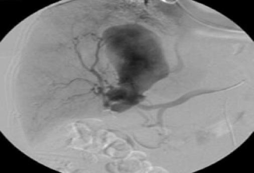
Interventional radiology was consulted for further evaluation. Catheterdirected angiography of the gastroduodenal artery demonstrated a giant pseudoaneurysm with active extravasation arising from the right gastroepiploic artery with fistulous communication with the inferior vena cava (IVC).
DISCUSSION:
A pseudoaneurysm of the right gastroepiploic artery (RGEA) is rare and potentially life threatening due to risk of rupture. And even rarer in the development of venous fistula. This can occur from pancreatitis, although it is uncommon, it is more common in men occurring at an average age of 65 years.
The process and products of inflammation, along with the high-pressure flow from arteries, weakens the vessel walls resulting in pseudoaneurysm formation, The greatest risk is rupture of the pseudoaneurysm and it increases with size, if rupture happened this will lead to shock.
Definitive diagnosis must be made with catheter angiography. Treatment options include surgery and endovascular procedures, and the treatment depends on whether the pseudoaneurysm has ruptured or not, and on the patient’s vital signs. In this case, coil embolization was the best treatment option, given the size and high probability of frank rupture, Endovascular treatment with coil embolization not only resolved the giant pseudoaneurysm, but prevented a more invasive treatment option such as laparotomy.
Axial CT angiogram demonstrates a giant subhepatic pseudoaneurysm. (B) Coronal CT Angiogram again demonstrates the giant pseudoaneurysm with active extravasation.

Catheter-directed angiography of the common hepatic artery demonstrates the giant pseudoaneurysm arising from the right gastroepiploic artery with a fistula to the IVC.


