3 minute read
AN UNUSUAL CASE OF TUBEROUS SCLEROSIS (TS
AN UNUSUAL CASE OF TUBEROUS SCLEROSIS (TS) By: Obada Al-Jayyousi
Case:
Advertisement
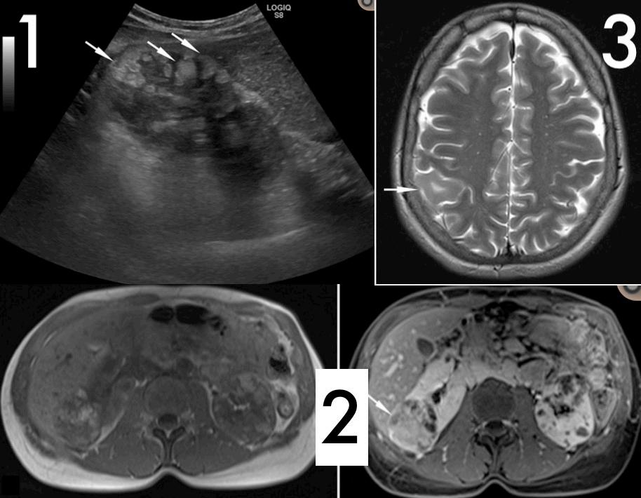
A 37-year-old female with mild abdominal pain performed a routine abdominal ultrasound that showed multiple, nodular and mainly hyperechoic renal lesions with regular margins and vascular signal on color Doppler indicating multiple angiomyolipomas, “AMLs” (Fig.1). The upper pole of the right kidney had the largest lesion with a diameter of 60mm. Physical examination revealed six rounded, reddish papules at the right nasal wing corresponding to the relapse of angiofibromas that have been surgically removed three years earlier. Neurological symptoms as well as personal and family history were negative. An MRI was then performed where several lesions were detected on both kidneys supporting the diagnosis of multiple and bilateral renal angiomyolipoma with the largest lesion on the upper pole of the right kidney showing some extent of hemorrhagic content (Fig.2). Furthermore, a contrast-enhanced brain MRI was performed that showed a single lesion in the cortical-subcortical side of right parieto-occipital cerebral cortex consistent with a cortical-subcortical tuber (Fig.3). Blood tests were normal besides an initial mild proteinuria. Otherwise, no remarkable findings were found (1).
Discussion:
Tuberous sclerosis is a rare inherited autosomal dominant disorder (with around 2 million cases worldwide) (2) with many manifestations in multiple organs, mainly hamartomas. It can be defined as a triad of: sebaceous adenoma, mental retardation, and seizures. CNS involvement is very common in the context of TS, occupying 85% of the cases (1). As mentioned earlier, the patient had a cortical-subcortical tuber in the right parieto-occipital side which is a hamartoma. Moreover, cutaneous involvement is one of the common manifestations of TS. This includes, pigmentation defects of the skin and nail fibrosis. Other manifestations could include liver and kidney manifestations. AMLs are the most common urinary system manifestation of TS. In fact, AMLs in female patients with TS are mainly found in those in their thirties and forties. In addition to that, AMLs show a higher female prevalence than males possibly suggesting hormonal influence to some extent. The diagnostic criteria of TS were finalized in a 1998 conference held by the Tuberous Sclerosis Association and the National Institute Of Health followed by a Consensus conference in 2012 (3)(4).TS definitive diagnosis requires at least two major criteria or one major criteria followed by two or more minor criteria.
Regardless of that being said, a mutation in TSC1 or TSC2 genes alone is sufficient for a TS definitive diagnosis. The patient in this case meets two major criteria: multiple and bilateral renal AMLs and at least six cutaneous angiofibromas with a corticalsubcortical tuber. Most TS cases are diagnosed during childhood due to early cutaneous and neurological symptoms. However, this case shows a late presentation due to the manifestations being primarily dermatologic, which makes this case unusual. Interventional radiology has provided a convenient treatment option for such cases including: angio-embolization, cryotherapy, and radiofrequency ablation. Studies have shown that symptomatic patients or those with masses larger than 4 cm are excellent candidates for arterial embolization as prophylaxis for rupture (5). What’s more, percutaneous ablation devices for the primary management of AMLs is possible depending on tumor size and location. However, more studies should be done to assess it fully. Another study also showed that, thirty-nine angiolipomas in 19 patients were successfully treated with selective arterial embolization (92% with primary success and 8% with secondary success) (6). Conclusion: This is an unusual case of Tuberous Sclerosis (mainly concerning the late presentation of the disease) that has been diagnosed and can be successfully treated with interventional radiology including arterial embolization and radio ablation. This field is expected to show promising results with the diagnosis and treatment of many disorders in the future as it is doing currently.
References: ___________________
1: 2020. [online] Available at: <https://jordan.gov.jo/wps/portal/Home/GovernmentEntities/Ministries/ Ministry/Ministry%20of%20Health?nameEntity=Ministry%20of%20Health&entityType=ministry> 2: 2020. [online] Available at: <http://apps.who.int/medicinedocs/documents/s17296e/s17296e.pdf> 3: 2020. [online] Available at: <http://khmc.jo/en/specialized-units/diagnostic-interventional-radiologydepartment/> 4: 2020. [online] Available at: <http://www.jordan-hospital.com/Contents/Radiology_Imaging_Department. aspx> 5: 2020. [online] Available at: <https://www.mefomp.com/Workshop-on-Advances-in-Radiology-andDiagnostic-Physics-in-Amman-Jordan_a6930.html> 6: 2020. [online] Available at: <https://www.mefomp.com/Computed-Tomography-Workshop-andQuality-Control-in-Amman-Jordan_a6931.html> 7: 2020. [online] Available at: <https://www.zawya.com/mena/en/press-releases/story/UAE_Ministry_of_ Health__Prevention_to_organize_regional_workshop_on_Interventional_Radiology_IR_and_Radiation_ Protection_RP-ZAWYA20190716110908/> 8: Northwestern University, Feinberg university of medicine, 2019: feinberg.northwestern.edu/ 9: Society of interventional radiology, 2019: sirweb.org/ 10: Review Committee for Radiology ACGME: 415InterventionalRadiologyFAQs2018.pdf


