18 minute read
The Jig is Up


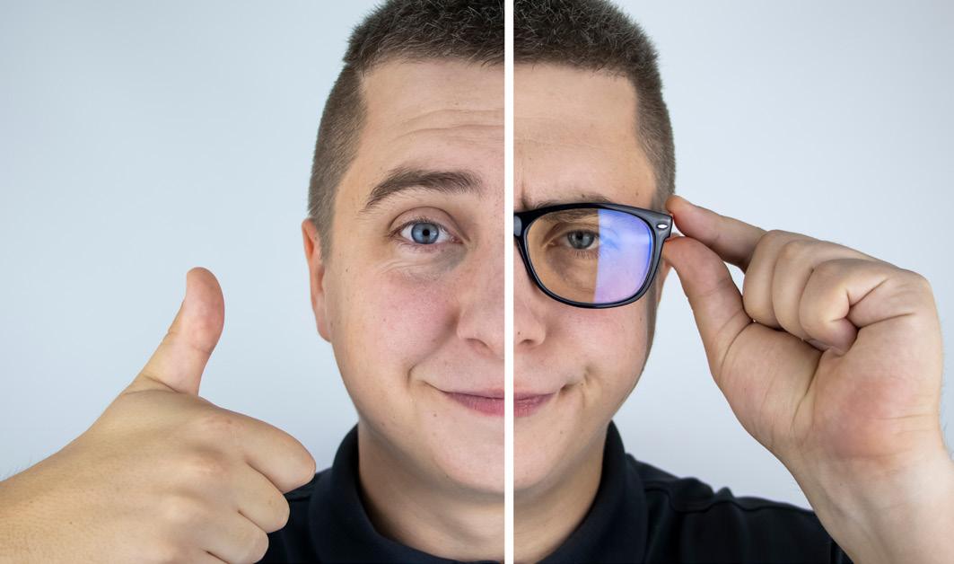
An Honest Comparison of Visual Performance in Multifocal IOLs by April Ingram
Advancements in intraocular lens (IOL) design and functionality have changed the outcomes and expectations of both surgeons and patients. Today, the decision of which lens to choose is far removed from the hardscrabble days of the Old West, where a cowboy simply had to decide between the saloon’s finest whiskey or ale to cap his journey. IOLs are way more complicated than that.
Once upon a time (in the West)
In a not so distant past, the new hightech IOLs were bifocal multifocal IOLs that provided two foci of sharp vision, distance and additional power for near. However, the image quality at intermediate distances was less than ideal. Eventually, trifocal multifocal IOLs came to the rescue and provided the intermediate focus that was lacking in the bifocal IOLs. This technology was also needed for intermediate tasks like computer use, while also delivering good visual acuity at both near and distance.
As technology improved, patient and surgeon expectations heightened. The three foci of sharp vision from trifocal IOLs came with disappointing gaps of poor image quality between them. Today, we have extended depth of focus (EDOF) lenses that come with the promise of an extended range of sharp vision across a continuum from far to intermediate distances. These premium multifocal IOLs provide the ability to compensate for the loss of near and intermediate vision due to presbyopia by restoring functional vision at several distances.
But hold your horses … there are still so many options of EDOF IOLs in the market. So, how do you choose? Are all EDOFs created equal?
Howdy! Doctors to the rescue!
Luckily, we have Dr. Juan F. Zapata-Díaz and colleagues from the Departamento de Investigaciones Clínicas, Vista Ircovisión Oftalmólogos, Murcia, Spain, to help us break it all down. Their study¹ was recently published in the Journal of Refractive Surgery.
“New IOL technologies are being designed to extend the range of vision of presbyopic patients after cataract surgery,” Dr. Zapata-Díaz explained. “The total depth-of-focus of the new multifocal implants is probably the most important information for surgeons and patients.”
Dr. Zapata-Díaz and his colleagues recognize that being spoiled for choice comes with its own challenges on how to select the best IOL for the patient and how to compare IOLs. “The market of multifocal implants is evolving rapidly with lots of new different technologies, making it difficult to do a direct comparison between them,” he said. “Hence, my colleagues and I want to give important information about some of the new premium multifocal intraocular lenses in the market.”
The team designed a study that compared the in vitro optical performance of five premium multifocal IOLs, using a single-valued metric that shows the total range of distances where a specific multifocal IOL generates an acceptable image quality. Dr. Zapata-Díaz explained: “Our new metric is intended to provide this information to the ophthalmology community.”
A new metric? Well, that’s a bee in your bonnet...
Why do we need a new metric? Don’t we currently have a way to compare? The authors explained in their publication the challenges of comparing subjective visual outcomes between multifocal IOLs due to inter-subject variability in the eye’s optical parameters.
The International Organization for Standardization provides guidelines to determine the image quality of ophthalmic implants, including two model eyes in which the IOL should be
inserted before measurements, and proposes the use of the modulation transfer function (MTF) as an image quality measure.
Through-focus MTF at 50 cycles/mm provides information for several defocus values and has been adopted in research as the standard objective metric to compare the optical quality of different multifocal IOLs.
Albeit interesting, does MTF provide useful information that can be interpreted outside of the research and allow us to predict clinical outcomes? Because as we know, the data can tell us what the optical quality should be, but a patient will tell you, with great honesty and possibly great volume, about their post-op quality of vision.
The new metric from Dr. Zapata-Díaz and colleagues compared the total depth of focus (TDOF) of different multifocal IOLs objectively, using optical bench measurements of the MTF at 50 cycles/mm for several defocus values. “The intention of the TDOF metric is to provide ophthalmologists and patients with further knowledge to make it easier to select the appropriate IOL to match patient expectations,” shared Dr. Zapata-Díaz. “We also provide threedimensional maps of the through-focus MTF at all spatial frequencies from 0 to 100 cycles/mm to illustrate the variation of performance of diffractive IOLs according to the size of the details within the object.”
– Dr. Juan F. Zapata-Díaz
Presenting, the five contenders
The researchers evaluated five multifocal IOLs: the Tecnis Symfony (Johnson & Johnson Vision, New Brunswick, Jacksonville, Florida, USA), FineVision Micro F (PhysIOL, Liege, Belgium), Acrysof IQ PanOptix (Alcon, Geneva, Switzerland), and Artis Symbiose Mid and Plus (Cristalens Industrie, Lannion France) and provide the technical specifications of each within the publication. In notable, breaking news, this was also the first measurement of in vitro optical performance of the Artis Symbiose Mid and Plus IOLs.
The study assessed the MTF measurements of each IOL for a 3mm pupil and a spherical aberration-free cornea from 0 to 100 cycles/mm, which corresponds to a visual acuity of 20/20 at distance. In order to cover all focal planes of the multifocal IOLs, throughfocus MTF was obtained in 0.10-D steps for approximately 5.00D. They used illustrative images of a United States Air Force resolution target for defocus values between 0.00 and -5.00D in 0.50-D steps. Additionally, in order to be sure to include the intermediate focus of the FineVision Micro F and the Tecnis Symfony, a -1.75D step was added.
Dr. Zapata-Díaz and colleagues found that as a result of the different optical designs, as might be expected, energy is distributed differently between far, intermediate and near focus for each multifocal IOL.
“The light distribution of the Symbiose Mid and Plus multifocal IOLs was similar, concentrating the energy into far focus and the intermediate into near focus, but extending the intermediate focus more (plus) or less (mid) toward the near focus,” Dr. Zapata-Díaz explained. Translating findings into TDOFs for each of the five IOLs, they were quite similar: 1.58D (FineVision), 1.71D (Tecnis Symfony), 1.73D (Artis Symbiose Plus), 1.74D (Artis Symbiose Mid), and 1.90D (Acrysof IQ PanOptix). The maximum difference was 0.32D.
REFERENCES:
1. Zapata-Díaz JF, Rodríguez-Izquierdo MA, Ould-
Amer N, Lajara-Blesa J, López-Gil N. Total Depth of Focus of Five Premium Multifocal Intraocular
Lenses. J Refract Surg. 2020;36(9):578-584. 2. Le Grand Y. La dioptrique de l’oeil et sa correction, 3rd ed. Optique Physiologique; Vol 1.
Editions de la Revue d’Optique, 1964.
Getting an overall idea
So how are these TDOF values applicable to clinical practice? As an example, the research team suggested that by implanting a combination of the Symbiose Mid and Plus IOLs, one in each eye, it would theoretically provide the largest clinical TDOF of 2.90D. This result was predicted by the United States Air Force resolution target image assessment as well.
What does a TDOF of 2.90D mean? Early studies from Le Grand et al. found that 3.00D represents an interval of vision large enough to cover all visual necessities of individuals with presbyopia, and presbyopia is usually defined when the amplitude of accommodation falls below 3.00D.²
This work introduces an objective metric based on a single value, the TDOF, that allows for eye care professionals to have a quick idea of the optical performance of a multifocal IOL for the whole range of vergences, useful in IOL selection.
Contributing Doctor
Dr. Juan F. Zapata-Díaz, PhD, even from a very young age — thrilled by rainbows, mirages and optical illusions — was already interested in physics and more specifically in optics. He studied optics and optometry at the University of Murcia (Spain), and at the suggestion of his professor, Norberto López-Gil, participated as an internal student in the CiViUM (Vision Science Research Group of the University of Murcia), an ongoing collaboration for more than 10 years. Dr. Zapata-Díaz completed his optometry PhD at the University of Manchester, working with the Physiological Optics Research Group, led by Hema Radhakrishnan. Upon returning to Spain, he became research manager at Dr. Lajara’s Vista Ircovisión ophthalmology clinic, working with IOL clinical and technical research, which led to a product manager position with IOL manufacturers, Cristalens. Currently, Dr. Zapata-Díaz combines his research work at the clinic with his position at Cristalens, remaining near to his patients and sharing his knowledge with the ophthalmology industry.
juanf.zapata.diaz@gmail.com

Learning from the Big Guns of Cataract Surgery
From India and Around the World by Elisa DeMartino
Recently, the All India Ophthalmological Society (AIOS) hosted its first-ever International Ophthalmic Conclave (IOC). And in true COVID-era format, the meeting was held digitally, spanning the weekend of February 19 to 21. Settling into the couch with a freshly brewed cup of coffee to binge-watch Sunday’s cataract surgery symposium was a pleasantly educational way to spend the day.
Cataract surgery: Still hard to access?
Cataract removal is common around the world, but it has particular significance in India. The country was the first to carry out cataract surgery, performed by ancient Indian physician Sushruta via couching in 700 B.C. However, today, cataracts are still the most common cause of treatable blindness — and the cause of up to 80% of bilateral blindness in the country, overall. Financial reasons, distance from clinics, lack of awareness about the condition, and seasonal temperatures can all be obstacles of access to care for patients and create somewhat of a backlog of patients needing care.* But that’s not too surprising — in fact, in every country in the world, there’s a backlog of patients needing some sort of medical care.
As AIOS President Dr. Mahipal Sachdev pointed out in his opening words during the symposium, India today performs more cataract surgeries than the U.S., Europe, and maybe China, combined. Knowing this made it particularly compelling to hear from the 18 experts on this topic.
Obviously, trying to cover each and every speaker’s presentation would not do a single one of them justice. So, at the risk of skipping over some fascinating demonstrations (which the AIOS has graciously posted online), let’s take a look at what three of these big guns had to offer when it comes to cataract surgery.
Dr. Steve Arshinoff, USA: On immediate sequential bilateral cataract surgery
One of the speakers to kick off the symposium was Dr. Steve Arshinoff, an American pioneer in bilateral cataract surgery. Dr. Arshinoff is president of the International Society for Bilateral Cataract Surgeons, which (probably by no coincidence) shares its acronym with immediately sequential bilateral cataract surgery (ISBCS). Many doctors have now adopted the Principles of Excellence for the surgery published by the organization. The expert offered preparation tips for embarking upon this surgery, including making sure small pupils can be managed well; understanding the phaco machine and its settings; making sure irrigation and aspiration are excellent; and overall, what machine or technique to use and when. Interestingly enough, Dr. Arshinoff presented studies by his own society and others, which use his guidelines in their procedures, that show that there is no increased incidence in infection with bilateral surgery.
ISBCS poses a lower cost to the patient and the clinic, and requires less healing time overall. “The reason I perform bilateral surgery in most of my cases — I think between 80% and 90% — is because it’s better for the patient and really, everybody else concerned,” said Dr. Arshinoff.
His topic became particularly interesting regionally because, as other members pointed out, some Indian doctors are still reluctantly doing bilateral surgeries full time. While they found Dr. Arshinoff’s methods to be convincing, they still have hesitations because, for instance, in tropical countries, the risk of infection is higher.
Another speaker, Dr. Jeewan Titiyal, pointed out that with COVID, bilateral surgeries at his practice and others in India are becoming more common for practical purposes: less staff is exposed to the patient, the patient is less exposed to the outside world, and less disinfecting has to be carried out at the clinic.
Dr. Mahipal Sachdev, India: On FLACS for challenging situations
About midway through the symposium, we heard again from Dr. Sachdev, whose self-proclaimed passion is routinely using femtosecond-laser-assisted cataract surgery (FLACS), particularly in posterior polar cataracts. Dr. Sachdev offered encouragement and advice on the use of femto lasers, explaining that if there is pre-existing weakness in the posterior capsule, the cortical cleaving hydrodissection can cause hydraulic rupture, but the femto laserassisted pneumodinealation eliminates
the need of doing hydrodissection or hydrodelineation. At the same time, he also recommended a study he worked on regarding this titled, Femtosecond laser-integrated anterior segment optical coherence tomography to detect preexisting posterior capsular dehiscence and increase safety in posterior polar cataracts.
Dr. Sachdev is a strong proponent of using femto, not only in routine cases but also in complex ones, such as those involving rock-hard cataracts and subluxations. According to him, the pros outweigh the cons. “There are various indications for the femto machine that can help you in these difficult and tough cases … things become much easier. One of the things that has been said about femto is that it slows you down. But, actually, if you have teamwork, you can have someone else doing the femto outside while you do the procedure inside and vice versa,” he explained. “My time has actually gone down by about 40%.”
Dr. Mohan Rajan, India: On managing rock-hard cataracts
Ending the session was Dr. Mohan Rajan’s rockin’ presentation on rockhard cataracts. He provided his “Top 10 Tips” for ophthalmologists when treating this condition. Some of his suggestions included good preoperative assessment with specular microscopy; using Trypan blue rhexis and sizing; using peribulbar or parabulbar anesthesia; using high phaco power at 80%-100% for trenching; and using minimal hydrodissection to avoid being aggressive.
Dr. Rajan also went over the ins and outs of the chopping technique for suprahard cataracts. “The Quick Chop Express makes life easy, effective and enjoyable both for the patient as well as for the surgeon,” he said. He also shared that a vertical chop is more effective than a horizontal chop in treating this condition.
A great day to learn from experts
It was a gift to observe these experts engage back and forth on the best way to do a certain procedure, or how they can improve a technique. Some of the other symposiums that day were on the topics of ophthalmic trauma, pediatric ophthalmology, updates on AMD/PCV and women in ophthalmology. No doubt all were fascinating!
Editor’s Note:
The All India Ophthalmological Society-International Ophthalmic Conclave (AIOS-IOC) 2021 was held from February 19-21, 2021. Reporting for this story also took place during the AIOS-IOC 2021 virtual conference.
A LASER TREATMENT FOR EACH GLAUCOMA STAGE
GLASS
TM
Glaucoma Laser Assisted SolutionS
Tango Reflex®
Optimis Fusion® + Vitra 2®
Vitra 810®


ABiC for Glaucoma

Navigating Uncharted Trabecular Meshwork
by Olawale Salami
In recent years, there has been an increasing interest in ab-interno canaloplasty (ABiC) as a treatment for mild to moderate primary open-angle glaucoma (POAG). This isn’t surprising considering the procedure’s ease of use, comprehensive approach and low-risk profile. However, while ABiC requires an understanding of the patency of the aqueous humour outflow system of patients’ eyes, there are, unfortunately, very limited practical options to enhance this.
Tracing canalogram patterns with trypan blue
In a paper* published in the Journal of Glaucoma, Gavin Docherty and colleagues at the Department of Ophthalmology, University of Calgary, elegantly described canalogram patterns observed during ab-interno canaloplasty with trypan blue. The primary aim of the study was to demonstrate canalogram patterns observed when trypan blue tracer was combined with an ocular viscoelastic device during ab-interno canaloplasty, and evaluate potential implications for diagnosis, prognosis and treatment of open-angle glaucoma.
In this retrospective case series, the authors performed ABiC on 5 eyes, and all surgeries were free from complications. Patients were followed up for 8 to 18 months, during which the surgeons recorded an average intraocular pressure (IOP) of 13mmHg in the treated eyes, down from an average preoperative level of 16.4mmHg. Furthermore, the average number of topical glaucoma medications was halved, from 3.6 preoperatively to 1.8 postoperatively. These results suggest that ABiC may play a vital role in our understanding of the aqueous outflow system and its role in the underlying pathophysiology of glaucoma.
– Dr. Ammar Khan
Hold your horses: Do not disturb the conjunctiva
Dr. Ammar Khan, from the Department of Ophthalmology, University of Calgary, shared his insights into the patient population that stands to benefit the most from this procedure.
“Patients who would benefit from ABiC are those with open-angle glaucoma with medically uncontrolled IOP, or those unable to tolerate topical drops but are not yet at the level of requiring trabeculectomy or glaucoma drainage device surgery,” Dr. Khan shared. “In these patients, the moderate reduction of IOP through ABiC would be beneficial as a surgical option, either performed
singly or in combination with cataract extraction.”
Furthermore, he said: “Additional patients who could benefit from this procedure are those with inadequate conjunctiva precluding successful trabeculectomy, as another advantage of this ab-interno procedure is the sparing of conjunctival tissues. This sparing of tissues allows preservation, should trabeculectomy or other interventions be required in the future.” Other benefits of this procedure include the avoidance of trabeculectomy and blebrelated complications, such as bleb leak and blebitis, and reduced risk of choroidal hemorrhage or effusions.
No odd stick: Tailoring patient management
Dr. Khan provided vital suggestions and tips to anterior segment surgeons who would be interested in this technique to optimize patient outcomes.
“Patient selection is crucial and ABiC should be considered in those with mild to moderate glaucoma,” he said. On preoperative screening, he noted that preoperative evaluation should include a thorough gonioscopy. “It is vital to identify those with a relatively functioning trabecular meshwork. This is because ABiC may not be successful in those with significantly diseased trabecular meshwork,” Dr. Khan explained. “A highly cohesive viscoelastic is helpful (but not required) to create space and optimize the angle structures. This may also be of benefit for canaloplasty, although this subject is not confirmed in the literature.”
Commenting on the tailored use of this procedure in patients, Dr. Khan said: “The extent of goniotomy can be modified on a case by case basis. For patients on anticoagulation, goniotomy should be avoided or minimized. In monocular patients, goniotomy is not preferable due to the risk of hyphema. If combined with glaucoma drainage device surgery, the goniotomy should be done inferiorly and the drainage device placed superiorly.”
Pushing the boundaries of aqueous humour outflow systems
What kind of research do we need to expand the frontiers of our current understanding of the aqueous humour drainage?
Dr. Khan shared: “Regarding the conventional pathway of aqueous humour drainage, further elaboration and understanding are required on the collector channel system, and whether earlier interventions are beneficial in preventing the collapse of the distal collector channels. Ideally, future research will develop ways to visualize the outflow system preoperatively.” He said this will allow surgeons to determine if outflow scarring is present, if patients with severe outflow scarring may benefit more from a primary filtering surgery as opposed to a minimally invasive canal-based surgery.
“Substantial research is required to delineate potential lymphatic channels in the eye and how this uveolymphatic pathway plays into aqueous humour drainage and regulation of intraocular pressures,” he explained. “If we can understand this system and assess it preoperatively, we may have improved ability to select a surgery with better outcomes for patients,” he concluded.
Like charting a course through high seas, determining the right procedure for glaucoma patients can be a tricky business.

– Dr. Ammar Khan
REFERENCE:
* Docherty G, Waldner D, Schlenker M, Crichton
A, Ford B, Ahmed IIK, WGooi P. Ab Interno
Canaloplasty in Open-angle Glaucoma
Patients Combined With In Vivo Trypan
Blue Aqueous Venography. J Glaucoma. 2020;29(12):e130-e134.
Contributing Doctor
Dr. Ammar Khan is a fifth year ophthalmology resident at the University of Calgary. He began his academic journey at Simon Fraser University in British Columbia, where he majored in biomedical physiology and kinesiology. During this time, he was introduced to anatomy and physiology, and subsequently decided to pursue these interests further in medical school at the University of Calgary. While studying medicine, he had the opportunity to gain exposure to the world of ophthalmology and decided this would be a rewarding and gratifying career path. He is currently in the final year of his residency and will continue his training with a fellowship in glaucoma surgery in Calgary this summer.
ammar34@gmail.com