5 minute read
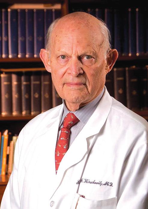
Who is this?


THE BEAT from page 1
Keeping the beat
Like just about everything else in the human body (and the natural world as a whole), the heart is extremely complex and sophisticated, and its mechanism of pumping is involved and elaborate. A simple lub-dub it is not.
Probably everyone reading this article has benefited from the medical advancements for which this man is responsible. After all, he devoted his career to studying the upper GI (gastrointestinal) tract, significant because surveys and research reveal 74 percent of Americans are living with unpleasant digestive symptoms of some kind: abdominal pain, gas, bloating, diarrhea, ulcers, constipation, acid reflux, hemorrhoids, hernias, diverticular disease, gallstones, et cetera, et cetera, et cetera. Where does it end?
Clearly, this doctor chose a career path with plenty of potential for patients, and an abundance of potential for relieving human discomfort and suffering
Born in South Africa in 1925, Dr. Basil Hirschowitz earned the American equivalents of M.D. and Ph.D. degrees in Johannesburg before leaving South Africa in 1950 to work in London. By 1953 he was a member of the Royal College of Physicians at Edinburgh. All of which makes it somewhat unexpected that the final decades of his career were spent in Birmingham.
Alabama. Not England.
Dr. Hirschowitz moved to the United States in 1953, where he enjoyed a gastroenterology fellowship at the University of Michigan and became a faculty member there in 1954. Five years later he accepted a position at the University of Alabama at Birmingham (UAB) as the founding faculty member of its Division of Gastroenterology.
While at Michigan in 1957 he and his colleagues invented a super-flexible hair-thin optical fiber of coated glass that permitted viewing internally and remotely, even around corners. The medical implications were enormous: unobstructed and illuminated views of hollow organs like the stomach, colon and esophagus were now possible. The endoscope was born, used for the first time at UAB in 1959. Minimally invasive surgery thereby took a huge step forward. Hirschowitz tested the prototype (which is in the permanent collection of the Smithsonian) by putting the optic fibers down his own throat.
As one of his colleagues observed upon Dr. Hirschowitz’ death in Birmingham in January 2013, he essentially built what gastroenterology is today, and along the way created a tool that has saved and improved countless lives all over the world in numerous medical disciplines, changing the way physicians diagnose and treat patients. The endoscope is considered by many to be one of the most important medical developments of the 20th century.
But there were even more advancements Dr. Hirschowitz was responsible for that are much more everyday than an endoscopic procedure: the research he did into ordinary gastric functions paved the way for the widespread use of medicines like Pepcid, Prilosec, and Nexium.
Yes, many of us benefit from Dr. Basil Hirschowitz’ research virtually every day. +
As suggested by the illustration on the front page, the heart has an electrical system. After all, it’s getting an EKG, the E of which stands for “electro.” (EKG stands for electrocardiogram.) That’s where the beat comes from, the regular pulse, the constant rhythm of our hearts: it’s a reaction to a mild electric shock that originates within the heart itself.
The mini-jolt is generated in the sinoatrial (SA) node located in the right atrium of the heart. The jolt is triggered when the right and left atria, the top chambers of the heart, fill with blood. “Full” activates the SA node, which zaps both atria, causing them to contract or squeeze their contents through one-way valves (the tricuspid valve on the heart’s right side, the mitral valve on the left) down into the right and left ventricles.

While the ventricles are filling with blood, the SA node’s electrical signal reaches a relay switch of sorts known as the atrioventricular (AV) node. The AV node interrupts the signal for an instant, just long enough for the ventricles to fill with blood. The momentarily delayed electrical signal arrives just as the ventricles complete their fill-up. At that instant the jolt of voltage reaches them, causing contractions that send the blood in the left ventricle through the aortic valve and off to the rest of the body, and a split-second later from the right ventricle through the pulmonary valve and into the lungs to be oxygenated.
As highly choreographed as it all sounds, what you have just read is like learning A, B and C compared with all the words in an entire encyclopedia.
WHAT DOES AN EKG MEASURE?
The “P wave” marks the contraction of the heart’s atria, pumping blood into the ventricles. The “Q wave” denotes the moment they’re full and the electrical current enters the Bundle of His. The “R wave” marks the contraction of Purkinje fibers around the left ventricle, the “S wave” the right ventricle. The “T wave” signifies the relaxation of the ventricles as they await the next signal.

How, for instance, does heart tissue that looks under a microscope like the dessert some people call Heavenly Hash create electricity? Alas, that is an incredibly complex recipe that defies simple description. Although this is an insult to the sophistication of the system, the best way to describe it simply is to say that tissue in the SA node is charged and then discharged chemically, and does it so quickly and efficiently that a healthy heart can often beat more than 100 times a minute. The entire system monitors demand continuously and adjusts automatically, speeding up if you hit a flight of stairs, slowing down for the night when you snuggle down under the covers.
Of course, creating the voltage is of little value if it can’t be delivered to the appliance where it is needed. In the heart, an elaborate network of nerves designed specifically to carry electrical impulses takes care of that. When each burst of current leaves the SA node, it first travels to the AV node, stopping briefly there as described above.
Leaving the AV node, the current has a big job ahead of it. No disrespect to the atria intended, but all they have to do is fill up and then open their trap doors to let the ventricles fill with blood. Gravity is their best friend. The ventricles, by contrast, have to pump with enough force to provide circulation for the entire body. They can’t simply be “poked” by an electrical current; they need to be squeezed.
To accomplish this, the heart’s wiring after it leaves the AV node has two branches to serve the large and powerful ventricles. They’re called the Bundle of His after the German cardiologist (Wilhelm His) who discovered them. The left and right branches of the Bundle of His travel down through the septum that separates the left and right ventricles, and then branches out in smaller nerves to almost encircle the ventricles. These smaller nerves are called Purkinje (purr-KIN-jee) fibers, and when the current shocks them, the ventricles squeeze their contents out for general circulation to the lungs and entire body. Then they can rela x before going to work when another jolt of electricity zaps them - in about half a second.
Editor’s note: this article appeared in the Medical Examiner six years ago today, on Feb. 3, 2017. It’s okay to steal an article if we wrote the original, right?


