List of Contributors
Abraini, J.H.
Université de Caen UMR 6232, CI-NAPS, Centre CYCERON, BP 5229, Boulevard Becquerel, 14074 Caen cedex, France.
E-mail: abraini@cyceron.fr
Amérand, A.
ORPHY EA4324, Universite Europeenne de Bretagne, Universite de Brest, UFR Sciences et Techniques, France.
E-mail: aline.amerand@univ-brest.fr
Aviner, B.
Department of Physiology, Faculty of Health Sciences,Ben-Gurion University of the Negev, Beer-Sheva, 84105, Israel.
E-mail: benaviner@gmail.com
Bartlett, D.
8750 Biological Grade, 4405 Hubbs Hall, Marine Biology Research Division, Center for Marine Biotechnology and Biomedicine, Scripps Institution of Oceanography, University of California, San Diego 92093-0202, La Jolla California, USA.
E-mail: dbartlett@ucsd.edu
Blumelhuber, G.
Sonnenweg 16, 85084 Reichertshofen, Germany.
E-mail: info@immobrau.com
Bode, C.
Institute of Biophysics and Radiation Biology, Semmelweis University, H-1444 Budapest P.O.Box 263.
E-mail: Csaba.Bode@eok.sote.hu
Castellini, M.
School of Fisheries and Ocean Sciences, University of Alaska Fairbanks, Fairbanks, Alaska, USA 99775.
E-mail : mikec@ims.uaf.edu
Chevalier-Lucia, D.
Assistant Professor, Department of Agro-resources and Biological and Industrial Processes, Mixed Research Unit of Agro-polymer Engineering and Emerging Technologies, Université Montpellier 2, Sciences et Techniques, Place E.Bataillon, 34095 Montpellier, France.
E-mail: Dominique.Chevalier-Lucia@univ-montp2.fr
Damasceno-Oliveira, A.
CIMAR-LA/CIIMAR, Universidade do Porto, Rua dos Bragas, 289 4050-123 Porto, Portugal.
E-mail: aol@ciimar.up.pt
Daniels, S.
Wales Research and Diagnostic Positron Tomography Imaging Centre School of Medicine, Cardiff University, Cardiff CF14 4XN, UK.
E-mail: danielss@cf.ac.uk
Dumay, E.
Professor, Department of Agro-resources and Biological and Industrial Processes, Mixed Research Unit of Agro-polymer Engineering and Emerging Technologies, Université Montpellier 2, Sciences et Techniques, Place E.Bataillon, 34095 Montpellier, France.
E-mail: Eliane.Dumay@univ-montp2.fr
Fraser, P.J.
Institute of Biological and Environmental Sciences, Zoology Department, College of Life Sciences and Medicine, Aberdeen University, Tillydrone Avenue, Aberdeen, UK, AB24 2TZ.
E-mail: p.fraser@abdn.ac.uk
Friedrich, O.
School of Biomedical Sciences, University of Queensland, Skerman Bldg, QLD 4072, St Lucia, Brisbane, Australia.
E-mail: o.friedrich@uq.edu.au
Grossman, Y.
Feldman Chair for Neurophysiology, Department of Physiology, Faculty of Health Sciences, Zlotowki Center for Neuroscience, BenGurion University of the Negev, Beer-Sheva, 84105, Israel.
E-mail: ramig@bgu.ac.il
Jammes, Y.
UMR MD2 P2COE, Faculté de Médecine, Université de la Méditerranée, France.
E-mail: jammes.y@jean-roche.univ-mrs.fr
Jebbar, M.
Laboratoire de Microbiologie des Environnements extremes, UMR 6197 (UBO, Ifremer, CNRS), Universite de Brest, France.
E-mail: mohamed.jebbar@univ-brest.fr
Kato, C.
Extremobiosphere Research Center, Japan Agency for Marine-Earth Science and Technology, 2-15 Natsushima-cho, Yokosuka 237-0061, Japan.
E-mail: kato_chi@jamstec.go.jp
Lange, R.
INSERM U710, Université Montpellier 2, Place Eugene Bataillon 34095 Montpellier cedex5, France.
E-mail: reinhard.lange@inserm.fr
Lavoûte, C.
Université dela Méditerranée et Institut de Médecine Navale di Service de Santé des Armées, UMR-MD2—Physiologie et physiopathologie en condition d’oxygenation extrême, Institut de Neurosciences Jean Roche, Faculté de Médecine Nord, Bd P. Dramard, 13015 Marseille France.
E-mail: cecile.lavoute@univmed.fr
Le Péchon, J-C.
Ingénieur Conseil, 94, rue de Buzenval, 75020 Paris, France.
E-mail: hyperbar@club-internet.fr
López-Pedemonte, T.
Assistant Professor, Departamento de Ciencia y Tecnologia de los Alimentos, Facultad de Quimica—Universidad de la República (UDELAR), Avenida General Flores 2124 – Montevideo, Uruguay.
E-mail: tlopez@fq.edu.uy
Marchal, S.
INSERM U710, Université Montpellier 2, Place Eugene Bataillon, 34095 Montpellier cedex5, France.
E-mail: stephane.marchal@inserm.fr.
Moisan, C.
ORPHY EA4324, Université Européenne de Bretagne, Université de Brest, UFR Sciences et Techniques, France.
E-mail: christine.moisan@univ-brest.fr
Mor, A.
Department of Physiology, Faculty of Health Sciences, Ben-Gurion University of the Negev, Beer-Sheva, 84105, Israel.
E-mail: morami12@gmail.com
Oger, P.
Laboratoire de Science de la Terre, UMR 5570 CNRS-ENSL-UCBL 46, allée D’Italie, F-69364 Lyon, France.
E-mail: poger@ens-lyon.fr
Péqueux, A.J.R.
Ecophysiology Unit, University of Liège 22,Quai Van Beneden B4020 Liège, Belgium.
E-mail: A.Pequeux@ulg.ac.be
Prieur, D.
Laboratorie de Microbiologie des Environnements extrémes, UMR 6197 (UBO, Ifremer, CNRS), Université de Brest, France.
E-mail: daniel.prieur@univ-brest.fr
Risso, J.J.
Université de la Méditerranee et Institut de Médecine Navale du Service de Santé des Armées, UMR-MD2—Physiologie et physiopathologie en condition d’oxgénation extrême IMNSSA, BP 84, 83800 Toulon Armées, France.
E-mail: j.jrisso@imnssa.net
Rostain, J.C.
Université de la Méditerranée et Institut de Médecine Navale du Service de Santé des Armees, UMR-MD2—Physiologie et physiopathologie en condition d’oxgénation extrême, Institut de Neur osciences Jean Roche, Faculté de médicine Nord, Bd P.Dramard 13015 Marseille, France.
E-mail: jean-claude.rostain@univmed.fr
Sébert, P.
ORPHY–EA4324, Université Européenne de Bretagne—UBO 6, Avenue Le Gorgeu, CS 93837, 29238 Brest Cedex3 France.
E-mail: Philippe.sebert@univ-brest.fr
Siebenaller, J.F.
Department of Biological Sciences, Louisiana State University, Baton Rouge, Louisiana 70803 USA.
E-mail: zojose@1su.edu
Smeller, L.
Semmelweis University, Dept. Biophysics and Radiation Biology, Tuzolto u. 37-47, Budapest IX, H-1444 Pf 263.
E-mail: laszlo.smeller@eok.sote.hu
Thom, S.R.
Professor of Emergency Medicine, Chief, Hyperbaric Medicine, University of Pennsylvania, 3620 Hamilton Walk, Philadelphia, PA 19104 USA.
E-mail: sthom@mail.med.upenn.edu
Tolgyesi, F.G.
Institute of Biophysics and Radiation Biology, Semmelweis University, H-1444 Budapest P.O.B. 263.
E-mail: Fer enc.Tolgyesi@eok.sote.hu
Torrent, J.
INSERM U710, Université Montpellier 2, Place Eugene Bataillon, 34095 Montpellier cedex5, France.
E-mail: joan.torr ent@inserm.fr.
Winter, R.
TU Dortmund University, Physical Chemistry 1, Biophysical Chemistry, Otto-Hahn Str. 6, D-44227 Dortmund, Germany.
E-mail: roland.winter@tu-dortmund.de
PRESSURE AND CELL COMPONENTS
1Protein Kinetics under High Pressure
Stéphane Marchal, Joan Torrent and Reinhard Lange
INTRODUCTION
High pressure affects protein structures at the secondary, tertiary and quaternary level (Gross and Jaenicke, 1994). Depending on the protein and on incubation conditions, exposure to pressure may thus convert a protein from -helical to -sheet structure, induce protein unfolding, or dissociate or aggregate proteins (Smeller et al., 2006). Of course, these effects are very important for a variety of applications, such as maintaining or optimizing the structural integrity of enzymes used as bio-catalysts under extreme conditions of temperature and pressure, developing new and safer food products, or controlling folding/misfolding reactions of proteins associated to systemic or neurodegenerative diseases. (Lange, 2006).
In the last decades, considerable progress was made in developing analytical instruments for recording protein structural changes under high pressure. The interest of using pressure as structure perturbation parameter is i) it provides complementary thermodynamic information to the more conventional parameters, such as temperature or chemical agents; ii) pressure is a relatively mild perturbant: irreversible structural changes, such as those induced by heat, can generally be avoided; iii) its use does not lead to complications such as specific interactions of protein with chemical denaturants.
However, this progress was mainly restricted to the study of pressure dependent reactions under equilibrium conditions. Only in the last decade, the idea of studying protein reaction kinetics under pressure became popular.
INSERM U710, Université Montpellier 2, Place Eugène Bataillon 34095 Montpellier cedex5, France.
E-mail: stephane.marchal@inserm.fr, joan.torrent@inserm.fr, reinhard.lange@inserm.fr
The underlying theory is simple, though. The pressure dependence of the rate constant (k) of an elementary process yields the activation volume, V‡, which is equal to the difference in volumes of the kinetic transition state, V‡, and the ground state, Vg. This relation is described in equations 1 and 2: ( lnk / p)T = –( G‡ / p)T/RT = – V‡ / RT(1) V‡ = V‡ – V g (2)
where p is the pressure, T the temperature, R, the gas constant and G‡ the activation free energy. Hence, any chemical reaction—and a fortiori one involving protein structural changes— is accelerated (for a negative value of V‡) or decelerated (for a positive value of V‡) by pressure depending on the relative volume of the kinetic transition state with respect to that of the ground state (Mozhaev et al., 1996). These differences in volume are due to transient changes of protein packing and hydration in the course of the reaction (Royer, 2005). As an example, a value of V‡ of 69 mL mol–1, found for invertase (Morild, 1981), gives rise to a more than 100-fold acceleration for a pressure increase from 0.1 to 200 MPa.
In practice, protein kinetics under high pressure have been studied mainly by two methods, using high pressure stopped flow and pressure jump devices. Here we will illustrate the use of these experimental set-ups for studying the mechanism of enzyme activity, of ligand binding to hemoproteins, and of protein folding. As our team is specialized in these approaches, most of the examples will stem from our laboratory in Montpellier.
EXPERIMENTAL
High pressure stopped flow technique
The high pressure stopped-flow technique (Fig. 1) was introduced by Heremans and further developed by Balny’s group (Balny et al., 1984, 1987) and Merbach (Pappenberger et al., 2000). It allows mixing, within less then 10 ms, of two solutions under defined pressure (up to 200 MPa) and temperature (between –20 and +40°C). The reactant solutions (5 mL) are kept in glass syringes. The mixing bloc and observation cell (1 cm optical path) are made of Teflon. The apparatus is equipped with quartz windows for absorbance and fluorescence measurements and interfaced via optical fibers to a BioLogic light source and detection system.
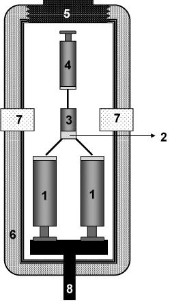
Figure 1. Schematic representation of a high pressure stopped-flow apparatus. The actual mixing device, consisting of mixing syringes (1), mixing chamber (2), observation cell (3), and waste syringe (4), is introduced via an opening screw (5) into a silicon oil containing cylindrical high pressure cell (6), equipped with quartz windows (7). Mixing of the reactants contained is induced by a pneumatically driven upward movement of a central piston (8), pushing upward the pistons of the mixing syringes.
Pressure-jump device to induce protein
structural relaxations
Figure 2 gives a schematic representation of a pressure-jump device connected to a fluorescence spectrometer, as it is used our laboratory. A limitation of this technique is the pressure-induced variation of temperature. During a pressure-jump, the temperature of the sample is heated (for upward jumps) or cooled (for downward jumps) by about 0.1°C for a pressure jump amplitude of 10 MPa. In order to minimize these temperature variations, experiments are therefore usually limited to pressure jump amplitudes of 50 MPa (Font et al., 2006b)
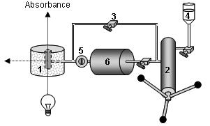
Figure 2. Schematic representation of the pressure jump device. The high pressure optical cell (1) allows the sample cell to be cooled or heated at a set temperature, and operates at a pressure of several hundred MPa (up to 700 MPa) without leakage, even at sub-zero temperatures. It holds a quartz sample cell, which is sealed with a polyethylene stretch, maintained by a rubber O-ring. This high pressure cell, equipped with sapphire windows, is easily interfaced to conventional spectrophotometers and fluorimeters. Pressure is generated by a manual pump (2) and controlled by HP valves (3). The pressure vector, water (4), is mediated through stainless steel capillaries. A home made pressure-jump device (Torrent et al., 2006a) can be connected to the high pressure cell. Pressure jumps are carried out by opening an electrically driven pneumatic valve (5) localized between the high pressure optical cell and the ballast tank (6). The pressure-jumps consist of sudden changes of pressure (up to 100 MPa) within a pressure range of 0.1–600 MPa.
THE USE OF HIGH PRESSURE TO STUDY THE MECHANISM OF ENZYME REACTIONS
The main problem for studying enzyme reactions under high pressure is the generally high reactivity of enzymes; the conversion of a substrate into a product starts immediately after mixing the enzyme with its substrate. If this mixing is done at atmospheric pressure, the time lap that is necessary to raise the pressure of the mixture precludes observation of early reaction stages. Moreover, in the case of rapidly reacting enzymes, the reaction may be even finished before the end of the pressurization time.
Fluorescence
Absorbance
A convenient means to overcome this difficulty is given by the stoppedflow technique under high pressure (see Fig. 1). This approach allows keeping enzyme and substrate separated during the pressurization time. They are mixed only once the final pressure has been reached. An alternative technical solution, optimized for steady state enzyme kinetics, has been developed by (Hoa et al., 1990). The use of high pressure stopped-flow devices for studying enzyme activity is illustrated by the following three examples. Frequently, enzyme catalyzed chemical reactions are carried out by oligomeric proteins. However, one may ask, what is the role of the oligomeric states? And are the monomers equally active as dimmers or tetramers? These questions have been dealt with by Kornblatt and co-workers for the case of yeast enolase (Kornblatt et al., 1998). This dimeric enzyme catalyzes the interconversion of 2-phosphoglyceric acid and phosphoenolpyruvate. Raising pressure induces the dissociation of the protein into monomers. This information could be deduced from a fourth derivative UV absorbance spectroscopic investigation (Lange et al., 1996) under pressure. Furthermore, as shown by stopped-flow results, the activity of the enzyme decreased as a function of pressure. As shown in Fig. 3, these two processes occurred concomitantly, suggesting that monomers do not retain native-like structures.
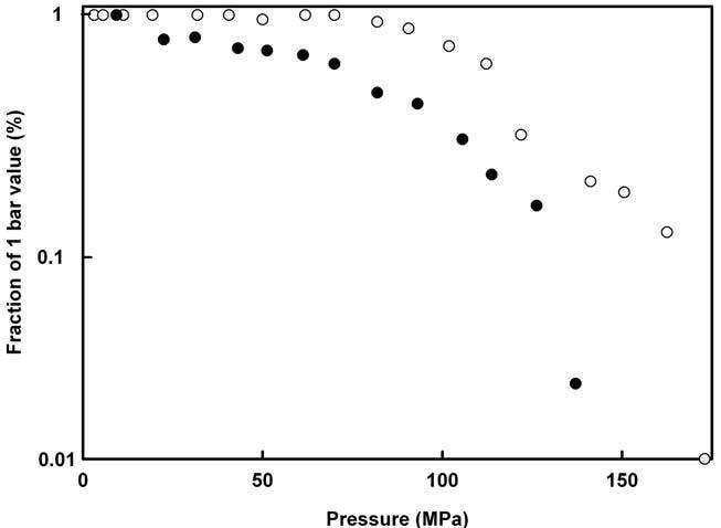
Figure 3. Effects of pressure on the quaternary structure and activity of yeast enolase. The quaternary structure (full dots) and the enzyme activity (open circles) are expressed as percentage of the respective values at atmospheric pressure. Adapted from (Kornblatt et al., 1998).
This study also showed that pressure-induced dissociation of yeast enolase in the presence of magnesium involves the loss of bound metal ion Mg(II). Interestingly, when magnesium was replaced by manganese, the application of pressure did not lead to a loss of the metal ion. Moreover, the pressure-induced monomers of Mn-bound enolase remained active and showed a native-like structure. Figure 4 resumes the effects of pressure on magnesium and manganese-bound yeast enolase (Kornblatt et al., 2004).
Step 1
Mg +2-dimers
Metal-free monomer* + Mg2+
Step 2
Mn +2-dimersMn +2-monomers
Step 3
Mn+2-monomers*
Step 4
Metal-free monomer* + Mn2+ (Species in bold are enzymatically active; monomers and monomers* indicate different conformations of the monomer)
et al., 2004).
Carboxypeptidase from the thermophilic archaebacterion Sulfolobus solfataricus is another example of successful application of high pressure to study enzyme activity. In the absence of glycerol and beta-mercaptoethanol, this otherwise heat-stable enzyme undergoes thermal inactivation at 50°C. However, this loss of activity can be inhibited when the enzyme is maintained at 200 MPa (Bec et al.,1996). As shown in Fig. 5, at higher temperatures, even higher pressures (up to 400 MPa) can be used to drastically reduce the thermal inactivation rate. This property of high pressure to stabilize proteins against thermal inactivation is interesting for the use of enzymes in biotechnological applications under high temperature conditions.
Using the same carboxypeptidase, pressure was used for investigating the role of individual non-covalent interactions in enzymatic reactions (Occhipinti et al., 2006). This carboxypeptidase turned out to be ideally suited for this investigation due to its very broad substrate specificity. This enzyme can remove any amino acid, with the exception of praline, from short peptides and N-blocked amino-acids, irrespective of the blocking group. This allowed a first comparative analysis of the effects temperature and pressure have on the interaction of an enzyme with a broad range of substrates. Furthermore, the pressure and temperature dependence of the enzyme kinetic parameters (turnover number and Michaelis constant) could be used to dissect the individual non-covalent interactions in substrate binding. Together with molecular docking experiments performed on a 3D
Figure 4. Effects of hydrostatic pressure on yeast enolase. Adapted from (Kornblatt
Figure 5. Effect of pressure on the inactivation rate of carboxypeptidase from S. solfataricus at different temperatures. Adapted from (Bec et al., 1996).
model of the enzyme, it turned out that both hydrophobic and energetic interactions (i.e., stacking and van der Waals) contribute most strongly to substrate binding.
As shown in Fig. 6, a strong correlation was observed between the volume changes associated with K m ( VKm) and kcat ( V‡kcat). Taking into account the kinetic reaction model of ordered Bi Bi system, describing Km and k cat in terms of individual rate constants, this correlation indicates that the pressure dependence of the kinetic substrate binding constant does not depend on the nature of the substrate. However, as shown in Fig. 7, the thermodynamic parameters describing the temperature dependence of the enzyme-substrate interaction and those describing its pressure dependence were not correlated. This suggests that—in contrast to the pressure dependence—the temperature dependence of the kinetic substrate binding constant depends on the nature of substrate.
LIGAND BINDING TO HEMOPROTEINS
In the field of hemoproteins, one of the key features of the puzzling mechanism of heme-type monooxygenases has been to identify the structural features responsible for binding and subsequent activation of molecular
Figure 6. Distribution of the volume changes associated with the Michaelis constants ( VKm ) versus the activation volumes ( V‡kcat ) of the catalytic step for different N-blocked amino acids used as substrates. Reprinted with permission from (Occhipinti et al., 2006).
Figure 7. Distribution of the enthalpy changes associated with the Michaelis constants ( HKm) versus the volume changes associated with the Michaelis constants ( VKm) for different N-blocked amino acids used as substrates. Reprinted with permission from (Occhipinti et al., 2006).
oxygen. The best-studied monooxygenase is cytochrome P-450, the generic name for a multigene family of heme-thiolate proteins that catalyze the oxidation of a variety of exogenous, as well as endogenous, substrates (Makris et al., 2002; Makris et al., 2003; Sligar, 1999). Closely related to P450 is nitric oxide synthase (NOS), a two-domains protein that consists of a cytochrome P450 reductase (Bredt et al., 1991) domain and an oxygenase domain that resembles cytochrome P450 (Marletta, 1994; McMillan et al., 1992; Stuehr and Ikeda-Saito, 1992; White and Marletta, 1992). For both classes of proteins, the heme moiety in its active form contains a pentacoordinated high-spin iron (FeIII) with an axial cysteine thiolate ligand. Oxygen can bind to the heme iron as a sixth ligand. This step is the essential prerequisite for its enzymatic function. Generally, observation of these binary complexes is very difficult because of their restricted stability in the timescale of measurement at ambient temperature (Gorren et al., 2000; Lipscomb et al., 1976; Peterson et al., 1972; Sligar et al., 1974). Besides oxygen, other small ligands can bind. For example, carbon monoxide (CO) induces a characteristic red shift of the Soret band to 450nm. Due to the chemical stability of the ferrous iron -CO complex, the latter is often used as a model for a better understanding of the interaction of cytochrome P-450 and NOS with oxygen. An example for a stopped-flow kinetics of CO-binding under high pressure to cytochrome P450 is shown in Fig. 8.
Several studies reported the importance of thiolate as a fifth proximal heme-iron ligand on O2 activation for cytochrome P450. Using high pressure stopped-flow kinetic experiments, Lange et al., 1994 have determined the activation volume for CO binding in several isoforms of cytochrome P450 in the absence of a substrate and compared the results with those obtained with histidine ligated hemoproteins such as haemoglobin and myoglobin. They pointed out that the pressure effect depended on the nature of the proximal axial heme ligand. Markedly negative values of the transition state volume were determined for histidine ligand proteins indicative of protein conformational changes and/or solvation processes of the transition state. In contrast, small positive activation volume changes led the authors to the conclusion that CO-binding transition state was only slightly affected by pressure for thiolate ligated proteins (Table 1). A first explanation of the origin of this behaviour was to suggest the presence of the negatively charged sulphur from cysteine ligand that produces specific electronic properties. Reconsidering this conclusion, Jung et al. (2002) showed that CObinding in cytochrome P450cam complexed with substrate analogues of camphor carrying methyl groups was also characterized by negative activation volumes in contrast to the results observed for the protein in the presence or in the absence of natural substrates (Jung et al., 2002). From these observations, the authors suggested that the accessibility of the protein heme centre can be modulated by substrate binding. The positive sign of the
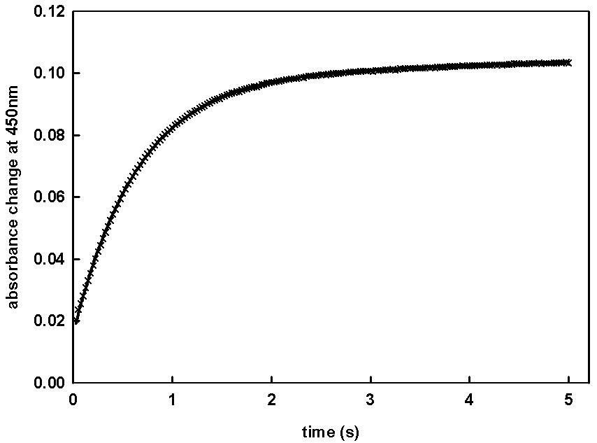
Figure 8. Time course of the CO binding to cytochrome P-450 BM3 after stopped flow mixing at 150 MPa. Kinetics were started by mixing equal volumes (60µl) of the reduced form of enzyme and CO solution in a thermostated high-pressure cell placed in Aminco DW2 spectrophotometer operating in dual wavelength mode. The experiments were setup with one syringe filled with enzyme and the other one containing the CO-saturated solution (1mM). Enzyme and gas-saturated solutions were prepared as described by (Gorren et al., 2000; Lange et al., 2001). The monoexponential fit is shown by the solid line through data points.
Table 1. Activation volume changes of the CO binding to various hemoproteins.
EnzymeLigand V ‡ (ml.mol – 1 )References
P-450sccS– (cys)2 ± 2(Lange et al., 1994)
P-450LM2S– (cys)3 ± 2(Lange et al., 1994)
P-450LM3S– (cys)6 ± 5(Lange et al., 1994)
P-450camS– (cys)4.6± 2.0 (slow phase)(Jung et al., 2002) 10.2± 2.1 (fast phase)
P-450cam + 1R-camphorS– (cys) –19.6± 0.9 (slow phase)(Jung et al., 2002) –13.2± 0.8 (slow phase)
P-450cam + norcamphorS– (cys)7.6± 2.0 (fast phase)(Jung et al., 2002)
P-450cam + norbornaneS– (cys)8.4± 0.8 (fast phase)(Jung et al., 2002)
chloroperoxidaseS– (cys)1 ± 2(Lange et al., 1994)
lactoperoxidaseN (his) –10 ± 3 (Lange et al., 1994)
Horseradish peroxidaseN (his)–24 (Balny and Travers, 1989) myoglobinN (his)–9 (Hasinoff, 1974) –18.8 (Adachi and Morishima, 1989)
hemoglobinN (his)–3 (Hasinoff, 1974) –36(Hasinoff, 1974)
activation volume for CO binding is rather indicative for solvent accessibility and flexibility of the protein than for diffusion-controlled CO binding or for the specific electronic structure of the thiolate proximal group.
With regard to the CO binding properties of cytochrome P450, it was surprising that the closely related NOS did not show the same behaviour. Indeed, using stopped-flow kinetic approach, Lange et al. (2001) ( pointed out that the rate of CO binding was strongly decreased by the presence of substrate. However, application of high pressure induced a strong increase of the rate constant, up to a value similar to that in the absence of substrate. In contrast, for cytochrome P450 BM3, a protein structurally homologous to NOS, CO-binding to the heme iron was not affected by pressure, whether in the absence or in the presence of substrate. Altogether, these observations indicated that the substrate binding site in BM3 would be large and flexible while that of NOS would be more rigid and narrow. Thus, the effect of pressure could be attributed to an expulsion of substrate of the active centre of NOS.
After instrumental optimization of the high pressure stopped-flow apparatus and a very careful design of anaerobic experimental conditions, it became possible to record oxygen binding kinetics under high pressure.
Figure 9. Effect of pressure on the rate of CO binding by BM3 (circles) and neuronal NOS (triangles). High pressure stopped-flow kinetics were carried out at 25°C in the absence (solid symbols) and in the presence of 100µM palmitate (open circles) with cytochrome P450 BM3 and with NOS, in the presence of 200µM substrate, L-arginine (open triangles up) or in the presence of both 200µM L-arginine and an excess of 25µM cofactor BH4 (open triangles down) for nNOS (Lange et al., 2001).
Accordingly, Marchal et al., 2003 successfully investigated the kinetics of CO and oxygen-binding to heme-thiolate proteins, the F393H mutant of cytochrome P450 BM3 and the endothelial NOS under pressure. These proteins were used as models because of their specific chemical properties, the formation of a stable ferrous-oxy complex for the former and the absence of a reductase domain for the latter, preventing the reduction of the oxycomplex by electron transfer. For BM3, rates and activation volumes of oxygen-binding matched the values measured for CO binding. In contrast, these properties differed for NOS, suggesting different binding mechanisms of CO and oxygen in NOS. Moreover, these results indicated that CO binding does not seem to be an appropriate probe for studying oxygen-binding. The analysis of the pressure dependence of the oxygen binding rates of NOS revealed biphasic profiles. A closer analysis of these profiles, in the presence of substrates, L-arginine and hydroxyl-arginine, and the cofactor tetrahydropterin or its inactive analogs, led to interesting results which could be interpreted by the transient formation of a new form of ferrousheme centre for NOS (see reviews (Gorren et al., 2005; Marchal et al., 2005)). These results were corroborated in more recent rapid-scanning stoppedflow experiments performed at low temperature (Marchal et al. 2004) which
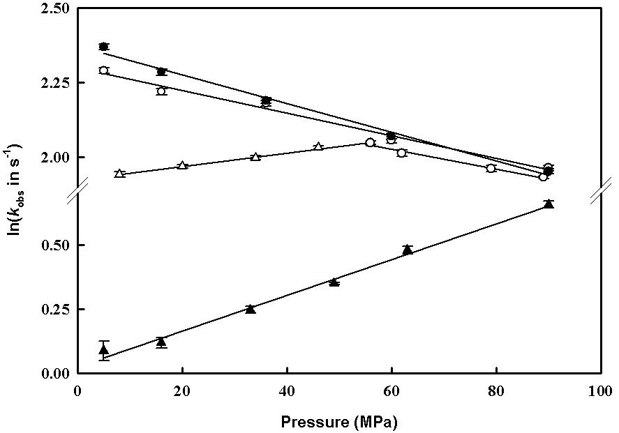
Figure 10. Pressure-dependence of ligand binding to the ferrous heme centre of P450 BM3 (circles) and eNOSoxy (triangles). CO binding rates (solid symbols) and O2 binding rates (open symbols) have been measured by rapid mixing of reduced proteins in the presence of substrate (60µM arachidonate for BM3) and inactive cofactor (0.5mM L-arginine, 50µM amino-BH2 for eNOS) with 0.5mM CO and O2, respectively, in appropriate buffer at pH 7.4 and at 4°C (Marchal et al., 2003).
showed that oxygen-binding to the reduced endothelial nitric oxide synthase (eNOS) resulted in two distinct species differing in their Soret and visible absorbance maxima, and in their capacity to exchange oxygen by CO.
Altogether, ligand-binding studies using high pressure which stopped flow technology appears to be informative for the characterization of elementary steps in the complex mechanism of several enzymatic systems.
PROTEIN FOLDING AND UNFOLDING
Protein folding/unfolding is a complex reaction that can be described properly only in terms of multidimensional energy surfaces (Dill and Chan, 1997; Socci et al., 1998). Determining the path and rate of this reaction remains a major experimental challenge. Nonetheless, there have been significant advances in experimental techniques. Apart from computational approaches, the p-jump method appears as an interesting tool. Over the past 15 years, the pressure-jump induced relaxation kinetics has yielded many significant insights in this research field, especially towards the understanding of folding and unfolding mechanisms (Desai et al., 1999; Font et al., 2006b; Herberhold et al., 2003; Mohana-Borges et al., 1999; Panick et al., 1999; Panick and Winter, 2000; Pappenberger et al., 2000; Tan et al., 2005; Torrent et al., 2006b; Vidugiris et al., 1995; Woenckhaus et al., 2001), and the structure of folding transition states (Font et al., 2006a; Jacob et al., 1999; Mitra et al., 2007; Perl et al., 2001).
The p-jump approach can be used to study both the unfolding reaction (by upward p-jump—pressurization) and the folding reaction (by downward p-jump—depressurization). These studies monitor the structural changes through relaxation of the protein’s spectroscopic properties. The data obtained, with a dead-time on the order of milliseconds, exhibit single- or multiple-exponential time-decay profiles, showing, respectively, that proteins can fold in a two-state process (between the native state and the unfolded state) or in an scenario involving intermediate states.
Generally, protein folding and unfolding reactions are triggered by rapid flow techniques, involving turbulent mixing with high concentrations of a chemical denaturant, and by laser-induced temperature-jump (T-jump). Although, the pressure-jump technique is used much less often, it offers several advantages: (1) pressure propagates rapidly so that sample inhomogeneity is a minor problem. (2) Pressure-jumps can be performed bidirectionally, i.e., by upward and downward p-jumps of magnitude between 10 and 100 MPa. (3) In the case of fully reversible structural changes of the sample, identical p-jumps can be repeated to allow for an averaging of the data over several jumps. (4) Pressure allows the activation volumes for folding and unfolding reactions to be determined, a parameter that informs about the hydration of the transition state.
Here, we discuss the application of this method to several model proteins: 33-kDa and 23-kDa proteins from photo-system II, a variant of the green fluorescent protein, and a fluorescent variant of ribonuclease A, and a set of seven hydrophobic variants of this protein. As illustrated by the examples shown thereafter, the pressure-jump relaxation method permits, on one hand, to reveal path dependent protein folding/unfolding reactions, which are hidden in equilibrium unfolding studies and barely accessible by other techniques; and on the other hand, to explore the folding transition state ensemble of a protein, giving direct information about its state of hydration.
Path dependent protein folding/unfolding reactions
Depending on the model protein and the experimental conditions, significant discrepancies were observed when comparing upward and downward p-jump induced kinetics. In fact, the recent landscape theories of protein folding suggest that individual proteins within a large ensemble may follow different routes in conformation space from the unfolded state toward the native state, and vice versa. The results are in line with a more complex free energy landscape involving multiple pathways.
The
33-kDa and 23-kDa
proteins from photo-system II
The thermodynamically predicted equivalency of upward and downward pressure-jump induced relaxation kinetics for a typical two-state protein was observed for the 33-kDa model. The situation becomes more complex with the 23-kDa protein. At 45°C, the relaxation profiles are fast, occurring on the timescale of seconds. Hence, the apparent rates for the folding and unfolding reaction are comparable. However, upon decreasing the temperature to 20°C, and further on to 10°C, increasing differences in the apparent reaction rates are revealed. At 10°C, refolding is about 20 times slower than unfolding.
A GFP variant
Intrigued by the precedent results, we extended our study to other proteins. The GFP variant used appeared to be a very suitable model. At pressures below the overall breakdown of the secondary structure of the native protein, partial protein unfolding results in a quenching of the fluorescence of its co-factor. The associated unfolding kinetics occurs on the millisecond to second time-scale, and is accelerated at high pressure, indicating that the activation volume is negative. In Fig. 11, the observed relaxation rates, kobs, are compared for upward and downward pressure jumps within a broad pressure range, from 150 to 500 MPa. We note that the observed rate constants kobs are not the
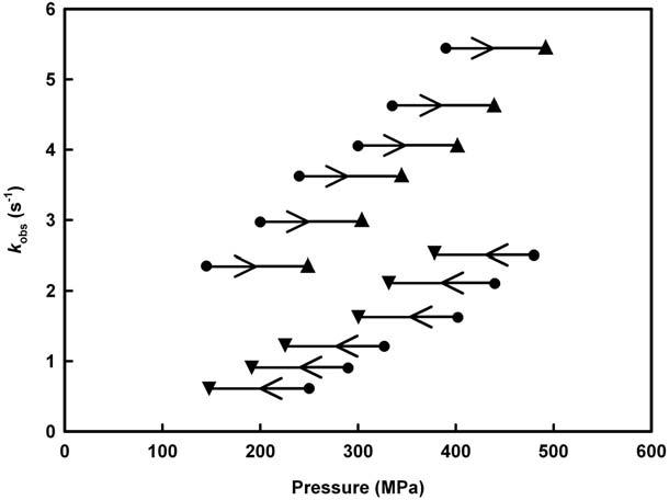
Figure 11. Pressure-induced shift of observed rate constants (inverse of relaxation times) for rsGFP after p-jumps of 100 MPa amplitude at pH 5.5 and 5°C. Triangle symbols ( ) and ( ) are the k obs values determined in the pressurizing (right-handed arrow) and depressurizing (left-handed arrow) direction, respectively. The length of the arrows refers to the p-jump amplitude. Figure adapted from (Herberhold et al., 2003).
same for forward and backward pressure-jumps, i.e., the process under consideration is path-dependent, indicating that p-jumps in opposite directions appear to induce kinetics involving different transition states.
A Fluorescent variant of ribonuclease A
Again, significant deviations from the expected symmetrical protein relaxation kinetics were observed. Whereas downward p-jumps always resulted in single exponential kinetics, the kinetics induced by upward p-jumps was biphasic in the low pressure range and monophasic at higher pressures (Fig. 12). The relative amplitude of the slow phase decreased as a function of both pressure and temperature. At higher temperatures (50°C), only the fast phase remained. These results were interpreted within the framework of a two dimensional energy surface containing a pressure- and temperature-dependent barrier between two unfolded states differing in the isomeric state of the Asn-113-Pro-114 bond. Analysis of the activation volume of the fast kinetic phase revealed a temperature-dependent shift of the unfolding transition state to a larger volume. The observed compensation of this effect by glycerol offered an explanation for its protein stabilizing effect.
Figure 12. p-Jump induced Y115W RNase A folding and unfolding relaxation kinetics at 30°C. Comparison of upward and downward p-jumps led to identical final pressures. The kinetics after a downward p-jump (upper x-axis) is fitted by a single exponential (A), that after an upward p-jump (lower x-axis) by a double (B) and a single exponential (C, dashed line). Figure adapted from (Font et al., 2006).
The folding transition state ensemble of a protein. The case of hydrophobic variants of ribonuclease A
Investigation of the transition state ensemble is experimentally extremely challenging, since—due to its transient nature—it can not be directly observed. However, some characteristics of this state can be deduced from kinetic experiments, especially by applying the -analysis (Fersht, 2004; Raleigh and Plaxco, 2005), which involves measuring the folding kinetics and equilibrium thermodynamics of variant proteins containing amino acid substitutions. Since experiments under pressure allow in determining reaction and activation volume changes (and hence the change in hydration of the protein upon unfolding), using the p-value analysis, the relative hydration of the TSE with respect to the folded and unfolded states can be evaluated. This is illustrated by the following example.
The role of hydrophobic interactions in the pressure-folding transition state of ribonuclease A was investigated using the -value and p-value methods. To this aim, the ratio between the folding activation volume and the reaction volume ( p-value), which is an index of the compactness or degree of hydration of the transition state, was calculated. The results obtained indicate a pressure-folding transition state for the main hydrophobic core of RNase A that is approximately halfway between the native and unfolded states. The results showed that the TSE was a relatively uniformly expanded form of the folded structure presenting a weakened
hydrophobic core. This strongly suggests a pressure-folding pathway following a nucleation-condensation mechanism. In addition, a direct comparison of the nature of the transition state inferred from pressureinduced folding studies and the results of the protein engineering method was presented. As shown in Fig. 13, a good correlation was found between the -values and the p-values.
The stage is now well set for more detailed and further work addressing these questions, but also for looking at more complex questions, such as the study of the folding reaction of oligomeric proteins as well as for studies of protein aggregation and the fibrillation phenomena. Of particular interest may be the application of this method to proteins which naturally fold into different thermodynamically stable conformations, such as prion proteins. In fact, for such proteins, identification of distinct path-dependent folding/ unfolding reactions might help defining strategies aimed at selecting or modulating particular folding routes.
Figure 13. Correlation between the fractional -values and the p-values of each variant. The solid line shows the best fit to a linear equation (r = 0.85). Reprinted with permission from Font et al., 2006a.
CONCLUSION
In this chapter we have analyzed the effects of pressure on several types of protein reactions. The necessity to incorporate pressure as a variable in the thermodynamic analysis of protein kinetics and for getting a closer insight into the structural features of their reaction mechanism have been shown.
Another random document with no related content on Scribd:
from eclampsia,
734 from gastro-intestinal affections,
734 from hydrencephaloid disease,
734 from typhoid fever, 733 Etiology, 724
Age and sex, influence of, 725
Exciting causes,
725
724
Hereditary tendency to tuberculosis and scrofulosis,
Season, influence of,
725
Pathological anatomy and duration, 729
Prophylaxis, 736
Prognosis, 735
Symptoms, 725
726
Abdomen, state of,
Cerebral, 726-729
Constipation, 725-727 , 729
Cry, 726 , 727
Decubitus, 727
Eyes, state of, 726-729
Headache, 725-727 in the adult, 737-739
Paralysis and convulsions, 727 , 728
Prodromal, 725
Pulse, respiration, and temperature, 726-728
Pupils, state of,
726-729
Sensitiveness to light, 725-727
Strabismus, 727
Stupor, 726-729
Tongue, state of, 726 , 729
Vomiting, 725 ,
Treatment,
735
736
Bromides, chloral, and hyoscyamus,
Counter-irritation, value of,
736
Diet in,
736
736
Iodide of potassium, use,
Menorrhagia and amenorrhœa in the insane, treatment of, 136
Menstrual disorders, influence on causation of hysteria, 220 in the opium habit, 656 , 658 insanity, 173
Menstruation, influence on recovery from epilepsy, 498 on causation of cerebral hyperæmia, 766
of spinal hyperæmia,
painful, influence on causation of hystero-epilepsy,
Mental condition in acute spinal meningitis,
750 in atrophy of the brain, 994 ,
in chorea,
in chronic hydrocephalus,
, 743 in delirium tremens, 628 in disseminated spinal sclerosis, 877 in hemiplegia, 956 in hysteria, 230 in tabes dorsalis, 836 in tumors of the brain, 1037
Defects
Deterioration,
Classification of,
Definition of, 99-105
Diagnosis of, 123-125
Exciting causes, 118-120
Heredity, 113
Morbid
Prevalence,
Prognosis,
Percentage of recoveries,
Asylums, methods of commitment to,
objection to,
question of removal to,
Galvanism, use,
Home, removal from, question of,
Mechanical restraint in,
Medicines, value of, in,
of cerebral hyperæmia in, 136 of constipation in, 137 of insomnia in, 137 of intercurrent diseases in,
136 of masturbation in,
137 of menorrhagia and amenorrhœa in, 136


