7 minute read
Cooperating on cataracts
by VetScript
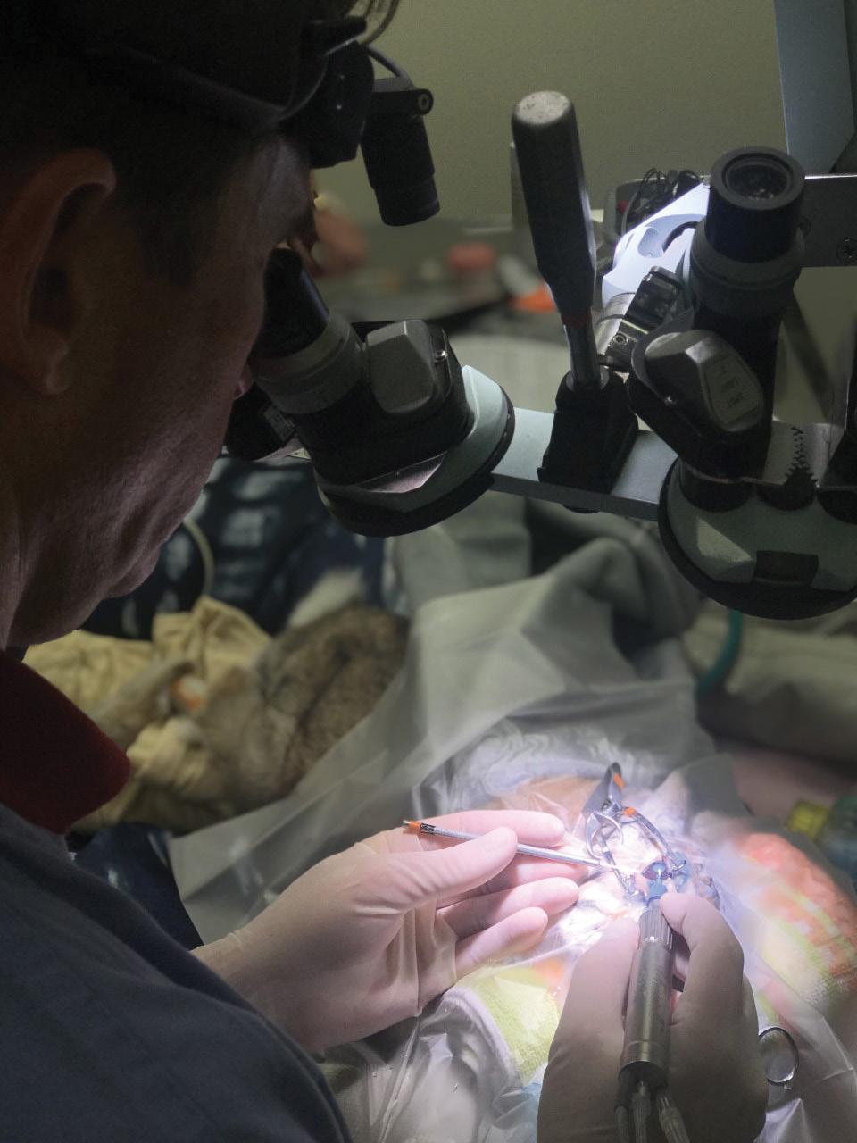
Steve Heap, a veterinarian with advanced qualifications in veterinary ophthalmology, from McMaster & Heap Veterinary Practice in Christchurch, describes a cataract surgery he successfully performed on a rabbit – and highlights the benefits of working collaboratively with colleagues.
Advertisement
IN JULY 2019 neighbouring Christchurch veterinary clinic At the Vets referred Coco, a three-year-old crossbred female rabbit with cataracts and vision loss, to McMaster & Heap Veterinary Practice.
The cataract in Coco’s left eye had initially been noticed two months previously and she had been authorised a course of oral fenbendazole for Encephalitozoon cuniculi. Although not confirmed as the cause in Coco’s case, this protozoa can cause cataracts in rabbits.
The owners noticed a change in Coco’s right eye and took her back to her primary veterinarian, Helen Keane, 10 days later. Sadly, it was also developing a cataract. Coco’s owners, wanting to do everything possible to improve her sight, were happy to be referred to me (given my training in ophthalmology) to see if it would be possible to remove the rapid-onset bilateral cataracts.
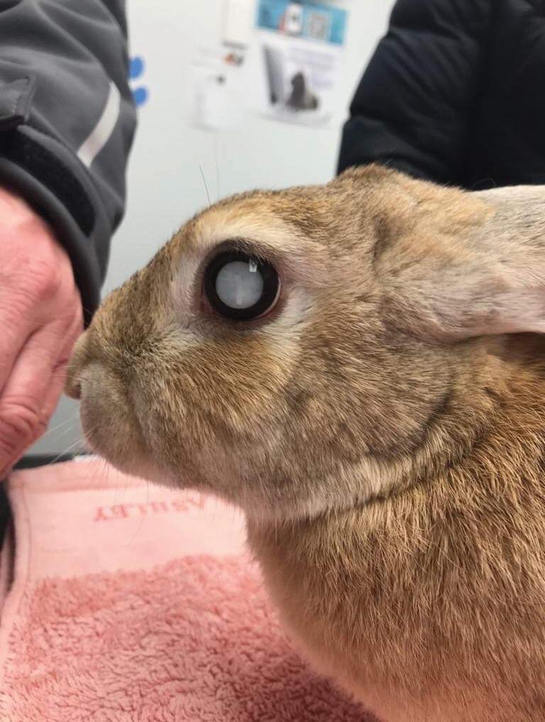
Coco presented with visual mistakes and had lost some confidence, but was otherwise well and on no current medication. Bilateral total mature cataracts were present, with some mild lens-induced uveitis.
Coco’s owners felt her quality of life was not as good as they would like it to be and were enthusiastic about the cataract surgery’s potential to enhance it. We had a thorough, and quite pessimistic, discussion about the possible problems with the surgery from an anaesthetic point of view and those associated with phacoemulsification cataract surgery itself,
Coco was completely blind when she was referred for surgery.
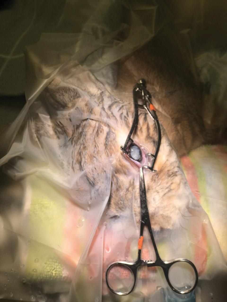
A video of the surgery can be viewed at www.facebook.com/ watch/?v=1381123515377460 &extid=yKI6BYT9D2wPPutd.
and the massive amount of postoperative care the owners would need to provide.
In spite of my apprehension, the cost and the potential problems, the owners were keen to proceed. I had performed more than 200 cataract operations in dogs and about 10 in cats, but this was to be my first cataract procedure on a rabbit.
Coco was started on ketorolac tromethamine (Acular) eye drops twice daily to both eyes, to control the lensinduced uveitis before surgery.
To anaesthetise Coco I called on Helen and veterinary nurse Emma Harre from At the Vets. Helen’s caseload largely consists of rabbits and she and Emma felt confident about performing the procedure. They also provided advice on postoperative pain relief and supportive care.
Phacoemulsification involves grooving and dividing the cataractous lens with a phaco handpiece and a second fine instrument to help ‘crack’ the lens nucleus. The separate lens segments are then removed, with the phaco needle vibrating, cutting and aspirating the usually very firm lens fragments.
Preoperative ocular medication involved a rotation of topical phenylephrine (Minims), tropicamide (Mydriacyl), chloramphenicol (Chlorafast), ketorolac (Acular) and prednisolone acetate (Pred Forte) eye drops every 10 minutes for three hours. Meloxicam and Depocillin injections were also given preoperatively. A 25-gauge cannula was placed in Coco’s marginal ear vein to deliver warmed intravenous (IV) Hartmann’s fluid pre- and postoperatively. We made sure to take care when securing the IV line as rabbits are masters at tearing them out if they are uncomfortable.
Coco was then anaesthetised using an intramuscular injection of buprenorphine, medetomidine and ketamine, and pre-oxygenated via mask while this took effect. This step is essential to prevent hypoxia, especially given that intubating rabbits is not as
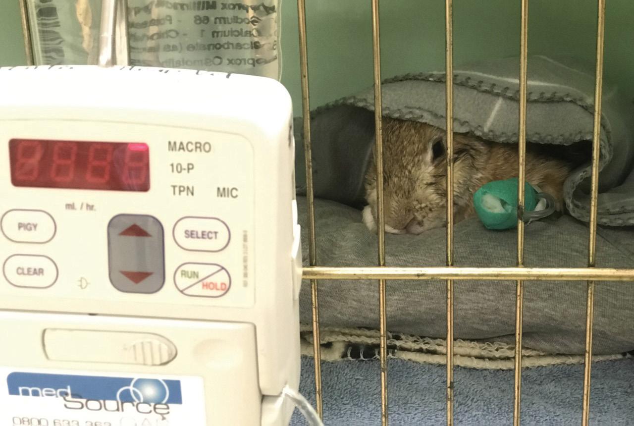

Top left: Coco recovers from surgery under the very watchful eyes of staff.
This page: Coco seven days postoperatively, now with functional vision.

straightforward as it is for cats and dogs and can take a few minutes.
Coco was blind-intubated by Helen then hooked up to oxygen and lowconcentration isoflurane, increasing the isoflurane as the medetomidine and ketamine wore off. If it can be achieved quickly, endotracheal intubation protects the airway from foreign objects going down, allows oxygen to be delivered reliably and allows intermittent positive-pressure ventilation (IPPV) if the rabbit becomes hypoxic or apnoeic.
Coco was connected to an Ayre’s T-piece circuit, which has the advantage of low resistance and little dead space. The use of inhalant agents alone as induction agents, whether administered via face mask or induction chamber, is contraindicated in rabbits because it is too stressful and patients can breathhold (leading to hypoxia, hypercapnia and bradycardia) and struggle, potentially injuring themselves. There are many safe anaesthetic protocols for use in rabbits, using a variety of drugs, and ultimately the choice of the most appropriate anaesthetic protocol depends on personal preference and familiarity with each of the drugs, the patient’s health status and the procedure to be performed.
Patient monitoring is essential during an anaesthetic procedure, and more so with rabbits than with other animals. Emma was on hand to constantly monitor Coco’s anaesthesia level, response to painful stimuli and vital parameters. Coco was attached to a pulse I HAD PERFORMED MORE THAN 200 CATARACT OPERATIONS IN DOGS AND ABOUT 10 IN CATS,
oximeter and a Doppler blood-pressure monitor, and had an oesophageal stethoscope placed. Her vital signs, including rectal temperature, heart and respiratory rates, oxygen saturation and blood pressure, were recorded through SmartFlow, a paperless patientmonitoring system.
All anaesthetics affect thermoregulation, and measures should be taken to avoid hypothermia. Rabbits are particularly sensitive to heat loss because of their small body mass and their high body surface-tovolume ratios. Hypothermia can have serious consequences and is one of the most common causes of mortality during anaesthesia in rabbits, along with respiratory compromise. Coco was positioned on a heat pad and had a fluid warmer attached, her extremities were wrapped in bubble wrap, she was wrapped in merino blankets and she had hotties placed around her.
She was under anaesthetic for a total of 120 minutes.
As Coco’s head shape and eye positions were very different from those of the average dog, positioning was critical to allow access of the phacoemulsification needle. The first eye was difficult due to the phacoemulsification needle’s different angle of entry to the anterior chamber and lens. Some positional adjustments made after doing the first eye made the second procedure significantly easier.
In both eyes the rabbit’s very thin, mobile iris prolapsed slightly into the corneal incision during phacoemulsification. Extra viscoelastic solution and lower fluid flow rates managed this to some extent.
Vigilant patient monitoring continued after the surgery, as more than 60% of deaths occur in the first three hours after an anaesthetic. The mucous membrane colour and Coco’s pulse and respiratory rate were monitored every five minutes until she was able to move around her cage. As heat should be provided until a rabbit is able to maintain their normal body temperature of 38.5–40°C, a Bair Hugger blanket was used on Coco for warmth in the immediate postoperative period. We continued warmed IV fluid and encouraged her to eat once awake. Coco recovered very well from her anaesthetic and surgery.
Postoperatively, the owners applied ciprofloxacin eye drops four times daily

SHE HAS VERY FUNCTIONAL VISION AND IS
for seven days, topical phenylephrine atropine daily for four days, ketorolac tromethamine twice daily and prednisolone acetate twice daily. All were applied to both eyes. Meloxicam was given orally for six weeks.
Coco developed a central superficial corneal ulcer in her left eye two days postoperatively. The prednisolone acetate was stopped in the left eye and ocular lubricants added. The ulcer resolved over two days. Prednisolone acetate was restarted and Aketorolac tromethamine and prednisolone acetate were tapered over the next two months.
Vision was present immediately after surgery. Anterior uveitis was controlled and minimal posterior lens capsular scarring developed after surgery.
One year after surgery Coco is still on ketorolac tromethamine daily to both eyes. Her owners have complied with the various medications and check-ups she has needed.
Coco now shows confidence in her environment and an enjoyment in playing again (toilet rolls are her favourite). She has very functional vision and is enjoying a full and active life. Her owners are thrilled.
This case highlights the benefits of working collaboratively with colleagues in using skills in different fields of interest to achieve positive outcomes.
Help us and help them live longer, happier and healthier lives
N e w d i a g n o s t i c t e s t i n g

B e tt e r t r e a t m e n t o p t i o n s H u m a n - p e t b o n d


I m p r o v e d c a r e


