6 minute read
Differential Diagnosis: Radiographic Lesion


ODA FEATURE
DIFFERENTIAL DIAGNOSIS: RADIOGRAPHIC LESION
Advertisement
By: Glen D. Houston, DDS, MSD (gdhdds@heartlandpath.com)
HISTORY
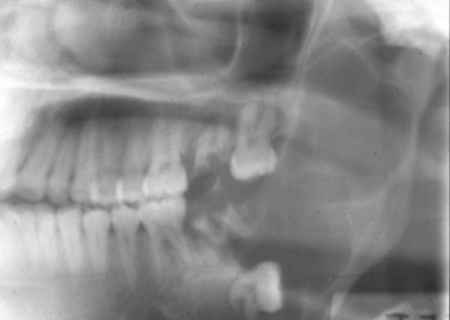
A 31-year-old female was referred by her family physician with a chief complaint of "facial swelling". Following the clinical history review and accomplishment of a radiographic survey, a mass involving the left posterior mandible was observed. According to the patient, the lesion was asymptomatic, and the duration was “many months”.
QUESTION #1
The radiographic appearance of this lesion is most accurately described as: a. A well-defined, expansible, multilocular, radiolucent lesion
b. A diffuse, invasive lesion
c. A unilocular, radiolucent lesion
d. A multilocular, diffuse, radiopaque lesion
ANSWER #1
The radiographic appearance of this lesion is most accurately described as a welldefined, expansile, multilocular, radiolucent lesion (a). Because of these observed features the other possibilities under consideration (b,c,d) are excluded in this radiographic assessment.
QUESTION #2
Your differential diagnosis for this case based on the clinical history and radiographic appearance might include: a. Dentigerous cyst b. Ameloblastoma
c. Odontogenic keratocyst d. Central (intraosseous) arteriovenous malformation
e. Cherubism f. Aneurysmal bone cyst
ANSWER #2
Your differential diagnosis based upon the clinical history and radiographic appearance should include all of the conditions listed. unerupted tooth. However, it is most often associated with the impacted mandibular third molar tooth. Additionally, it is most frequently observed in young patients between the ages of 10 and 30 years of age. Extensive lesions may produce facial asymmetry and radiographically present as unilocular radiolucent lesions. Occasionally multilocular lesions have been observed.
The ameloblastoma (b) is an odontogenic neoplasm that shows an approximately equal prevalence in the third to seventh decades of life. This lesion is usually asymptomatic and typically presents as an expansile, multilocular, radiolucent ("soap bubble" or “honeycomb”) lesion involving the posterior mandible. In many cases, an unerupted tooth is associated with this radiolucent lesion.
The odontogenic keratocyst (c) is a distinctive developmental odontogenic cyst found in patients between 10 and 40 years of age. The mandible is involved in 60-80% of the cases, especially the posterior region. This lesion presents as a well-defined radiolucent area that may be unilocular or multilocular in appearance and is associated with an unerupted tooth in 25-40% of the cases.
The central (intraosseous) arteriovenous malformation (d) is a vascular lesion produced by the abnormal communication between the arterial and venous circulation, bypassing the capillary bed. A thrill or bruit over the area may be detected and the overlying mucosa or skin is significantly warmer than the adjacent tissue. This lesion usually presents as a unilocular or multilocular radiolucent area.
Cherubism (e) is a rare familial jaw condition that occurs in children. It typically is asymptomatic and presents as bilateral, expansile, multilocular radiolucent areas of the mandible with occasional involvement of the maxilla. These lesions may be associated with unerupted teeth and simulate the appearance of a dentigerous cyst. There are rare reports of unilateral involvement.
The aneurysmal bone cyst (f) is typically observed in the long bones or vertebral column in patients under the age of 30. Those that occur in the jaws usually are seen involving the posterior mandible. They may be painful and present radiographically as an expansile, multilocular, radiolucent area. There is a female predilection observed with this lesion.
QUESTION #3
The appropriate procedure(s) to arrive at a definitive diagnosis include: a. Aspiration of the area in question b. Biopsy c. Advise the patient that this is probably a dentigerous cyst and, unless the area becomes symptomatic, no treatment is indicated
d. Tell the patient she has cherubism and a biopsy is not necessary in order to make the diagnosis
ANSWER #3
The following procedures are indicated in this case:
(a) Aspiration of the area in question
(b) Biopsy Aspiration of the lesion (a) is indicated in order to rule out a central vascular lesion (i.e. arteriovenous malformation). A biopsy (b) is necessary in order to establish a definitive diagnosis. To advise the patient that this is probably a dentigerous cyst and, unless the area becomes symptomatic, no treatment is indicated (c) and telling the patient she has cherubism and a biopsy is not necessary in order to make the diagnosis (d) would have no clinical support
QUESTION #4
Microscopically, the following features are noted: There is a prominent cystic structure in which numerous, small, discrete islands and strands of odontogenic epithelium are observed arising from the epithelial lining of this cyst and infiltrating into the fibrous capsule. Each of these islands and strands exhibits a peripheral layer of cells with polarized nuclei and encloses a central region composed of stellate-shaped epithelial cells. The correct diagnosis is: a. Odontogenic keratocyst b. Dentigerous cyst c. Ameloblastoma arising in a dentigerous cyst d. Adenomatoid odontogenic tumor
ANSWER #4
The lesion is correctly diagnosed as ameloblastoma rising in a dentigerous cyst (c)--see "Discussion" section. The other possibilities are not considered here because the odontogenic keratocyst (a) is composed of a thin wall of fibrous connective tissue (capsule) which exhibits a thin, uniform layer of luminal epithelium that exhibits a basal layer composed of cells with palisaded, hyperchromatic nuclei and a corrugated layer of parakeratin along the luminal surface. The dentigerous cyst (b) is also composed of a thin fibrous connective tissue capsule in association with a layer of luminal epithelium composed of two to four layers of non-keratinized stratified squamous epithelial cells. Inflammatory cells are usually present within the fibrous capsule. Finally, the adenomatoid odontogenic tumor (d) exhibits a thick, fibrous connective tissue capsule. This neoplasm is composed of spindle-shaped epithelial cells that are arranged into sheets, strands, or whorled masses. Tubular or duct-like epithelial structures and foci of calcification are also observed.
DISCUSSION
The ameloblastoma is a benign neoplasm arising from enamel organ tissue which does not undergo differentiation to the point of enamel formation. This tumor has been described by Robinson as "usually unicentric, nonfunctional, intermittent in growth, anatomically benign, and clinically persistent". The first thorough description of the ameloblastoma in the literature was by Falkson in 1879. The ameloblastoma may arise from any source of odontogenic epithelium. Approximately 17-30% are associated with the crown of an impacted tooth, usually a mandibular third molar tooth. While the ameloblastoma usually affects the middle-aged adult, those that arise from the dentigerous cyst are typically observed during the second or third decades. There is an even distribution between males and females.
Whereas the pericoronal radiolucent lesion may be small, most cases of ameloblastoma arising in a dentigerous cyst exhibit a classic radiographic pattern characterized by a large, unilocular radiolucent lesion involving the entire ramus and extending to the coronoid notch and process. They are most frequently associated with an impacted, displaced third molar tooth demonstrating incomplete root formation. Occasionally, these tumors are multilocular and exhibit scalloped margins. When expansion occurs, clinically observable deformity or facial symmetry may be apparent. Pain and/or paresthesia are generally not evident. An ameloblastoma arising in a dentigerous cyst is radiographically indistinguishable from the odontogenic keratocyst, typical dentigerous cyst, and various other odontogenic and non-odontogenic lesions mentioned in Question 2. A biopsy is required for a definitive diagnosis. The ameloblastoma is composed of islands of odontogenic epithelium demonstrating a peripheral layer of cells with polarized nuclei that resemble ameloblasts. The central portion of these islands is composed of loosely arranged, stellateshaped cells resembling stellate reticulum. Ameloblastomas are treated by a variety of surgical means ranging from enucleation and curettage to block resection based upon the anatomic location and size of the lesion. Recurrence rates of 50% - 90% have been reported.
REFERENCES Small IA, Waldron CA: Ameloblastoma of the jaws, Oral Surg Oral Med Oral Pathol 8:281-297, 1955.
Hong J, Yun P-Y, Chung I-H, et al: Long-term follow up on recurrence of 305 ameloblastoma cases, Int J Oral Maxillofac Surg 36:283-288, 2007.
Dhanuthai K, Chantarangsu S, Rojanawatsirivej S, et al: Ameloblastoma: a multicentric study, Oral Surg Oral Med Oral Pathol Oral Radiol 113:782788, 2012.