21 minute read
Smooth Muscle Cells
3.2 Acquisition of Intracellular Calcium Whole-Cell Images
4. After the loading period, carefully recover or the coverslip and wash the cells three times with Tyrode-BSS buffer. 5. Leave the loaded cells for an additional 15 min period to ensure complete hydrolysis of acetoxymethyl ester groups.
Advertisement
1. After cleaning the objective lens, a drop of nonfluorescent oil is applied and then the chamber containing the loaded cells is placed in contact with the objective lens. Using the lowest possible intensity of the mercury lamp of the microscope, we choose a cell that presents a stable fluorescence level and that does not have a flat appearance. This should be done without exposing the chosen cell for a long period of time to mercury lamp light in order to avoid photobleaching. 2. The mercury lamp is turned off and the shutter of the microscope is set in the position allowing the passage of the laser excitation waves. 3. At first, a continuous section scanning is done in order to estimate the basal level of cytosolic and nuclear calcium. The cytosolic level should be between 50 and 100 nM, and the nuclear level should be all the time higher than that of the cytosol (near 300 nM) according to the calibration curve already determined for the calcium fluorescent dye. This permits all the cells used to have nearly the same starting normal calcium.
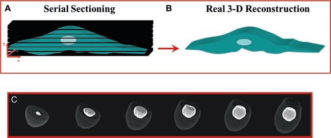
During this process, the focus should be adjusted with the fine adjustment knob. 4. When the continuous sectioning is terminated, we proceed to the determination of the thickness of the cell by performing a vertical scan which allows the determination of the starting section (just above the cell) and the number of sections needed to scan the whole cell. The step size should be kept at the minimum value. 5. Serial Z-axis optical scans (section series) taken for the intracellular calcium of the cell are captured by a photodetector, digitalized, and saved (Fig. 1a). The captured section series can then be presented either as 2D or real quantitative 3D reconstructions using various angles of rotation and inclination as well as a variety of cutting planes. These real quantitative 3D reconstructions of cells (Fig. 1b, c) are then used for the measurement of basal level fluorescence intensity and/or the cellular response after the addition of different agents [1–5]. 6. At the end of each experiment, the nucleus is stained with 100 nM of the live nucleic acid stain Syto-11 (Fig. 1c) (Molecular Probes, OR, USA) [1–5]. Serial Z-axis optical scans are taken after development of the stain (3–5 min) while maintaining positioning, number of sections, and step size identical to those used throughout the experiment. Nuclear labeling is
Fig. 1 Serial sectioning and real 3D reconstruction. (a) Using confocal microscopy, cells (and nuclei) are subjected to sequential serial scans, which permits the recording of fluorescence from conjugated antibodies and/ or probes which target intracellular proteins (receptors and ligands), membranes, organelles, and ions. (b, c) Serial sections captured by confocal microscopy are reconstructed in real quantitative 3D using the ImageSpace software. The real quantitative 3D reconstruction allows measuring the total fluorescence (per μm3) of a given target in volume. It also allows to designate the locations of organelles in the cell (c: 10-day-old single embryonic chick ventricular myocyte) and the specific fluorescence related to them
3.3 Rapid Scan of Intracellular Calcium
important because it permits the isolation or extraction of the nucleus from the cytoplasm by setting a lower-intensity threshold filter to confine relevant pixels [1–5]. Fluorescence intensities of the calcium probe can then be measured in the entire volume of each compartment (cytosol and nucleus) separately to provide quantitative 3D information (Fig. 1).
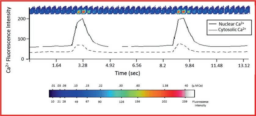
The confocal setting can be changed to the rapid-scan feature. This rapid-scan technique permits measurements of temporal rapid changes of intracellular calcium (Fig. 2). It is employed to visualize transitory variations of calcium (Fig. 2) which cannot be otherwise visualized by the real quantitative 3D scan given the fact that the duration of image acquisition in 3D can surpass that of the studied phenomenon. The rapid scan generates consecutive images of the section at the focal plane of the sample, with a speed of 0.320 s/image in the case of the Molecular Dynamics Multiprobe 2001 confocal microscope and 1.61 s/image for the MRC1024 Bio-Rad confocal microscope. With these speeds, even the smallest changes in fluorescence intensity, which would otherwise be undetected with conventional fluorescence microscopy tools, can be captured (Fig. 2). Up to 200 frames can be recorded. Each frame consists of 32 lines/scan, with a 512×512 pixel resolution. Graph tracings of fluorescence variations in the cytosol and the nucleus give the corresponding exact pattern of registered Ca2+ variations that occur spontaneously or as a consequence of pharmacological interventions (Fig. 2). Since the rapid scan is a result of 2D section image (Fig. 2), it cannot be used for measuring the level of calcium. However, it is an excellent tool to measure the kinetics of intracellular calcium variations as well as the frequency and relative amplitude of spontaneous calcium waves (Fig. 2).

Fig. 2 Rapid scan of spontaneous cytosolic and nuclear calcium waves. Representative figure illustrating a continuous rapid scan of a section of heart cells contracting spontaneously and the corresponding graphic representation of the variation of cytosolic (discontinuous line) and nuclear (continuous line) using confocal microscopy. Images are generated continuously at the speed of 0.320 s/image at a resolution of 256 × 256 pixels of 0.7 μm each. At the end of the experiment, the nucleus is labeled with Syto-11. The pseudocolor bar represents the intensity of Ca2+ fluorescence and the absolute concentration of Ca2+ measured using the calcium calibration method described in Subheading 3.6
3.4 Intracellular Calcium Sparks and Puff Measurements Using Line-Scan Technique
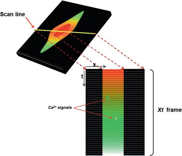
The line scan is a versatile technique on the MRC1024 confocal microscope which allows to capture and visualize transient ion movements such as calcium sparks, puffs [14, 15], and waves occurring locally at a certain subcellular location in a cell. 1. After the right sample is chosen and set in focus, the scan line is placed at the desired location of the sample (e.g., in the cytosol, the perinuclear area, or the nucleoplasm) (Fig. 3). Upon execution, the system excites and scans the sample only along the designated scan line in the focal plane (Fig. 3). Up to 3000 line scans can be recorded in around 6 s, with a speed of 2 ms/line. 2. The images of the scan lines are stacked to generate an xt frame, where x is the scan line and t is time. Any change of fluorescence intensity along the line, in this case as a result of increase or decrease of Ca2+, and any passage of Ca2+ transients through the line are therefore recorded and presented in the xt frame. 3. Using an appropriate image treatment software such as the
ImageSpace software, the xt frames can be presented topographically, as a surface plot of fluorescence intensities, which allows a better appreciation of small Ca2+ signals (sparks and puffs). The frequency, amplitude, and kinetics of calcium sparks and puffs could be determined.

Fig. 3 Principle of line-scan technique for spontaneous Ca2+ sparks and puffs. Ca2+ probe-loaded cells are mounted on the metallic chamber of the confocal microscope, and after a sample cell is chosen, the scan line is positioned in the desired area of the cell (red arrow). Upon execution, up to 3000 scans can be performed at the line position, and the images of the recorded lines are stacked adjacently next to each other. The resultant is an xt frame, which depicts any temporal changes of fluorescence (sparks, puffs, waves) occurring at the line position or crossing through it
3.5 Volume Rendering for Cytosolic and Nuclear Calcium Measurements
1. Scanned images are transformed onto a Silicon Graphics O2 analysis station equipped with Molecular Dynamics Imagespace 3.2 analysis and volume workbench software modules. Reconstruction of quantitative real 3D images is performed on unfiltered serial sections (Fig. 1). All quantitative 3D reconstructions are presented using the “maximum intensity” top-view image format which produces high-contrast images and is sensitive to noise. It is worth noting that images are presented as pseudocolored representations according to an intensity scale of 0–255, with 0 intensity in black indicating the absence of fluorescence and 255 in white indicating the maximal fluorescence intensity [4]. 2. The quantitative 3D reconstructions (section series) of cells are used for the measurement of whole-cell fluorescence intensities (per μm3) where cells are simply delimited. 3. When the quantitative measurement of the nuclear and cytosolic fluorescence intensities is needed to be done separately, the nuclear area (following Syto-11 staining) is isolated from
3.6 Calcium Calibration Curve for Fluo-3
4 Notes
the rest of the cell by setting a lower-intensity threshold filter to confine relevant pixels. 4. A 3D binary image series of the nuclear volume is generated for each cell, using the exact same x, y, and z set planes. Thus, by applying these binary image patterns, which now serve as a cookie cutter, to their corresponding cells (but labeled for calcium), two new real 3D projections are created depicting fluorescence intensity levels in the nucleus and cytosol separately [1–5]. 5. Intensities are measured in the entire volume of sections for each compartment (cytosol and nucleus) to provide quantitative real 3D information. Since the measurements are done over the whole volume of the cell (per μm3), and not in one or a few sections (per μm3) for the entire cell volume, they eliminate any possible problem concerning drifting of the Z line.
1. For each concentration of Ca2+ of kits No. 2 and No. 3, a final concentration of 13.5 μM Fluo-3 acid is added. 2. The cell membranes are perforated by exposing the cell to the 0.1 % Triton solution for 10 min and then washed with the zero Ca2+ buffer of kit No. 2. 3. The confocal system is set for measuring Fluo-3 Ca2+ fluorescence as described before. 4. The perforated cells are then exposed to each different concentration of Ca2+ buffer containing 13.5 μM Fluo-3 acid, and the fluorescence intensity is recorded in the whole area under the field of the microscope. 5. At the end of the experiment, the fluorescence intensity of the last sample (1 mM free Ca2+) is recorded and then Syto-11 is added to delimit the nuclear regions. 6. The calibration curve is plotted on intensity vs. concentration axes, and the results are fitted to the pseudocolor fluorescence intensity bar of the calcium dye (Fluo-3) (Fig. 3).
In order to overcome the limitation of using fluorescence imaging for the detection of intracellular calcium distribution and measurement in the cytosol and nucleus, the following should be taken into consideration:
1. For ionic probes, optimal loading should be ensured by determining the relative concentration of the bound and non-bound probe by using ionomycin to perforate the cell membrane.
This also could be used at the end of each experiment (in the absence of Syto-11) in order to determine if the probe is completely bound to calcium.
2. A minimum of 36 serial sections and preferentially near 75–150 serial sections of the cell (depending on the morphology) should be taken at rest for quantiative real 3D imaging. 3. In order to avoid measurement problems due to difference in volumes between cells or between organelles, the fluorescence intensity should be normalized and expressed per μm3 .
Furthermore, intensity measurement in a single section of a cell is that of a surface (μm2) and does not represent absolute levels of intensity. 4. It is recommended to verify the distribution of the Ca2+ probe when a new type of cell is used. 5. Fluo-3 and other Ca2+ probes such as Indo-1, Fluo-4, Ca2+ green, yellow cameleon, and Fura-2 are equally distributed in the cytosol and the nucleus of the heart, vascular smooth muscle cells (VSMCs), vascular endothelial cells (VECs), and endocardial endothelial cells (EECs) of human (h), rat, rabbit, hamster, and mouse origins [2, 3, 5, 10, 12, 13]. Thus, these fluorescent probes can be used safely in these cell types. 6. For experiments longer than 20 min, it is absolutely important to replace the medium in order to avoid the effect related to evaporation of the experimental solution. If a drug is already present in the experimental bath, the replacement solution should already contain this drug. At least 2 min is necessary before continuing the experiment.
All of these aspects in addition to others should be established in order to accurately determine the fluorescence distribution and density of a fluorescent probe.
Acknowledgments
This study was supported by grants from the Canadian Institutes of Health Research (CIHR), the Natural Sciences and Engineering Research Council of Canada (NSERC), and the Heart and Stroke Foundation of Quebec (HSFQ) (grants awarded to Drs. Ghassan Bkaily and Danielle Jacques).
References
1. Bkaily G (1994) Ionic channels in vascular smooth. In: G. Bkaily (ed) Molecular Biology
Intelligence Unit, Austin 2. Bkaily G, Gros-Louis N, Naik R, Jaalouk D,
Pothier P (1996) Implication of the nucleus in excitation contraction-coupling of heart cells.
Mol Cell Biochem 154:113–121 3. Bkaily G, Pothier P, Orleans-Juste P, Simaan
M, Jacques D, Jaalouk D, Belzile F, Hassan G,
Boutin C, Haddad G, Neugebauer W (1997)
The use of confocal microscopy in the investigation of cell structure and function in the heart, vascular endothelium and smooth muscle cells. Mol Cell Biochem 172:171–194
4. Bkaily G, Jacques D, Pothier P (1999) Use of confocal microscopy to investigate cell structure and function. Methods Enzymol 307:119–135 5. Bkaily G, Nader M, Avedanian L, Choufani S,
Jacques D, D’Orléans-Juste P, Gobeil F,
Chemtob S, Al-Khoury J (2006) G -proteincoupled receptors, channels, and Na+-H+ exchanger in nuclear membranes of heart, hepatic, vascular endothelial, and smooth muscle cells. Can J Physiol Pharmacol 84:431–441 6. Bkaily G, Avedanian L, Jacques D (2009) Nuclear membranes’ receptors and channels as targets for drug development in cardiovascular diseases. Can
J Physiol Pharmacol 87(2):108–119 7. Bkaily G, Avedanian L, Al-Khoury J, Provost
C, Nader M, D’Orleans-Juste P, Jacques D (2011) Nuclear membrane receptors for
ET-1 in cardiovascular function. Am J Physiol
Integr Comp Physiol 300(2):R251–R263 8. Bkaily G, Avedanian L, Al-Khoury J, Ahmarani
L, Perreault C, Jacques D (2012) Receptors and ionic transporters in nuclear membranes: new targets for therapeutical pharmacological interventions. Can J Physiol Pharmacol 90:953–965 9. Avedanian L, Jacques D, Bkaily G (2011)
Presence of tubular and reticular structures in
the nucleus of human vascular smooth muscle cells. J Mol Cell Cardiol 50(1):175–186 10. Jacques D, Abdel Malak NA, Sader S (2003)
Angiotensin II and its receptors in human endothelial endothelial cells: role in modulating intracellular calcium. Can J Physiol
Pharmacol 81:259–266 11. Abdel-Samad D, Perreault C, Ahmarani L,
Avedanian L, Bkaily G, Magder S, D’Orléans-
Juste P, Jacques D (2012) Differences in neuropeptide Y-induced secretion of endothelin-1 in left and right human endocardial endothelial cells. Neuropeptides 46:373–382 12. Abernica B, Gilchrist JS (2000) Nucleoplasmic
Ca2+ loading is regulated by mobilization of perinuclear Ca2+. Cell Calcium 28:127–136 13. Abernica B, Pierce GM, Gilchrist JS (2003)
Nucleoplasmic calcium regulation in rabbit aortic vascular smooth muscle cells. Can
J Physiol Pharmacol 81:301–310 14. Cheng H, Lederer WJ, Cannell MB (1993)
Calcium sparks: elementary events underlying excitation-contraction coupling in heart muscle. Science 262:740–744 15. Yao Y, Choi J, Parker I (1995) Quantal puffs of intracellular Ca2+ evoked by inositol trisphosphate in Xenopus oocytes. J Physiol 482:533–553
Chapter 15
Ivana Y. Kuo and Caryl E. Hill
Abstract
Patch clamp electrophysiology is a powerful tool that has been important in isolating and characterizing the ion channels that govern cellular excitability under physiological and pathophysiological conditions. The ability to enzymatically dissociate blood vessels and acutely isolate vascular smooth muscle cells has enabled the application of patch clamp electrophysiology to the identification of diverse voltage dependent ion channels that ultimately control vasoconstriction and vasodilation. Since intraluminal pressure results in depolarization of vascular smooth muscle, the channels that control the voltage dependent influx of extracellular calcium are of particular interest. This chapter describes methods for isolating smooth muscle cells from resistance vessels, and for recording, isolating, and characterizing voltage dependent calcium channel currents, using patch clamp electrophysiological and pharmacological protocols.
Key words Voltage dependent calcium channel, Patch clamp electrophysiology, Vascular smooth muscle cells
1 Introduction
Peripheral resistance vessels play an important role in the regulation of blood pressure and exist in a state of partial constriction known as vascular tone. The maintenance of vascular tone results from elevation of the intracellular calcium concentration of smooth muscle cells, following influx of extracellular calcium or release of calcium from intracellular calcium stores. The myogenic response is an important regulatory response, which links increases in intraluminal pressure to vasoconstriction through depolarization of vascular smooth muscle cells and influx of calcium through voltage dependent calcium channels (VDCCs) [1, 2]. As these channels inactivate at various rates, the ability of VDCCs to contribute to the myogenic response results from the persistent calcium influx that occurs over the range of voltages where the steady-state activation and inactivation curves overlap [3]. This is known as the window current.
Rhian M. Touyz and Ernesto L. Schiffrin (eds.), Hypertension: Methods and Protocols, Methods in Molecular Biology, vol. 1527, DOI 10.1007/978-1-4939-6625-7_15, © Springer Science+Business Media LLC 2017 189
The VDCC superfamily is comprised of five families; the L- (Cav1), N-, P/Q-, R- (Cav2), and T-type (Cav3) channels. Of the 10 molecular subtypes of VDCCs comprising these families, the L-type channel, CaV1.2, is the main contributor to vascular tone [4] and increased activity of these channels has been linked to hypertension and cerebrovascular disease [5]. The resting membrane potential of smooth muscle cells of pressurized arteries and arterioles in vivo and in vitro is relatively depolarized; lying in the range, −45 to −30 mV [4, 6–10]. This voltage range encompasses the window current of L-type channel splice variants isolated from small cerebral arteries [11, 12]. However, recent studies have provided evidence for the expression of other VDCCs, particularly the T-type channels, Cav3.1 and Cav3.2, in the vasculature (see [13]). Unfortunately, defining the contribution of T-type channels to vascular tone has been complicated by the inability of many of the commonly used T-type channel antagonists to clearly select between L- and T-type effects [13]. Nevertheless, patch clamp electrophysiology of isolated vascular smooth muscle cells provides a very useful technique to assist in identifying a role for T-type channels in physiological and pathological states, such as hypertension, since this technique enables investigation of the differing biophysical characteristics of the endogenous currents. Unlike the L-, N-, P/Q-, and R-type VDCCs, the T-type channels are low voltage activated, i.e., they are activated at more hyperpolarized potentials than the other VDCC superfamily members.
The patch clamp technique, developed by Sakmann, Neher, and colleagues in the late 1970s, is a powerful tool to measure currents produced by the opening of ion channels in cells [14]. In this technique, a tight seal is formed between a glass micropipette filled with solution and the membrane of a cell. To obtain the whole-cell configuration, the cell membrane at the mouth of the pipette is disrupted by suction and the membrane reseals around the mouth of the pipette, providing electrical access to the whole cell. Another configuration, which allows study of individual ion channels, involves isolation of a patch of the cell membrane in the mouth of the pipette. Through the single electrode, either current or voltage steps can be delivered to the cell or the membrane patch, and the resultant changes in voltage (current clamp) or current (voltage clamp) can be recorded. The reader is directed to the following resources for a review of the theory behind the patch clamp electrophysiological technique [15, 16].
In this chapter, we describe procedures for the isolation of vascular smooth muscle cells from cerebral arteries and application of the whole-cell patch clamp configuration in voltage clamp mode to measure and characterize the VDCCs of these cells. Several prominent laboratories have studied VDCCs and other ion channels expressed in vascular smooth muscle cells and references to some of these studies are provided in Table 1.
It should be noted that the protocols described here have been developed to study T-type calcium channel currents in cerebrovascular smooth muscle cells. Since T-type channels are low voltage activated, investigation of these channels involves the use of protocols that employ voltage steps from membrane potentials (holding potentials) that are more negative than those likely to be experienced by smooth muscle cells of pressurized vessels. We therefore also focus on the pharmacological tools that can be used to assist in the differentiation of L- and T-type calcium channels in smooth muscle cells [13].
Table 1 Examples of studies of ion channels in vascular smooth muscle cells
Reference (first author) Year Animal Age/weight Vessel Channel
Droogmans [19] 1987 Rabbit 1–2 kg Ear VDCC
Wang [20] 1989 Rat 100–200 g Tail VDCC Simard [21] 1991 Guinea pig 200–500 g Basilar VDCC Worley [22] 1991 Rabbit N/A Basilar Includes VDCC
Langton [23, 24] 1993 Rat N/A Basilar VDCC
Quayle [25] 1993 Rat 16–25 Weeks PCA VDCC Ohya [26] 1993 Rat 4–5, 16–18 Weeks Mesenteric VDCC McHugh [27] 1996 Guinea pig 350–400 g Basilar VDCC Yokoshiki [28] 1997 Rat 250–350 g Mesenteric VDCC Petkov [29] 2001 Rat Adult Tail VDCC Matchkov [30] 2004 Rat 12–18 Weeks Mesenteric VDCC, Cl− Nikitina [31] 2007 Dog Adult Basilar VDCC Zhang [32] 2007 Mouse 12–18 Weeks Mesenteric VDCC Criddle [33] 1994 Rat 150–200 g Mesenteric Potassium Jackson [34] 1997 Rat Hamster 250–450 g rats 80–150 g hamster Cremaster Potassium
Park [35] 2007 Rabbit 1–2 kg Basilar Potassium
Wu [36] 2007 Rat 10–12 Weeks Basilar Potassium Morita [37] 2007 Rat 10–12 Weeks PCA, MCA TRP Welsh [38] 2000 Rat 12–16 Weeks Cerebral, PCA Cl− Berra Romani [18] 2005 Mouse, rats 3–4 Month mice 200–250 g rats Mesenteric NaV
VDCC voltage dependent calcium channel, TRP transient receptor potential channel, Cl− chloride channels, Nav voltage dependent sodium channels, PCA posterior cerebral artery, MCA middle cerebral artery, N/A not available
2 Materials
2.1 Isolation of Cerebral Blood Vessels
2.2 Enzymatic Dissociation of Blood Vessels
2.3 Patch Clamp Electrophysiology
All solutions should be prepared using MilliQ distilled water (18 MΩ at 25 °C). Solutions should be stored at 4 °C, unless otherwise stated.
1. Isoflurane vaporisor, or other suitable, approved short-acting anesthetic. 2. Surgical instruments: rongeurs, forceps, large and small scissors, scalpel handle and clean blade, iridectomy scissors, No. 5 watchmakers forceps. 3. Dissecting microscope. 4. Dissecting dishes (5 cm glass dishes) with a layer of sylgard polymer set in the base. 5. Fine pins, cut to size from a roll of tungsten wire (0.05 mm diameter).
1. Physiological Saline Solution: (PSS, mM): NaCl 140, KCl 5,
MgCl2⋅6H2O 1, HEPES 10, Glucose 10 (see Note 1). Add 1 % albumin (endotoxin-free) and 0.01 mM CaCl2. Adjust pH to 7.3 at room temperature with NaOH, filter at 0.22 μm. Check osmolarity (315–325 mOsmol). 2. Enzyme Solution A: Papain 0.5 mg/ml, dithioerythritol (DTE) 1.6 mg/ml, DNase1 5 U/ml in PSS. Adjust pH to 7.3 at room temperature with NaOH, filter at 0.22 μm. Make up fresh. 3. Enzyme Solution B: Collagenase 1.5 mg/ml (Type IV;
Worthington), DNase1 5 U/ml in PSS (see Notes 2 and 3). Adjust pH to 7.3 at room temperature with NaOH, filter at 0.22 μm. 4. Acid washed coverslips for plating cells: Wash glass coverslips in 1 M HCl overnight with shaking. Rinse twice in distilled water over 30 min. Dry in oven. Coverslips can be cut into small pieces so that 4–6 pieces would fit in a single tissue culture dish (see below). 5. Tissue culture grade petri dishes (35 mm diameter) for plating cells. 6. Glass Pasteur pipettes for dissociation of blood vessels; tips are fire polished to prevent damage to tissues and cells during trituration.
1. Electrodes: pulled from borosilicate glass (GC150 F-15, Clark
Electromedical Instruments; outer diameter 1.5 mm, inner diameter 0.8 mm), tips are fire polished to facilitate high resistance seals. Tip resistance is 5–7 MΩ in bath solution and filled with Electrode solution (see below). Tips may be coated with sylgard to minimize noise, if necessary.
2.4 Pharmacology
3 Methods
3.1 Blood Vessel Dissection
2. Vapor pressure osmometer (see Note 4). 3. Electrode solution (mM): CsCl 110, Na2phosphocreatine 20,
EGTA 10, Na2ATP 5, MgCl2 5, NaGTP 0.2, HEPES 10, adjusted using CsOH to pH 7.3, 315–325 mOsmol. Filter at 0.22 μm. 4. Barium Extracellular Bath Solution (mM): NaCl 135, BaCl2 10,
TEA-Cl 10, HEPES 10, CsCl 5, Glucose 10, pH 7.3, 315–325 mOsmol (see Note 5). Store at 4 °C. Filter before use. 5. Calcium Extracellular Bath Solution: as above, except 10 mM
BaCl2 is replaced with 2 mM CaCl2 and 5 mM 4-aminopyridine. 6. Inverted phase contrast microscope with 10×, 20×, and 40× lenses; Faraday cage; perfusion system for fast exchange of solutions. 7. Electrode puller. 8. Axopatch or equivalent amplifier. 9. Analogue-to-digital converter. 10. Computer and software capable of delivering electrophysiological protocols (e.g., Axograph, pClamp).
1. Nifedipine/nimodipine: Dissolved in DMSO (stock concentration 10 mM), store at −20 °C. As dihydropyridines undergo photodegeneration, these drugs were protected from light and fresh stocks made weekly. The final concentration of DMSO in bath solution not greater than 0.1 % v/v. 2. Mibefradil/NNC 55-0396/NiCl2/diltiazem: Dissolved in water. 3. Efonidipine: Due to the limited solubility of the efonidipine enantiomer in aqueous solution, stock solutions of 400 μM efonidipine were made in DMSO; however, the final concentration was 5 % DMSO when used at 20 μM. 4. See Table 2 for suggested concentrations and comments for
L-type and T-type channel antagonists.
1. Anesthetize and decapitate rat/mouse according to approved animal ethics protocols. 2. Remove skin from dorsal part of skull. Use rongeurs to remove the dorsal skull from the neck to the optical orbits. Use scalpel to cut through the brain anteriorly, remove brain from the skull by gently cutting through the cranial nerves as they exit the skull. Transfer brain into beaker of prechilled PSS.


