3 minute read
Cerebellum


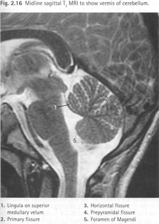
olives give a square configuration to the medulla on this section. The fourth ventricle is seen posteriorly, especially on sagittal sections, w i th its midline foramen leading to the vallecula. The most inferior part of the fourth ventricle is called the obex. This may be sealed during surgery of the cervical cord syrinx.
In the lowest images the hypoglossal nerve may be seen entering the hypoglossal canal of the occipital bone.
Advertisement
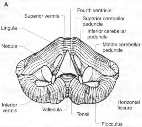
Cervical myelography Contrast medium may extend into the cranium and outline the medulla anterior to the cisterna magna. The expanded upper medulla w i th its nerve rootlets can be seen. CEREBELLUM (Figs 2. 15 and 2. 16) The cerebellum lies in the posterior fossa. It is separated from the occipital lobe by the tentorium and from the pons and midbrain by the fourth ventricle. It is connected to the brainstem by three pairs of cerebellar peduncles: • Superior cerebellar peduncles (brachium conjunctivum) to the midbrain; • Middle cerebellar peduncles (brachium pontis) to the pons; and • Inferior cerebellar peduncles (restiform body) to the medulla.
The cerebellum lies on the occipital bone posteriorly and close to the mastoid anteriorly. It is related to the dural sinuses, especially the sigmoid sinuses.
The superior surface of the cerebellum slopes upwards from posterior to anterior. A CT slice of upper cerebellum, therefore, contains cerebellum anteriorly and occipital lobes posterolaterally.
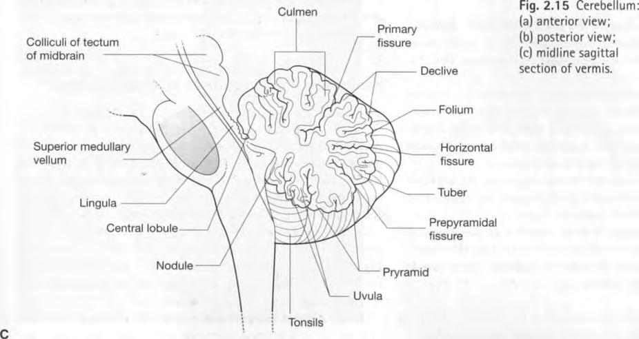
The surface of the cerebellum has numerous shallow sulci that separate tiny parallel folia. Deep fissures separate the hemispheres into lobules.
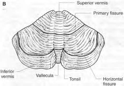
There are two hemispheres w i th the midline vermis between.
Hemispheres
The two hemispheres extend laterally and posteriorly. These are separated by a shallow median groove superiorly and by a deep groove inferiorly. A midline posterior cerebellar notch lodges the falx cerebelli.
The hemispheres are divided into lobules by fissures, the deepest of which is the horizontal fissure, which extends from the convex posterior border into the depth of the hemispheres. The primary fissure (fissura prima) divides the superior surface and defines a small anterior lobe and a larger posterior lobe.
On each side below the middle cerebellar peduncle is the flocculus. This is a small ventral portion of the hemisphere that is almost completely separate from the remainder. It extends laterally from the midline as a slender band w i th an expanded end. It lies close to the lateral recess and choroid plexus of the fourth ventricle. With the nodule, the most inferior part of the vermis, it makes up the flocculonodular lobe, the smallest lobe of the cerebellum.
The tonsils are the most anterior inferior part of the hemispheres and lie close to the midline.
The cerebellum, like the cerebrum, has a cortex of grey matter and deeper white matter. There are deep nuclei within the hemispheres, the largest of which is the dentate nucleus.
Vermis
The vermis is the narrow midline portion of the cerebellum. Superiorly this is merely a low median elevation not clearly separated from the hemispheres; however, inferiorly the vermis is quite separate and lies in a deep cleft called the vallecula. The vermis is separated into named parts by fissures, each part except the lingula corresponding to an adjoining lobule of the cerebellar hemispheres. The most anterior part of the superior vermis is the lingula, which lies on the superior medullary velum (a thin sheet of white matter between the superior cerebellar peduncles). The most anterior part of the inferior vermis is the nodule.
Subdivisions of the cerebellum
• Anterior lobe: lingula, central lobule, culmen • Posterior lobe: declive, folium, tuber, pyramid, uvula • Flocculonodular lobe
The function of the cerebellum
The cerebellum is important in muscle coordination and in the regulation of muscle tone and posture. Unilateral lesions of the cerebellum affect the ipsilateral side of the body.
Arterial supply
Three pairs of arteries supply the cerebellum, namely: • Posterior inferior cerebellar arteries (PICA) from the vertebral arteries supply the inferior vermis and the inferior part of the hemispheres. • Anterior inferior cerebellar arteries (AICA) from the basilar artery supply the anterolateral aspect of the undersurface of the hemisphere. • Superior cerebellar arteries, also from the basilar artery, supply the superior aspect of the cerebellum.
Venous drainage
(See section on veins of the posterior fossa. )
The precentral cerebellar vein and the superior vermian vein drain to the great cerebral vein. The remainder of


