2 minute read
The azygos system


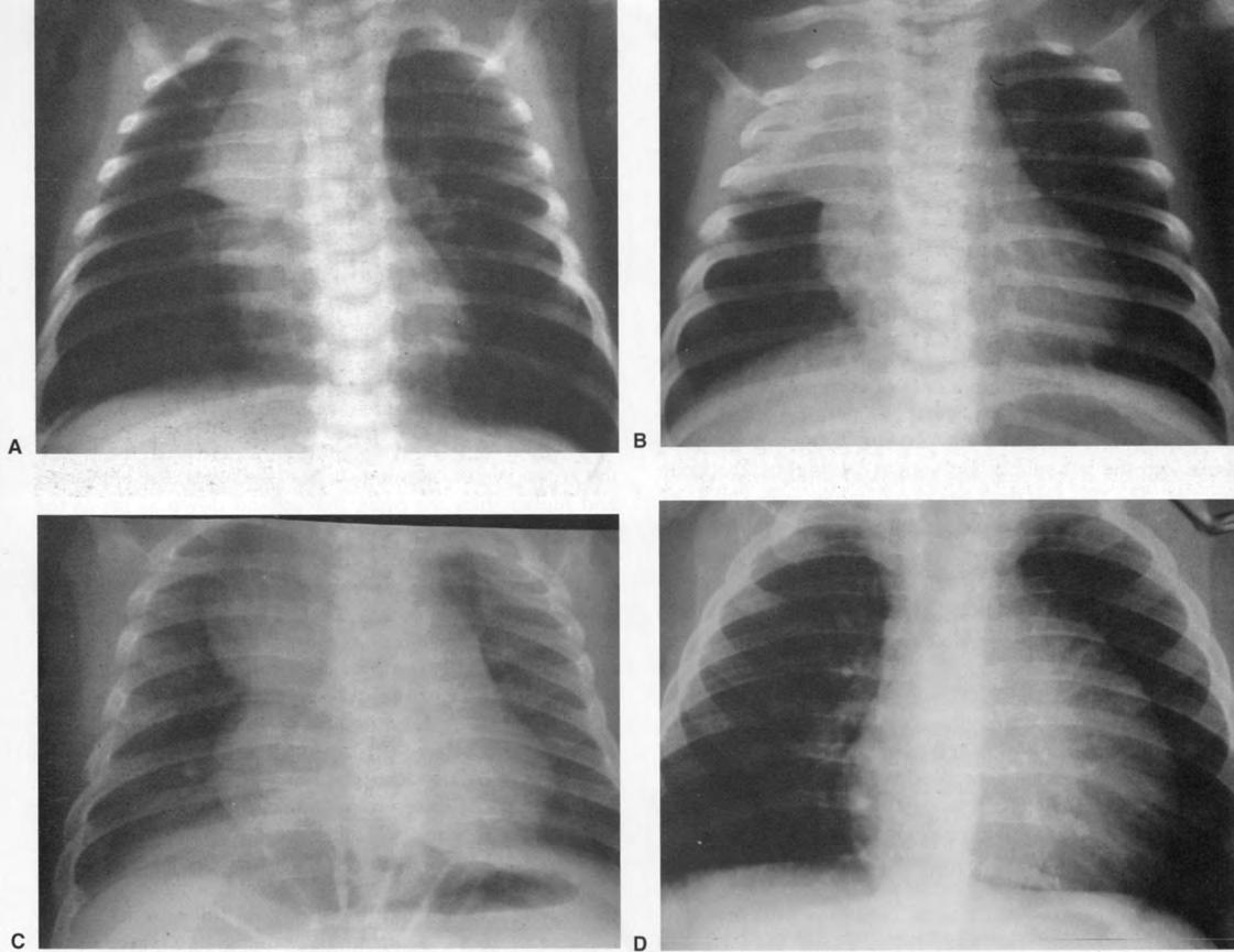
Fig. 4.34 Variable appearance of normal thymus on chest radiographs of young children: (a) sail-like; (b) like consolidation of right upper lobe; (c) like mediastinal mass; (d) like cardiomegaly.
CT and MRI
Advertisement
The normal thymus can be seen on CT or MRI long after it is no longer visible on a radiograph. On CT it is homogeneous, w i th a density similar to that of muscle. On T1and T2-weighted MRI the thymus has a signal greater than that of muscle.
Ultrasound
Using high-frequency high-resolution ultrasound the internal architecture of the thymus can be seen. One can see the vascular septa dividing parenchymal lobules, which have an echogenic medulla centrally and a relatively hypoechoic cortex.
THE AZYGOS SYSTEM
(Figs 4.21, 4.32, 4.36, 4.37, 4.43 and 4.44) These veins are found in the posterior mediastinum. The system consists of the azygos vein on the right and the hemiazygos and accessory hemiazygos azygos veins on the left.
The azygos vein commences variably anterior to the L2 vertebra, either as a branch of the inferior vena cava or as a confluence of the right ascending lumbar vein and right subcostal vein. It ascends to the right of the aorta and thoracic duct, anteriorly to the bodies of T12-T5 and the right posterior intercostal arteries, which cross behind it. The right lung and pleura are to its right. It usually passes behind the right crus of the diaphragm to enter the thorax (see Fig. 4.4). At the level of T4 the azygos vein arches


anteriorly, passing over the hilum of the right lung w i th the trachea and oesophagus medially from the posterior side to the anterior side. The azygos vein drains into the superior vena cava just above the upper limit of the pericardium, behind the third costal cartilage.
The azygos vein receives all but the first intercostal vein on the right. The second, third and fourth intercostal veins usually drain via a common channel - the right superior intercostal vein. The hemiazygos and accessory azygos veins drain into the azygos vein at midthoracic level. The right bronchial veins drain into the azygos vein near its termination. It also receives tributaries from the oesophagus, pericardium and mediastinum.
The hemiazygos vein commences in a similar manner to the azygos vein on the left side of the aorta. Its origin may communicate w i th the left renal vein, and it ascends anterior to the vertebral column to the midthoracic level, then passes behind the aorta to drain into the azygos vein. It receives the left ascending lumbar and lowest four posterior intercostal veins, as well as mediastinal and oesophageal veins.
The accessory hemiazygos vein receives the fourth to the eighth posterior intercostal veins. (The first to third posterior intercostal veins drain into the left brachiocephalic vein.) It may also receive the left bronchial veins. It descends on the left side of the vertebral column to the midthoracic level, then crosses to the right behind the aorta to drain into the azygos vein. The hemiazygos and accessory hemiazygos veins may drain into the azygos vein separately or via a common trunk.


