and other diagnostic tests and radiological procedures

Product # 04E
1 Table of Contents 2-3 A heart cath 4-5 At the hospital 6 Before the cath 7-9 The cath lab 10-11 The catheter and dye 12-15 After the cath 16 Going home 17 Results 18-21 Treatment 22-30 Other heart tests 22 – ECG (electrocardiogram) 22-23 – Exercise stress test 24 – Echo (echocardiogram) 25-26 – Nuclear scan 27-28 – Cardiac Computed Tomography (Cardiac CT) 29-30 – Magnetic Resonance Imaging (MRI) 30 Getting ready Icons used in this book: i Special notes and information Caution and warning Check off or fill in with your specific information !
A heart cath
Cardiac catheterization* is when a tube (catheter) is put in an artery and/or vein and moved along until it reaches the heart. Once the tube is in the heart, X-ray dye (contrast) is injected. The contrast is used to highlight the vessels and any issues within them.
A heart cath is done to see if there are problems in the heart valves, chambers or main blood vessels (aorta or pulmonary artery). It is also done to see if there is fatty buildup (atherosclerosis) in the heart arteries.
There also may be calcium deposits that block arteries. These can be cleared by a heart cath.
iNote: Ask your doctor which areas of your body will be used for your cath. The doctor will choose a backup site in case there is a need to change the access point.
neck (internal jugular) veins
Arm
brachial
radial
Arteries: Leg Artery: left and right femoral arteries
* Other terms used to describe cardiac catheterization are coronary arteriogram, coronary angiogram or dye study of the heart.
2
needle guide wire skin blood vessel
During a cardiac cath, pressure readings are made inside the heart.
dye enters heart’s left chamber (to assess the pumping ability of the heart)
right coronary artery
catheter through the aorta catheter
left main artery blockage
left circumflex coronary artery
left anterior descending coronary artery
Also, moving pictures are taken of one or more of the heart chambers and coronary arteries. These arteries carry blood to the heart muscle. Often a cardiac cath shows that the heart and major blood vessels are normal. But when problems are found, plans for treatment can be made. The heart cath lasts between 1 and 2 hours, but may run longer for some people.
3
AORTA
AOR T A
At the hospital
Some people have a cardiac cath during a hospital stay. Most of the time, a heart cath is done while you are an outpatient. Here’s what to expect.
Medical history & tests
You will be asked about your health, both past and present. You will also have blood tests and an ECG (electrocardiogram—see page 22).

Be sure to tell your doctor of any allergies (food, clothing, medicine, metal, x-ray dye [contrast], or latex) and how you react to each one.
Note:
iIf you are pregnant or think you might be, tell your doctor or nurse.
4
Your medicines
Take your medicine bottles or a list of them with the dosages to the hospital. Follow your doctor’s advice about which medicines to stop and which to take on the day of your cath.
Anticoagulants (blood ‘thinners’)
Anticoagulants (blood “thinners”) like warfarin, Pradaxa® (dabigatran), Xarelto® (rivaroxaban), Eliquis® (apixaban), Savaysa® (edoxaban) and Lovenox® are often STOPPED 1 or more days before a heart cath.
Anti-platelet (ATP) drugs
Many doctors ask their patients to CONTINUE their daily anti-platelet (ATP) drugs before a heart cath. Examples of ATP drugs include aspirin, Plavix® (clopidogrel), Effient® (prasugrel) and Brilinta® (ticagrelor).
Oral hypoglycemics
Diabetics are often told not to take any oral hypoglycemics* on the day of the cath.
ED (& lung high blood pressure) drugs
Tell your doctor or nurse if you’ve used one of these in the past 24 hours: a sex-enhancing drug like Viagra®, Cialis® or vardenafil or a drug for lung high blood pressure (Revatio® or Adcirca®). NTG causes serious blood pressure changes if any of these are still in your body.
Nitroglycerin (NTG)
Note:
Always let the nurse know if you have headaches or any symptoms of chest, arm, neck or jaw discomfort while you are in the hospital. Only use NTG under his or her direction.
*Some oral hypoglycemics are stopped a few days before and after procedures using X-ray dye. These include the generic drug metformin (Fortamet®) as well as drugs that include metformin.
5
iNot following the doctors advice about some medicines could result in your procedure being delayed.
Before the cath
No food or drink (NPO)
Do not eat or drink anything for the hours your doctor specifies before a cath. This lowers the chance of nausea or vomiting during the procedure. Most people can eat and drink right after the cath.
Clip & scrub
The catheter(s) will be inserted into a blood vessel in your leg, neck and/or arm. Any hair over the area where the catheter will be placed may be shaved or clipped away. Then the skin is scrubbed with a special soap.
The bare essentials
Before leaving your room, you will be asked to empty your bladder in the bathroom and put on a hospital gown. Leave your slippers, clothing (including underwear) and any necklaces with your family. Ask if you can wear your glasses, rings, watch, dentures or your own socks.

Intravenous (IV) access
A tiny IV catheter will be placed in an arm vein. Often it is attached to a bag of IV fluid. Medicines can then be given right into the bloodstream as needed.
6
The cath lab
When you get to the lab, you will lie on a firm bed under an X-ray camera. In most labs, the camera moves on an arm over the bed. You will see TV screens nearby. The room is usually cool to keep computers that control the X-ray equipment from overheating. Sterile bags and covers are draped over the equipment to protect you from infection.

7
ECG (electrocardiogram)
Sticky pads (electrodes) will be placed on the side of your body. These monitor your heartbeat. You may also be able to see your ECG or the pictures as they are taken of the heart and arteries. Sometimes you can hear a beeping sound with each heartbeat.
Oxygen Saturation
A device that looks like a piece of tape (or a device that clips onto your finger) will measure the amount of oxygen in your blood.
Fluoroscopy
Contrast (dye) is pushed into your body through the catheter. This allows your healthcare team to see your vessels on pictures.
If you are awake, you may be able to see the pictures. During this test, it is important to be quiet and as still as possible. You may speak during the test, but it's best to keep talking limited to any discomfort you may be having.
Cath medicine
A combination of relaxation drugs may be used to help your procedure go as well as possible. If you are allergic to X-ray dye you may be given a special medicine to reduce or prevent a reaction.
8
Not surgery
A cardiac cath is not surgery, but most times the nurses and doctor wear gowns and masks. These help keep the equipment free of germs (sterile). The site(s) where the catheter will be inserted is scrubbed again with a special soap. Sterile towels and a sheet are then placed around the site(s). The X-ray camera and other equipment are prepared for the procedure.
The catheter will be inserted in an artery or vein in the groin, arm or neck*. The skin over the chosen blood vessel will be numbed with medicine. Your skin may burn or sting as the medicine is injected. When the skin is numb, a needle is placed in the artery or vein. You may feel some pressure when this is done, but pain is not likely. Once the tip of the needle is in the blood vessel, a guide wire is moved through the needle into the artery or vein.
* Sometimes a small skin cut is made to find the desired blood vessel in the arm.
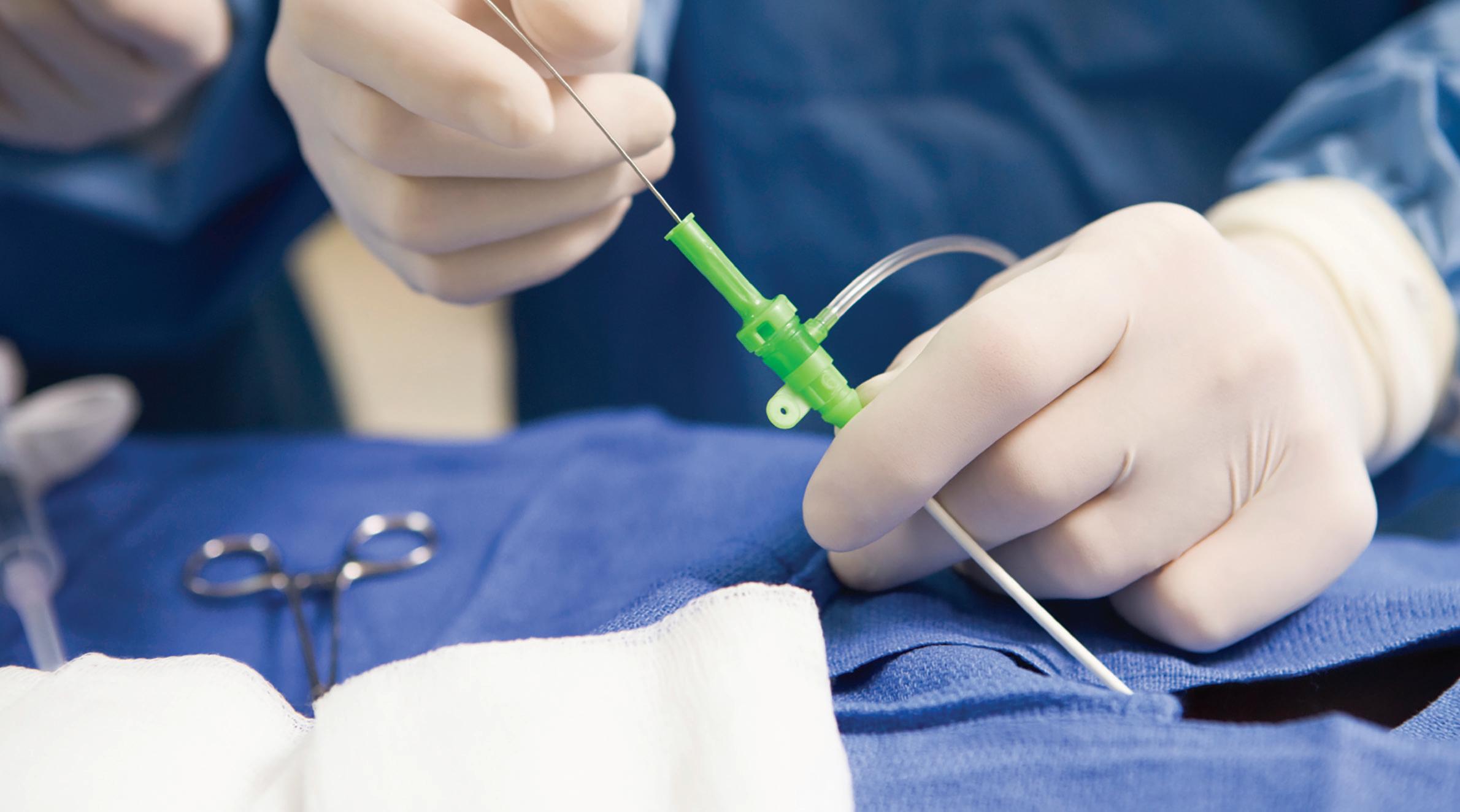
9
needle blood
skin
vessel guide wire
guide wire
The catheter and dye
The catheter is a long, very thin tube. There should be no pain as the catheter passes over the guide wire to the heart.
Most of the time, the catheter is first placed in the left heart pumping chamber, and X-ray dye is injected. You may be asked to cough to help move the catheter into place. Moving pictures are made as the heart squeezes the dye into the main blood vessel (aorta).
You will have a warm or hot feeling throughout your body for 30 seconds or less. Some people also have slight nausea or extra heartbeats. You may also feel a false need to urinate (pee). These feelings should pass quickly and not come back.
If the TV screen is nearby, you may be able to watch your doctor guide the catheter into place in your heart. Most of the time, the lights are dimmed during the cath to make the screen clearer.
10
AOR T A
dye enters heart’s left chamber
catheter through the aorta
After pictures of the pumping chamber are made, the catheter is placed in each of the coronary artery openings. Smaller amounts of dye are pumped into the arteries a number of times. Moving pictures then record the flow of dye into the smaller branches. Extra blood flow readings and closeup pictures of any narrowed place in your vessels may be needed. Your doctor will study the pictures to see if any of the heart arteries are blocked with fatty buildup (atherosclerosis) and/or a clot (coronary thrombosis).
If an artery is prone to spasm, injection of the dye sometimes allows the doctor to see the spasm happen. Nitrates or a calcium channel blocker may be given to relax the vessel spasm.
The total heart cath lasts about 1 to 2 hours. At times you will be asked to take a deep breath, cough, turn to one side or not speak for a few minutes while pressures are being measured.
left main artery
left circumflex coronary artery (LCX)
blockage left anterior descending coronary artery (LAD) right coronary artery (RCA)
11
AORTA
catheter
After the cath
When the X-ray pictures have been taken, the catheter, wire and sheath are removed.
Leg artery
If the groin artery was used, the hole in the artery can be sealed by one of these:
Firm hand pressure followed by a tight bandage and 2-4 hours of bed rest with the hip and knee of the cath leg straight
A closure device (collagen plug, a purse-string metal clip or other material) left in the groin artery to reduce the amount of time needed to stay still and lie flat

Stitches placed inside the artery (with bed rest as needed to recover from sedatives used during the procedure)
No matter how the artery is sealed, you will be asked to slide from the cath table onto a stretcher or bed without bending your groin. Follow your nurse and doctor’s advice about the amount of bed rest needed to prevent bleeding at your puncture site.
Your movements during the bed rest will be limited. Do not get out of bed or bend the knee of the affected leg. You may move your arms and other leg freely.
12
The head of your bed will be raised slightly, but you will be asked not to sit up or turn to the side for a period of time. Your nurse can tilt your bed slightly so that you won't feel that you are lying completely flat. In some cases you may be asked not to lift your head off the pillow for part of the bedrest. The purpose of this is to prevent pressure from building up in your groin, possibly causing an injury to the access site. When needed, you must use a urinal or bedpan without bending your groin. If you have trouble doing this while lying down, let your nurse know.
Hold firm pressure over the bandage for extra support while you cough, laugh or sneeze. Your family or a nurse can help you cut up foods to make them easier to eat while you are lying down.
You may have back discomfort during the bed rest. It can be eased by: bending the knee of the unaffected leg often a small towel or pillow under the back and/or a back massage (by nurse)
mild to moderate pain medicine
Neck vein
Note:
iYou may bend your foot or wiggle your toes on the leg used for the heart cath. You don’t have to hold the leg stiff, just straight.
Your cath team will hold pressure on your neck until they are sure your bleeding has stopped. A bandage or gauze may be added for more pressure. After a couple of hours, you will be allowed to go home. You can sit up or walk normally once you leave the hospital.

13
Arm artery
Many physicians are using the radial artery near the thumb for cardiac cath. After a radial artery cath, a small compression pillow/ strap is often placed over the site for several hours. If an artery or vein near the elbow (brachial) is used, a few stitches may be needed in the blood vessel and skin afterwards. An armboard may be placed on your arm to remind you not to bend your elbow or wrist for several hours.
When an arm artery is used for the cath, you can walk to the bathroom and around the room (with help) within 1 to 2 hours. Remember that the arm may not be used for lifting or pushing for the rest of the day after the cath. (This includes pushing yourself to get out of a chair, off the bed, etc.) Also, frequent arm motions like typing, knitting or chopping vegetables may be off limits for another 24 hours.
Caution!
If you:
feel sudden pain at the cath site
notice warm, sticky or wet feelings near the site that was used
see your hand or arm turning blue or getting painful and cold to the touch


Notify the nurse at once!
If the groin artery (femoral) was used, pressure is often needed right away to stop bleeding. More bedrest is likely as well.
14
compression strap (filled with air)
!
Other things to know about cardiac cath
After the cath, a nurse will check your vital signs often. The nurse will also check the pulse in your foot or arm and check your bandage for bleeding. (Bleeding rarely occurs.)
You may feel drowsy as the medicine given during the heart cath continues to work. Also, it is normal to feel soreness at the puncture site as the “numbing” wears off.
Your body gets rid of the dye used during the cath through your kidneys, making extra urine. You may be asked to drink more fluids after the cath.
During your stay, tell the nurse of any discomfort in your chest, neck, jaw, arms or upper back. Also let your nurse know if you feel short of breath, weak or dizzy. These are not common, and they may be relieved with medicines.
A cardiac cath is considered to be a safe procedure, but any work done inside a blood vessel carries a small risk of problems. These include reactions to the dye, bleeding, heart attack or stroke. Your doctor will discuss any risks that cardiac cath might hold for you and ask you to sign a written consent.

15
Going home
Your doctor will most likely let you get out of bed once your bedrest is complete. When you first get up, a nurse should be present in case dizziness or bleeding occurs.

Unless you need other treatment right away, most people go home after the cath. You will likely be released once you can walk around the room with no weakness or bleeding from the groin site. You will need a ride, since most hospitals ask that you not drive for the rest of that day. Fatigue is common for a day or two. You can return to your normal routine the next business day unless your doctor tells you not to. If you have a bruise and/or a small lump under the Band-Aid® where the catheter was put in, you may be asked to delay any hard physical activity (tennis, running, etc.) for a longer period. Change the Band-Aid® daily until the site is healed. Tell your doctor right away if you notice new swelling or signs of infection (drainage or hot/tender feelings) at the cath site. Also, tell your doctor if you have constant itching, rash or fever.
You will be told when it’s OK to shower or take a tub bath. Showers are often allowed after 24 to 36 hours. Tub baths may be delayed up to a week.
If an elbow artery was used, there may be stitches on the skin. If so, you should protect the site with a plastic covering when taking a shower or bath. Do this until the stitches have been removed (6 to 10 days later in your doctor’s office).
16
Results
Many times a heart cath shows that the heart and blood vessels are normal. But if the cath shows that there is a problem, your doctor can use these drawings to show you what can be done about it.
17
pulmonic valve mitral valve tricuspid valve aortic valve
1 Right Coronary Artery
2 posterior descending artery (PDA)
Left Coronary Artery
3 left main
4 circumflex artery (Cx)
5 obtuse marginal (OM)
AORTA 1 4 3 2 6 5
6 left anterior descending (LAD)
Treatment
Some of the treatments for heart problems include medicines, changes in lifestyle, angioplasty/stents and surgery.
Medicines
Heart medicines may be prescribed to:

keep blood lipids normal: total cholesterol less than 200 mg/dL*, HDL over 40 mg/dL in men and over 50 mg/dL in women, LDL less than 100 mg/dL* and triglycerides less than 150 mg/dL*
stabilize and reduce fatty plaque relax the blood vessels produce a regular, steady heartbeat make the pumping action of the heart stronger
take excess fluid from the blood
replace minerals (potassium)
Note:
iYour ethnic group and genes play a large role in your lipid numbers. You can help with a healthy diet, exercise and prescribed medicines.
*Some people are told to aim for even lower numbers: total cholesterol less than 180 mg/dL, LDL’s less than 70 mg/dL, and/or triglycerides of 100 mg/dL or less. Your doctor will tell you the ‘right’ lipid goals for you based on the latest statistics and how any of your drug(s) are helping lower your risk of heart disease.
18
Changes in lifestyle
The exact causes of fatty buildup in the arteries (atherosclerosis) are not known. But there are some risks that make a person more likely to have buildup in the arteries. For this reason, most doctors will ask that you: quit smoking
keep your resting blood pressure in the range experts have defined as normal: less than 120 (top number) and less than 80 (bottom number)*. Doctors often use a blood pressure goal of less than 130 and less than 80 for persons with hypertension AND one or more of these: diabetes, heart failure or chronic kidney disease
eat less unhealthy fat
exercise (your doctor will tell you what kind and how much)
lose weight (if you are overweight)

keep blood glucose under good control (if you are diabetic)

reduce tension and stress
Changes in activity
Sometimes the cardiac cath shows recent heart damage or an overall heart weakness. If so, activity limits may be needed for a time so healing can take place. Your doctor will let you know which activities to avoid.
*Source: American Heart Association, Inc. http://hyper.ahajournals.org/content/guidelines2017

19
Coronary angioplasty/stents
If the cardiac cath shows clogged arteries or vein bypass grafts, angioplasty may be needed. Angioplasty is when one or more catheters (like the balloon catheter shown below) are guided under X-ray to the clogged artery or vein bypass to clear or compress the blockage so more blood can flow through. Often a piece of metal wire mesh (stent) is placed in a narrowed artery to prop it open. This is left in place to help keep it open.
Some stents are bare metal. Newer stents are partially or fully absorbable. The most often used are drug-coated stents which slowly release a drug that helps keep tissue from buildling up (restenosis) as the artery lining heals.
Anti-clotting medicines are needed to prevent blood clots inside all stents. Studies show blood clots can occur even a year after a drugcoated stent is put in. So people who have these stents are often told to take an aspirin a day for life. And a second anti-clotting drug like Plavix® (clopidogrel), Effient® (prasugrel) or Brilinta® (ticagrelor) is often needed for months or years. These are known as antiplatelet drugs since they help blood platelets resist ‘clumping’—the start of a blood clot. Follow your doctor’s advice about the medicines you take.
20
after before during artery open
stent
artery narrowed by fatty buildup
balloon catheter
Bypass surgery
This surgery is advised for some people with blockages of the heart arteries. A leg vein or an artery from the chest (internal mammary) is used for the bypass graft. When a leg vein is used, one end is sewn to the aorta (main blood vessel) and the other end to the coronary artery just below the blockage. When a chest artery is used, one end is left attached to a branch of the aorta. The other end is sewn to the coronary artery below the blockage. The kind of bypass graft depends on the number of blockages and where they are.
internal mammary artery (chest) bypass graft
saphenous vein (leg)
blockage
Sometimes heart surgery is needed to replace a scarred heart valve. Many years may pass before enough scarring occurs to need heart surgery. Heart valve surgery can also be needed if the heart chamber(s) enlarges or stretches out. In some cases, a catheter is used to put in a new heart valve. Examples include TAVI® /TAVR® (transcutaneous aortic valve implant or replacement) and/or the Mitra® clip for mitral valve repair. This offers an option for those who have a scarred heart valve but need less invasive surgery.
Other problems in the valves, heart chambers or major blood vessels can be present at birth. These may require surgery in childhood or later as an adult.
21
Other tests
You may also have one or more of these cardiac diagnostic tests or radiological procedures before your heart cath:
ECG (electrocardiogram)
This test records the electrical impulses moving through the heart muscle. The ECG can show abnormal heartbeats (arrhythmias or dysrhythmias) and any damage or enlargement in the heart muscle. (These problems can also occur without changes on an ECG.)
An ECG does not hurt and lasts only a few minutes. You are asked to remove stockings or socks and upper body clothes. ECG pads are placed on your chest, arms and legs, and you are asked to lie quietly. If you move or talk, it is harder to record the heart’s electrical signals.
Exercise stress test
An exercise ECG (stress test) is a test done while you are walking on a moving belt (treadmill) or riding a stationary bike. (If necessary, medicines can be used to mimic the effects of physical activity.) During the test you are connected by ECG pads to a machine that records your heart’s electrical activity. As you exercise, the doctor watches for changes in the ECG. Changes in the ECG during exercise can occur with heart problems.
After the stress test, you return to a table for a post-exercise ECG and to relax. Sometimes a nuclear scan is done along with the exercise (see page 25).
22
Caution!
BEFORE having a stress test: do not eat heavy foods or drink a lot of liquids wear comfortable clothes (well fit but loose sweatpants are ideal) and good walking shoes follow your doctor’s advice about which medicine to take avoid caffeine and nicotine
Caution!
DURING a stress test, it is important to let the doctor or nurse know if you feel:
any chest, arm, jaw or back discomfort
a severe fatigue
If any skin irritation occurs at home where the ECG pads were placed, keep the places dry. Don’t scratch. If the irritation is severe, call your doctor for advice.
Note:
Tell your doctor or nurse if you’ve used one of these in the past 24 hours: a sex-enhancing drug (Viagra®, Cialis®, or vardenafil) or a drug for lung high blood pressure (Revatio® or Adcirca®). NTG can cause serious blood pressure changes if any of these are still in your body.
23
i ! !
Echo (echocardiogram) or stress echo

The echo uses sound waves to look at your heart’s size and how the chambers and valves are working. It can be used to check on abnormal openings between chambers and fluid in the sac around the heart.
Having an echo does not hurt and takes about 20 to 30 minutes. You remove upper body clothes and stockings and lie quietly on a table or bed. A technician moves a scanner (transducer) over your chest, taking pictures and recording them on video.
You must lie quietly so that the scanner can be placed at the exact angles needed. You may be asked to turn to your side or briefly hold your breath while the best transducer position is found.
A stress echo is done to see if there are any problems when your heart is beating harder and faster. Some patients get an IV medicine to dilate an artery or a drug to cause the heart to work harder and faster (dobutamine stress echo or DSE). Other patients exercise on a treadmill.
24
heart chest scanner sound waves
Nuclear scan
A cardiac nuclear scan is a type of X-ray. In this test, a small amount of radioactive material is injected into a vein. These scans include:
PET scan (positive emission tomography)

Thallium scan
First pass scan
MUGA scan (multiple gated acquisition)
During a nuclear scan, a tiny IV catheter is put in an arm vein, and sticky ECG pads are placed on the upper chest. Radioactive material is injected through the IV. (Some nuclear scans involve a number of these injections.) The radioactive material moves through the heart chambers and/or heart muscle while a scanning camera records pictures of the heart. Total exposure to radiation is limited (less than with a chest X-ray).
25
You will be asked to limit or delay eating and drinking until the scan is finished. Your doctor may also ask that you not take certain medicines before the test.
Sometimes exercise on a treadmill or bike is part of the test. If exercise is part of your scan, wear socks and shoes for easier pedaling or walking.

As the camera scans the movement of the radioactive material, keep your upper body as still as you can. Also, let your doctor know if you have any feelings in the chest, arms, neck, jaw or upper back that feel strange to you (even if they do not seem important).
pregnancy
If you are pregnant or think you might be, let your doctor or nurse know. Ask your doctor to discuss any effects a nuclear scan may have for you.

Note:
Tell your doctor or nurse if you’ve used one of these in the past 24 hours: a sex-enhancing drug (Viagra®, Cialis®, or vardenafil) or a drug for lung high blood pressure (Revatio® or Adcirca®). NTG can cause serious blood pressure changes if any of these are still in your body.
26
i
Cardiac Computed Tomography (Cardiac CT)

Ultrafast Electron Beam CT (EBCT or EBT)
Cholesterol deposits that build up into fatty plaque contain calcium. Ultrafast CT looks for calcium in the heart arteries before it causes symptoms of heart disease. Ultrafast CT involves lying still for a few minutes with the X-ray camera over your chest. It is painless and requires no special preparation or follow-up. No X-ray dye is used.
Multi-slice Cardiac CT (MS-CT) — or multi-detector CT or MDCT
Multi-slice CT takes X-ray pictures of the beating heart, major blood vessels, lungs and the sac around the heart. If iodinebased dye (contrast) is given by IV, pictures are also made of the heart arteries (coronary CT angiography or CTA). When dye is used, you will likely feel warmth as the dye passes through your body. Tell your doctor if you have allergies to shellfish, iodine or X-ray dye.
27
The CT Scanner is a large machine with a hollow, circular tube in the middle. You will lie on your back on a sliding table that goes in the tunnel-like machine. Many pictures are made while you’re in the scanner (multi-slice) and a computer puts them together in one image. The technician controls the scanner from the next room but can see and talk to you through an intercom at all times.
Cardiac CT usually takes 15-30 minutes. You will take off all jewelry and clothing from your waist up and wear a hospital gown. Small sticky patches are placed on the skin for ECG monitoring. Since the best pictures are made between heartbeats, you will be asked to lie still and briefly hold your breath. A fast heartbeat or body movement causes blurred pictures. Not drinking caffeine before the test can help slow your heartbeat. Sometimes an IV drug (beta-blocker) is needed.
Your doctor will explain the risks of cardiac CT and get your written consent. Since X-rays may harm a developing fetus, cardiac CT is not done if you are pregnant or suspect pregnancy. Cardiac CT is painless. Allergy symptoms can occur in some people if X-ray dye is used to show the heart arteries. Persons with asthma or emphysema can have short-term breathing problems if a beta-blocker is used during cardiac CT. Serious complications of cardiac CT are rare.
Although cardiac CT scans involve radiation, many of today’s CT scanners can use smaller amounts of radiation but still offer good images. This further reduces the small chance that radiation with a CT scan could lead to cancer.
Note:
Your doctor chooses the test that offers the most benefit to you at the lowest risk.
28 i
Magnetic Resonance Imaging (MRI)
or Angiography (MRA)
Magnetic Resonance Imaging (MRI) uses a magnetic field to take detailed pictures of the heart. MRI studies of the heart can show problems with the heart’s function and shape. When used to detect heart muscle damage due to poor coronary blood flow, it is called MR angiography (MRA).
During an MRI, you lie very still on a narrow table that slides into a long tube containing a round, donut-shaped magnet. Earphones can be used to block out some of the noise and let you hear and talk with the technician or doctor. You will be asked to remove any metal you are wearing (jewelry, belts, etc.).
Caution!
If MRI is being considered, remind your doctor if you have any of these:
Internal metal fragments (metal prosthesis or bullets in your body or if you worked as a metal grinder)
Aneurysm clips
A cardiac electrical device (pacemaker and/or ICD)
Show your cardiac device ID card to your doctor. Some newer pacemakers or internal cardioverter-defibrillators (ICDs) ARE approved for MRI as long as they are paired with a suitable lead system. If the device OR lead system is NOT approved for MRI, many hospitals will NOT do the MRI. If MRI became crucial at some point, your doctor may bring in an electrophysiologist (EP doctor)
29
!
Getting ready
Before your test:
Bring a list of all the medicine and dosages you take (including over-the-counter and herbal supplements).
Tell your doctor of any allergies (food, clothing, medicine, metal, X-ray dye [contrast], or latex) and how you react to each one.
Tell your doctor if you are pregnant or think you might be.
Do not eat or drink anything for the hours your doctor tells you before your test.
Sign the consent form.
Arrange for someone to drive you home.
This book should not replace the advice or treatment your doctor gives you. It is only to add to what you are now learning about cardiac catheterization (a heart cath) and other cardiac diagnostic tests and radiological procedures.

30
Suggested Questions
It’s important to talk with your doctor about your heart tests. Here are a few questions to get you started:
• What kinds of exercise can I do after my cath or test?
• Where will the catheter enter into my body?
Written by: Julia Ann Purcell, RN, MN, FAAN Clinical Nurse Specialist, Cardiology
1968-1996, Emory University Hospital, Atlanta, GA
• Can I not even drink water before my test(s)?
Reviewed by: Jason Johnson, MHA, MSN, RN, CCT Northside Hospital Johnson Health Homes, LLC Atlanta, GA
• When can I return to work? Am I limited in what I can do?
• How long should I expect to be at the hospital?
• Do I need to stop or continue any medicines?
Please let us know how this booklet is helping you (or your patients). Share your comments at http://p-h.com/04 3440
Copyright © 2023 by Pritchett & Hull Associates, Inc. All rights reserved. No part of this book may be photocopied, reprinted or otherwise reproduced without written permission from Pritchett & Hull Associates, Inc.
1-800-241-4925

OAKCLIFF
ROAD, NE, SUITE 126 ATLANTA, GA 30340-3006
• www.p-h.com
