Phenotype | Issue 19 | Michaelmas Term 2014

Page 1

PHENOTYPE Issue 19 | Michaelmas Term 2014

www.phenotype.org.uk

The Birthday Issue: OUBS is 50!

Prof Tony Watts celebrates with reminiscences of our achievements

Stimulating Simulations

Dr Matthieu Chavent

Understanding membrane proteins through molecular dynamics

New hope through natural immunity

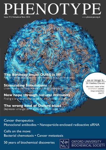
cover image by this issue’s winner of the SNAPSHOT scientific image competition page 31

Finding a long sought-after target for anti-malarial vaccines

The wrong kind of Oxford blues

Depression amongst DPhil students and how to get help

Cancer therapeutics: Monoclonal antibodies • Nanoparticle-enclosed radioactive siRNA Cells on the move: Bacterial chemotaxis • Cancer metastasis 50 years of biochemical discoveries

OXFORD UNIVERSITY BIOCHEMICAL SOCIETY


We Care - about Innovation

VACUETTE速 www.gbo.com/preanal y t i cs

Our Preanalytics Product Range Blood Collection Paediatric Blood Collection Urine Collection Saliva Collection Transport Line Safety Products to help prevent Needlestick Injury

Greiner Bio-One Ltd. Brunel Way, Stroudwater Business Park, Stonehouse, Glos. GL10 3SX. Tel: 01453 825255 | Fax: 01453 826266 | email: info@uk.gbo.com

i va

l

l b a

e

m co ' . o e gb typ . uk o @ hen ns s tio le 'P di n a co l s ng d ly ai oti an app s m em qu er

m a S

pl

es

A

t


Issue 19 | Michaelmas 2014

EDITORIAL Another year begins in Oxford and another thrilling issue of Phenotype for the new term. Welcome to our celebratory issue for the 50th anniversary of the Oxford University Biochemical Society (OUBS), noting as well the 5 year anniversary of the New Biochemistry building and the 15 year anniversary of co-education colleges in the University. As usual our magazine is packed full of articles contributed by PIs, research staff and students from across the University, showcasing some of the hot topics being studied right here in Oxford. In celebration of the 50th anniversary, check out our electrifying line-up of speakers detailed on our double-spread of featured seminars on pages 20-21. Don’t miss Prof Tony Watts’ article on the history of OUBS and some of the great moments of its first 50 years. And while you’re in a historical frame of mind, see if you agree with our choice of the top ten biochemical discoveries over the last half century, shown in our research comic on page 16. Congratulations to Dr Matthieu Chavent, the winner of last issue’s SNAPSHOT competition, with his stunning visualisation of lipid movement in a membrane vesicle. Further details of his research and career can be found on page 31. Fortuitously, our PI article this issue originates from the same research group, with Prof Mark Sansom’s illustration of molecular dynamics simulations of membrane proteins. On the subject of immunology, Caitlin Clunie-O’Connor describes the discovery of a new target for malarial vaccines, found by examining people that harbour natural immunity against the disease. Dr Thierry Deltheil also expounds on the world of monoclonal antibodies used in treating cancer, and Dr Jessica Stolp explains how regulatory B cells can both contribute to and fight against disease. Also in this issue we look at cells that move, beginning with Andrea Szöllössi’s article on bacterial chemotaxis on page 22 and then Dr Ana Gil-Bernabé’s blood-curdling description of the promotion of metastasis by factors involved in coagulation. But it’s not just cells that move; like miniature postmen, Michael Delazzari’s nanoparticles deliver radioactive siRNA to cancer cells, so check out his article on page 13. Our Science and Society section highlights the importance of recognising depression as a DPhil student and getting the right kind of help in Madeleine Pope’s personal article on page 24. We also look at the participation of men and women in all levels of academia with a new infographic on page 26; while you think whether you’ve got what it takes to make it to PI, read our interview with Dr Michael Kohl on page 30. To help you on your way to success, get the low-down on the fantastic new Longitude prize on page 27 – could you be the winner? If the Longitude prize is a little out of your scope, at least have a go at this issue’s revolutionary evolutionary crossword on the back cover. Be sure to send us your answers for a chance to win one of the excellent Wiley-Blackwell textbooks that we have reviewed on pages 28-29 of this issue! Remember our beautiful magazine doesn’t grow on trees! If you’re interested in science communication, writing, publishing or making an article leap out of the page with stunning design work, why not join us on the Phenotype team? Contact us at oubs@bioch.ox.ac.uk! We are always looking for more writers, editors and designers to help us with the next issue. Or why not help with getting Phenotype out to our audience by assisting with our sponsorship, distribution or social media presence?

PHENOTYPPE HENPHE PNHOETNYOPTEYPE OTY NO PETY PHE P Issue 15 | Trinity Term

2013

Issue

Issue 13 | Michaelmas

Issue 12

| Trinity

Term 2012

17 | Hila

ry Term

Issu

2014

e 18

| Trin

ity Ter

m 201

4

Term 2012

www.ph

enotype

.org.uk

www.p

hen

Toxo Can par plas asites mosis En affect Turn ginee our beh ri Gen aviing Gene regulation ourpla ? nt cellng Tas What etics metab ti Dr Korneel Hens uses and Keep traits high-resolu wil olism er T Ho basis of homeostat techniques image by w me ing to study the molecular l your offFa utionic regulation tion m cover revol into om ming spring ily chan You ieu O a ga ato gram e by Olivia Berthoum besi an r H ism exhibitPl stron HIV ty an cover The cell repro Finimage cover imag of the Wh ng ea by? s ni of ric omices required Sleigh this issue´s winnerscientific y are d Developing an effective d e su d A ing Jamesvaccine Dr quescov Wolf Ev No human embryos some Do Huetteroth SNAPSHOT of the rviva olna winner t er the ity popula this issue´s ut Spring on the scientific commun turio ropy image competition l couldbove this issue´s scientific winner image Mar al ofna the tion page 31Fr Nex pleiotthe Arab pla Prescr s After tition SNAPSHOT SNAPSHOT revolution of ry mo feed Wat ia Kuzma-Kuz by iption ing nt scientific am compe B effects im genom His image oimage inhere comp this issue investigate the the Treating cancers individually page 31 es niarska n recompetition its eyes Dr Rob eshift Cu tot ry Proclacavefish lost We worlder ntly sus ou ’s winn nd SNAPSH ert Gilb s anWhat haes page in 29th er of s still re ia cep se the ? OT scien How the spread disea d tibl to ert right aemas image or M ppen e toha migrationConditioned eD on his an Chag ve a competit tific obe on risky ia vs leme disease uses modern use of asteed sitypla tropical ion How does the brain taste aversion r be A ntati ? ce hind ark Re Manlar page stru remember Pl age iron supp in y future gu 31 of is to ctu the clo avoid an drug sickness-inFu rne ral la sedto Whe antibiotics of ts ducing iotics come turefoods? e jou disco dors antib s colicins, the protein in networks Big tic sensing Plan and th biology in und ors in Ther redibl agne very? prote Kleanthous introduce inc geom t Colin d Ap e and ers e the ap Prof fla Th iology behin tandin M geneticpharmaceutical vono Brain: lex Are : g RNuseum companies ‘big badtamers • Hi iesSe The phys ids • A fun of trolbiology to unravel compe capital istse wolfs’? cr jac Antisen OXFORD ctio con Na et kin Pr A ns tur transcription rabi of Ch cyclek usesInterv g ths eof ni systems iew with a ventur al Hi Cell dops UNIVERSITY ris Po OXFORD injec Nová story trtis Tree OXFORD ateom Prof Béla is in crowd nting theern discussi ment SciBar: Science MICAL ons tran BIOCHE ofopLi UNIVERSI SITY at theHo Wisdomtoofgov e •spCu A onTY pub w cle UNIVER fe: se nce SOCIETY ortres fo lzheimer Prof BIOCHEMICAL en ar ar ICAL Sylvia McLain5´ with... aceceho Drley ing scie HEMMatthew Freeman reve r HI e your 5´ with... BIOC ’s re sswpu ord? IETY CaSE: Tak aled V? SOCIETY sear data Hagan Bay plbli anca the cryptic crossw SOC by cr ProfCan ch? crack presen tstio youfere nce evnol 5´ with... ysta ted? ? ved l stru r own con crossword ct ptic Start you cry ure crack the OXFO Can you RD BIOCH

cove r Lu

imag

e

ke He by this issu aton SNAP e’s win SH ner of image OT scie the com ntific petitio page n 31

Joel

E

otype.

org.uk

Beevers Editor

OXFORD UNIVERSITY BIOCHEMICAL SOCIETY

UNIV EMIC ERSIT Y AL SO CIET Y

Phenotype is also available to read online via our website: www.phenotype.org.uk


Issue 19 | Michaelmas 2014

CONTENT

OUBS SEMINARS

TEAM

regulars 3 5 20 28

30 31 32

Editorial Joel Beevers Research Highlights Dr Ruth Faram Featured Seminars Helen Thompson Book Reviews Dr Mariam Ayub, Emilia Milne, Jamie Mawhinney, Anna Sigurdsson 5’ with… Dr Michael Kohl Dr Ruth Faram and Dr Óscar Cordero Llana SNAPSHOT Jan Soroczynski Crossword

science & society

24 Second year blues: One student’s journey through depression and out the other side… Madeleine Pope 26 The £10 million prize: Solving antibiotic resistance Laura Pritchard 27 Infographic Sofia Hauck

features 6 10 12 13 14 16 18 22 23

Membrane proteins in context Prof Mark Sansom Targeted cancer immunotherapy Dr Thierry Deltheil Harnessing natural immunity against malaria Caitlin Clunie-O’Connor Nanoparticle-enclosed radioactive siRNA Michael De Lazzari The proof that B (cell) does not always mean bad Dr Jessica Stolp Research Comic Jessica Beevers OUBS: the last 50 years Prof Tony Watts Bacteria on the move AndrWea Szöllössi Bloody metastasis! Dr Ana Gil-Bernabé

4 | Oxford University Biochemical Society

EDITOR Joel Beevers DPAG FEATURES EDITORS Christopher Hillyar MRC Radiation Oncology Rachel Patel Undergraduate REGULARS EDITORS Lauren Chessum MRC Harwell Emilia Milne Undergraduate SCIENCE & SOCIETY EDITOR Sofia Hauck Zoology DESIGN & PRODUCTION Dr Maria KuzmaKuzniarska NDORMS Andrea Szöllössi Biochemistry Sharon Ruane DPAG ADVERTISING Prashanthini Shanthakumar MRC Harwell ILLUSTRATIONS Dr Óscar Cordero Llana DPAG WEB EDITOR Dr Alexander Kurze Biochemistry ONLINE MEDIA EDITOR Sofia Hauck Zoology

PAGE EDITORS Kinda Al-Hourani MRC Human Immunology Dr Felicity Alcock Biochemistry Dr Jennifer Badger Alumna Louise Bowler Life Sciences Interface DTC Dr Óscar Cordero Llana DPAG Iona Easthope Biochemistry Dr Ruth Faram DPAG Dr Alexander Feuerborn Pathology Samuel Grayer Chemistry Andy Hall-Ponselè Plant Sciences Suvi Honkanen Plant Sciences Angela Hoslin MCR Harwell Manisha Jalan Biochemistry Melissa Jansen van Rensburg Zoology Stephanie Kapsetaki Zoology Olga Kuznetsova Pathology Katarzyna Kozyrska Biochemistry Rupal Mistry Biochemistry Madeleine Pope MRC Harwell Rosalind Roberts DPAG Anna Sanniti Clinical Medicine Max Sloan DPAG Michael Song Biochemistry Dr Jessica Stolp Surgical Sciences Terence Tang Undergraduate Dr Garrick Taylor Biochemistry Anne Turberfield Chromosome & Dev Biol Dr Hayley Tyrer Alumna Evelyn Tzika Visiting Student, Pathology Dr Roderick Walker DPAG Beth Watts Biochemistry Caroline Woffindale DPAG

Featured Seminar See the article on page 20 for information on some of our exciting guest speakers.

For a full list of the Monday seminars, please check the OUBS website: http://www.bioch. ox.ac.uk/oubs/

This issue is supported by: Greiner bio-one Anachem Mabtech Medical Sciences ‘Skills Training Fund’

To advertise in

Phenotype

please contact us by email:

please contact us by email:

oubs@bioch.ox.ac.uk


A selection of recent life sciences research from the University of Oxford

RESEARCH HIGHLIGHTS

by Dr Ruth Faram

Sattlecker M, et al. (2014) Alzheimers Dement doi:10.1016/j.jalz.2013.09.016

Geibel M, et al. (2014) Nat Commun 5:3427.

Alzheimer’s disease biomarker discovery using SOMAscan multiplexed protein technology

Ablation of TrkB signalling in CCK neurons results in hypercortisolism and obesity

Research into the molecular pathology of Alzheimer’s disease (AD) is rapidly progressing. However, current treatments are unable to directly modify disease pathology before clinical symptoms appear. The ‘pre-dementia’ phase needs to be targeted and biomarkers for this stage would highlight potential avenues for the development of early intervention drugs.

The neurotrophin receptor TrkB and its endogenous ligand brain derived neurotrophic factor (BDNF) are both associated with metabolic regulation in the adult mouse brain. Dysfunction of TrkB or BDNF results in overeating and obesity. Cushing’s syndrome (CS), a disorder caused by excessive cortisol, manifests itself through weight gain, skin thinning/bruising, muscle weakness and hirsutism. Glucocorticoids, such as cortisol, control the hypothalamicpituitary-adrenal (HPA) axis, a neuroendocrine feedback loop that responds to stress and activity. To investigate if TrkB is implicated in CS, Geibel et al. used Cre-Lox recombination to delete the gene encoding TrkB receptors (Trkb or Ntrk2) specifically within cholecystokinin (CCK)expressing GABAergic inhibitory neurons, known for their role in modulation of HPA axis activity. They produced a mouse line expressing Cre-recombinase under the control of the CCK promoter and crossed these with mice carrying Trkb flanked by loxP sites, producing a CCK-specific Trkb knockout.

To examine the effect of TrkB deletion on CCK neurons, Geiber et al. analysed their synaptic outputs. CCK cells normally inhibit the paraventricular nucleus (PVN), preventing its release of corticotropin-releasing hormone and thus reducing subsequent release of ACTH and glucocorticoids. However, Geibel et al. show that CCKspecific Trkb knockout mice have reduced inhibitory control over the PVN, resulting in upregulation of both the HPA axis and circulating corticosteroids. Together, these data demonstrate that specific deletion of the TrkB receptor from CCK-expressing neurons disrupts the HPA axis glucocorticoid feedback loop and results in CS phenotypes in mice. This work identifies a novel cellular mechanism critical for the modulation of HPA axis activity and physiological stress responses.

Le t

us G tw ot e a @ et ne O yo w xP ur pa he O pe no xfo r o ty rd ut? pe re se ar c

As some of these associations reconfirmed previous findings in the literature, the authors also discuss the suitability of these proteins for use as AD biomarkers. Testing of blood plasma for these biomarkers would be less invasive compared to current diagnostic methods, such as cerebrospinal fluid collection or positron emission tomography (PET) imaging.

Trkb gene deletion in CCK neurons resulted in agedependent obesity, elevated adrenocorticotropic hormone (ACTH) corticosterone secretion and several other phenotypes symptomatic of CS, demonstrating that deletion of TrkB from CCK neurons may directly affect the HPA axis. Mirroring the human CS phenotype of age-dependent obesity associated with glucose intolerance and a fatty liver, older obese Trkb knockout mice secreted elevated levels of serum insulin and leptin.

h!

Sattlecker et al. measured 1001 proteins within a cohort of 691 individuals and identified potential biomarkers for AD. The study led by Simon Lovestone, who recently joined Oxford University from King’s College London, used blood plasma from four groups: patients with mild cognitive impairment (MCI); MCI patients that subsequently developed AD within one year; AD patients and controls. By employing SOMAscan multiplexed proteomic technology, a technique that uses modified fluorescent nucleotides as 3D aptamers to detect specific proteins, the team were able to screen proteins under a variety of clinical parameters. These parameters included diagnosis, conversion from MCI to AD, rate of cognitive decline, and atrophy of the hippocampus and entorhinal cortex in the brain. Each patient and parameter subset was associated with a characteristic protein, which each may represent important changes in molecular pathology at stages of disease progression. Among the strongest AD-associated proteins were: the prostate-specific antigen complexed to α1-antichymotrypsin, characterising AD diagnosis; the pancreatic pro-hormone, also identifying AD diagnosis as well as atrophy of the left entorhinal cortex/hippocampus; clusterin, which associated with the rate of cognitive decline; and fetuin B, associated with left entorhinal atrophy. Interestingly, the proteins were largely associated with the left side of the brain, which may be related to changes in the left-right symmetry seen in AD progression.

Michaelmas 2014 | PHENOTYPE | 5


Membrane proteins in context M by Prof Mark Sansom

embrane proteins are central to many aspects of biochemistry, playing roles in processes such as solute transport, energy transduction, signalling and development.Their important roles are reflected at the genome level: in most organisms, approximately 25% of genes encode membrane proteins. For a long time, membrane proteins have presented considerable difficulties to structural biology. However, due to advances in membrane protein expression, detergents for solubilisation, and crystallographic and NMR methods, there are now around 2,000 known membrane protein structures; although this still corresponds to less than 2% of the entries in the Protein Data Bank. From a biomedical perspective, membrane proteins, especially G protein-coupled receptors and ion channels, are of considerable importance, currently forming around 40% of drug targets.

Membrane proteins do not function in isolation, but are either embedded within (integral membrane proteins) or bound to (peripheral membrane proteins) cell membranes. It has been known for some time that functionally important, specific interactions between membrane proteins and lipids may occur. A membrane protein may rely on interactions with lipids for aspects of its structure and stability. Lipids may also modulate membrane protein activity; for example, both glycolipids and phosphatidylinositol 4,5-bisphosphate (PIP2) allosterically regulate the epidermal growth factor receptor via direct interactions with its transmembrane and juxtamembrane domains respectively. It is therefore important to understand the interactions of membrane proteins with their lipid bilayer environment. Furthermore, lipid interaction sites may be important drug targets. The interactions of membrane proteins with lipids can be studied via molecular simulations. Following the award of the 2013 Nobel Prize in Chemistry “for the development of multi-scale models for complex chemical systems�, it is timely to discuss multi-scale computer simulations of membrane proteins and their interactions. What do such simulations have to offer to biochemical studies of membrane proteins and lipids? There are already structural and biophysical approaches: lipids are sometimes observed bound

to membrane protein crystal structures, and lipidprotein interactions can be probed by spectroscopic methods such as NMR. What are the advantages of a simulation-based approach? As I hope to show, molecular simulations can complement experimental studies of membrane proteins, and allow us to probe dynamic and transient interactions with a level of detail often difficult to achieve experimentally. Current simulation methods are: (i) sufficiently accurate to reproduce experimental determinations of membrane proteinlipid interactions; (ii) sufficiently fast to allow us to predict interactions across the membrane structural proteome; and (iii) sufficiently powerful to allow us to model complex, crowded cellular membranes, thereby bridging structural and cell biological perspectives. Overall, we can use simulations to take the structure of a membrane protein and put it back where it belongs – in the membrane (1). Membrane Proteins and their Lipids We have developed and evaluated a multi-scale simulation approach which enables us to embed an integral membrane protein within a model lipid bilayer. We take the structure of a membrane protein, self-assemble a lipid bilayer around the protein, and simulate and analyse the lipid-protein interactions. To test the accuracy of this protocol, we focussed on Aquaporin-0 (Aqp0) from the lens of the eye. Aqp0 is a member of the aquaporin family of membrane proteins, which allow water and small polar molecules to pass through membranes. Aqp0 is one of the few membrane proteins for which there exists a very high-resolution crystal structure of the protein within a complete lipid bilayer. This structure is unusual for a membrane protein, as it provides a complete description of the phospholipid molecules interacting with the surface of Aqp0.

Figure 1: Predicting the interactions of membrane proteins with lipids. The principal lipid interactions of Aqp0 are shown. Figure modified from (2).

6 | Oxford University Biochemical Society


Figure 2: Membrane protein interactions with PIPs. (A) Interaction of PIP2 with a Kir channel as predicted by multi-scale MD simulations and subsequently confirmed by X-ray crystallography. Phosphates are shown as orange spheres and the bilayer-water interface is indicated by a horizontal broken line. Figure modified from (1). (B) Interaction of PTEN with the surface of a phospholipid bilayer (shown in grey and red) containing PIP3 (phosphates shown as orange spheres). The catalytic (green) and C2 (cyan) domains are bound to the lipid bilayer surface. Figure modified from (4). The predictions from our simulations for Aqp0 (Figure 1) were in excellent agreement with the experimental structural data. The phospholipids which formed ‘strong’ (close and long-lasting) interactions with the membrane-exposed protein surface in the simulations were also seen to interact with key interfacial residues in the crystal structure. In particular, the head-groups of strongly interacting lipids formed either hydrogen bonds with amphipathic aromatic side chains, such as tryptophan and tyrosine, or electrostatic interactions with basic side chains, especially those of arginine. This is in agreement with general biophysical models of how tryptophan and arginine residues ‘lock’ membrane proteins into their correct position in a lipid bilayer. We used further simulations to explore whether the identified interactions were important in other aquaporins. By developing a simulation pipeline, we performed simulations on all 40 aquaporins with known structure – from bacteria, plants, and animals – and demonstrated conservation of the sequence motifs that lock these proteins into the bilayer (2). Thus we not only accurately reproduced a crystallographic ‘gold standard’ in terms of proteinlipid interactions, but also demonstrated evolutionary conservation of these interactions across a family of membrane proteins. This general approach is now being extended to all membrane proteins of known structure and the predicted protein-lipid interactions will be made available online. More Complex Lipids: PIPs More recently, we have focussed on determining how more complex lipids interact with membrane proteins. Of particular interest are the phosphatidylinositol phosphates (PIPs). These lipids

have polyanionic head-groups, and play key roles in signalling and in membrane protein targeting in eukaryotic cells (Figure 2). Our interest in PIPs emerged from collaborations with Frances Ashcroft of DPAG and Stephen Tucker of Biological Physics. We used simulations to explore structure-function relationships in inward rectifier potassium (Kir) ion channels, which modulate cellular electrical activity and are directly activated by binding of PIP2. Using our serial multi-scale simulation approach, we were able to define the nature of the PIP2 binding site on Kir6.2 and related channels. This computational discovery was later confirmed when Rod MacKinnon’s lab at the Rockefeller University determined the crystal structure of a mammalian Kir channel with a shorttail analogue of PIP2 bound to the protein. Again, the agreement between the computational prediction and the subsequent structure was excellent (1,3). Having shown that simulations can predict PIP2 interactions with Kir channel proteins, we turned our attention to the plethora of domains which enable peripheral membrane proteins to interact with PIP-containing or anionic membranes in eukaryotic cells. Our test system was the pleckstrin homology (PH) domain from GRP1, which specifically recognises PIP3. Working in collaboration with Tatiana Kutateladze in Colorado, we showed that simulations could reproduce and extend her NMR experiments, defining a dual recognition mode of binding of the GRP1 PH domain to PIP3containing lipid bilayers. This provided proof-ofprinciple that simulations could unmask the details of membrane recognition by peripheral membrane protein domains.

Michaelmas 2014 | PHENOTYPE | 7


a bilayer on a nanoscale, and how the formation We used this approach to define the membrane of lipid clusters correlates with local fluctuations interactions of the tumour suppressor protein in membrane curvature (5). It is known from PTEN. PTEN binds to PIP3-containing biophysical studies that the functional properties or membranes, catalysing the hydrolysis of a localisation of certain membrane proteins may be phosphate group to give PIP2. Impaired PTEN sensitive to changes in local membrane curvature. activity results in elevated levels of PIP3 in the These very large scale simulations membrane, resulting in also allow us to explore the dynamic the over–activation of behaviour of proteins and lipids downstream signalling in crowded membranes – by pathways. Indeed, cross-sectional area, 40% of a cell PTEN is a commonly membrane may consist of proteins. lost tumour suppressor We can show that protein crowding in human cancers. We in membranes leads to reduced therefore wished to use diffusion of both lipids and proteins. simulations to probe This indicates that membrane PTEN interactions at proteins, under cellular conditions, the surface of a PIP3do not float in an unimpeded containing membrane, fashion in a fluid bilayer. and to examine whether these interactions were Using these methods, we can study impaired by diseaseFigure 3: Model of (A) an influenza A ever more complex membrane causing mutations. virion (yellow = proteins, purple = models. One such system is a glycolipids) shown docked onto (B) the complete membrane envelope Our simulations have complex curved surface of a model of the influenza A virion. This revealed how both the mammalian cell membrane. Figure and envelope seems to have exceptional C2 and phosphatase data from simulations by Dr Tyler Reddy physical stability, conferred by its domains of PTEN (influenza virion) and Dr Heidi Koldsø high cholesterol content. These contribute to membrane studies are technically demanding binding. They also concur (complex bilayer). – modelling a membrane with with neutron scattering approximately 100 proteins and 50,000 lipids, plus studies. Simulations revealed how PIP3 accesses surrounding water requires us to simulate about five the catalytic site of membrane-bound PTEN (4), million particles, which brings challenges in terms of and how bound PTEN can cluster PIP molecules analysis and visualisation of the results. We are now around itself in the membrane. We also showed simulating viral encounters with target membranes conservation of the membrane binding mode in (Figure 3), and are just beginning to glimpse the related proteins containing the PTEN domain, future scale and importance of computational studies including auxilin-1, which is involved in clathrinof membranes and their associated proteins. mediated endocytosis, and in a voltage-sensitive phosphatase. Analysis of the pattern of interaction of PTEN with membranes has enabled us to explain References 1. Stansfeld PJ & Sansom MSP (2011) Molecular how certain cancer-causing mutations may alter the simulation approaches to membrane proteins. balance between cytoplasmic and membrane bound PTEN forms. This has been subsequently confirmed Structure 19(11):1562-1572. experimentally by researchers at Johns Hopkins 2. Stansfeld PJ, et al. (2013) Multiscale simulations University. reveal conserved patterns of lipid interactions with aquaporins. Structure 21(5):810-819. More Complex Membranes … and Viruses 3. Schmidt MR, et al. (2013) Simulation–based We are currently using simulations to explore prediction of phosphatidylinositol 4,5-bisphosphate larger, more complex membrane systems containing binding to an ion channel. Biochemistry 52(2):279-281. hundreds of proteins and tens of thousands of 4. Kalli AC, et al. (2014) Interactions of phosphatase lipids. This is made possible by access to powerful and tensin homologue (PTEN) proteins with supercomputers at both national and European phosphatidylinositol phosphates: insights from levels, which enable us to perform simulations on molecular dynamics simulations of PTEN and voltage very large systems in order to explore the effects sensitive phosphatase. Biochemistry 53(11):1724-1732. of crowding and compositional complexity on the 5. Koldsø H, et al. (2014) Lipid clustering correlates dynamics of membrane proteins and lipids. We with membrane curvature as revealed by molecular are currently exploring how lipids cluster within simulations of complex lipid bilayers. PLoS Comput Biol (in press).

Professor Mark Sansom is both a group leader in and the Head of the Department of Biochemistry.

8 | Oxford University Biochemical Society


Rainin Pipet-Lite XLS+ ™

NEW

Rainin’s New Pipet-Lite XLS+ manual pipettes combine cutting-edge technology and innovation to deliver exceptional high performance with extraordinary comfort and control. These new pipettes feature state-of-the-art wet seals for stunningly smooth operation, corrosion resistant tip ejectors and liquid ends that can be autoclaved

NEW

Up to 35% Lighter

NEW

State-of-the-Art Wet Seals

NEW

Autoclavable Liquid End

NEW

Improved Tip Ejector

Contact us to book a demo or to find out more about the new XLS+ pipette family including the new E4 XLS+ electronic pipettes

Tel 01582 455135

Fax 0116 234 6715

Email orders@anachem.co.uk

www.anachem.co.uk

IL-8 (CXCL8) ELISA & ELISpot This chemokine has been suggested to be an important early marker for bacterial infection as well as connected to proangiogenic functions. Our monoclonal antibodies to Human IL-8 also detect IL-8 from Monkey, Cow and Dog.

Mabtech develops and manufactures monoclonal antibodies and kits specifically suited for ELISA, ELISpot and FluoroSpot. For 25 years we have maintained a strong research focus and been a leader in advancing these methods. Mabtech offers a wide portfolio of reagents and kits for research in human, monkey and mouse models as well as the veterinary field. For a complete listing please visit our website. SWEDEN (Head Office): Tel: +46 8 716 27 00 E-mail: mabtech@mabtech.com

www.mabtech.com


Targeted cancer immunotherapy T by Dr Thierry Deltheil

he first attempt to fight cancer using immunotherapy dates back to the late 19th century, when Héricourt and Richet tried to produce tumour-specific antibodies by injecting cancer cells into animals (1). This method was quickly abandoned in the early 20th century because it was too unreliable. It was not until the advent of hybridoma technology in 1975 that monoclonal antibody production became robust enough for anticancer treatment.Twenty-two years later, rituximab was approved by the US Food and Drug Administration for the treatment of non-Hodgkin lymphoma. The number of clinically approved antibodies is growing and today 17 monoclonal antibodies are approved by the European Medicines Agency (EMA).This article reviews the mechanisms of action of these antibodies, with clinical examples of their use for anti-cancer treatment. Blocking growth factor signalling One of the hallmarks of cancer is sustained cell proliferation, driven by the activation of growth factor receptors. Inhibition of cellular proliferation with antibodies is possible due to antibodies targeting members of the human epidermal growth factor receptor family of growth factor receptors. An antibody can act as an antagonist to a growth factor receptor by sterically blocking the ligand-receptor interaction (Figure 1A). Cetuximab does this when it inhibits the response of epidermal growth factor receptor (EGFR) to stimulation by its ligand, EGF. Cetuximab is used clinically for the treatment of squamous cell carcinoma of the head and neck and metastatic colorectal cancer and prolongs patient survival when combined with chemoradiotherapy (2).

Figure 1: Mechanisms by which antibodies cause toxicity to tumour cells. Adapted from (3).

Heterodimerisation of growth factor receptors initiates downstream signalling. Inhibition of dimerisation reduces growth factor signalling and cell proliferation (Figure 1B). When pertuzumab interacts with the EGFR relative HER2, it prevents HER2 heterodimerization with other receptors. These monoclonal antibodies, where used in pre-clinical models without chemoradiotherapy, still produce a good therapeutic response. In the clinic, however, pertuzumab is used in combination with another monoclonal antibody called trastuzumab, which also binds HER2, for the targeted treatment of HER2 positive breast cancer.

Recruiting complement and phagocytic cells When bound to their target membrane-receptors, some monoclonal antibodies aid the recruitment of immune effector cells, specifically neutrophils and macrophages. Once the receptor-specific variable region is bound to the target, the constant region, which is oriented away from the cancer cell membrane, is recognised by an effector cell via its FcγRIIIa receptors. FcγRIIIa receptors are antibody receptors expressed at the surface of effector cells. The antibody-antibody receptor interaction promotes the effector cell-mediated destruction of the cancer cell (Figure 1C). This is, in part, how rituximab works. After rituximab targets CD20 – an antigen expressed at the surface of B-cell lymphomas – it is recognised by effector cells via their membrane-antibody receptors. Two variants of antibody receptors exist: a low affinity receptor and a high affinity receptor. Lymphoma patients whose effector cells express more of the high affinity variants have better disease free survival than patients whose effector cells express more low affinity variants. Rituximab indirectly causes cellular toxicity by activating a system of soluble plasma proteins called complement. Complement proteins are part of the innate immune system and become activated to enhance the destruction of cancer cells. After antibody binding at the cancer cell surface, the exposed tail or Fc (fragment crystallisable) region of the antibody becomes bound by the soluble complement components. This activates the complement cascade, which leads to the insertion of lytic pores in the cancer cell membrane (Figure 1D). Activated complement recruits neutrophils and macrophages that ingest cellular debris. Activating T and B cells Immune cell-mediated anti-cancer effects are an important consideration for cancer therapy. Monoclonal antibodies are able to enhance the activation of the immune system via two different pathways: monoclonal antibodies either block a suppressor component of the immune system or they activate a stimulator (such as a receptor). An example of blocking a suppressor is the use of antiCTLA-4 monoclonal antibodies that bind to CTLA4 expressed on the surface of T cells. CTLA-4 is

10 | Oxford University Biochemical Society


an inhibitory receptor that, when stimulated by antibody binding, tunes down the immune response. Ipilimumab targets CTLA-4 and is used clinically to prolong the survival of advanced stage metastatic melanoma patients (Figure 1E).

Bevacizumab is used clinically for the treatment of colorectal cancer, lung cancer, renal cancer, glioblastoma and ovarian cancer. The combination of bevacizumab with chemoradiotherapy increases disease free survival (Figure 1G) (3).

Monoclonal antibodies can also block interactions Delivering toxins to the tumour between immune-inhibitory receptors and their Monoclonal antibodies can be decorated with either ligands. For example, PD-L1 is the ligand that binds a toxin or a radionuclide. Due to its specificity for a to the inhibitory receptor PD1. PD1 is expressed tumour-specific antigen, a monoclonal antibody is on the surface of activated T and B cells and, to capable of delivering its toxic payload to a vascularised avoid immune-mediated destruction, cancer cells tumour. By using this combination of antibody and toxic upregulate the transmembrane protein, PD-L1, agent, off-target effects of the toxin can be minimised, on their cell surface. When PD-L1 interacts with while potent anti-tumour effects can be achieved. For its receptor PD1, inhibitory signals override the example, trastuzumab emtansine comprises an agent anti-cancer immune response due to the activation capable of blocking cell proliferation that is delivered of T cell receptors. Anti PD-L1 antibodies have to the tumour by an anti-HER2 antibody (Figure 1H). been developed for This combination was a certain type of able to increase survival by lung cancer and are almost six months in breast Antibodies target specific currently in clinical cancer patients (4) trials (Figure 1F). antigens or deliver toxins or Conclusion The opposite strategy Monoclonal antibodies radionuclides to the tumour – activating the offer the ability to target immune system – is specific antigens or deliver also being explored. To become fully activated, T toxins or radionuclides to a tumour. These targeted cells require simultaneous co-stimulatory signals: therapeutics produce potent anti-cancer effects. Their one from the T cell receptor (TCR) and one from a pharmacological profile means monoclonal antibodies co-receptor, CD28, which interacts with an antigen display few side effects compared to traditional presenting cell. Some antibodies that target CD28 treatments such as chemoradiotherapy. circumvent the requirement for TCR stimulation and activate T cells in the absence of a TCR signal. Monoclonal antibodies are therefore a promising Although a promising approach, severe toxic effects alternative anti-cancer treatment. However recent were unfortunately reported in healthy volunteers statistical analysis of clinical trials suggests that their use in a first-in-human phase I clinical trial. has given patients minimal improvement in morbidity. Another issue is their high cost, which has meant that Restraining angiogenesis healthcare providers such as the NHS have been unable Angiogenesis, the development of new blood to offer some monoclonal antibody therapeutics. Future vessels, plays a crucial role in the growth of solid research in this area needs to be directed at developing tumours. Without a blood supply, tumours are low cost strategies against suitable tumour targets that incapable of becoming overt cancers beyond 1 deliver measurable improvement to patient morbidity as mm3 in size due to insufficient supply of nutrients well as mortality. and oxygen. One anti-angiogenic therapeutic References strategy relies on restraining the vascularisation of 1. Héricourt J & Richet C (1895) Physiologie Pathologique the tumour, so that metabolic demands outstrip – De la Sérothérapie dans le traitement du cancer. C R supply. Hebd Seanc Acad Sci 121:567-569. 2. Glassman PM & Balthasar JP (2014) Mechanistic In order for a tumour to promote the growth of considerations for the use of monoclonal antibodies for new blood vessels, pro-angiogenic factors, such as cancer therapy. Cancer Biol Med 11(1):20-33. vascular endothelial growth factor (VEGF), are 3. Modjtahedi H, et al. (2012) Therapeutic application of secreted into the extracellular matrix. Thus, VEGF monoclonal antibodies in cancer: advances and challenges. diffuses to, and stimulates, the VEGF receptor Br Med Bull 104(1):41-59. (VEGFR) on endothelial cells. This causes new 4. Verma S, et al. (2012) Trastuzumab emtansine for blood vessel tubes to sprout due to the growth and HER2-positive advanced breast cancer. N Engl J Med migration of the endothelial cells, resulting in a 367(19):1783-1791. new network of tumour blood vessels that supplies the tumour with all of its necessary nutrients. To block this process and delay, or even reverse, tumour development, a monoclonal antibody called bevacizumab is used to ‘mop up’ secreted VEGF. A bevacizumab-VEGF complex is formed Dr Thierry Deltheil is a post-doctoral researcher in the Wade-Martins that cannot interact with VEGFR and inhibits group in DPAG. the growth and migration of endothelial cells.

Michaelmas 2014 | PHENOTYPE | 11


Harnessing natural immunity against malaria T by Caitlin Clunie O’Connor

he malaria parasite, Plasmodium falciparum, is one of the biggest causes of death and disease in the developing world. Risk of infection is highest in Sub-Saharan Africa and, despite the existence of effective drug programmes, those at risk often have limited access to treatment. Disease control is further hampered by the continued emergence of drug-resistant strains (1). Severe malaria can cause death within hours if untreated. Children suffer most, with one child dying of malaria every minute (2). However, a study in Tanzania has indicated that a small number of children can develop natural immunity and avoid infection despite living in high risk areas (1). Researchers at Rhode Island Hospital hypothesised that these naturally immune children must be producing antibodies that recognise key malarial antigens. Identification of these antigens could be used to create a vaccine to immunise the rest of the population. During vaccination, a recognisable but non-infectious pathogenic antigen is introduced into the body and stimulates antibody production. Then in the case of a real infection the pathogen can be immediately recognised and destroyed by the immune response.

Figure 1: A simplified version of the life cycle of the malaria parasite, with the proposed point of action of PfSEA-1 indicated.

To test the hypothesis, a library was generated containing over 100,000 protein products of the malaria genome. These were incubated with blood plasma from immune and from malaria-susceptible children. Proteins bound by antibodies in the plasma of immune but not susceptible children were identified as antigen candidates for vaccine development. One such candidate was the protein PfSEA-1 (1). During the life cycle of the malaria parasite, thousands of red blood cells are infected by parasite ‘merozoites’ which replicate until the cells burst, releasing thousands more merozoites into the bloodstream (Figure 1). The number of infected blood cells is directly related to disease severity (3). A strain of malaria with mutated PfSEA-1 is unable to replicate in the blood. Additionally, the introduction of antibodies against PfSEA-1 into infected blood samples prevents red blood cell rupture and merozoite release. Though the mechanism is not yet understood, it seems that targeting the PfSEA-1 protein traps the parasite inside

Caitlin Clunie O’Connor is in the 2nd year of her DPhil in cardiovascular medicinal chemistry.

12 | Oxford University Biochemical Society

blood cells, where it cannot cause further damage. In theory, infected cells can then be filtered out of the body by the spleen. In a further study, mice were immunised with PfSEA-1 before being infected with deadly rodent malaria. Immunised mice had increased resistance to parasite loading and greatly increased survival rates. This was the first successful example of vaccine-induced protection in this disease model. PfSEA-1-induced immune protection is an exciting development, particularly as most vaccine candidates so far have been designed to prevent merozoites from entering red blood cells, rather than inhibiting their release. This may prove to be a winning approach. In blood samples, antibodies for PfSEA-1 can lead to a 75% reduction in parasite replication at significantly lower concentrations than for other trialled vaccine candidates. Although the vaccine is yet to be tested in humans, PfSEA-1 was identified from a human immune response, which is more clinically relevant than an animal model. Furthermore, studies have demonstrated that children who naturally produce PfSEA-1 antibodies “never contracted severe malaria’’ (4). Development of a malaria vaccine has been a largely fruitless effort thus far, in part due to its complicated, multistage life cycle. However, with hundreds of millions of malaria related deaths reported per year, there is an urgent need for an effective vaccination program. It will take several years to determine whether PfSEA-1 can be developed into a successful vaccine, but the results are encouraging and give hope that there may soon be a means of protection against this debilitating disease. References 1. Raj DK, et al. (2014) Antibodies to PfSEA-1 block parasite egress from RBCs and protect against malaria infection. Science 344(6186):871-877. 2. Malaria No More (2014) The Challenge. Available at http://www.malarianomore.org/pages/the-challenge [Accessed July 2014]. 3. Haque A & Engwerda C (2014) Hepatocytes break the silence during liver-stage malaria. Nat Med 20(1):1719. 4. Connor S (2014) High hopes for new malaria vaccine based on blood protein. Available at http://www. independent.co.uk/news/science/high-hopes-for-newmalaria-vaccine-based-on-blood-protein-9420800.html [Accessed July 2014].


Nanoparticle-enclosed radioactive siRNA

M

utations that occur during tumour development can lead to the activation and overexpression of oncogenes. When overexpressed, oncogenes give a cell survival advantages over its neighbours. The step-by-step accumulation of many survival advantages by tumour cells makes cancer a difficult by disease to treat. In order to maximise therapeutic effects whilst minimising toxicity, new cancer therapies aim to specifically target the features that are altered by oncogene Siamak activation.

Redhai

Small interfering RNAs (siRNAs) are short, doublestranded RNA molecules. siRNAs can reduce the production of specific proteins by causing the destruction of targeted mRNA transcripts. Thus, siRNAs reduce the expression of or ‘silence’ genes, and can be used to stifle the survival advantages bestowed upon cancer cells by oncogene activation. That said, the cell membrane presents a barrier to siRNAs. Nanoparticles are nanometre-sized materials, usually comprised of lipids or polymers, which can self-assemble around anti-cancer agents. In 2013, the first clinical trial showed that siRNAs produced anti-cancer effects in liver and endometrial cancer when delivered using nanoparticles (1). The nanoparticles also protected siRNAs from destruction in the blood, metabolism in the liver, and excretion via the kidneys. This study presented proof-ofprinciple of the siRNA approach for cancer treatment. Approximately half of all cancer patients receive radiotherapy during their course of treatment. In addition to external beam radiotherapy, radioactive isotopes (radionuclides) have been investigated for use as molecular radiotherapy agents. When radionuclides decay, ionising radiation is deposited in tumour tissue. An under-researched strategy is the combination of siRNA and radionuclide therapy; nanoparticles can be used to deliver both of these simultaneously to cancer cells (Figure 1). Since 2006, four different radionuclides, 18F, 111In, 99mTc and 177 Lu, have been attached to siRNAs, and pre-clinical studies have demonstrated that cellular uptake of radiolabelled siRNAs is feasible (2). Careful nanoparticle design is important for delivery to tumour tissue. The optimal nanoparticle size is 10-100 nm; small enough to leak from poorlyformed tumour blood vessels into the spaces between tumour cells, but large enough not to leak out of the normal vascular system. Functionalising nanoparticle surfaces with a slight positive, slight negative, or neutral charge discourages nanoparticlenanoparticle interactions, preventing aggregation that could otherwise reduce targeting of all the cells in a tumour. Nanoparticle stability and solubility can be optimised by attaching hydrophilic polymers, whilst tumour targeting can be achieved by attaching receptor ligands. The fast release of siRNAs from nanoparticles results in short-term gene silencing. Better than

by Michael De Lazzari and Chris Hillyar

this, nanoparticles can be engineered to provide slow, sustained siRNA release in order to silence genes over longer periods. Some nanoparticles can release siRNAs over periods of up to four weeks (3). For successful slow-release radiolabelled-siRNA therapy, the distance over which radionuclides emit should be considered, to ensure that it is not too short. For example, the short-range emissions (1 nm to 1 µm) of 111In may not cause sufficient tumour radiotoxicity, because slow release of the radiolabelled siRNA would result in the majority of decay occurring inside the nanoparticle rather than inside a tumour cell. The longer-range emissions (1 to 11 mm) of 131I and 90Y are more likely to reach tumour cells, despite any decay that does occur inside nanoparticles. In conclusion, radiolabelled siRNAs are a promising strategy for cancer therapy, but require the development of slow-release nanoparticles for longer potency. As cancer research becomes increasingly collaborative, this may soon become less of a dream and more of a reality. References 1. Tabernero J, et al. (2013) First-in-humans trial of an RNA interference therapeutic targeting VEGF and KSP in cancer patients with liver involvement. Cancer Discov 3(4): 406-417. 2. van de Water FM, et al. (2006) Intravenously administered short interfering RNA accumulates in the kidney and selectively suppresses gene function in renal proximal tubules. Drug Metab Dispos 34(8):13931397. 3. Shi J, et al. (2014) Hybrid lipid-polymer nanoparticles for sustained siRNA delivery and gene silencing. Nanomedicine 10(5):897-900.

Figure 1: Nanoparticles enclosing radiolabelled siRNA are envisaged as anti-cancer agents, which can both silence activated oncogenes and inflict DNA damage via decay of the siRNA radiolabel.

Michael De Lazzari is an undergraduate in Biology. Chris Hillyar is completing a DPhil in the Oxford Institute for Radiation Oncology.

Michaelmas 2014 | PHENOTYPE | 13


The proof that B (cell) does not always mean bad T

by Dr Jessica Stolp

he immune system, crucial for our survival, comprises cells that circulate around our bodies in the blood on the lookout for invading viruses and bacteria. These cells are often collectively referred to as white blood cells, but in fact include many different types of cells with specific roles in the joint effort to keep us healthy. B cells are a large population of white blood cells, famous for their ability to produce antibodies. During and after an infection or vaccination, B cells release antibodies as a way for the body to remember previous illnesses and to be able to mount a fast response following reinfection with the same pathogen. In addition to cells that fight viruses or bacteria, our immune system has special regulatory cells that control immune responses. These regulatory cells are responsible for ensuring that our immune system does not attack our own tissues, or get out of control. A subpopulation of differentiated B cells fulfills this key function and although they were first hypothesised to exist in the 1970s regulatory B cells (Bregs) were not formally described in an immune-related disorder until 1996 (1). Although relatively newly discovered, they are now the focus of many branches of immunology research including autoimmune diseases and transplantation. Bregs were first identified in a mouse model of multiple sclerosis (1). When this disease was induced in B cell-deficient mice, they developed a more severe form of multiple sclerosis that did not enter spontaneous remission. Further experiments established that this exacerbated disease state was caused by a deficiency of an immunosuppressive cytokine called interleukin-10 (IL-10). IL-10 is produced by a subset of B cells: the Bregs. Bregs also play an important role in spontaneous type 1 diabetes (2).

Figure 1: Mechanisms of suppression. Bregs can suppress different arms of the immune response using many different mechanisms. These include secreted cytokines such as IL-10 and TGFß, or cell surface receptors such as CD40, CD80, and CD95L.

While it is not perfectly clear what is required to initiate and drive the development of Bregs, most data published to date point to activation through various receptors on the cell surface, including tollTh1 cells

Th17 cells

rorγt

IL-10

IL-10

Effector T cell CD80/CD86 CD95L TGFβ

CTLA4 & CD80/CD86 CD40L & CD40 IL-10 Naive T cell

Breg cell CD1d

Regulatory T cell

14 |

IFNγ TNFα

NKT cell

IL-10

IL-4 DC

IL-12

like receptors, co-stimulatory receptors and the B cell receptor (3) (Figure 1). Toll-like receptors are a family of proteins found on the surface of immune cells that respond to microbial products. These molecules can signal danger and initiate an immune response. Co-stimulatory receptors are important for the interaction between B cells and other immune cells such as T cells. It is thought that a certain level of stimulation through these molecules can trigger an immuno-suppressive response rather then an immuno-activating one. B cell receptors make each B cell unique, as each B cell is specific for a particular invading pathogen. It is hypothesised that only B cells with specificity for a certain antigen are able to suppress the immune reaction to that antigen. The exact origin of Bregs is controversial, with various laboratories around the world identifying different B cell populations with immunomodulating functions (3) (Figure 2). In mice, these populations are mainly located within the splenic B cell compartment with only small numbers located in the blood (Figure 2A). In humans, Bregs have only been identified within the peripheral blood (Figure 2B), although other populations may also exist. After a B cell becomes a Breg, it can suppress the immune response in numerous ways. The majority of Bregs secrete special factors called cytokines, especially IL-10 and TGFß, which prevent other immune cells from participating in the immune response. They can also kill other immune cells directly through the interaction of special ligands on their surface with the cognate receptor on their target cell. Bregs are also capable of inducing another major immuno-suppressant population of T cells called regulatory T cells (Tregs). Thus it appears that Bregs have multiple weapons up their sleeves to combat an activated immune response (Figure 1). Bregs have been shown to have an important role in diverse disease types, including autoimmunity, parasite infections and cancer. Mice deficient in B cells develop exacerbated symptoms during an infection with the worm Schistosoma manson

Figure 1: Mechanisms of suppression. Breg can suppress different arms of the immune response using many different mechanisms. These include Oxford University Biochemical Society secreted cytokines such as IL-10 and TGFβ, or cell surface receptors such as CD40, CD80 CD95L.


A) Breg development in mice.

B) Breg development in Humans

Bone marrow

Blood

Immature B cells in the spleen

Bone marrow

Follicular B cell

Marginal zone B cell

CD19+ CD40+ CD80+ CD86+ CD1d+ CD5+ IL-10+

Immature Marginal zone B cell

Breg B cell

CD19+ CD20+ CD10+ CD27CD24hi CD38hi

Immature B cells in blood are Breg B cells

Immature Breg

Figure 2: (A) Bregs are found in the spleens of mice. They are thought to arise from both an immature Breg subset as well as another type of B cell called a Marginal zone B cells. (B) Bregs have been identified in human blood. It is unknown whether any Breg subsets exist in the spleen.

and increased numbers of IL-10 producing Bregs a genetic screen of human kidney transplant A) Breg are found in the spleens of mice. They are thoughtthat to arise both an function immaturewithout are found in worm-infected humans. Bregs also recipients had from stable graft Breg subset as well as another type of B cell called a Marginal zone B cells. B) Breg have play an important role in autoimmune diabetes. immunosuppressant drugs showed an abundance been identified in human blood. It is unknown whether any Breg subsets exist in the spleen. on this Transfusion of Bregs into pre-diabetic mice prevents of B cell related genes. Further experiments the spontaneous development of type-1 diabetes special group of patients showed that these patients (2), suggesting that Bregs release anti-inflammatory have increased numbers of Bregs. factors to protect against beta cell destruction due to an activated immune response. The regulatory role of B cells is one of the most striking findings to emerge from immunology These findings have lead the way for the research in the past two decades. B cell subsets investigation of Bregs in human patients suffering with regulatory phenotypes have been identified from autoimmune diseases (3,4). One study has in numerous mouse models and human patients of identified a subset of Bregs in patients suffering autoimmune diseases, cancer, parasite infections from systemic lupus erythematosus that are and transplants. Further research is required to fully defective in the production of immunosuppressing investigate the roles of these Breg populations and cytokine IL-10. In contrast, another study showed the precise mechanisms by which they function. that patients suffering from rheumatoid arthritis, The use of regulatory immune cells as a therapy is systemic sclerosis and systemic lupus erythematosus currently being investigated in numerous diseases had increased levels of IL-10 producing Bregs. This and in transplantation. It is exciting to think that, increased production of an immunosuppressive in the future, Bregs could be used to suppress the factor is most likely a compensatory mechanism immune system in these diseases, preventing the for the chronic immune response associated with need for long-term medication in patients. autoimmunity in these patients. References In cancerous cells, Bregs interact with specific 1. Wolf SD, et al. (1996) Experimental autoimmune molecules expressed on the surface of tumours, encephalomyelitis induction in genetically B cellleading to an increased production of IL-10. This deficient mice. J Exp Med 184(6):2271-2278. suppresses the active immune response against the 2. Tian J, et al. (2001) Lipopolysaccharide-activated tumour. An agent called Rituximab that removes B cells down-regulate Th1 immunity and prevent all B cells is now being widely used as a treatment autoimmune diabetes in non-obese diabetic mice. J of non-Hodgkin’s lymphoma and the success of this Immunol 167(2):1081-1089. therapy may be due to the removal of Bregs from 3. Mauri C & Ehrenstein MR (2008) The ‘short’ these patients. history of regulatory B cells. Trends Immunol 29(1):3440. The role played by Bregs in the acceptance of a 4. Yang M, et al. (2013) Regulatory B cells in transplanted organ is also a much-discussed topic autoimmune diseases. Cell Mol Immunol 10(2):122(5). Studies in both humans and rodents suggest that 132. under certain conditions B cells have the capacity to control the immune response mounted against 5. Stolp J, et al. (2014) B cells with immune-regulating the transplanted organ. Bregs have been identified function in transplantation. Nat Rev Nephrol as crucial preventers of graft rejection in numerous 10(7):389-397. animal models of transplantation. The first indication of the beneficial role of Bregs came from rat models of heart transplantation, where injection Dr Jessica Stolp is a post-doctoral fellow within the Transplant Research of B cells from the donor was able to increase the Immunology Group in the Nuffield Department of Surgical Sciences. life span of the transplanted heart. More recently,

Michaelmas 2014 | PHENOTYPE | 15


TTTTHTTTTTTTTTTTT

TTTTT6T4TTTTTTTTTTHT4T TKhemistryHTTTTTTTTTATTTT OX/OR!T7T 4IOKH7MIK2TKI7TK7L74R2T7TQ5T72RSHTTTTTTTTT TTTTTTATTTTTTTTTTTTTTTTTTTTTTTTTTTTTTTTTTTTTTTTTTTTTTTTTTTTTTTTTT4eevers

0(-5

4T7STRIKT7NZYM7T TT7V7RS7T2NSKRIPT2S7T 7NZYM7TMTTTTTT TTMTTTT characterisedA

5

OUBS

0(''

/T2KTTTHTT G7N7TIKTKO!7TTTAT/TT TTTHTTTT TTTA

0((5 0(-2hHT!N2T7QU7NKINGHTTTTTT you6T/TNTMTT TTTTHTTTT!N2T seqHTTTTTA

0(PG

POLYM7R2S7TKH2T72KT IPKRVTTTTT!N2T TT0(PGHTTTT sideMTTTMT TTTA

0(P-

TTHTTTT conversationHT2//T KHROM2TOGR2TTT TTTTTT TA

PDB structures 1ERI and 1GFL


TTTTTTTTHTT T77T/LUOR7SK7T7TIG/PVT TTTTTTTTT0((JTT TTTTTT0((qTTHTT TTTTTTTTA

TTT KT

Q5T T

ATTGCCGATAGCCGTAT ATTGCCGA TTCGCAATCGACTTTCA TTCGCAAT GGACACAAGTCCATTGCGGACACAA CGATAGCCGTATTTCGC CGATAGCC AATCGACTTTCAGGACA AATCGACT CAAGTCCATTGCCGATA CAAGTCCA GCCGTATTTCGCAATCG GCCGTATT ACTTTCAGGACACAAGT ACTTTCAG ATTGCCGATAGCCGTAT ATTGCCGAT TTCGCAATCGACTTTCA TTCGCAATC GGACACAAGTCCATTGCGGACACAA CGATAGCCGTATTTCGC CGATAGCC AATCGACTTTCAGGACA AATCG

0((JL(q

T2T7NOM7T7KTTT TTTTTTTT TTTTTTT TATTTTT TTTTTA

J555s

TTTTTTTTT TTT2T7R/7R7NK7TT TTTTTHTT TTTTATTT TT2HT2HTT2TTT TTA

TNTTTTT TTTTTT TTT!UK7!T PLURIPOT7T7TK7TTT TATT6 TO

J505 J55'

TNTNTTTTHTTTKT TTTT/TKK7SS/T SYNTH7TIKT7NOM7TTTTA


OUBS: the last 50 years by Professor Anthony Watts

Figure 1: Brian Kobilka, who gave the OUBS Annual Lecture in 2012, and his wife Tong Sun who accompanied him to Oxford (Stanford News). OUBS Anniversary logo by Dr Óscar Cordero Llana

T

5

OUBS

he Oxford University Biochemical Society (OUBS) was founded on 2nd December 1964 by a group of Oxford Biochemistry graduate students. They established a constitution stating that “the Society exists to invite speakers from outside Oxford.” Sir Hans Krebs, Nobel Laureate, was Head of Department at that time, and founding officers included Tony Roberton (Merton; now at the University of Auckland) as President, Nick Kuhn (Balliol; subsequently a lecturer at Birmingham University) as Treasurer, Frank Rolleston (Linacre) as Secretary and Dr MW Whitehouse as the Senior Member.

Other early officers include Paul Engel (Keble; Lecturer at Sheffield and now Head of Department at University College Dublin) and Carole Start (Merton), who co-authored the book “Regulation of Metabolism” with Eric Newsholme, something of a best-seller at the time. A number of officers went on to stay in academia, including Geoff Moore (Wadham; now University of East Anglia), Lucy Forrest (Wolfson; now National Institute of Health), Mark Howarth (St John’s; now a Lecturer in the Department) and Zareen Ahmed (St Hugh’s), who was until recently the Educational Coordinator for the Oxford University Mathematical, Physical and Life Sciences Division. The Department benefits from the support of many of these alumni at Departmental Alumni events, notably Sia Marshal (Queen’s), David Lancaster (Linacre), Kate Bingham (Wadham) and Alison East (St Hugh’s). OUBS started with a subvention of £100 from the Biochemical Society, which was not enough to support their activities. Entrance fees were therefore collected at organised lectures. Paul Engel tells the story of a plan to make significant amounts of money from one particular lecture.

Figure 2: An OUBS dinner at a College with the speaker Prof JR Postgate, FRS (far right), who spoke on “The enzymology of nitrogenase”, with Prof Sir Hans Krebs (second from left) and other guests.

18 | Oxford University Biochemical Society

“The superstar of the moment was Jacques Monod, who had ‘invented’ the operon and allostery within a couple of years of one another, and transformed what we had to study. We only had to bring him from Paris, not, for example, California. So the plan was that if we could get him to come, we’d get the largest hall we could and absolutely rake it in on the entrance fee. I wrote the invitation letter and unbelievably the great man ‘would be delighted’. We booked our hall, plastered posters around the science area and pulled in a huge audience – about 200 as I recall. Just two things went wrong. Firstly, Prof Monod thought he was being invited by the Department and was very put out when he was welcomed by a couple of scruffy students, and was only partially mollified when we took him straight up to see Prof Krebs. Secondly, and worse still, under the impression that Heathrow was very close to Oxford he’d taken a taxi all the way, which neatly ate up all our takings! I do vividly remember Carole (Start) and me taking him for dinner, and the two of us sitting very quietly in the presence of not merely one, but two Nobel Laureates (Profs Monod and Krebs).” Since 1980, sponsorship has enabled OUBS to operate without a membership fee. A range of societies have sponsored OUBS lectures, including the British Biophysical Society, the Biochemical Society and the Royal Society of Chemistry. Significant industrial sponsorship has come from local spin-outs, including Oxford Glycosystems, Oxford Nanopore and Oxford Gene Therapy, as well as from multinational pharmaceutical companies, including Lilly, AstraZeneca, GSK and Pfizer. Although metabolism was never a popular undergraduate subject, the Pfizer lecture “The metabolism of nitrous oxide” was delivered by David Brown of Pfizer soon after the release of Viagra, and drew a large audience. Amongst the list of Nobel Laureate speakers who have been invited to the Department are Jens Skou (Aarhus, Denmark), Sir John Walker (Cambridge, and an Oxford Alumnus), Sir James Watson (Cold Spring Harbour Laboratories),


Rodney Porter (Oxford), George Porter (London), Sydney Brenner (Salk Institute), Paul Nurse (London), Tim Hunt (Cambridge) and, most recently, Brian Kobilka (Stanford) – a couple of months before being awarded his Nobel Prize in 2012 for work on GPCRs (Figure 1). One feature of the OUBS lectures is dinner with the speaker for the commitee. Venues for the dinner have varied and their selection has depended on available funds. Colleges often provide good, special venues for speakers (Figure 2). However, local and less salubrious venues, such as noodle bars, pizza houses and The King’s Arms, have also featured. OUBS ‘Careers Days’ were organised over a period of about 20 years, and proved very successful, drawing audiences from across several departments. Chris Evans (later Sir Chris), at the time of Merlin Ventures, was very popular and spoke on “Molecules, Medicines and Money - Is there life in the UK Bioscience Industry?”. He described how he had started seven different pharmaceutical companies and revitalised the UK Bioscience Industry. His arrival was conspicuous; his British Racing Green Bristol car, with white leather upholstery, required two parking spaces and caused the administration some consternation. The programmes for the Careers Days focussed on drawing Alumni back to describe how their careers had developed since leaving the Department. Katy Gearing (St. Catherine’s; Research Director, GSK), Peter Howes (Syngenta), Kate Bingham (Wadham; Schroeder Ventures) and Fiona Walker (St Hugh’s; Goldman-Sachs) were notable highlights. Nature Publishing, investment banks, pharmaceutical companies and Oxford spin-outs have all sponsored free lunches at these events, which provided networking opportunities that led to employment offers for some attendees. OUBS always has a Senior Member who must be an academic member of the University. The first was MW Whitehouse, who was followed by Charles Pasternak, Keith Dyke (Wadham), Iain Campbell (St John’s) and John Knowland (Pembroke) (Figure 3). Anne Marie Seymour (St Hugh’s; now Hull) took over for one year in 1987. Having been the Senior Member since 1988, I have seen OUBS being driven by its constituent officers. My role has simply been to monitor financial activity, mediate disputes and make suggestions about society activities and sponsorship.

The role of OUBS in the Departmental seminar programme has caused discussion over the years, not least because prominent speakers seem unable to say no to student societies. On more than one occasion, it has been necessary to help out financially, as this email from Andrew Carter (Queen’s; now Laboratory of Molecular Biology, Cambridge) about Jens Skou’s lecture (Figure 4) proves:

Figure 3: Former Senior Members of OUBS: Charles Pasternak (1966-1970), Keith Dyke (1970-1979), Iain Campbell (1979-1981), John Knowland (19841987) and AnneMarie Seymour (1987-1988).

Date: 15 Feb 1999 Dear Prof Watts Excellent to hear Skou will be able to come. Thank you very much. Any Thursday in Michaelmas would be fine (see below). If you were able to cover some of his air fares it would be very helpful as we find running a full seminar program is draining the resources rather. Yours sincerely Andrew Carter I have very much enjoyed trying to give a light touch in guiding OUBS through its development and its ups and downs. Certainly, the creation of Phenotype, and its proven success and high quality, is an impressive recent venture, and a superb advertisement for our student body to Alumni and the community as a whole. I will be stepping down from my Senior Member position very soon and will be sad to leave, as I have enjoyed the time immensely. However, I do need to pass the job on to someone else after such a long while. Of course, I wish OUBS every success for the future.

Figure 4: Jens Skou, Nobel Laureate (at 82 years of age; in centre), with his wife, Janet Hyde (centre left), Atlanta Cook (right) and Tony Watts (centre right) at the Parsonage in 1999.

Prof Anthony Watts is a group leader in Biochemistry and has been the OUBS Senior Member from 1988 to the present.

Michaelmas 2014 | PHENOTYPE | 19


OUBS Featured Seminars T

by Helen Thompson

his term the Oxford University Biochemical Society (OUBS) celebrates its 50th anniversary. To mark this we are delighted to welcome Prof Gerard Evan and Prof Sir Alan Fersht, leading cancer biologists at Cambridge University, and Prof Dame Linda Partridge, director of the UCL Institute of Healthy Ageing.

5

OUBS

Choosing a target: the molecular basis of cancer Gerard Evan’s research career has revolved around elucidating the molecular basis of human cancers. Evan and his team study the aberrant processes and pathways in tumours, aiming to define new potential drug targets. Specifically, the group focuses on certain ‘master switches’: proteins whose functionality, if restored, could both arrest cell proliferation and initiate the breakdown of the specialised tumour microenvironment. Most cancers arise when a single genetic mutation allows a cell to reproduce more rapidly than other cells in the tissue. Tumour evolution continues as daughter cells acquire additional genetic defects that enable them to overcome the regulatory barriers that normally limit their proliferation. Thus, cancer arises through a multi-step mutagenic process whereby cells develop a common set of properties including self-sufficiency in growth signals, unlimited proliferation potential and resistance to anti-proliferative and apoptotic cues. Tumours also evolve to stimulate angiogenesis, evade immune recognition, promote genetic instability and metastasise. By the time a cancerous tissue is detected, it often has numerous mutations. Without a systematic approach, determining the optimal drug target in deregulated pathways is very difficult. Evan employs genetically engineered mice (GEM), in which specific oncogenes or tumour suppressor genes can be switched on and off in vivo by the application of an external substance. This technique avoids many limitations associated with traditional germ line genetics including embryonic lethality, developmental compensation and adaptive degeneracy, which can lead to the misinterpretation of a protein’s normal function.

Since starting a post-doctoral position at the University of California, Evan has studied the pleiotropic leucine-zipper transcription factor Myc. Myc is deregulated in most human cancers and is implicated in cell cycle progression. In the early 1990s, investigation into Myc oncogenesis by Evan highlighted another of its roles, inducing cell death, a fate originally dismissed as a tissue culture artefact. After better exploration using time-lapse video microscopy, Evan proposed that the dying cells were being induced to undergo apoptosis by Myc. Evan proposed that the protein modulated two overlapping set of genes: one required for continued proliferation, and the other in programmed cell death. Evan’s 1992 paper led to the notion that a cell’s default pathway, should it not receive appropriate signals for growth, is death. Cancer, therefore, not only relies on growth promoting mutations, which are fairly common, but also on a separate mutation in the normal cell suicide pathway. In 2008, Evan’s group explored the potential of pharmacologically targeting Myc (1). Despite the concern that inhibition of Myc may cause detrimental side effects by inhibiting proliferation in normal tissues, the team’s work using their GEM model seemed promising. Myc inhibition in the models triggered rapid regression of Rasinduced lung adenocarcinomas and the effects on normal tissues were surprisingly well tolerated (2). Targeting Myc with drugs, since it is a common downstream regulator for many oncogenic signals, may prove to be very successful in the future.

References 1. Soucek L, et al. (2008) Modelling Myc inhibition as a cancer therapy. Nature 455(7213):679-683. 2. Soucek L, et al. (2013) Inhibition of Myc family proteins eradicates KRas-driven lung cancer in mice. Genes Dev 27(5):504-513. 3. Arbely E, et al. (2011) Acetylation of lysine 120 of p53 endows DNA-binding specificity at effective physiological salt concentration. Proc Natl Acad Sci USA 108(20):8251-8256. 4. Fontana L, et al. (2010). Extending Healthy Life Span–From Yeast to Humans. Science 328(5976):321-326.

20 | Oxford University Biochemical Society


Restoring p53 function: the guardian of our genomes Our second speaker, Sir Alan Fersht, heads a group at the MRC Laboratory of Molecular Biology at Cambridge University. Fersht studies protein folding, misfolding, activity and mutation. He pioneered the protein engineering method Phi-value analysis: a technique used to study the structure of folding transition states in small protein domains. The Phi-value analysis involves comparing the conformational folding stability of the wild-type protein with that of point mutants. From this comparison, a Phi-value – which aims to measure the mutated residue’s energetic contribution to the folding transition state from the free energies of the unfolded state, the folded state, and the transition states – is generated for the wild-type and mutant proteins. Recently, Fersht has investigated how mutations in the tumour suppressor protein p53 can lead to loss of its function. p53, like Myc, is a transcription factor that regulates the expression of many genes. In its normal form p53 either helps initiate repair and survival of damaged cells or promotes death of damaged cells by senescence or apoptosis. Which pathway it takes depends on a number of factors such as tissue type, external signalling and the extent and nature of damage to the cell. In 2011 Fersht

found that acetylation of a specific lysine residue in p53 occurred following DNA damage (3). This modification initiates a target search by the transcription factor for specific DNA binding sequences. It was also suggested that an added acetyl group allows oligomerisation or interaction with other domains, both of which could convey binding specificity. Fersht’s group is currently trying to elucidate the 3D structure of p53 and two of its binding partners, Mdm2 and Mdm4, both negative regulators of p53. The tertiary structure of p53 is complex and contains both folded and intrinsically disordered domains. These unfolded regions give p53 the flexibility it needs to act as a ‘hub protein’ and interact with numerous other molecules. The team found that roughly 30% of p53 mutations in cancer are temperature sensitive and thus structurally minor. In theory, this means the correct function may effectively be restored using small molecules. The Fersht group is investigating the stabilising effects of small molecules on mutant p53 by first defining the interactions that occur during aggregation and denaturation to explore how these interactions can be disrupted.

The Pathways of Ageing: Can we grow old without growing ill? Many of the most prevalent diseases in the western world are age related, including cancer, cardiovascular disease and neurodegenerative disease. While many researchers have approached age-related illness on a disease-bydisease basis, scientists at the Max Planck Institute for Biology of Ageing and at UCL Institute of Healthy Ageing, both directed by Dame Linda Partridge, target evolutionarily conserved ageing pathways in the hope that this approach could potentially lead to broad-spectrum preventative treatments that tackle many age-related conditions at once. More specifically, Partridge has explored the roles of nutrient-sensing pathways and dietary restriction in the context of ageing. Since ageing is such a complex process, Partridge’s groups study simple organisms such as yeast, C. elegans and Drosophila sp. before moving into mammalian species (4). Many pathways are evolutionarily conserved and our understanding of the process can be built up rationally. Partridge is particularly interested in the interconnected signalling pathways of insulin/insulin-like growth factor (IIS) and target of rapamycin (TOR), which are involved in sensing nutrient status and regulating the nutrient consumption of the organism. A breakthrough discovery found that monogenic mutations, which dampen signalling through these pathways, can extend C. elegans lifespan and improve age-associated phenotypes in rodents. Specifically, genetic modifications that reduce the expression and availability of insulin-like peptides (ILPs), over-expression of ILP binding proteins and removal of endocrine cells that

produce ILPs can increase lifespan in Drosophila. In addition to global deletion of the IGF1 receptor and the insulin receptor substrate protein 1 (IRS1) in the TOR pathway, complete deletion of ribosomal S6 kinase (S6K1) was shown to correlate with increased life span in mice. These mutations tend to decrease activity of the pathway, a phenomenon that can be replicated by dietary restriction – a reduction in food intake without malnutrition. In humans, dietary restriction has been shown to reduce risk of diabetes, cardiovascular disease and cancer, but this approach may have additional problems such as increased incidence of osteoporosis, infertility and immune deficiencies. Potentially, drugs may be a more realistic and safe way of improving health in old age, and this is another area of investigation for Partridge’s laboratory. Another part of Partridge’s work involves studying the evolutionary theory of ageing. In a paper published last year, she outlined routes whereby ageing could have evolved. First, mutations that only lower fitness in older people will reach a higher frequency in the population than those that affect younger individuals. Second, any mutation that increases the fitness of the young, irrespective of its cost on the rate of ageing, will propagate by natural selection. The process of ageing, therefore, is a side effect of a mutation that benefits the young rather than a process which itself confers a selective advantage.

Michaelmas 2014 | PHENOTYPE | 21


Bacteria on the move by Andrea Szöllössi

Figure 1 The E. coli chemotaxis system. Inner membrane (IM) embedded chemoreceptors (MCPs) bind attractant molecules and, through CheW, regulate the activity of CheA. Increased CheA kinase activity, leads to increased levels of CheY-P and CheB-P. CheY-P binds to the flagellar motor and causes the cell to change direction. CheB-P and CheR enable adaptation to persistent chemoeffector binding. Signal termination is achieved through dephosphorylation by CheZ.

T

hings have come a long way since 1676, when Antoine van Leeuwenhoek peered down his handcrafted microscope at a sample of plaque from his teeth and found “very little living animalcules, very prettily a-moving” (1).

It wasn’t until two centuries later that the term ‘bacterium’ was coined to refer to van Leeuwenhoek’s “animalcules”. In the 1880s, TW Engelmann and WF Pfeffer noted the directed movement of bacteria towards certain attractive substances and away from toxic ones. It was around this time that the word ‘chemotaxis’ (chemo – chemical; taxis – movement in response to a stimulus) started being used to describe directed movement along a chemical gradient. Chemotaxis is very important for bacterial survival and plays key roles in symbiosis, biofilm formation and pathogenesis. Not all bacteria are capable of motion (motile); however, those that are achieve this in various ways. For simplicity, we will focus on motile bacteria that swim through liquid propelled by the rotation of one or more filaments. These filaments are known as flagella, and they typically extend over several cell body lengths. One end of the flagellum is attached to a rotary motor, which is embedded in the cell envelope and is powered by the flow of H+ and/or Na+ ions into the cell. Life is very different when you’re a thousandth of a millimetre in length. At this small size, the physics of swimming is very different to that experienced on a human scale. To bacteria, water appears very viscous – imagine trying to swim through honey, for example. Despite this viscosity, the flagellar motor spins 1,000 times every second, enabling the bacterium to swim surprisingly quickly: Escherichia coli, the well-known gut bacterium, can travel around 35 times its length in a single second (2). For comparison, Usain Bolt – regarded as the world’s fastest human – set the world record for the 100 m sprint by running five times his body length in a second.

Tracking the movement of a bacterium under a microscope reveals a characteristically erratic trajectory: it is unable to move straight towards a food source. What it can do, however, is assess the presence of attractants or repellents by comparing the current conditions to those experienced last. If the bacterium finds itself in less favourable conditions, it will randomly change direction, but if conditions are improving, it will continue swimming in the same direction. This movement pattern is called a biased random walk and causes the bacterium to move, on average, towards the food source. Chemotaxis is therefore a lot like tracing a smell whilst blindfolded! Bacteria sense environmental attractants and repellents through a large protein array embedded in the cell envelope. This array is a hexagonal lattice formed by three core proteins: transmembrane chemoreceptors, CheA kinases and CheW adaptor proteins. Chemoreceptors recognise chemicals present in the environment and relay this information to the cell’s interior by controlling the activity of the CheA kinase. In turn, the active kinase transfers a phosphate group to two types of response regulators that are responsible for controlling the flagellar motor (CheY-P) and adapting to changes in environmental conditions (CheB-P) (3). In his lecture “There’s plenty of room at the bottom”, Richard Feynman considered the “weird possibility” of “swallowing the doctor” – a concept involving building a tiny surgical robot to be swallowed. Nobody has yet attempted to implement Feynman’s thought experiment but one can imagine how, in the future, adapting a chemotaxis system to a capsule could create a vehicle for the ‘doctor’ enabling diagnosis or targeted drug delivery. References 1. Dobell C (1960) Antony van Leeuwenhoek and his “little animals”. New York: Dover Publications. 2. Berg HC (2000) Motile behaviour of bacteria. Physics Today 53(1):24-29. 3. Porter SL, et al. (2011) Signal processing in complex chemotaxis pathways. Nat Rev Microbiol 9(3):153-165. Andrea Szöllössi is a DPhil student in the Armitage group of the Department of Biochemistry, and is part of the Systems Biology Doctoral Training Centre.

22 | Oxford University Biochemical Society


Bloody metastasis! R

esearch is turning cancer from a fatal to a chronic disease. While mortality rates in the UK have been decreasing, metastasis – the spread of a primary tumour to a secondary organ – still accounts for 90% of cancer deaths. Therefore, a major hurdle for improving cancer outcome is the effective treatment of metastasis. One possible therapeutic approach is to interfere with the mechanisms that metastatic cells use to colonise secondary organs. An alternative approach is to make those secondary organs more hostile to metastatic cells. Both approaches could unveil mechanisms responsible for the development of metastasis. Blood clotting, or coagulation, promotes metastasis. Armand Trousseau first suggested that the appearance of phlegmasia alba dolens – white painful oedema, nowadays called deep vein thrombosis – was the first symptom of occult cancer (1). More recent clinical and experimental research confirms that coagulation strongly supports metastasis (2). Tissue factor (TF), one of the main initiators of coagulation, is normally expressed in the inner layers of blood vessels, but upon blood vessel injury becomes exposed and triggers coagulation. TF is also expressed by some tumour cells, in particular metastatic tumour cells. Indeed, high levels of TF correlate with poor prognosis. How does TF help tumour cells to form metastasis? TF expressed on the plasma membrane of tumour cells triggers the formation of platelet clots, which facilitate a flattening of tumour cells (3) and their adherence to the endothelium, as well as conferring protection from shear stress. Work in our lab showed that these clots also serve to recruit immune cells, particularly a subset of monocytes/macrophages, that promote tumour cell survival and metastasis (4-5). When we interfered with the formation of clots induced by tumour cells, we reduced the recruitment of these immune cells to the tumour cells and so inhibited the development of metastasis. If, on the other hand, we let the tumour cells form clots on their surface, but impaired the function of the recruited immune cells, similar results were achieved. The underlying molecular mechanisms behind the pro-survival effects of the recruited immune cells on tumour cells remain undiscovered. Nonetheless, disruption by anticoagulants or by inactivating the interacting immune cells could potentially be of therapeutic use in metastasis. Targeting metastatic cells at the early stages of metastasis, however, may be too late for the clinic, where patients frequently present with welldeveloped metastatic lesions. Alternatively, secondary organs could be made hostile to incoming metastatic cells. Coagulation also participates in the formation of the pre-metastatic niche (5) – a preconditioning of secondary organs that facilitates metastasis – by enabling the recruitment of a similar population of immune cells. Disruption of the coagulation cascade abrogated their recruitment to the pre-metastatic

by Dr Ana Gil-Bernabé

niche. In the clinic, anticoagulation treatments could be used in prophylaxis for patients in high-risk of developing metastasis, such as those in relapse after treatment. Finally, recent studies have shown a protective role for aspirin in the incidence of cancer and metastasis (2, 6). Our lab is addressing whether the anticoagulant and anti-inflammatory effects of aspirin are responsible for its inhibition of metastasis. Our research is making steady improvements in the treatment of metastasis, a major barrier towards improving cancer mortality rates. References 1. Trousseau A (1865) Phlegmasia alba dolens. In Clinique Medicale de l’Hôtel-Dieu de Paris: Tome Troisième (2nd ed., pp. 654-712). Paris: J-B Baillière et fils. 2. Gil-Bernabé AM, et al. (2013) Coagulation and metastasis: what does the experimental literature tell us? Br J Haematol 162(4):433-441. 3. Im JH, et al. (2004) Coagulation facilitates tumor cell spreading in the pulmonary vasculature during early metastatic colony formation. Cancer Res 64(23):86138619. 4. Ferjancic Š, et al. (2013) VCAM-1 and VAP-1 recruit myeloid cells that promote pulmonary metastasis in mice. Blood 121(16):3289-3297. 5. Gil-Bernabé AM, et al. (2012) Recruitment of monocytes/macrophages by tissue factor-mediated coagulation is essential for metastatic cell survival and premetastatic niche establishment in mice. Blood 119(13):3164-3175. 6. Algra AM & Rothwell PM (2012) Effects of regular aspirin on long-term cancer incidence and metastasis: a systematic comparison of evidence from observational studies versus randomised trials. Lancet Oncol 13(5):518527.

Figure 1 (left) TF-expressing tumour cells form platelet clots on their surface that recruit metastasis– promoting monocytes/ macrophages. (right) Tumour cell (blue) surrounded by a cluster of monocytes/ macrophages (green, CX3CR1; red CD11b).

Dr Ana Gil-Bernabé is a post-doctoral fellow in the Mechanisms of Metastasis Group in the CRUK/MRC Oxford Institute for Radiation Oncology.

Michaelmas 2014 | PHENOTYPE | 23


SCIENCE and SOCIETY Second year blues: One student’s journey through depression and out by Madeleine Pope the other side… Nothing’s working. There are no meaningful data here. This is all pointless.

D

Phils are tough, especially in the seemingly endless middle year that many reading this will be familiar with. Everything seems to go wrong, and the combination of fellow high-achievers and our own impossible standards creates a constant pressure to work harder, longer, better. But when does a sense of exhaustion and frustration become something more serious, and how can we spot it? Stress and depression are becoming increasingly common amongst postgraduate students, with one recent article in The Guardian suggesting that half of academics show signs of psychological distress. Yet we still struggle to realise when it is affecting us or our colleagues. I’m hoping that by sharing my own experience, I can help others spot the signs and relieve the feeling of being alone with it all. It’s not like you wake up one day with depression. It creeps up on you, often so subtly that you might not even be the first one to notice. That’s how it was for me. I spent almost a year feeling something I thought was par for the DPhil course and not a disorder. Moreover, I did not for a moment consider how treatable it was, and how quickly I could feel better. I thought I knew what being depressed should feel like. You want to lie in bed all day, you can’t sleep at night, you don’t want to eat anything. You stop caring about your appearance and turn up to work in your pyjamas with unwashed hair. Right? In some cases that may be true. But I did still care about my appearance, I enjoyed my food and thought that I slept okay. However looking more closely, something wasn’t quite right. I cared about my appearance, yes, but maybe a bit too much. Sometimes just trying to do my hair in the morning would reduce me to tears. And each night it could take hours

before I could quiet my mind enough to fall asleep. And then there was that perpetual thought in my head – the one that told me I hated science, that I wasn’t good enough to do well at it, and that I’d never get a job or earn any money and that I would lead a miserable life struggling to make ends meet. Thoughts like that are key traits that can so easily be missed or ignored, because it is in the very nature of depression to be selfdismissive.

Woman Combing Her Hair, Edgar Degas (c. 1894).

24 | Oxford University Biochemical Society

So exactly what are the hallmarks of depression? This is a tricky one, because the disorder will manifest itself differently in each person. Mind, a mental health charity, offers a comprehensive list of thoughts and behaviours associated with depression on their website, which is an excellent port of call for anyone worried about their own or a colleague’s mental health. I’d like to highlight a couple of key traits that are of particular relevance to students like me.


SCIENCE and SOCIETY Useful resources Mind, the mental health charity- www.mind.co.uk Mind Your Head Oxford - http://mindyourheadoxford.org/index.html Nightline - http://users.ox.ac.uk/~nightln/, 01865 270 270 Samaritans - http://www.samaritans.org/, 08457 90 90 90 List of college doctors and nurses - http://www.ox.ac.uk/students/welfare/health/doctors List of useful resources on mental health - http://www.ox.ac.uk/students/welfare/resources Students Against Depression - http://studentsagainstdepression.org/student-blogs/

Firstly, the sense of isolation. Depression is often characterised by a feeling that no one is interested in your problems, which can lead to sufferers isolating themselves to an increasing degree. Not great when the thing they need most is to talk to someone. In an environment such as the lab, which requires a lot of independent work and that can be fairly lonely, it is particularly important to seek help when we need it. For this reason, it’s vitally important that we look out for one another, and if we notice a colleague becoming withdrawn and isolated, to get them talking.

Sometimes just trying to do my hair in the morning

would reduce me to tears

Secondly, the loss of productivity that can come with depression can be very damaging if left unchecked. I certainly suffered from a sizeable drop in the time that I spent in the lab, and would while away hours at my desk staring at the computer, not achieving much at all. This can lead to a downward spiral in which we feel overwhelmed by the sheer workload ahead, yet not feel equipped to tackle it. Next, there are the suicidal thoughts. With universities such as Oxford having historically high suicide rates, it is crucial to stay aware of this devastating facet of depression. I would not say that I ever felt suicidal, more that I didn’t want to be alive anymore. The distinction is that I knew that I’d never do anything to end my own life. However, I have learned that thoughts like this are not meant to pop into our heads, and we should take them very seriously if they do.

Thankfully I was able to rationalise: my life isn’t bad – I have a nice home, family and friends who love me, I’m able to do a lot of things I love and I really do enjoy science. So why do I feel like everything is so awful? Making this realisation was a huge turning point – in that moment I knew that this wasn’t me. So, after almost a year, I sought help. Perhaps the saving grace of mental health disorders being relatively common at top universities is that as a result, lots of help is available. College Welfare Officers and nurses are ideally situated as first points of contact for any queries about mental health, and are incredibly well-equipped to deal with them. In my case, I contacted my College Welfare Officer who immediately offered to meet with me and was absolutely wonderful. There are also web-based services such as Mind Your Head, which contain helpful information and some enlightening blogs. You can also talk to your college GP. I’ve been lucky that my journey through depression was relatively easy once I sought help. Of course, seeking that help in the first place is arguably the most difficult part, but to do so relieves the huge burden we bear when we are depressed. I was also lucky that my supervisor was incredibly understanding and approachable, and since being prescribed medication I have returned to my happy, productive self. Others may find medication less effective or prefer to seek talking therapy, but the important thing to note is that the help is there if we ask. When it works, it is a revelation. Since beginning to take medication, my hair has not once induced a tearful episode.

This article is written with great thanks to the University Mental Health Service and to my supervisor, Dr Andy Greenfield, for their invaluable help and support.

Madeleine Pope is a third year DPhil Student at the MRC Mammalian Genetics Unit, Harwell

Michaelmas 2014 | PHENOTYPE | 25


SCIENCE and SOCIETY by The £10 million prize: Laura Pritchard

solving antibiotic resistance

I

n 1714 the British Government passed the Longitude Act. Determining the precise coordinates of a ship was a major problem for the British Navy and the Act offered a £20,000 prize to anyone who could accurately establish longitude at sea. The winner was eventually John Harrison, a working-class carpenter and clockmaker, who invented the marine chronometer. 300 years on, the Longitude Prize has been revived to help tackle one of the world’s most pressing problems: antibiotic resistance. The 2014 Longitude Prize, funded by the innovation charity Nesta, is a £10 million prize to solve one of the most pressing scientific challenges of our time. The six topics nominated by an expert panel represented a broad spectrum of issues: dementia care, sustainable food production, clean drinking water, paralysis treatment, air travel, and antibiotic resistance. Following a vote by the general public, antibiotic resistance was chosen as the focus of the prize. A decade ago the result of the vote might well have been different, but media coverage has increased public awareness of the prevalence and danger of bacterial resistance to antibiotics, particularly following highly publicised hospital outbreaks of MRSA (meticillin-resistant Staphylococcus aureus). Professor Dame Sally Davies, Chief Medical Officer for England, has been a prominent advocate Photo by James Gathany, CDC PHIL. of the urgent need to address this, and Prime Minister David Cameron commissioned a review of the antibiotics crisis in July. This review will examine the overuse of antibiotics and the prevalence of drug-resistant bacterial strains, as well as why no new classes of antibiotics have been developed in the last 25 years. The Longitude Prize will focus on overuse, which is at the heart of the resistance problem. The prize will be awarded for the development of a cheap, reliable and rapid point-of-care test for bacterial infection, which would allow doctors to prescribe suitable antibiotics when, and only when, necessary. Without reliable diagnosis there is a bias towards administering antibiotics as a precaution. This is especially true when sepsis, which causes 37,000 deaths in the UK annually, is suspected. Previous research has identified biomarkers of infection, such

Laura Pritchard is a fourth year DPhil student in the Crispin group in Biochemistry. 26 | Oxford University Biochemical Society

as acute phase proteins, that can be rapidly assayed. However, many of these markers suffer from a lack of sensitivity and specificity. It is hoped that advances in proteomics and transcriptomics may help overcome the limitations of analysing single biomarkers (1). Other approaches involve testing for genetic markers of bacterial infection directly from blood samples, for example through multiplex PCR or hybridisation microarrays. While the level of possible diagnostic information is vast, sensitivity is still an obstacle given the high background of human DNA (1). While improvements in diagnostics will help tackle the plague of drug-resistance, a long-term solution to the crisis will also need to address the failing drug pipeline, which has seen a marked decrease in the rate of new drug approvals in recent years (2). With some pharmaceutical companies, including Pfizer, shutting down their antibiotic development R&D facilities, incentives need to be offered to companies who undertake research in this area. The EU-backed Innovative Medicines Initiative (IMI) has launched a “New Drugs for Bad Bugs” programme, which aims to promote collaboration between industry, academia and biotech companies in order to accelerate the discovery of new antibiotics. Changing public attitudes towards health policies can often be as challenging as the underlying science, and expectations surrounding antibiotic prescriptions will need to be moderated in future, as well as campaigns addressing their improper use. However, the result of the Longitude Prize public vote is a positive indicator that there is an understanding of the dangers associated with failing to act now. Entries for the 2014 Longitude Prize open this autumn. More information can be found on the website: http://www.longitudeprize.org/. References 1. Reinhart K, et al. (2012) New approaches to sepsis: molecular diagnostics and biomarkers. Clin Microbiol Rev 25(4)609-634. 2. Hede K (2014) An infectious arms race. Nature 509(7498):S2-3.


SCIENCE and SOCIETY percentage female per stage at Oxford Uni 10

30

50

Progression of PhD candidates in academia in the UK

by Sofia Hauck

55%

admitted to PhD course (calibrated to 100%) 46%

35%

30%

11%

While Oxford celebrates forty years since the introduction don't finish within 7 years of male-female doctoral graduates of enrollment (29%) co-education in (71%) five of its constituent colleges, the university is hard at work on another initiative for gender balance: the Athena SWAN Charter. Despite over half of current go on to wor k outside researchers undergraduates being women, of science (37%) (34%) fewer than a third of researchers in academia and barely over 10% of professors are female. The Athena SWAN charter has three goals: to promote women in academic roles, to ensure progress private sector (13%) of students into academia and to improve the working environment for all staff. Oxford University is implementing the Athena SWAN Charter in all researchers in departments of the Mathematical, Physical and Life Sciences division and the Medical Sciences division. Of the 28 university academia (21%) departments in these two divisions, 20 currently hold an Athena SWAN award, up from a single award in 2010. The university as a whole also holds a Bronze Award, the lowest of three levels. The application for this award is publicly available on the university’s lecturers website. It includes a comprehensive audit of the current gender balance, (2.5%) and explains the steps being taken to improve it. For example, after a small but significant number of women were found to not return to work after maternity leave, grants of up to £10,000 were introduced to allow these professors women to catch up on research upon their return. The success of this scheme has encouraged the university to consider introducing it as a general ‘returners’ (0.3%) scheme’. Likewise the university is expanding its paternity leave and ensuring that men are aware when it is provided. To read more about Athena SWAN at Oxford, visit http://www.ox.ac.uk/about/oxfordpeople/women-at-oxford.

References 1. The Royal Society (2010) The Scientific Century: securing our future prosperity. Available at: https://royalsociety.org/~/media/ Royal_Society_Content/policy/publications/2010/4294970126.pdf [Accessed August 2014]. 2. HEFCE (2005) PhD research degrees: entry and completion. Available at: http://webarchive.nationalarchives.gov. uk/20100202100434/http://www.hefce.ac.uk/pubs/hefce/2005/05_02/05_02.pdf [Accessed August 2014].


BOOK REVIEW Drugs of Abuse: Pharmacology and Molecular Mechanisms

Molecular and Cellular Toxicology: An Introduction

More than three million people in the UK use illegal drugs. For the vast majority of users recreational drug use is a phase, mainly during their younger years. However, approximately 10% of users are considered ‘problem drug users’, defined as those who inject drugs or are considered dependent, facing serious social and health consequences as a result.

Despite being a fairly common concept in medical sciences, it is easy to overlook the complex and challenging subject of toxicology, relegating it to the background of research and looking at more glamorous areas of biology. Molecular and Cellular Toxicology: An Introduction gives this complicated subject the attention it deserves, tackling it in both depth and breadth.

Drugs of Abuse: Pharmacology and Molecular Mechanisms presents an interesting, and often amusing, history of commonly abused drugs. For example, a French chemist, A Mariani, led the introduction of cocaine to Europe. He imported coca leaves, extracted them and added this extract to wine, calling it ‘coca vin’, a remedy for physical and mental fatigue. Coca wine became so popular that even the Pope presented a medal of appreciation to Mariani in the middle to the nineteenth century.

The book begins with a basic revision of the normal cellular response to damage and the consequence of tissue injury, followed by a basic definition of toxicology. From the start, the tone of the text is set by outlining the need for the field of toxicology to move from older techniques into a new era of technology-focused, ‘animal light’ toxicology. This is driven by the need to find objective, standardised measures of assessing toxicity amidst increasingly public concerns about the growing use of animals in research and the requirement to implement the regulations outlined by the European Union’s 7th Amendment to the Cosmetics Directive (2003/15/CE) and Registration, Evaluation, Authorisation and Restriction of Chemicals (REACH). With that in mind, a recurring theme of the book is the concept of ‘the three Rs’: Replacement, Reduction and Refinement of techniques involving animals. This scheme is considered from a number of angles, principally looking at alternatives for predicting in vivo toxicity. These include in vitro and in silico models, and how computer-based methods can complement existing models.

Sherrel Howard ISBN: 978-1-118-28845-0, Wiley-Blackwell (2014) Hardcover, 208 pages, £53.50 Reviewed by Emilia Milne

The book specifically covers in detail the mechanism of action of these drugs, both on the central and peripheral nervous system. Clinical and non-clinical use is then addressed, along with the development of tolerance and dependence. The first two chapters are introductory, providing background information regarding the drugs covered and the basic biochemistry of neurotransmission. The next nine chapters individually examine the different categories of drugs and the final chapter discusses the failures and successes of various treatment programs. The book provides a clear and well-organised read, with consideration of mechanistic similarities and differences between classes of drugs. The author readily points out areas where lack of information has caused a gap in our understanding of a drug’s activity and goes on to provide likely explanations. However, the extensive use of abbreviations becomes quite tiresome; a paragraph often contains just a few unabridged words, requiring frequent referrals to earlier passages. As such, a glossary of abbreviations would be useful. A discussion of why there are varying effects between individual users would also be an interesting addition. Overall, Drugs of Abuse: Pharmacology and Molecular Mechanisms provides features to interest even the least scientific of minds, but the reader would need, at a minimum, an undergraduate’s level of understanding to appreciate fully its detailed mechanistic insights.

28 | Oxford University Biochemical Society

Lesley Stanley ISBN: 978-1-119-95206-0, Wiley-Blackwell (2014) Paperback, 434 pages, £45 Reviewed by Jamie Mawhinney

In addition to the emphasis on moving away from animalcentric research, the advent of highly sophisticated, technology-centred methods of research is shown as a way of improving the quality of scientific research and turning the field into a more exact science. These techniques are presented in a way that is easily translatable to the environment of research laboratories. The author focuses on ways in which such techniques could synergise and scale up to produce vast quantities of data in a short time period. Throughout, the author directs the reader towards the important questions in toxicology, including how to manage large quantities of data. The practical aspects of toxicology are covered with references to real-life examples, often in the context of medical conditions and diseases. In short, Molecular and Cellular Toxicology: An Introduction is a technical read that is a great addition to the shelf of anyone interested in the mechanics and practicalities of toxicology, and would be perfect to inspire any final year undergraduate or postgraduate student planning on entering this most fascinating and challenging field.


BOOK REVIEW Cyanobacteria: An Economic Perspective

MicroRNAs in Medicine

Edited by Naveen K. Sharma, Ashwani K. Rai and Lucas J. Stal ISBN: 978-1-119-94127-9, Wiley-Blackwell (2014) Hardcover, 376 pages, ÂŁ90 Reviewed by Anna Sigurdsson

Edited by Charles H. Lawrie

As the first book of its kind, Cyanobacteria: An Economic Perspective brings together the present knowledge of cyanobacteria with their biotechnological potential from an economic perspective, emphasising commercial exploitation and industrial-scale cultivation. The book is aimed at students, researchers, and biotechnology engineers. It successfully caters to these audiences by balancing the basics with more technical details, and also includes extensive reference lists for those who wish to delve further. This allows those not strictly in the field to enjoy the book, although some prior knowledge of biological terminology and concepts might be helpful.

MicroRNAs in Medicine provides an up-to-date overview of current literature on microRNAs and their medical relevance, and is an ideal read for clinicians, researchers and students interested in this emerging field.

The book comprises five parts with 21 well-ordered chapters. Part I (Chapters 1-2) provides a thorough introduction to the diverse characteristics of cyanobacteria and insight into the ongoing discussion regarding their taxonomic classification. Part II (Chapters 3-5) discusses the ecological aspects of cyanobacteria, such as carbon sequestration, their role in the weathering of stone monuments, which is more a loss of cultural value than monetary worth, and conservation strategies. Part III (Chapters 6-14) deals with areas of both current and potential cyanobacteria exploitation and application, and provides a plethora of examples, many of which are delightfully unexpected, especially for the layperson. These include therapeutic applications, use as a nutritional/ food supplement (e.g. Spirulina), and the potential use of cyanobacteria for biofuel production and an alternative source of plastics. On a more sombre note, part IV (Chapters 15-16) deals with the harmful aspects of cyanobacteria, such as cyanobacterial blooms and the potential subsequent production of cyanotoxins in aquatic environments. Here too, emphasis is placed on the economic impact of such events and the need to develop low-cost monitoring systems to mitigate economic and social complications. Lastly, part V (Chapters 17-21) covers tools and techniques related to the commercial and industrial production aspects of cyanobacteria, e.g. tools available for their genetic engineering and the use of photobioreactors. The final chapter discusses how the increasing number of patent applications reflects the considerable potential of cyanobacteria and cyanobacterial products.

ISBN: 978-1-118-30039-8, Wiley-Blackwell (2013) Hardcover, 720 pages, ÂŁ126.00 Reviewed by Dr Mariam Ayub

The book opens with a brief introduction as to what microRNAs are and how they function. The remainder of the book is then divided into six parts, each of which contains shorter chapters that describe the latest microRNA research and are written by international leaders in the field. Together, these explore the role of microRNAs in normal physiological functions, as well as in infectious and non-infectious diseases and cancer. Their potential as non-invasive biomarkers and novel therapeutics is also discussed. Each chapter introduces theories and places them in their relevant historical and scientific context. This provides sufficient background even for readers who may be unfamiliar with this field, and in doing so, allows the reader to compare and contrast the role of microRNAs in differing disciplines. For example, a haematologist may recognise the central role of a particular microRNA in lymphoid differentiation and malignancy but may not realise its importance to other pathologies, such as breast cancer, colorectal carcinoma or even schizophrenia. Importantly, the book provides material in an easy-to-digest format by using flow diagrams, tables and images. In summary, this book aspires to provide experts and non-experts alike with an understanding of the excitement, importance, breadth and potential of microRNAs in modern medicine. I would recommend this book to other readers as a teaching aid, as it provides a comprehensive coverage of the world of microRNAs and serves as a foundation for further investigation and novel research directions.

This book could have profited from a concluding chapter to further drive home its message. There is also sometimes a topical overlap between chapters, e.g. Chapter 7 on Spirulina and Chapter 18 on the large-scale culturing of Spirulina. However, this does not detract significantly from the overall reading experience. In conclusion, Cyanobacteria: An Economic Perspective is a book that can, and should, be read and appreciated by many.

Michaelmas 2014 | PHENOTYPE | 29


5’ with ... Dr Michael Kohl M ichael Kohl is an Early Career Research Fellow in the Department of Physiology, Anatomy and Genetics. He studied neuroscience at University College London before undertaking a DPhil in Physiology at the University of Oxford as part of the Oxford Ion Channel Initiative (OXION). Following post-doctoral studies in Cambridge, Stanford and Berkeley, Michael returned to Oxford in 2013 where his research focuses on information encoding in the brain.

What do you like the most about being a research scientist? Significant discoveries. After those many late nights in the lab, you might be the only person in the world that knows about that new finding, the only one that knows the answer to that particular question. It is a great feeling. A good scientist is someone who is passionate and fearless about asking questions and finding answers to them. When did you know you wanted to study neuroscience? I always loved the natural sciences, but for a long time wanted to study more lofty things, like philosophy and music. When I was 17, my uncle, a physiologist, suggested I consider neurophysiology as an option. I started reading popular science articles and books about the brain and got fascinated with neuroscience. The field of optogenetics – selectively controlling and monitoring neuronal activity with light – has recently expanded. Where do you think it’ll be in the next decade? Do you think it could be used clinically? Optogenetics is just one flavour of genetics. The combination of optical methods with genetics has proven a fantastic tool and will remain key to our work over the next decade. In the clinical setting, other approaches, like chemical genetics might be more suitable. The transition from post-doc to group leader happened very fast for you. Did you find this difficult? It came pretty naturally. I was always quite independent and started supervising other students before I finished my DPhil. I had several side projects during my DPhil and post-docs, and was always keen to talk and work with other researchers – one of the reasons I came back to Oxford. Perhaps the most boring part is the amount of paperwork to sort out, but that’s easily offset by the excitement of getting the first experiments underway.

What would you consider to be your biggest finding? That’s a difficult question! The tools we used and our optogenetic approach allowed us to unequivocally prove that long-term potentiation is different in the right and left hippocampus, but the concept of an asymmetric brain was already in the literature. I do not find this so surprising. We have evolved two separated hemispheres, why should we expect them to do exactly the same things? But my first, very own finding was a firing frequency-dependence of mGluR4 receptors in the cerebellum. It got me hooked on the whole science thing. 3.79 million animal procedures were carried out in the UK in 2011, the highest number in the last 25 years. What are your thoughts? I do not know the actual statistics but I suspect that the increase in the animal procedures is due to the breeding of new mouse lines and genetic modifications, not necessarily an increase in surgical procedures. We should try to reduce, refine and replace animal experiments, but also concentrate on communicating this, and our science, to the general public. Do you have any funny tales from the lab? All sorts of curious things happen. My first lab blew up: when I was a teenager, I got hold of an 1880’s edition of Die Kleine Feuerwerkerei (The Little Pyrotechnist) and used good contacts and improvisation to study inorganic chemistry in our garden shed. Do you have a hero? My grandfather. He was a chemist, but basically a natural sciences polymath. With him, we’d fashion a bow and arrow from wood to go hunting for imaginary mammoths in the morning, build a telescope in the afternoon and then observe the stars in the evening. Interviewed by Dr Ruth Faram and Dr Óscar Cordero Llana

Write for Phenotype? • The deadline for article submissions is Friday of 8th week, 5 December 2014 • We accept articles on any aspect of biological sciences research, books or science education • For next issue we particularly welcome articles related to plant science or the environment • Articles can be either 650 or 1300 words If interested, please get in touch: oubs@bioch.ox.ac.uk.

Work for Phenotype? If you’d like to get involved in editing, production or management of Phenotype, please get in touch: oubs@bioch.ox.ac.uk.


SNAPSHOT

Research Image Competition

This issue's winner is... Dr Matthieu Chavent Research Image Competition

Dr Matthieu Chavent is a post-doctoral research associate in the Structural Bioinformatics and Computational Biochemistry (SBCB) unit of Prof Mark Sansom’s group in the Department of Biochemistry. His main research focus is on large membrane systems and protein-membrane interactions involved in cell signalling, currently funded by the Wellcome Trust. The winning image shows a visualisation of collective lipid motions in a spherical vesicle membrane bilayer model of a human influenza A virion. The visualisation is based on a model developed by Dr Syma Khalid, a senior lecturer in computational chemistry at the University of Southampton.

Matthieu completed his PhD at the Lorraine Research Laboratory in Computer Science and its Applications (LORIA) in Nancy, France, where he became interested in developing new ways of visualising molecular systems. While there he published an article about the visualisation of molecular surfaces using graphics processing unit (GPU) technology to accelerate the rendering time. This work was extended during his post-doctoral position at the Theoretical Biochemistry Laboratory in Paris to unify different representations of inter-atomic bonds in molecular dynamics simulations, generating a new methodology to display bonds using hyperboloids rather than the ball-and-stick representation. Matthieu and his colleagues in Paris continued to explore new ways of displaying molecular assemblies using the Unity3D engine that is normally used to create video games. They developed a user-friendly molecular viewer and their work was published last year in PLoS One. With a background in molecular graphics, Matthieu’s expertise complemented those of the SBCB unit and he joined the department as a post-doctoral scientist in October 2011. Matthieu’s current research is being carried out in collaboration with Dr Bruno Jobard at Pau University in France and Dr John Stone at the University of Illinois. Their latest work, from which the winning image originates, was recently published in the Royal Society of Chemistry’s Faraday Discussions. The cell membrane is a complex and crowded environment with different types of molecules, such as lipids and proteins, in dynamic equilibrium. It is important to understand how these molecules interact in order to study key biological phenomena such as virus entry into cells or drug permeation through cell membranes. It is now possible to simulate membrane systems that mimic bacterial and viral envelope membranes. Molecular dynamics (MD) simulations provide a valuable tool for studying membrane models; however, effective and meaningful data visualisations are needed to complement these experimental approaches. As computational power grows, researchers can model and simulate larger and larger molecular complexes, and it is imperative that researchers have access to the tools necessary to analyse increasingly complex data sets. Inspired by approaches used in physics to model wind or ocean currents, Matthieu and his colleague Tyler Reddy have developed a new way to display collective movements of lipids molecules constituting the membrane. The visualisation above shows how it is possible to represent the flow-like movements of lipids as nanovortices. Matthieu hopes that this visualisation will help non-specialists to understand the complex dynamics of lipid membranes better and to provide other researchers with new ideas. The result of their work,Visual Molecular Dynamics (VMD), is now a popular molecular visualisation programme and is freely available at the following address: http://sbcb.bioch.ox.ac.uk/flows/

Win a £50 book voucher kindly provided by Oxford University Press! Do you have an image from, or inspired by your research? Why not enter it in SNAPSHOT? We are now accepting entries for pictures to be featured on the cover of Phenotype Trinity 2014.

SNAPSHOT

Research Image Compettion

To enter, send images to oubs@bioch.ox.ac.uk with a brief description (maximum 100 words). Please get permission from your supervisor before sending any images. The deadline for the competition is 14 March 2014.


PHENOTYPE

crossword Enter the competition by sending your answers to oubs@bioch.ox.ac.uk or leave a paper copy in a sealed envelope in the OUBS pigeonhole at the New Biochemistry reception. Entries received by Friday 12th December 2014 will be entered into the prize draw. Check out the answers to last issue’s crossword at the bottom of the page. Fish challenges Phenotype readers to this latest cryptic crossword on the theme of evolution. Can you crack it?

The winn er can choose o ne of the four books rev iewed in this issue, gen erously p r ovided by WileyBlackwell .

ACROSS

1 Voltaire’s hero speaks sweetly (7) 5 Types of agents who get around the EC (7) 9 A woman in a state runs back and forth (3) 10 A short 5 across and one who 14s is - no, don’t tell me! (7) 11 Loud also-ran (he took second) is one who gets the bit between their teeth? (7) 12 Sound describing “as” in “least”? (5) 13 Neutering sometimes involves their removal? (5) 14 Cockney isn’t taking no additive, but will cover it in oil! (6) 17 She packs a representative basket (6) 18 Disulphide bond in aluminium sheet used by 4 as evidence of 6 (6) 21 Property document to outline size, boundaries, but to remove the core (6) 25 “First, dress as Sauron’s army!” say the nerds (5) 26 Four fish (5) 27 Climb - not the downs! - with an easy stride (7) 28 Out and about, about (about) - go about? (7) 29 “No!” says the horse (or the Scots?) (3) 30 see 8 31 Nothing changes in these organs (7)

DOWN Congratulations to Jenna Schwarz from DPAG who won the Trinity ’14 crossword competition.

Answers to the crossword from issue 18 | Trinity ‘14 Across: 1 Mendel; 4 Brassica; 9 Notch; 10 analgesic; 12 chlorophyll; 14 pea; 15 testicle; 18 cardio; 20 lignin; 22 symposia; 25 die; 26 chloroplast; 29 navigable; 30 spear; 31 bestride; 32 Persia. Down: 1 monocotyledon; 2 nettles; 3 ether; 5 ready; 6 sigil; 7 insipid; 8 rappel; 11 cyanobacteria; 13 locum; 16 tan; 17 conch; 19 rio; 21 greaves; 23 yarned; 24 stamens; 26 cigar; 27 lobed; 28 paste.

1 God of desire take direction from a canine (6) 2 Oxymoron: Spooner’s tumour . . . (8) 3 . . . is permitted in the pancreas (5) 4 Father of 6 had an inward struggle (6) 5 Expedition of note is wearing Indian dress (6) 6 Revolt and remove the crown to bring change (9) 7 On foot together (6) 8,15,30 Over time, fit thus lives over fat - providing the mechanism of 6 (8,2,3,7) 15 see 8 16 Makes one’s death monumental? (9) 17 Restrain reaction of hydrogen and CuF2 (8) 19 Special forces primarily use grit to erode ridges in the snow (8) 20 Meryl is confused but already determined! (6) 22 12s performed tacet? (6) 23 European scientific organisation sees arginine and tyrosine absorbtion by foetus (6) 24 Gaze at these oranges (6) 26 Land of fear, say (5)


Turn static files into dynamic content formats.

Create a flipbook
Issuu converts static files into: digital portfolios, online yearbooks, online catalogs, digital photo albums and more. Sign up and create your flipbook.