SJAMS - By Dr Prabhu Dayal Sinwar

Page 1

Scholars Journal of Applied Medical Sciences (SJAMS) Sch. J. App. Med. Sci., 2014; 2(5A):1565-1568

ISSN 2320-6691 (Online) ISSN 2347-954X (Print)

©Scholars Academic and Scientific Publisher (An International Publisher for Academic and Scientific Resources) www.saspublisher.com

Research Article

Evaluation and Management of Splenic Injury in Blunt Trauma Abdomen Prabhu Dayal Sinwar1*, S.P.Chouhan2, R.K.Kajla3 Senior Resident General Surgery, S. P. Medical College, Bikaner-334003, Rajasthan, India 2 Professor and head of department General Surgery, S. P. Medical College, Bikaner-334003, Rajasthan, India 3 Associate Professor, S. P. Medical College, Bikaner-334003, Rajasthan, India 1

*Corresponding author Prabhu Dayal Sinwar Email: Abstract: The spleen is an important organ in the body’s immune system. It is the most frequently injured organ in blunt abdominal trauma. Over the past several decades, diagnosis and management of splenic trauma has been evolved. Focused assessment with sonography for trauma (FAST) examination has replaced diagnostic peritoneal lavage as diagnostic modality. In our study we have focused to manage patient with non-operative management. Keywords: Blunt trauma, Splenic injury, FAST, MDCT, Non-operative management, Splenectomy INTRODUCTION The spleen is one of the most commonly injured intra-abdominal organs. The diagnosis and prompt management of potentially life-threatening hemorrhage is the primary goal. The preservation of functional splenic tissue is secondary and in selected patients it may be accomplished by using non-operative management or operative salvage techniques [1]. Liver and spleen are the two most common organs that are injured following blunt abdominal trauma [2]. Nonoperative management of these injuries has evolved

over the past two decades [3]. Hemodynamically stable patients with liver and/or spleen injuries detected by CT are managed non-operatively. Anatomical CT grading was an ineffective exclusion criterion for NOM or embolisation for splenic or hepatic trauma [4]. Focused assessment with sonography for trauma (FAST) examination has replaced diagnostic peritoneal lavage as diagnostic modality. In hemodynamically stable patients with intra-abdominal fluid detected with FAST, MDCT scanning with intravenous contrast is now the gold standard diagnostic modality [5].

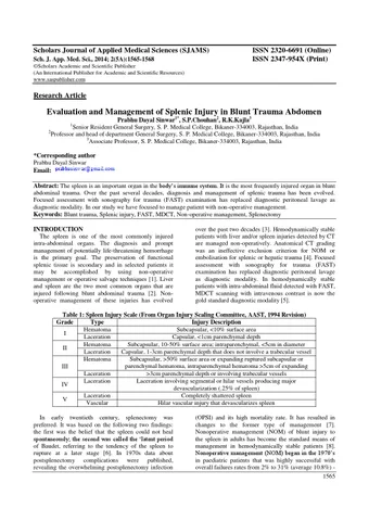
Table 1: Spleen Injury Scale (From Organ Injury Scaling Committee, AAST, 1994 Revision) Grade Type Injury Description Hematoma Subcapsular, <10% surface area I Laceration Capsular, <1cm parenchymal depth Hematoma Subcapsular, 10-50% surface area; intraparenchymal, <5cm in diameter II Laceration Capsular, 1-3cm parenchymal depth that does not involve a trabecular vessel Hematoma Subcapsular, >50% surface area or expanding ruptured subcapsular or parenchymal hematoma, intraparenchymal hematoma >5cm of expanding III Laceration >3cm parenchymal depth or involving trabecular vessels Laceration Laceration involving segmental or hilar vessels producing major IV devascularization (.25% of spleen) Laceration Completely shattered spleen V Vascular Hilar vascular injury that devascularizes spleen In early twentieth century, splenectomy was preferred. It was based on the following two findings: the first was the belief that the spleen could not heal spontaneously; the second was called the ‘latent period of Baudet, referring to the tendency of the spleen to rupture at a later stage [6]. In 1970s data about postsplenectomy complications were published, revealing the overwhelming postsplenectomy infection

(OPSI) and its high mortality rate. It has resulted in changes to the former type of management [7]. Nonoperative management (NOM) of blunt injury to the spleen in adults has become the standard means of management in hemodynamically stable patients [8]. Nonoperative management (NOM) began in the 1970’s in paediatric patients that was highly successful with overall failures rates from 2% to 31% (average 10.8%) 1565


Sinwar PD et al., Sch. J. App. Med. Sci., 2014; 2(5A):1565-1568 with the majority of failures occurring in the first 24 hours [9]. Sclafani first described the use of transcatheter arterial embolization in the treatment of splenic injuries. After that, it has been employed by many to achieve increased rates of splenic salvage [10]. Two primary techniques of splenic artery embolization have been described: proximal splenic artery embolization (PSAE) and superselective distal embolization [11].

The currently available modalities for evaluating the splenic injury after blunt trauma include  Physical examination,  Diagnostic peritoneal lavage,  Abdominal ultrasonography,  Plain X-ray,  Computerized tomography and Diagnostic laparoscopy.  Intra operative findings

NOM of splenic injury includes observation (OBS) or angiography and embolization (AE). OBS involves admission to a unit with monitoring of vital signs, strict bed rest, monitoring of red blood cell count with serial abdominal examinations [12, 13]. van der Viles reported that in recent years interest in the NOM of blunt traumatic injury has been increased because of the progress made in the quality and availability of the multidetector computed tomography (MDCT) scan and the development of minimally invasive intervention options such as angioembolization [5]. Fig. 1: Ligation at splenic pedicle Purpose of our study is the analysis mode and nature of injury, evaluation of severity related to clinical presentation, evaluation of various investigations and their role in finding extend of injury, identify the factors that influence the choice of treatment for spleen trauma and outcomes of different management modalities. MATERIALS AND METHOD This study was conducted on 24 cases in the Department of General Surgery, S.P. Medical College, Bikaner during January 2012 to December 2013. Inclusion criteria Patients of blunt trauma abdomen having splenic injury on clinical and various investigation procedures Exclusion criteria Patients with penetrating injury, major head injury, thorax injury, bone injury leading to hemodynamic unstability, age group <14 year.

Fig. 2: Removed splenectomy specimen with a large avascular area (>25%)

Table 2: Distribution and management of cases according to CT scan grading of injury Grade Management Total Conservative Operative No % No % No % I 6 35.3 0 6 25.0 II 8 47.1 0 8 33.3 III 3 17.6 4 57.1 7 29.2 IV 0 3 42.9 3 12.5 V 0 0 0 Total 17 100 7 100 24 100 Mean 1.82 3.43 SD 0.73 0.53 t 5.253 p <0.001 1566


Sinwar PD et al., Sch. J. App. Med. Sci., 2014; 2(5A):1565-1568 RESULTS AND DISCUSSION Most of splenic injury occur in <30yr age group patients with 62.5% of total patients. (86.66% patients in this age group managed successfully with conservative management and 13.33% patients require operative intervention.) Majority of the patients are male sex with 91.7%of total patients (68.18% of them managed conservatively and 31.81% required operative intervention.) Majority of patients belongs to rural residential area with 66.7% of total patients (68.75% of them managed successfully with conservative management and 31.25% patients required operative intervention.) Most common mode of injury is RTA constitutes 75% of total (77.8% of them managed successfully with conservative management and 22.2% patients required operative intervention.) Pain abdomen is most common clinical symptom observed in 100% patients followed by vomiting in 37.5% patients and respiratory distress in 25% patients. Guarding and rigidity indicate sever splenic injury high probability of operative intervention (p<0.001). Distension present in 41.7% patients and rebound tenderness present in 4.2% patients. Patient require operative intervention have high mean pulse rate (p=0.018 significant), low mean systolic blood pressure (p=0.029significant) ,low mean diastolic blood pressure(p=0.395 not significant) and high mean respiratory rate(p<0.001 significant). FQA have sensitivity of 41.7% in detection of splenic injury patients with significantly high predictive value for operative intervention (p<0.001). Sensitivity of USG/FAST in detecting hemoperitoneum and splenic injury is100% as compared to Rozycki GS et al. study [14] shows 90-93% sensitivity. CT scan of abdomen in detecting hemoperitoneum and splenic injury have sensitivity of 100%. Total non-operative managed patient is 70.83% and operative intervention done in 29.16% patients as compared to multi institutional EAST trial in which 61.5% managed nonoperatively with 10.8% failure rate [15]. Velmahos GC et al. [16] manage approx. 85% patients non-operatively with 838% failure rate. Table 3: Rate of operative intervention Grade Grade I Grade II Grade III Grade IV Grade V

In present study 0% 0% 57.14% 100% -

Multi-institutional study of EAST [16] 4.8% 9.5% 19.6% 33.3% 75%

Patients who have operative intervention have significantly high complication during hospital stay in form of wound infection, fever and cough. Mean duration of hospital stay is high 24.29 days (p<0.001significant) in operative patient in comparison to non operative management.

CONCLUSION From our prospective study it is concluded that young age group and male patients of rural population are commonly involved in splenic injury in BTA. Common Modes of injuries are RTA and patient usually present with pain abdomen. On initial clinical examination if patient have tachycardia, hypotension, tachypnea, guarding, rigidity and respiratory distress than it indicate severe splenic injury and high probability for operative intervention. Positive FQA and associated injury on x-ray (chest and FPA) also indicate underlying severe splenic injury. USG abdomen is initial investigation of choice than CT scan abdomen is best investigation to grade the injury. Higher grade (III, IV, V) on CT scan increase probability for operative intervention and associated morbidity and hospital stay in compare to lower grade (I, II). With increased use of splenic artery embolization rate of non operative managed patients can be increased. Also with further advance in diagnostic technique for splenic injury in blunt abdominal trauma in form of diagnostic and therapeutic laproscopy, rate of negative laprotomy can be decreased. REFERENCES 1. Maung AA, Kaplan LJ, Frankel HL, Collins KA; Management of splenic injury in the adult trauma patient. Available from http://www.uptodate.com/contents/managemen t-of-splenic-injury-in-the-adult-trauma-patient 2. Visrutaratna P, Na-Chiangmai W; Computed tomography of blunt abdominal trauma in children. Singapore Med J., 2008; 49 (4): 352– 358. 3. Koo TY, Ra YM, Lee SE, Choi IS, Yoon DS, Jo YJ et al.; Extension of nonoperative management on spleen injury with judicious selection and embolization; 10 years of experience. J Korean Surg Soc., 2011; 80: 5660. 4. Cohn SM, Arango JI, Myers JG; Computed tomography grading systems poorly predict the need for intervention after spleen and liver injuries. Am Surg., 2009; 75: 133-139. 5. van der Vlies CH, Olthof DC, Gaakeer M, Ponsen KJ, van Delden OM, Goslings JC; Changing patterns in diagnostic strategies and the treatment of blunt injury to solid abdominal organs. International Journal of Emergency Medicine, 2011; 4: 47. 6. Peitzman AB, Ford HR, Harbrecht BG, Potoka DA, Townsend RN; Injury to the spleen. Curr Probl Surg., 2001; 38: 932–1008. 7. Holdsworth RJ, Irving AD, Cuschieri A; Postsplenectomy sepsis and its mortality rate: actual versus perceived risks. Br J Surg., 1991; 78(9): 1031–1038.

1567


Sinwar PD et al., Sch. J. App. Med. Sci., 2014; 2(5A):1565-1568 8.

9.

10.

11.

12.

13.

14.

15.

16.

Ahmed N, Vernick JJ; Management of liver trauma in adults. J Emerg Trauma Shock, 2011; 4(1): 114–119. Beuran M, Gheju I, Venter MD, Marian RC, Smarandache R; Non-operative management of splenic trauma. J Med Life, 2012; 5(1): 4758. Sclafani SJ; The role of angiographic hemostasis in salvage of the injured spleen. Radiology, 1981; 141(3): 645-650. Raikhlin A, Baerlocher MO, Asch MR, Myers A; Imaging and transcatheter arterial embolization for traumatic splenic injuries: review of the literature. Can J Surg., 2008; 51(6): 464–472. Pachter HL, Guth AA, Hofstetter SR, Spencer FC; Changing patterns in the management of splenic trauma: the impact of nonoperative management. Ann Surg., 1998; 227(5): 708– 717. Rutledge R, Hunt JP, Lentz CW, Fakhry SM, Meyer AA, Baker CC et al.; A statewide, population-based time-series analysis of the increasing frequency of nonoperative management of abdominal solid organ injury. Ann Surg., 1995; 222(3): 311–322. Rozycki GS, Ochsner MG, Schmidt JA, Frankel HL, Davis TP, Wang D JA et al.; A prospective study of surgeon-performed ultrasound as the primary adjuvant modality for injured patient assessment. J Trauma, 1995; 39(3): 492–498. Peitzman AB, Heil B, Rivera L, Federle MB, Harbrecht BG, Clancy KD et al.; Blunt splenic injury in adults: Multi-institutional Study of the Eastern Association for the Surgery of Trauma. J Trauma. 2000; 49(2):177-187. Velmahos GC, Zacharias N, Emhoff TA; Management of the most severely injured spleen : a multicenter study of the Research Consortium of New England Centers for Trauma (ReCONECT). Arch Surg., 2010; 145(5): 456–460.

1568


Turn static files into dynamic content formats.

Create a flipbook
Issuu converts static files into: digital portfolios, online yearbooks, online catalogs, digital photo albums and more. Sign up and create your flipbook.