ISSN 2348-313X (Print)
International Journal of Life Sciences Research ISSN 2348-3148 (online)
Vol. 10, Issue 4, pp: (73-81), Month: October - December 2022, Available at: www.researchpublish.com

ISSN 2348-313X (Print)
International Journal of Life Sciences Research ISSN 2348-3148 (online)
Vol. 10, Issue 4, pp: (73-81), Month: October - December 2022, Available at: www.researchpublish.com
Aneeza Attique1, Uzma Hanif 1, Ghazala B. 2, Muzammal Abbas1 , Muhammad Fahad Shakeel1 , Ifra Aslam3, Misha Arshad4, Shumaila Rasheed1
Department of Botany Government College University Lahore, Pakistan 1 , Institute of Botany, University of the Punjab, Pakistan2 , Federal Medical College, Islamabad3, Pakistan, Sustainable Development Study Center, Government College University Lahore, Pakistan 4 DOI: https://doi.org/10.5281/zenodo.7466955
Published Date: 21-December-2022
Abstract: Free radicals interfere with the equilibriumof cells and tissues, which can lead to cancer. Fresh water algae such as Microspora tumidula and Lyngbya kützingii is a great source of secondary antioxidant metabolites. These metabolites most likely work well in the therapy of cancer. Algae exhibit huge variety of pigments not only chlorophyll, Carotenoids, phycobilins, and xanthophylls are the most prevalent of these. In the beginning, the medicinal effect of microalgae biomass was studied when it was used as pills, powder, and water additives. More and more studies in recent years have focused on finding and using useful medicinal components in algae. Aim of this study to evaluate the antioxidant role of algae in pharmaceutical industries. In the current investigation, the algal extracts were prepared by using three solvents methanol, chloroform and n-hexane to know about antioxidant potential of algae of specific area. To evaluate the antioxidant activity different test were performed such as DPPH, TAA, TPC, FRAP and MC. In Microspora tumidula 15.16% DPPH highest value was shown by methanolic extract. In FRAP Lyngbya kützingii showed maximum value in methanolic extract 64 µM Trolox mg-1 . While the highest value of TPC by Lyngbya kützingii was shown in chloroform extracts 37.5µg GAE /mg. The results of total antioxidant activity (TAA) were showed that Lyngbya kützingii and Microspora tumidula both exhibited the highest value 154mg /g and 152mg/g respectively in methanolic extract. The result of metal chelating test showed highest value in chloroform extract 10.44% by Lyngbya kützingii. So both these algal species showed antioxidant potential.
Keywords: Antioxidant potential, pharmaceutical industries, Evaluation, secondary metabolites, Cancer, Solvents extract.
Algae are photosynthetic and considered as special in nature because they show different phytometabolic contents and chemicals having various different structure and variety of biological functions [1]. Algae in addition to nutrient value also restrain various bioactive compounds like pigments (carotenoids and chlorophyll) amino-acids, phenolic compounds, mono and poly phenols [2]. These substances are vital in protecting algal cells from stressful and tension situations such as UV radiation, temperature changes, and nutrient rises and falls. In order to survive in these unfavorable conditions, they adapt by creating a large spectrum of secondary metabolites that are not seen in other species [3] A term generally used as substance that protects cells from the damage caused by free radicals (unstable molecules made by the process of oxidation during normal metabolism) However, synthetic antioxidant may have a negative effect on consumer’s health and have become limited or restricted in some countries. Because of its putative and detrimental actions, such as carcinogenesis, the usage of synthetic antioxidants has declined [4]
More recent studies have been planned to explore the useful products from algal compounds not only to check the nature of these products but also investigate some specific applications for humans use in different fields of interest. Now an alga is also considered as rich source of for market benefits. These metabolites gain the special attention in the production of new products for pharmaceutical, cosmetic and food industries. Free radicals cause disease by causing oxidative damage [5].
ISSN 2348-313X (Print)
International Journal of Life Sciences Research ISSN 2348-3148 (online)
Vol. 10, Issue 4, pp: (73-81), Month: October - December 2022, Available at: www.researchpublish.com
The discovery of natural substances with antioxidant capabilities is regarded as a major accomplishment [6]. Some interactions produce reactive oxygen species (ROS) such as (OH), superoxide (extremely reactive), and peroxyl (RO2) radicals. Then, through the process of oxidation, they cause damage to DNA molecules, proteins, and lipids, resulting in a variety of disorders in the human body [7]. Antioxidants play a vital role in slowing down the destructive effects of free radicals, reducing the risk of mutations, and so helping to stop cancer and heart syndrome [8]. There are three types of antioxidants: Primary antioxidants have a most important goal of preventing the creation of new free radicals. Enzymes and cytosol proteins are example. Secondary antioxidants are chemicals that have the ability to neutralize newly produced free radicals before they cause harm. Ascorbic acid, reduced gluthathione and uric acid are members of this group. Tertiary antioxidants help to restore cellular components that have been damaged by free radicals.
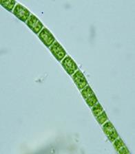
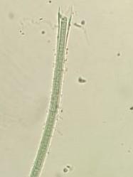
Lyngbya is unicellular autotrophs and provides the base for food chain. Long, unbranched filaments with a mucilaginous sheath cover Lyngbya species. Sheaths can form tangles or mats when mixed together with other phytoplankton species [9] Their filaments separate from one another and each cell produce a new filament. These mats can be found in both salt marshes and freshwater [10]. It has a lot of biodiversity and is rather safe, as evidenced by antioxidants and other activities. Lyngbya was found to be a good source of food, pharmaceuticals, and other industrial purposes [11].
Microspora is fresh water algae and belongs to division chlorophyta and most abundant present during dry season. They are autotrophic protists that are known for their many segments. Microspora species are green algae that are unbranched filamentous in nature [12], [13] Usually, a single dense net-like chloroplast fills the cell with no pyrenoid. Bulbous or barrel-shaped cells are the most common. The existence of an H-shaped wall section, which can typically be seen in the filament by focusing under a light microscope, is the most immediately distinguishing feature. This characteristic is most noticeable toward the filament's end.Besides this beings used as food source microspora species are believed to have many health benefits because they have many useful bioactive compounds such as antioxidants [14].
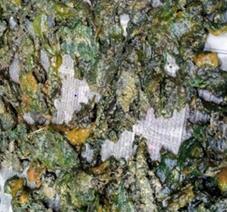
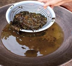
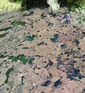
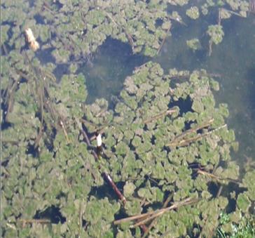
Algal samples were collected from tube well and fresh water pond in the village of Kasur during November, 2020. The samples werecollected andstored inplasticbags.Thenthealgae werebroughttothePhycologyLab,DepartmentofBotany, GCU Lahore. All samples were carefully washed with tap water to remove all debris. To remove extra water, the material was spread out on blotting paper. An alga was almost entirely dried in 3-4 days.
The small amount ofalgal sample was taken inplastic bottle contains water for preservation ofalgae. An alga was preserved for identification purpose. Later on slides were made and observed under light microscope to confirm its name.
ISSN 2348-313X (Print)
International Journal of Life Sciences Research ISSN 2348-3148 (online)
Vol. 10, Issue 4, pp: (73-81), Month: October - December 2022, Available at: www.researchpublish.com
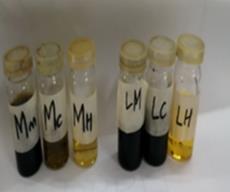
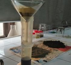
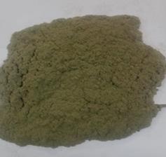
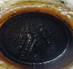
After dry, the collected samples were ground into a fine algae powder using a pestle and mortar or a grinder. Then, using a weighing balance, the algal powder was weighed; it was around 300g for Lyngbya kützingii and 350g for Microspora sp. This algal powder was placed in a 1000ml beaker with around 500 ml of n-Hexane. Cling film was used to properly cover it. This solvent was extracted in a 250 ml measuring flask using a funnel and Whatsmann filter paper no. 1 after 7 days. The flask was then placed in a rotating evaporator for roughly 2 hours, yielding pellets. For refine crude, this method was performed roughly three times. This crude extract was dried one more before being dipped in 500ml of chloroform. Cling film was used to properly cover it. The sample was extracted using Whatsmann filter paper no. 1 after 6-7 days. A rotary evaporator was used to get crude. Then, for one week, this algal sample was immersed in methanol. Cling film was used to properly cover it. The sample was extracted using Whatsmann filter paper no. 1 after 6-7 days. A rotary evaporator was used to get crude. The same method was used to obtain algae powder and crude for further research.
Using forceps, crude of methanolic extract (0.0075g) was placed in a petriplate, and a small amount of pure methanol was progressively added to it. The solvent was appropriately mixed until the algal crude was totally dissolved in it. This solvent was placed into a vial, the Petri plate was adequately rinsed, and the total volume was raised to 10ml with methanol. And it was properly covered with a lid. For the preparation of chloroform and n-hexane dilutions, the same process was used with crude from both species using these respective solvents. The dilutions were then kept at 200°C temperature
2.5.1.
Total antioxidant activity (TAA) is a chemical test that is often used to assess the antioxidant response to free radicals created as aresult ofvarious diseases [15]. Totalantioxidant activity was evaluated usingthe phosphomolybdenumcomplex production technique [16]. The reagent solution was prepared by mixing 2.47g ammonium molybdate and 5.32g sodium phosphate, raising the final volume to 100ml, then adding 16.7ml sulphuric acid and distilled water to raise the final volume to 500ml. Then, in a test tube, 1 ml of algal extract dilution was combined with 4 mL of the reagent solution. Only 4ml of reagent solution was taken as control. Cotton plug was used to properly cover it. Incubated for 90 minutes in a water bath at 950°C. At 695nm, the absorbance was measured. The total antioxidant activity is expressed as milligram, per gram of ascorbic acid, was calculated by the following equation derived from the calibration curve of ascorbic acid at different concentrations.
ISSN 2348-313X (Print)
International Journal of Life Sciences Research ISSN 2348-3148 (online)
Vol. 10, Issue 4, pp: (73-81), Month: October - December 2022, Available at: www.researchpublish.com
�� = ��+00328 00112
X= mg/ml of Ascorbic Acid
Y= Sample Absorbance
2.5.2.
The ability of a sample to convert ferric ion (Fe3+) TPTZ into ferrous ion (Fe2+) TPTZ at low pH, resulting in a blue color product, is determined by the FRAP capacity at 593nm, where it shows maximum absorbance [17]. The FRAP Assay was carried out according to Benzie and Strain's 1996 technique. Three compounds were required to make the FRAP reagent:
• 2.5 mL TPTZ solution (10 mM)
• 25 mL Acetate buffer (300 mM)
• 2.5 mL ferric chloride (20 mM)
The test tube was filled with 2.990l of FRAP reagent and 1.0l of algal extract solution. After 30 minutes in the dark, the sample was allowed to react with the FRAP reagent. In a spectrophotometer, the absorbance was measured at 593nm wavelength. By calculating with a standard calibration curve created for different concentrations of Trolox, the results were indicated in micromoles of Trolox Equivalents (TE) per ml of the sample. The following equation was used to compute FRAP values derived from standard calibration curve. �� = �� 0069 0002
X= Trolox Equivalent (µM/ml) Y= Sample Absorbance
The Folinciocalteu method is used inthe TPC assay. FC reagent contains Phosphomolybdic acid compound. The maximum absorbance of the reduced FC reagent was measured at 710nm using a spectrophotometer. The total phenolic content was determined according to following method [18]. In a test tube, 1ml of algal dilution was combined with 2.8ml of 10 percent Na2CO3and0.1mlof2NFolin-Ciocalteureagent(FC).For40minutes,the sample wasincubated at250°C.Theabsorbance was then measured in a spectrophotometer at 725nm. TPC was calculated using a standard calibration curve and expressed as milligrams of Gallic acid equivalent (GAE) per milliliter of sample. For varying amounts of Gallic acid, a standard calibration curve was created. The following equation, which was generated from a standard calibration curve, was used to calculate the TPC values that were stated in GAE g/ml. �� =0006X+0139
X= Total phenolic Content Value
Y= Sample Absorbance
2.5.4
The iron Ferrozine absorbance at 562nm was measured in the metal chelating test. The chelators have the ability to bind Fe2+. The red ferrous ion Ferrozine complex is generated in this test. If a chelators is present in the sample during complex formation, the red color fades significantly, indicating antioxidant potential [19]. Using a micropipette, 200 µl of Ferrozine solution and 50 µl of 2 mM ferrous sulphate were put to a test tube. It was filled with 100 liters of algal extract and methanol to bring the final volume up to 4 liters. Cotton plugs were used to cover all test tubes appropriately. The solution was then maintained at room temperature for 15 minutes before being tested for absorbance at 562nm. The following formula was used to calculate the percentage inhibition of Ferrozine ferrous complex formation:
%inhibition = Absorbance b Absorbance s Absorbance b ×100
ISSN 2348-313X (Print)
International Journal of Life Sciences Research ISSN 2348-3148 (online) Vol. 10, Issue 4, pp: (73-81), Month: October - December 2022, Available at: www.researchpublish.com
Ab = Absorbance of blank solution
As = Absorbance of the algal extract
2.5.5.
DPPH is a radical scavenger that is employed in a variety of samples. It has a purple color to it. At 517nm, it exhibits the highest absorption [20], [21]. DPPH free radical scavenging activity of different algal extracts was determined by modified form of Brand Williams et al., 1995. Different concentrations of fractions were mixed with 3ml of methanolic solution off DPPH (0.1mM). After shaking well the mixture then kept at room temperature for an hour to complete reaction. Blank sample was contained only methanol while control sample contained only DPPH solution. Absorbance was measured against methanol as a blank at 517nm. Each sample was run in three replicates. The percentage of DPPH discoloration of the sample was evaluated by following formula.
Absorbance c Absorbance s Absorbance c ×100
AC = Absorbance of control
AS = Absorbance of sample
3.1.
Graph 1 shows the DPPH radical scavenging activity of Lyngbya kützingii and Microspora performed on various solvent extracts. The maximum scavenging activity of Lyngbya kützingii shown chloroform extract at maximum concentration 1 is 3.738%. In case of Microspora the maximum scavenging activity is shown by methanolic extract 15.160% at 0.125 concentrations.
The methanolic extract of Lyngbya kützingii has highest potential 64±0.0026 µM Trolox/mg, followed by chloroform extract shows 42.5±0.00882 µM Trolox/mg while lowest FRAP activity is observed in n-hexane extract 29±0.008 µM Trolox/mg. Microspora sp. has highest potential 54.5±0.0020 µM Trolox/mg in methanol while lowest FRAP activity is observed in chloroform extract 34.5±0.0015 µM Trolox/mg
TABLE 1: Ferric Reducing Antioxidant Power assay of different solvent extracts of Lyngbya kützingii and Microspora tumidula
Lyngbya kützingii Solvents Mean Absorbance (593nm) FRAP (µM Trolox/ mg)
Methanol 0.197±0.002 64 n-hexane 0.127±0.0008 29 Chloroform 0.154±0.0008 42.5 Microspora tumidula
Methanol 0.178±0.0020 54.5 n-hexane 0.147±0.0005 39 Chloroform 0.138±0.0015 34.5
Lyngbya kützingii has antioxidant capacity against MC for methanol (10.010%), n-hexane (5.438%), and chloroform (10.443%). In comparison to methanol and n-hexane, chloroform extract showed the highest value, indicating that this solvent extract has a high iron bound capacity. Microspora sp. shown highest value in chloroform extract 19.91%, as compared to the n-hexane 16.950% and methanolic extract 9.142%.
ISSN 2348-313X (Print)
International Journal of Life Sciences Research ISSN 2348-3148 (online) Vol. 10, Issue 4, pp: (73-81), Month: October - December 2022, Available at: www.researchpublish.com
TABLE 2: Metal Chelating Activity (% inhibition) of different solvent extracts of Lyngbya kützingii and Microspora tumidula
Lyngbya kützingii
Solvents Mean Absorbance (562nm) Metal chelating inhibition (%)
Methanol 2.697±0.0394 10.010 n-hexane 2.834±0.0021 5.438 Chloroform 2.684±0.0093 10.443
Microspora tumidula
Methanol 2.723±0.0115 9.142 n-hexane 2.489±0.0105 16.950 Chloroform 2.40±0.0011 19.919
Lyngbya kützingii shows the highest value 154±0.02µg GA/mg in methanolic extract as compared to chloroform that is 62.57±0.0011µg GAE/mg. on the other hand the lowest value is observed in n-hexane extract 33.28±0.0005µg GAE/mg. In Microspora the methanolic extract shows the highest value 154±0.02µg GA/mg as compared to chloroform that is 62.57±0.0011µg GAE/mg.
200
150
100
50
Concentration Absorbance at 695nm
0
Methanol Chloroform n-hexane
Fig.3: Graphical representation of total antioxidant activity exhibited by extracts of algal species.
3.5. Total Phenolic Content (TPC):
The chloroform extract has the total phenolic potential 54µg GAE/mg whereas the n-hexane and methanolic extracts have TPC values of 8.77µg GAE/mg and 0.16µg GAE/mg, respectively. In Microspora n-hexane extract has the highest total phenolic potential 171µg GAE/mg and the lowest value is observed in chloroform extract 120µg GAE/mg.
50
40
30
20
10
Concentration Absorbance at 725nm
Lyngbya kützingii Microspora sp. 0
Lyngbya kützingii Microspora sp.
Methanol Chloroform n-hexane
Fig.4: Graphical representation of TPC (µg GAE/mg) of different solvent extracts of algal species.
ISSN 2348-313X (Print)
International Journal of Life Sciences Research ISSN 2348-3148 (online)
Vol. 10, Issue 4, pp: (73-81), Month: October - December 2022, Available at: www.researchpublish.com
Algae play an important and beneficial role in aquatic environment by producing oxygen and consuming carbon dioxide. Algal species have a high potential for food production and serve as the foundation for the food chain in fresh water, as well as acting as photosynthesizing organisms [22]. Algae have a lot of potential when it comes to producing some secondary metabolites which later act as antifungal, antibacterial, antiviral, antioxidant, and cytotoxic agents [23]. An alga is now widely used as a natural source of antioxidants.
The range of antioxidant activity of fresh water algae Microspora tumidula in different solvents (methanol, n-hexane and chloroform) against DPPH radical scavenging assay showed that methanolic extract exhibited highest % inhibition which was 15.16% while the lowest values showed by n-hexane extract 4.88%. This observation is in line with the earlier studies done on the solvent extraction system on the antioxidant potential of Microspora floccose by [24]. In their study the values of DPPH• varied from 19.7% in n-hexane extract to 9.3% (in methanol extract). On the other hand, Lyngbya kützingii also exhibited antioxidant activity against DPPH radical scavenging assay. The maximum value showed by chloroform extract 3.73% and lowest value was observed in methanolic extract which was 1.93%.
FRAP Assay of Lyngbya kützingii was done by using 3 different solvents extracts. The highest value was shown by methanolic extract 64 µM Trolox mg-1 followed by chloroform extract 42.9 µM Trolox mg-1. This observation was also done by Sea et al., 2019 on Lyngbya major Microspora tumidula showed maximum value in methanolic extract 54 µM Trolox mg-1 . Accordingto Bulent Microspora sp. extract in methanol showed 60.03 µM Trolox mg-1 value as compared to other solvent extracts [25].
Total phenolic content and extraction yield also depends upon chemical constituent’s solubility and polarity of extracting solvents. The highest value of TPC by Lyngbya kützingii was shown by chloroform extracts 37.5µg GAE /mg, and lowest value by methanolic extracts 6.835µg GAE /mg. According to seal Lyngbya major showed 15.89 µg GAE /mg. while the highest TPC value in Microspora tumidula was 10.5µg GAE /mg and 8.8 which was shown by n-hexane and methanolic extracts, respectively [26].
Total antioxidant activity was done by using the extractions of three solvent. The result showed that Lyngbya kützingii exhibited the highest value 154mg/gin methanol extract as compared to n-hexane and methanolic extract. While the highest value was observed in methanolic extract 152mg/g by Microspora tumidula. In the same way chloroform and n-hexane extract of this specie exhibited minimum value for this test.
The result of antioxidant activity against metal chelating by Lyngbya kützingii showed highest value in chloroform extract 10.44% and lowest value in n-hexane which was 5.43%. In same way Microspora tumidula also exhibited maximum value by chloroform extract 19.9% against metal chelating followed by methanolic extract and lowest value showed by n-hexane extract.
The goal of this present study is to determine the antioxidant potential of Microspora sp. and Lyngbya kützingii by using various solvent extracts. The chemical composition and biological potential indicated the existence of a variety of bioactive chemicals in various solvent extracts. In comparison to n-hexane and methanol, chloroform extract of Lyngbya kützingii has a high antioxidant potential. While methanolic extract has maximum potential in Microspora sp. algae. Macrolagae secondary metabolites and other bioactive chemicals may contribute in the treatment of a variety of diseases. So, according to results Microspora tumidula and Lyngbya kützingii both have high antioxidant potential due to the presence of bioactive compounds. As a result boththese species are apromisingsource ofnatural antioxidants and further used for pharmaceutical aspects in search of new drugs and medicines
[1] S. Singh, N. B. Kate and U. C. Banerjee. “Bioactive compounds from cyanobacteria and microalgae: an overview’’. Critic. Rev. biotech, Vol. 25, no. 3, pp. 73-95, 2005.
[2] S. Cox, N. A. Ghannam and S. Gupta. “An assessment of the antioxidant and antimicrobial activity of six species of edible Irish seaweeds.” Int. J. Food Res., Vol. 17, no. 1, pp. 205-220, 2010.
[3] A. Gonulol, E. Ersanli and O. Baytut. “Taxonomical and numerical comparisonof epipelic algae fromBalikand Uzun lagoon, Turkey”. J. Environ. Biol.,Vol. 30, pp. 777-784, 2009.
ISSN 2348-313X (Print)
International Journal of Life Sciences Research ISSN 2348-3148 (online) Vol. 10, Issue 4, pp: (73-81), Month: October - December 2022, Available at: www.researchpublish.com
[4] S. Maqsood, S. Benjakul, A. Abushelaibi, A. Alam. “Phenolic compounds and plant phenolic extracts as natural antioxidants in prevention of lipid oxidation in seafood: a detailed review.” Com. Rev. Food Sci. Food Saf.,Vol. 13, no. 6,pp. 1125-1140, 2014.
[5] M. Namiki . “Antioxidant or antimutagens in food” Critic. Rev. Food Sci. Nutr., Vol. 29, no 4, pp. 273-300, 1990.
[6] W. J.Craig . “Phytochemicals: guardians of our health”. J. American Diet. Assoc., Vol. 97, no.10, pp. S199-S204, 1997.
[7] G. C. Yen and H.Y. Chen . “Antioxidant activity of various tea extracts in relation to their antimutagenicity.” J. Agri. Food Chem.,Vol. 43, no. 1, pp. 27-32, 1995.
[8] X. Yan, Y. Chuda, M. Suzuki and T. Nagata T. “Fucoxanthin as the major antioxidant in Hijikia fusiformis, a common edible seaweed.” Biosci. Biotech. Biochem.,Vol. 63, no. 3, pp. 605-607,1999.
[9] M. L. Cornish and D. J. Garbary. “Antioxidants from macroalgae: potential applications in human health and nutrition.” Algae, Vol. 25, no. 4, pp. 155-171, 2010
[10] N. J. Turner and V.P. Aderkas. “The North American guide to common poisonous plants and mushrooms.” Timber Press, Vol. 115, pp. 403-406, 2009.
[11] G. C. Wang, X. G. Fei , J. M. Song, H.Y. Hu, Y. F. Yang and I. K. Chung. “Growth of Gracilaria lemaneiformis under different cultivation conditions and its effects on nutrient removal in Chinese coastal waters.” Aquaculture, Vol. 254, no. 1-4, pp. 248-255, 2006.
[12] A. Batista, M. Charles, J. Mancini-Filho and A. Vidal. “Las algas marinas como fuentes de fitofármacos antioxidants”. Rev. Cubana Plant Med., Vol. 14, pp. 1-18, 2009.
[13] M. Zubia, D. Robledo and Y. F. Pelegrin. “Antioxidant activities in tropical marine macroalgae from the Yucatan Peninsula, Mexico.” J. Appl. Phycol.,Vol. 19, no. 5, pp. 449-458, 2007.
[14] C. P.Rubio, J.Hernandez-Ruiz, S. Martinez-Subiela, A. Tvarijonaviciute and J.J.Ceron.“Spectrophotometric assays for total antioxidant capacity (TAC) in microslgae, an update.” BMC Vet. Res., Vol. 12, no. 166, pp. 1-7, 2016.
[15] P. Prieto, M. Pineda and M. Anguilar. “Spectrophotometric quantitation of antioxidant capacity through the formation of a Phosphomolybdenum Complex: Specific application to the determination of Vitamin E.” Anal. Biochem., Vol 269, pp. 337-341, 1999.
[16] M. Antolovich, P.D. Prenzler, E. Patsalides, S. McDonald and K. Robards. “Methods for testing antioxidant activity.” Analyst Vol. 127, no. 1, pp. 183-198, 2002.
[17] H. P. S. Makkar, M. Bluemme, N.K. Borowy and K. Becker. “Gravimetric determination of tannins and their correlations with chemical and protein precipitation methods.” J. Sci. Food. Agric., Vol. 61, pp. 161-165, 1993.
[18] J.P. Adjimani and P. Asare. “Antioxidant and free radical scavengingactivityofironchelators”. Toxicol. Rep., Vol. 2, pp. 721-728, 2015.
[19] K.Thaipong,U.Boonprakob,K.Crosby,L. Cisneros-ZevallosandD.H. Byrne. “ComparisonofABTS,DPPH,FRAP and ORAC assays for estimating antioxidant activity from guava fruit extracts”. J. Food Compos. Anal.,Vol. 19, pp. 669-675, 2006.
[20] A. M. Pisoschi, M. C. Cheregi and A. F. Danet. “Total antioxidant capacity of some commercial fruit juices: electrochemical and spectrophotometrical approaches”. Molecules, Vol. 1, pp. 480-493, 2009.
[21] K. Miyashita, M. Airanthi, K. Widjaja-Adhi, M. Abe M and Hosokawa. “ Algal carotenoids as potent antioxidants. Handbook of Marine Macroalgae” Biotech. Appl. Phyco., PP. 1-7, 2007
[22] R. Laungsuwon and W. Chulalaksananukul W. “Antioxidant and anticancer activities of freshwater green algae, Cladophora glomerata and Microspora floccosa, from Nan River in northern Thailand.” Maejo Int. J. Sci. Tech., Vol. 7, no. 2, pp. 181, 2013.
ISSN 2348-313X (Print)
International Journal of Life Sciences Research ISSN 2348-3148 (online) Vol. 10, Issue 4, pp: (73-81), Month: October - December 2022, Available at: www.researchpublish.com
[23] G. G. Satpati and R. Pal. “Biochemical composition and lipid characterization of marine green alga Ulva rigida- a nutritional approach.” J. Algal Biomass Util., Vol. 2, no. 4 pp.10-13, 2011.
[24] G. Sanger, L. J. Damongilala, B. E. Kaseger, L. Montolalu, L. K. Rarung. “Hytochemical constituents and antidiabetic activity of edible algae”. IOP Conf. Series: Earth Envi. Sci., Vol. 278, pp. 1-6, 2009.
[25] A. K. A. R. Bulent, A. K. A. R Zeynep and B. Sahin “Identification of Antioxidant Activity by Different Methods of a Freshwater Alga Microspora Sp. Collected From a High Mountain Lake.” Hittite Journal of Science and Engineering., Vol. 6, no. 1, pp. 25-29, 2019.
[26] T, Seal, N. Halder, K. Chaudhuri and S. N. Sinha. “Evaluation of antioxidant activities of algae and effect of solvent extraction system”. International Journal of Pharmaceutical Sciences and Research, Vol. 6, no. 3, pp. 1273, 2015.