ISSN 2348-313X (Print)
International Journal of Life Sciences Research ISSN 2348-3148 (online)
Vol. 10, Issue 2, pp: (44-54), Month: April - June 2022, Available at: www.researchpublish.com

ISSN 2348-313X (Print)
International Journal of Life Sciences Research ISSN 2348-3148 (online)
Vol. 10, Issue 2, pp: (44-54), Month: April - June 2022, Available at: www.researchpublish.com
1. Biomedical and Public Health Research Unit, Water Research Institute, CSIR, Ghana.
2. Department of Microbiology, Parasitology and Immunology, College of Veterinary and Medical Sciences, Sokoine University of Agriculture, Morogoro, Tanzania
DOI: https://doi.org/10.5281/zenodo.6637969
Published Date: 13-June-2022
Abstract: The existence of genetic substructure in E. coli was revealed by Whittam and colleagues (1983) and was later confirmed by Chaudhuri and Henderson (2012). This research is the first to investigate the various phylogroupsthatexistamongamongstAPECstrainsfromscavenginglocalchickeninMorogoro regionofTanzaniabased on the modern quadruplex method. All eight phylo-groups were detected among the APEC strains in varying percentages. Among the APEC isolates, 63.2%, 68.4%, 78.9%, and 73.7% were found to belong to phylogroups D and B2, C and F respectively, whiles isolates in groups A, B1 and E were 15.8%,10.5% and 5.3% respectively. There was a strong simultaneous occurrence of several iron acquisition genes with of structural gene for microcin iss thus (iss/iuCD), outer membrane protein(ompA) thus (ompA/sitEp). Iron acquisition genes and serum resistance genes recorded significant (P<0.05) differences between APEC and non-APEC strains and can thereby be used as markers for APEC isolates identification. The toxin and invasion genes, astA and ibeA which are normally recorded in minimal percentages were found to be significant determinants of APEC in the study. Funding: This work was supported by the intra ACP mobility scheme
Keywords: APEC, scavenging local chicken, revised phylo-grouping.
Escherichia coli are members of the natural microbiota of domestique animals, including chicken (Tenaillon et al., 2010) Although, these bacteria are usually harmless, but part of their population can become extraintestinal pathogenic E. coli (ExPEC). ExPEC have a fecal origin and occur asymptomatically in the intestinal tract. They can also colonize extraintestinal niches and cause serious diseases (Starcˇicˇ et al., 2015). Pathogenic strains of E. coli have been divided into intestinal (diarrheagenic; DEC) and extra-intestinal (ExPEC) pathogenic E. coli. Avian pathogenic E. coli (APEC), a subdivision of ExPEC that produces a systemic disease in poultry and also could serve as a potential zoonotic hazard to humans (Dziva and Stevens, 2008) APEC does not only colonize the intestinal tract of the chicken but also has the ability to disseminate systemically either through intestinal or respiratory mucosa (Leitner and Heller, 1992) The implementation of PCR methods to screen and identify the common virulence genes and phylogroups between different isolates is very critical (Moriel et al., 2010) Virulence genes (VGs) which causes pathogenicity are usually encoded on pathogenicity islands (PAIs), plasmids, and other mobile genetic elements, and hence can be transmitted via horizontal gene transfer (HGT) between various E. coli strains (Köhler and Dobrindt 2011)
ISSN 2348-313X (Print)
International Journal of Life Sciences Research ISSN 2348-3148 (online) Vol. 10, Issue 2, pp: (44-54), Month: April - June 2022, Available at: www.researchpublish.com
The population structure of E. coli is mainly clonal and strains can be classified into eight phylogenetic groups: A, B1, B2, C, D, E, F, and cryptic clade I (Clemont et al., 2013) Factors responsible for the phylogenetic structure of E. coli include: environment, gut anatomy, physiology, and diet. In animals, a critical force influencing the phylogenetic structure of the E. coli is the domestication status of the host (Escobar-Páramo et al., 2006). Domesticated animals have a lower proportion of B2 and A strains than their wild counterparts (Gordon and Cowling 2003).
The study area chosen for this research is the Morogoro municipality. The percentage of the populace that is engaged in livestock keeping and subsistence farming is 33% (Region and District Project Volume XII, 2011). A total of 400 swabs were collected from six different households within the Morogoro municipality. Specifically, oral-pharyngeal and cloacae swabs were collected from each of 200 scavenging local chicken. Half of these birds were kept semi-intensively whiles the remaining were on free-range. The swabs were collected and kept in transport media and transferred into the laboratory on ice
Isolation of E. coli and DNA extraction
Procedures used were as described in the Bacterial Analytical Manual (BAM 2007). The organisms were grown on MacConkey and Blood Agar media (OXOID, Hampshire, England) for the isolation.The following biochemical tests were performed to confirm the suspected isolates: Indole, Methyl Red, Voges-Paskeur, Citrate, Triple Sugar Iron and Motility test.
The positive Escherichia coli genomic DNAs were extracted by boiling. With modifications was necessary, rapid DNA extraction was done as protocol by Zhang et al. (2004).
Polymerase chain reaction
Virulence factor profiling to detect APEC strains
The positive E. coli strains, 192 in number, were investigated for various virulence genes by multiplex PCR, with protocol based on Ewers et al. (2007). The procedures were performed in 25µlreaction mixture. This includes: 12.5 µl of Taq polymerase (Dream Tag PCR Master mix, InqabaBiotec East Africa Ltd), 0.5 µl of each 100Mm dNTP, 0.1µl (100pmol) oligonucleotide primer pair, 6.9 µl of nuclease-free water and 4µl of template DNA. Primer concentration is 0.4 M. Conditions of the reaction mixtures include: 5mins at 95ºC initial denaturation,94ºC of denaturation for 30s, annealing at 56ºC for 30s, elongation at72ºC for 3minutes at 25 cycles, a final elongation at 72ºC for 10 minutes and a hold at 4ºC. List of primers used is shown in the appendix
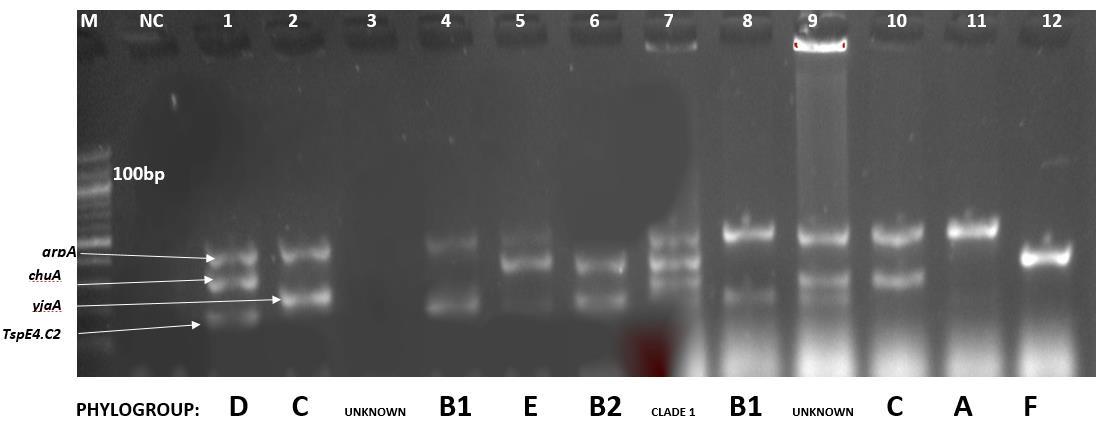
All E. coli strains, 192 in number, were screened for the presence of the genes in a quadriplex pcr reaction. The original triplex PCR method byClemont et al., (2000) had three primers: chuA, yjaAand TspE4.C2. This method was later modified by adding arpA targeted primer to act as an internal control for DNA quality and also to distinquish strains belonging to phylo-group F which was earlier misclassified as D. The protocol based on Clemont et al. (2013). The procedures were performed in 20µlreaction mixture. This includes: 10 µl of Taq polymerase (Dream Tag PCR Master mix, Inqaba Biotec East Africa Ltd),, 0.4µl (100pmol) oligonucleotide primer pair, 5.9 µl of nuclease-free water and 3µl of template DNA. Primer concentration is 0.4 M. Conditions of the reaction mixtures include: 4mins at 94ºC initial denaturation,94ºC of denaturation for 30s, annealing at 57ºC for 20s, elongation at72ºC for 3minutes at 30 cycles, a final elongation at 72ºC for 5 minutes and a hold at 4ºC. List of primers used is shown in table 1 The isolates whose genotype combinations give 2 possible phylotypes are run on pcr using specific primers. Thus the allele specific for phylo-group E and C are ArpAgpE.f/r and TrpAgpC.f/r respectively. The primer for internal control of E and C specific reaction is trypBA.f/r
The quadruplex genotypes combinations required for assigning phylo-groups are recorded in table 2.
ISSN 2348-313X (Print)
International Journal of Life Sciences Research ISSN 2348-3148 (online) Vol. 10, Issue 2, pp: (44-54), Month: April - June 2022, Available at: www.researchpublish.com
Table 1: Primer sequences and sizes of PCR products used in the extended quadruplex phylo-typing method.
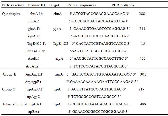
Table 2: Quadruplex genotypes required for assigning phylo-groups Quadruplex genotype
Quadruplex genotype Phylogroup
ArpA (400bp) ChuA (288bp) YjaA(211bp) TspE4.C2(152bp)
+ - - - A + - - + B1 - + - - F - + + - B2 - + + + B2 - + - + B2 + - + - A or C + + - - D or E + + - + D or E + + + - E or Clade 1 + - + - Unknown - - - - Unknown
+ Presence of the target amplicon; - Absence of the target amplicon. The isolates whose genotype combinations give 2 possible phylotypes are run on pcr using specific primers
Statistical analysis was done by use of Statistical Parkage for Social Sciences (SPSS) The presence of virulence genes among E.coli isolates were categorized as 1 = yes and 0 = no. Pearson’s correlation was used to access the frequency of occurrence of the various virulence genes. This frequency was rate from 0 to 1. The range of 1 to 0.1 was considered that highest rate of occurrence and given a color code of red, 0.099 to 0.001 was considered to be mild and given a pale pink
ISSN 2348-313X (Print)
International Journal of Life Sciences Research ISSN 2348-3148 (online)
Vol. 10, Issue 2, pp: (44-54), Month: April - June 2022, Available at: www.researchpublish.com
color code. The least occurrence rate was <0.001 and was assigned a white color. Proportions of various characteristics were tested by use of the chi-square test (x2). The threshold for statistical significance was indicated in the table with a P < 0.05 reflected statistical significance
After primary isolation of samples using Macconkey and blood agar media; 192, out of 400 samples were positive for Escherichia coli. The suspected positive Escherichia coli isolates were all confirmed in the biochemical tests. All suspected isolates, 192, were confirmed to be positive for E. coli. Virulence factor profiling revealed that all 192 E.coli isolates harbored at least of the 16 virulence genes. Also, 19 (9.8%) of them harbored at least 4 of the virulence genes and assigned as APEC isolates (Dziva and Stevens, 2008).
The remaining isolates, 173(90.2%), each containing less than 4 virulence genes were regarded as non-APEC.
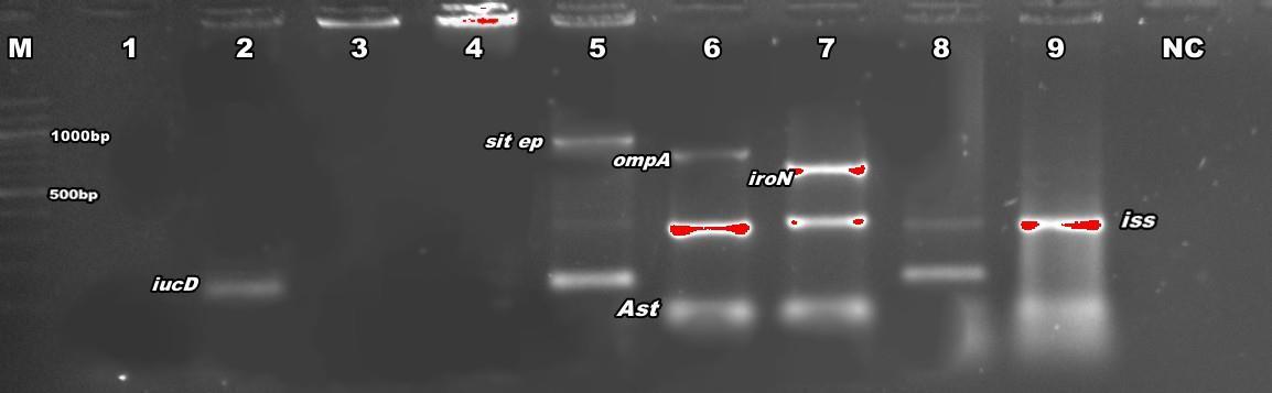
Figure 1: PCR detection of virulence genes sit ep(1032), omp(919)A, iroN(553), astA(116), iss(323) and iucD(269) NC is negative control. M is marker (100bp).
Out of the 16 virulence factors employed in this study, 12.5% of them were invasions (ibeA and gimB), 12.5% were adhesions (papC and tsh), 12.5% were toxins(EAST-1 and vat), 37.5% were for iron acquisition (chuA, OmpA, sit ep, iucD, iron and sit chr) and the remaining 25% were for serum resistance (iss, tra T, and cvi/cva), (Table 3).
The most prevalent virulence genes detected were tra T, iss and ibeA; these were found in 62.3%,78.9% and 84.2% among APEC isolates and 30.6%, 24.9% and 20.2% among non-APEC strains respectively. The least virulence genes were papC, cvi/cva, vat each recording 10.5%, 5.3% and 10.5% among APEC isolates and 0.6%, 1,2% and 0.6% among non-APEC isolates respectively (figure 2).
The prevalence in the virulence genes between APEC and non-APEC were compared; of the 16 genes, 7 of the recorded a significant difference (p<0.05). These are chuA, ibeA,traT, iroN, ompA, astA and irp2 (Table 3)
3:
among APEC and other ExPEC E.coli
ISSN 2348-313X (Print)
International Journal of Life Sciences Research ISSN 2348-3148 (online) Vol. 10, Issue 2, pp: (44-54), Month: April - June 2022, Available at: www.researchpublish.com
5(26.3) 2(1.2) 0.1006 1(5.3) 14(8.1) 0.0691 4(21.1) 2(1.2) 0.1068
Cvi/cva 1(5.3) 2(1.2) 0.2262 Iss Omp A 15(78.9) 43(24.9) 1 Tra T 4(21.1) 6(3.5) 0.0160* Adhesins Pap c 12(62.3) 53(30.6) 0.0001* Tsh Toxins Ast A 2(10.5) 1(0.6) 0.3714 Vat Invasins 2(10.5) 0(0) 9(47.4) 14(8.1) 0.0001* 2(10.5) 1(0.6) 0.3714
Gim B 1(5.3) 7(4) 0.1312 Ibe A 16(84.2) 35(20.2) 0.0001*
Figure 2: prevalence of virulence genes among APEC and non-APEC strains
ISSN 2348-313X (Print)
International Journal of Life Sciences Research ISSN 2348-3148 (online) Vol. 10, Issue 2, pp: (44-54), Month: April - June 2022, Available at: www.researchpublish.com
Figure 3: Statistical association between virulence genes of the E. coli isolates.
The statistical association between the various virulence genes were on figure 3. There were very strong associations were found between the genes ibeA and iroN, sitEp and ompA, OmpA and tra T, tra T and sit Ep, irop and vat and iss and iucD recording co-efficients of 1.05, 1.00, 0.206, 0.150, 0.172 and 0.119 respectively. Moderate associations, with co-efficient between 0.099 to 0.001 were observed among a number of pairs of virulence genes. These include chuA and iroN, gimB and traT and astA and iss with co-efficients of 0.094, 0.087 and 0.095 respectively. Weak associations, with coefficients≤0.001, occurred between the remaining virulence genes (figure 3)
Phylogroup profiling
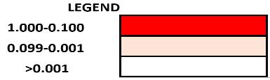
Figure 4: PCR detection of virulence genes arpA, chuA, yjaA and TspE4.C2 NC is negative control. M is marker (100bp). The various combination of these genes and their corresponding phylo-groups: A, B1,B2,C,D,E,F, and clade 1
ISSN 2348-313X (Print)
International Journal of Life Sciences Research ISSN 2348-3148 (online) Vol. 10, Issue 2, pp: (44-54), Month: April - June 2022, Available at: www.researchpublish.com
Table 4: Distribution of virulence genes in extended phylogenetic structure among APEC isolates.
Number of APEC isolates within phylogenetic groups
VG category A, 15.8% (n=3)
B1 10.5% (n=2)
B2 68.4% (n=13)
C 78.9% (n=15)
D 63.2% (n=12)
E 5.3% (n=1)
F 73.7% (n=14)
Clade 1 5.3% (1)
The 19 APEC isolates were screened with primers used in the quadruplex phylo-typing method listed in table 1 and their phylogenetic groups were determined base on the quadruplex genetypes profile described in table 2
Among the 8 phylogroups in the revised Clemont et al phylogrouping; group C recorded the highest number of APEC isolates, 78.9% and the least was clade 1 with 5.3% of the APEC isolates. Phylogroups F, B2, D, B1, E and A recorded 73.3%, 68.4%, 63.2%, 10.5%, 5.3% and 15.8% of the isolates respectively.
To the best of our knowledge, this research represents the very first time the revised Clemont phylo-genetic typing method have been used to examine the extended phylogenetic structure of E. coli amongst scavenging local chicken. Research of phylo-grouping among chicken have always targeted broilers The research thereby gave us a deeper understanding of the genetic make-up of this population as well as their relation with virulence genes and can serve as a baseline studies for more researches into the pathogenicity of E.coli in scavenging local chicken
Generally, our results showed that phylogroups D, B2, C and F are known to be associated with virulence strains whereas phylogroups A and B1 and E are known to be identified with commensal or less virulent strains Among the APEC isolates, 63.2%, 68.4%, 78.9%, and 73.7% were found to belong to phylogroups D and B2, C and F respectively, whiles isolates in groups A, B1 and E were 15.8%,10.5% and 5.3% respectively (table 4) Several similar researches carried out revealed that, the frequency of phylogroups do vary from different geographical regions (Gordon and Cowling, 2003, Coura et al., 2017). When Coura et al., 2017, investigated the phylo-groups in Tocantis state, central Brazil, they observed that phylo groups B2, C, D, E, and F are not common E. coli phylogroups isolated from poultry. Several reports from different geographical locations are consistent with their findings; Japan (Hiki et al., 2014), Iran (Bagheri et al., 2014) and Australia (Obeng et al., 2012). All of them stressed particularly that phylogroups B1 and A are the most common among poultry. However, contradictory reports were made from several researches at different locations In Egypt, Ramadan et al., 2016 revealed a significantly higher occurrence of phylogroups D and B2. Similar researches where phylo-group D dominated includes: Italy (Pasquali et al., 2015), Canada (Aslam et al., 2014), China (Wang et al., 2010). These studies indicated that most highly virulent ExPEC strains belong to either group B2 or D. These strains have been known to harbor more virulence strains than those of A and B1 (Cortes et al. 2010, and Ramadam et al., 2016).
In majority of the above researches, the assigning phylo-groups were based on the old triplet method of phylo-grouping, but interestingly, their results were consistent with ours. This lays lays credence to an assertion by Lougue et al., 2017; when the quadruplex system was applied, most isolates retain the same phylo-group they had in the old triplet method. In their study, Louge et al., (2017) observed that the expanded phylogenetic typing scheme was accurate and about 75% of isolates identified in the original typing scheme retained their original phylogroup.
The redistribution of the isolates in the new quadruplex system is associated with change in pathogenicity levels of the groups (Clemont et al., 2013, Logue et al., 2017). Phylo-group C is known to be originated from phylo group A, and phylogroup F from phylo group D Logue et al., (2017). Emerging phylo-groups are known to be associated with higher virulent strains than their original groups (Logue et al., 2017).In line with this, 78.9% of the APEC isolates belong to phylo group C whiles a relatively lower (15.8%) belong to group A. Same redistribution occurred between F and D F phylogroup habored more APEC isolates (73.7%) than the original D group (63.2%) (Table 4) This assertion was initially made by Logue et al., (2017) when they conducted an extended phylo-typing on human and avian ExPEC and commensal E. coli Several studies confirmed this assertion (Bok et al., 2020) In their study, Lougue et al., 2017 compared the previous and revised methods of phylo-grouping and realized that 75% of the isolates maintained their phylo-groups.
Horizontal transfer of the virulent genes occurs via mobile genetic elements; pathogencity island (PAI) and virulence plasmids. Analysis of association between the various virulence genes may identify which gene occur on the same genetic
ISSN 2348-313X (Print)
International Journal of Life Sciences Research ISSN 2348-3148 (online) Vol. 10, Issue 2, pp: (44-54), Month: April - June 2022, Available at: www.researchpublish.com
element (Bonnet et al., 2009) A study of phylogenetic group assignment by content of virulence, resistance, replicon and pathogenicity island genes in APEC reveals that insertion of pathogenicity islands into the genome appears to correlate closely with revised phylogenetic assignment (Johnson et al., 2006))
This research observed strong simultaneous occurrence of several iron acquisition genes with of structural gene for microcin iss thus (iss/iuCD), outer membrane protein(ompA) thus (ompA/sitEp) (figure 3). As observed by Bonnet et al., 2009 and Johnson et al., 2006, these are indicative of the presence of virulence plasmid pAPEC-O2-ColV Another plasmid of interested that showed its presence in this research is pAPEC-O2-R plasmid. It has been known to be associated with the complement resistance protein, tra T gene, and was originally isolated from an APEC strain (Johnson et al., 2005). In line with this, this gene was present in a higher percentage of APEC isolates than non-APEC.
Several virulence genes recorded significant (P<0.05) differences in their numbers between APEC and non-APEC strains. These can thereby be used as markers for APEC isolates identification. Among these, iron acquisition genes and serum resistance genes recorded the highest among isolates. Together, genes from these two groups were present in 80% of the isolates (table 3) Same conclusion was made by made by Paixao et al (2016), they observed that virulence genes fromthese two groups were most prevalent amongst APEC strains.
An important virulence determinant for APEC strains is the ability to survive and grow in serum, where the concentration of free iron is extremely low. This also plays a role in the pathogenesis of colibacillosis (Gao et al., 2012). Thus, serum resistance is a vital factor to APEC survival. The low concentration of iron in the serum forces APEC to synthesize ironuptake proteins which offers resistance to oxidative stress (Martinez et al., 2000; Janben et al., 2001). This study recorded significant differences between APEC and non-APEC isolates in the following iron-acquisition genes: chuA, iroN and irp2. Iron acquisition protects bacteria from host humoral immunity and the accumulation of these genes is a potential risk factor for APEC infection (Janben et al., 2001). The existence of several iron-related genes in APEC isolates is an indication that iron acquisition proteins play an important role in APEC pathogenicity, especially in sepsis-causing bacteria. Strong associations were identified between the genes within iron acquisition and serum resistance categories or between these two categories. The following pairs of genes; ompA and iss, iucD and iss, ompA and sitEp, ompA and traT and traT and sitEp are strongly associated, with co-efficient between 1.00 and 0.1 (figure 3) These genes usually occur together in the conserved virulence plasmidic (CVP) region, typical for the ExPEC virulence-associated plasmids. Earlier reports investigating the distribution of extra-intestinal pathogenic strains in these phylotypes confirmed this result. (Bonnet et al.2009 and Cortes et al 2010).
The toxin and invasion genes, astA and ibeA which are normally recorded in minimal percentages were found to be significant determinants of APEC in the study (figure 2) The astA and ibeA were significant (p<0.001, Table 3). All the literature reviewed and discussed in this study were of broilers because information of scavenging local chicken were very scant and virtually non-existent. We would then like to propose that there may be high correlation between APEC of SLC and toxins and invasion; a kind that doesn’t exist in broilers and other fowl. Since this is the first line of research on APEC phylo-groups of SLC, it would be our recommendation that more researches are carried out on Avian E. coli surveillance with SLC as it reference point since it is one of the most consumed chicken in Tanzania
The research represent the first time the revised Clemont phylo-grouping system has been applied on scavenging local chicken in Tanzania. Among the APEC isolates, 63.2%, 68.4%, 78.9%, and 73.7% were found to belong to phylogroups D and B2, C and F respectively, whiles isolates in groups A, B1 and E were 15.8%,10.5% and 5.3% respectively There was a strong simultaneous occurrence of several ironacquisition genes with of structural gene for microcin iss thus (iss/iuCD), outer membrane protein(ompA) thus (ompA/sitEp). Iron acquisition genes and serum resistance genes recorded significant (P<0.05) differences between APEC and non-APEC strains and can thereby be used as markers for APEC isolates identification The toxinand invasion genes, astA and ibeA which are normallyrecorded in minimal percentages were found to be significant determinants of APEC in the study.
Since this is the first line of research on APEC phylo-groups of SLC, it would be our recommendation that more researches are carried out on Avian E. coli surveillance with SLC as it reference point since it is one of the most consumed chicken in Tanzania
ISSN 2348-313X (Print) International Journal of Life Sciences Research ISSN 2348-3148 (online) Vol. 10, Issue 2, pp: (44-54), Month: April - June 2022, Available at: www.researchpublish.com
[1] Aslam M, M. Toufeer, C. Narvaez Bravo et al., “Characterization of Extraintestinal Pathogenic Escherichia coli isolated from retail poultry meats from Alberta, Canada,” International JournalofFoodMicrobiology,vol.177,pp.49–56,2014
[2] Bagheri M,R.Ghanbarpour,andH.Alizade,“Shigatoxinand beta-lactamases genes in Escherichia coli phylotypes isolated from carcasses of broiler chickens slaughtered in Iran,”International Journal of Food Microbiology,vol.177,pp.16–20,2014
[3] Bonnet C, Diarrassouba F, Brousseau R, Masson L, Topp E, Diarra MS (2009) Pathotype and antibiotic resistance gene distributions of Escherichia coli isolates from broiler chickens raised on antimicrobial-supplemented diets. Appl Environ Microbiol 75: 6955-6962.
[4] Campos TA, Lago JC, Nakazato G, Stehling EG, Brocchi M, Castro AFP, Silveira WD (2008) Occurrence of virulencerelated sequences and phylogenetic analysis of commensal and pathogenic avian Escherichia coli strains (APEC). Pesq Vet Bras 28: 533-540.
[5] Cortés P, Blanc V, Mora A, Dahbi G, Blanco JE, Blanco M, López C, Andreu A, Navarro F, Alonso MP, Bou G, Blanco J, Llagostera M (2010) Isolation and characterization of potentially pathogenic antimicrobial-resistant Escherichia coli strains from chicken and pig farms in Spain. Appl Environ Microbiol 76: 2799-2805
[6] Clermont O, Bonacorsi S, Bingen E (2000) Rapid and Simple Determination of the Escherichia coli Phylogenetic Group. Appl Environ Microbiol 66: 4555-4558
[7] Clermont, O.; Christenson, J.K.; Denamur, E.; Gordon, D.M. The Clermont Escherichia coli phylo-typing method revisited: Improvement of specificity and detection of new phylo-groups. Environ. Microbiol. Rep. 2013, 5, 58–65
[8] Chaudhuri,R.R.,and Henderson ,I.R.(2012)The evolution of the Escherichia coli phylogeny. Infect Genet Evol 12: 214– 226.
[9] Gao, Q., W. Wang, H. Xu, Y. Xu, J. Ling, D. Zhang, S. Gao, and X. Liu.2012.Rolesofironacquisitionsystemsin virulenceofextraintestinal pathogenic Escherichia coli: salmochelin and aerobactin contribute more to virulence than heme in a chicken infection model. BMC Microbiol. 12:143. doi:10.1186/1471-2180-12-143
[10] Ghanbarpour R,M.Sami,M.Salehi,andM.Ouromiei,“Phylogenetic background and virulence genes of Escherichia coli isolates from colisepticemic and healthy broiler chickens in Iran,”TropicalAnimalHealthandProduction,vol.43, no.1,pp. 153–157,2011.
[11] Gordon D M and A. Cowling, “The distribution and genetic structure of Escherichia coli in Australian vertebrates :host and geographic effects,”Microbiology,vol.149,no.12,pp.3575–3586, 2003.
[12] Hiki M, M. Usui, T. Akiyama et al., “Phylogenetic grouping, epidemiological typing, analysis of virulence genes, and antimicrobial susceptibility of Escherichia coli isolated from healthy broilersinJapan,”IrishVeterinaryJournal, vol.67,no.1,article 14,2014
[13] Dziva F, Stevens M (2008) Colibacillosis in poultry: unravelling the molecular basis of virulence of avian pathogenic Escherichia coli in their natural hosts. Avi Pathol 37: 355-366
[14] Escobar-Páramo, P.; Le Menac’h, A.; Le Gall, T.; Amorin, C.; Gouriou, S.; Picard, B.; Skurnik, D.; Denamur, E. Identification of forces shaping the commensal Escherichiacoli genetic structure by comparing animal and human isolates. Environ Microbiol. 2006, 8, 1975–1984.
[15] Gordon,D.M.;Cowling,A.ThedistributionandgeneticstructureofEscherichiacoliinAustralianvertebrates:Hostand geographic effects. Microbiology 2003, 149, 3575–3586.
[16] Janben, T., C. Schwarz, P. Preikschat, M. Voss, H. C. Philipp, and L. H. Wieler. 2001. Virulence-assocated genes in avian pathogenic Escherichia coli (APEC) isolated from internal organs of poultry having died from colibacillosis. Int. J. Med. Microbiol. 291:371– 378.
ISSN 2348-313X (Print) International Journal of Life Sciences Research ISSN 2348-3148 (online) Vol. 10, Issue 2, pp: (44-54), Month: April - June 2022, Available at: www.researchpublish.com
[17] Johnson,J.R., A. C. Murray, A.Gajewski,M. Sullivan,P.Snippes,M. A.Kuskowski,and K.E.Smith. 2003.Isolation and molecular characterization of nalidixic acid-resistant extraintestinal pathogenic Escherichia coli from retail chicken products. Antimicrob. Agents Chemother. 47:2161–2168.
[18] Johnson, T. J., K. E. Siek, S. J. Johnson, and L. K. Nolan. 2005. DNA sequence and comparative genomics of pAPECO2-R, an avian pathogenic Escherichia coli transmissible R plasmid. Antimicrob. Chemother. 49:4681– 4688.
[19] Johnson, T. J., K. E. Siek, S. J. Johnson, and L. K. Nolan. 2006. DNA sequence of a colV plasmid and prevalence of selected plasmid-encoded virulence genes among avian Escherichia coli strains. J. Bacteriol. 188:745– 758
[20] Köhler, C.D.; Dobrindt, U. What defines extraintestinal pathogenic Escherichia coli? Int. J. Med. Microbiol. 2011, 301, 642–647
[21] Leitner G, Heller ED (1992) Colonisation of Escherichia coli in young turkeys and chickens. Avi Dis 36: 211-220
[22] Logue, C.M.; Wannemuehler, Y.; Nicholson, B.A.; Doetkott, C.; Barbieri, N.L.; Nolan, L.K. Comparative analysis of phylogenetic assignment of human and avian ExPEC and fecal commensal Escherichia coli using the (previous and revised) Clermont phylogenetic typing methods and its impact on avian pathogenic Escherichia coli (APEC) classification. Front. Microbiol.
[23] Martinez, J. J., M. A. Mulvey, J. D. Schilling, J. S. Pinkner, and S. J. Hulltgren. 2000. Type 1 pilus-mediated bacterial invasion of bladder epithelial cells. EMBO J. 19:2803–2812.
[24] Moriel DG, Bertoldi I, Spagnuolo A, Marchi S, Rosini R, Nesta B, Pastorello I, Corea VA, Torricelli G, Cartocci E, Savino S, Scarselli M, Dobrindt U, Hacker J, Tettelin H, Tallon LJ, Sullivan S, Wieler LH, Ewers C, Pickard D, Dougan G, Fontana MR, Rappuoli R, Pizza M, Serino L (2010) Identification of protective and broadly conserved vaccine antigens from the genome of extraintestinal pathogenic Escherichia coli. Proc Natl Acad Sci USA 107: 90729077.
[25] ObengAS,H.Rickard,O.Ndi,M.Sexton,andM.Barton,“Antibioticresistance,phylogeneticgroupingandvirulence potential of Escherichia coli isolated from the faeces of intensively farmed and free range poultry ”VeterinaryMicrobiology, vol.154,no.3-4,pp.305–315,201
[26] Pasquali F, A. Lucchi, S. Braggio et al., “Genetic diversity of Escherichia coli isolates of animal and environmental origins from an integrated poultry production chain,”VeterinaryMicrobiology,vol.178,no.3-4,pp.230–237,2015
[27] Starcˇicˇ, E.M.; Žgur-Bertok, D. Virulence potential for extraintestinal infections among commensal Escherichia coli isolated from healthy humans The Trojan horse within our gut. FEMS Microbiol. Lett. 2015, 362, fnu061
[28] Tenaillon, O.; Skurnik, D.; Picard, B.; Denamur, E. The population genetics of commensal Escherichia coli. Nat. Rev. Microbiol. 2010, 8, 207–217
[29] V.L.Koga,G.R.Rodrigues,S.Scandorieiro et al.,“Evaluation of the antibiotic resistance and virulence of Escherichia coli strains isolated from chicken carcasses in 2007 and 2013 from Paran´ a, Brazil,”Food borne Pathogens and Disease,vol.12,no.6,pp.479– 485,2015.
[30] Wang X M, X.-P. Liao, W.-J. Zhang et al., “Prevalence of serogroups, virulence genotypes, antimicrobial resistance, and phylogenetic background of avian pathogenic escherichia coli insouthofChina,”FoodbornePathogensandDisease, vol.7,no. 9,pp.1099–1106,2010.
[31] Whittam, T.S., Ochman, H., and Selander, R.K. (1983) Geographic components of linkage disequilibrium in natural populations of Escherichia coli. Mol Biol Evol 1: 67–83
ISSN 2348-313X (Print)
International Journal of Life Sciences Research ISSN 2348-3148 (online) Vol. 10, Issue 2, pp: (44-54), Month: April - June 2022, Available at: www.researchpublish.com
Table 1: List of primers used for virulence genes identification of APEC strains
Virulence factor Target Primer sequences PCR product
Iron Aquisition Chu A(278) s: GACGAACCAACGGTCAGGAT a:TGCCGCCAGTACCAAAGACA iro N 553 s: ATCCTCTGGTCGCTAACTG a:CTGCACTGGAAGAACTGTTCT irp 2 (413) s: AAGGATTCGCTGTTACCGGAC a:TCGTCGGGCAGCGTTTCTTCT IucD(269) s:ACAAAAAGTTCTATCGCTTCC a:CCTGATCCAGATGATGCTC sit Chr(553) s: ACTCCCATACACAGGATCTG a: CTGTCTGTGTCCGGAATGA sit ep(1032) s: TTGAGAACGACAGCGACTTC a: CTATCGAGCAGGTGAGGA
Serum resistance cvi/cva (1181) s: TCCAAGCGGACCCCTTATAG a: CGCAGCATAGTTCCATGCT Iss (323) s: ATCACATAGGATTCTGCCG a:CAGCGGAGTATAGATGCCA OmpA(918) s: AGCTATCGCGATTGCAGTG a: GGTGTTGCCAGTAACCGG tra T(430) s: GTGGTGCGATGAGCACAG a:TAGTTCACATCTTCCACCATCG Adhesins pap C (501/328) s: TGATATCACGCAGTCAGTAGC a:CCGGCCATATTCACATAAC Tsh (824) s:ACTATTCTCTGCAGGAAGTC a:CTTCCGATGTTCTGAACGT Toxins ast A (116) s: TGCCATCAACACAGTATATCC a:TAGGATCCTCAGGTCGCGAGTGACG C
Vat(980) s: TCCTGGGACATAATGGCTAG a: GTGTCAGAACGGAATTGTC Invasins GimB (736) s: TCCAGATTGAGCATATCCC a:CCTGTAACATGTTGGCTTCA ibe A(341) s: TGGAACCCGCTCGTAATATAC a:CTGCCTGTTCAAGCATTGCA