ISSN 2348-313X (Print)
International Journal of Life Sciences Research ISSN 2348-3148 (online) Vol. 10, Issue 4, pp: (92-96), Month: October - December 2022, Available at: www.researchpublish.com

ISSN 2348-313X (Print)
International Journal of Life Sciences Research ISSN 2348-3148 (online) Vol. 10, Issue 4, pp: (92-96), Month: October - December 2022, Available at: www.researchpublish.com
FAHAD ABDULAZIZ ALKHARAAN1*, NAWAF TURKY ALENIZI2 , MOHAMMED HADI ALHABABI3, NORA MUNAHI ALMUTAIRI4 , Abdullah Abdulrzaq Aljuwayi5
1 * Corresponding Author: HEALTH ADMINISTRATION SPECIALIST, GENERAL DIRICTORATE OF HEALTH AFFAIRS IN RIYADH REGION, Riyadh, SA
2 HEALTH INFORMATIC SPECIALIST, GENERAL DIRICTORATE OF HEALTH AFFAIRS IN RIYADH REGION, Riyadh, SA
3
RADIOLOGY TECHNICIAN, GENERAL DIRICTORATE OF HEALTH AFFAIRS IN RIYADH REGION, Riyadh, SA
4 NURSE TECHNICIAN, GENERAL DIRICTORATE OF HEALTH AFFAIRS IN RIYADH REGION, Riyadh, SA 5 Medical technologist II, KFMC, Riyadh, SA
DOI: https://doi.org/10.5281/zenodo.7488592
Published Date: 28-December-2022
Abstract: Serum 25-hydroxyvitamin D3 [25(OH)D3] concentrations are currently recognized as the functional status indicator for vitamin D. Evidence is reviewed that shows that serum 25(OH)D3 concentrations of < 80 nmol/L are associated with reduced calcium absorption, osteoporosis, and increased fracture risk. For typical older individuals, supplemental oral intakes of ~1300 IU/d are required toreach the lower end of the optimal range. Evidence of substantial problems in routine clinical measurement of serum 25(OH)D3 concentrations among patients is cited. There is great need for standardization and improved reproducibility and sensitivity of measurements of serum 25(OH)D3 concentrations.
Keywords: Rickets, Osteomalacia, Osteoporosis, Calcium absorption, Fractures, Serum 25-hydroxyvitamin D3
Initsrecent reviewofrecommended intakes ofbone-related nutrients, the Food and NutritionBoard (FNB) identified serum concentrations of 25-hydroxyvitamin D3 [25(OH)D3] as a suitable functional indicator of vitamin D status (1) . However, based on the evidence available at the time, the FNB Panel on Calcium and Related Nutrients was unable to associate specific serum 25(OH)D3 concentrations with various health and disease states. The panel report also recognized that solar vitaminD synthesisinthe skinisanimportant sourceofvitaminD,butthedataavailableat the timedidnotallowestimation of the usual or optimal ratios of intakes from dermal and ingested sources. p> However, the increase in intake recommendations from 200 IU/day (5 µg/day) before age 50 to 600 IU/day (15 µg/day) at age ≥70 years reflects the realization that the contribution from cutaneous sources decreases with age. In addition, in the absence of the required information, the FNB again used the absence of rickets and osteomalacia as de facto indicators of vitamin D adequate intake. Other health or disease-related consequences were not considered in the vitamin D intake recommendations.
Although much additional work remains to be done, enough information has been developed over the past 8 years to fill some of the information gaps faced by the Panel on Calcium and Related Nutrients in its deliberations in the mid 1990's This brief overview highlights certain aspects of this new information.
ISSN 2348-313X (Print)
International Journal of Life Sciences Research ISSN 2348-3148 (online)
Vol. 10, Issue 4, pp: (92-96), Month: October - December 2022, Available at: www.researchpublish.com
Although rickets (in children) and osteomalacia (in adults) have long been considered index diseases of vitamin D deficiency, there is a growing belief that milder degrees of deficiency can also cause skeletal disorders. Vitamin D's canonical function is to facilitate the active transport component of intestinal calcium absorption, and there has never been any evidence that absorption is optimal at vitamin D concentrations just sufficient to prevent thisrickets or osteomalacia. In 1990, based on his extensive experience with histomorphometric analysis of adult bone specimens, Parfitt (2) introduced a heuristically important reconceptualization of bone diseases due to vitamin D deficiency, for which he coined the term hypovitaminotic osteopathy D. He identified 3 stages of the disease associated with increasing levels of vitamin D deficiency. In stage 1, the only detectable pathophysiological change was reduced intestinal calcium absorption , with the consequent decrease in skeletal calcium stores and associated osteoporosis.
On biopsies, stage 1 bone showed no evidence of osteomalacia. In stage 2 hypovitaminosis D, as in stage 1, there was reduced intestinal calcium absorption and reduced bone mass, but early osteomalacia was identifiable on biopsy, ie increased bone coverage by the osteoid and reduced rate of mineral apposition.
atients with stage2 disease had no clinical or laboratoryevidence ofosteomalacia. Its onlyclinical manifestationwas reduced bone mass, ie osteoporosis. In stage 3 hypovitaminosis D, there was persistent calcium hypoabsorption and clinically evident osteomalacia. biochemical and histological. The significance of this redesign is that the traditional index disease for vitamin D deficiency has been clearly delineated as representing only the most extreme degree of deficiency out of Lower degrees have been suggested to produce osteoporosis, which is silent until fracture occurs, as has long been recognized. Therefore, the presence of osteoporosis and its association with vitamin D status would have gone undetected. 25(OH)D3 of the patients who provided biopsy samples for analysis, Parfitt (2) was unable to quantitatively relate his 3 stages to specific values for what the FNB would later refer to as functional indicator . Parfitt's work made it clear that the then recommended daily dose for adults (200 IU/day), which was barely sufficient to prevent clinical osteomalacia, was insufficient to prevent stage 1 or 2 vitamin D deficiency osteopathy to protect.
Only now is it possible, at least tentatively, to assign specific serum 25(OH)D3 concentrations to the boundary between disease stage 1 and the normal state, and to estimate the vitamin D intake required to achieve such concentrations in this delineation of normal and deficient concentrations, it should be noted that a growing body of evidence summarized in other reports of this symposium points to a role for vitamin D not only in calcium metabolism but also in a variety of muscleand/ormuscleand/orvitaminDlevelsareindicativeofneuromuscularfunctionsandinthecontrolofcellproliferation and differentiation (with implications for oncogenesis). these limits. Bischoff et al. (3), in a recent analysis of data from the National Health and Nutrition Examination Survey, showed that lower extremity muscle function improved with increased serum concentrations of 25(OH)D3 , at least at levels in the 80-100 nmol/L range .
In addition, there is at least one prospective study linking prostate cancer risk to serum 25(OH)D3 concentrations, showing an inverse risk within the range of commonly observed serum concentrations for 25(OH)D3 (4). Estimates of the optimal 25(OH)D3 serum concentrations for such health outcomes may soon be available.
It is generally accepted that serum 25(OH)D3 concentrations of < 20 nmol/L is associated with clinical osteomalacia in adults. Most laboratory reference ranges, on the other hand, range from the lower limits of 37.5 or 40 nmol/L to just over 100 or 120 nmol/L. The range between 20 nmol/L (threshold for rickets/osteomalacia) and the lower end of the reference range is commonly referred to as vitamin D deficiency, in recognition of its suspected insufficiency for optimal functioning of the
Savings of vitamin D and calcium. (The avoidance of the term deficiency for values in this range reflects the generally implicit but common premise in nutritional science that inadequate intake of any nutrient causes only disease; therefore, patients who do not have osteomalacia cannot be "deficient." .) The expected physiological response to inadequate calcium absorption (due to either decreased vitamin D status or low calcium intake) is increased activity of the parathyroid hormone (PTH)-calcitriol-axis. Many studies reported the expected inverse association between serum 25(OH)D3 levels and serum PTH levels (5–7).
In most of these analyses, PTH concentrations tended to nadir at serum 25(OH)D3 concentrations of 70-110 nmol/L. Only the data from Lips et al. (8) showed a value belowthis range. Elevated PTH levels are indicative of a physiological response to calcium deficiency and could therefore be considered an appropriate response to a physiological stressor rather than an indicator of deficiency (9). However, PTH is the main determinant of bone remodeling,
ISSN 2348-313X (Print)
International Journal of Life Sciences Research ISSN 2348-3148 (online) Vol. 10, Issue 4, pp: (92-96), Month: October - December 2022, Available at: www.researchpublish.com
Figure 1. Calcium absorption fraction as a function of serum 25(OH)D3 concentrations, from3published reports [☐,study by Bischoff etal (15); E, study by Heaney et al (13); ‚, study by Barger-Lux et al (14)]. Error bars indicate 1 SEM.
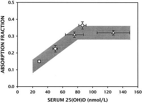
and it is now clear that a high remodeling rate is an important and perhaps the main determinant of osteoporotic bone fragility (10-12). Therefore, it can hardly be considered a benign condition. Concentrations of 25(OH)D3 on the role of calciumabsorptionhave recentlyemerged. Heaneyet al. (13)and Barger-Luxand Heaney(14),in2 complementarystudies, showed that fractional calcium absorption increased with 25(OH)D3 serum concentrations within the reference range up to ~80 nmol/L and stabilized above this level. These studies have shown that the reference range should not be taken as an indication of the physiological normality of the measured results
Values Bischoff et al. (15, 16), in a paper linking vitamin D status to falling propensity, provided data suggesting even lower absorption in people with 25(OH)D3 serum concentrations below the target reference range Clues. Figure 1 presents the data from these 3 studies and suggests an apparent threshold response, with absorption efficiency being maximized at concentrations of ~80 nmol/L or higher. Such physiological evidence, while strongly suggestive, proves no association with morbidity. The publication of a large UK vitamin D intervention study in 2003 provided crucial evidence needed.
Using a randomized, placebo-controlled design, Trivedi et al. (17) 100,000 IU of vitamin D3 every 4 months (average ~800 IU/day) for 5 years to 2,686 healthy UK participants aged 65-85 years of age. age. Serum 25(OH)D3 concentrations were measured for a subgroup of the cohort and averaged 53 nmol/L for the placebo group and 74 nmol/L for the vitamin Dtreated group. The risk of all fractures was reduced by 22% among the individuals reduced supplement treated and typical osteoporotic fractures, considered group , reduced by 33%. study were within the reference range; In fact, the two studies covered almost the same range of values (50 and 53 nmol/L).for untreated subjects in the 2 studies and 74 and 86 nmol/L for treated subjects).
Figure 2. Suggested mapping of the principal vitamin D-related bonediseases onto the serum 25(OH)D3 concentration continuum. (To convert values to nanograms per milliliter, divide values by 2.5.)
These recently published studies clearly demonstrate calcium malabsorption and an increased risk of fracture at serum 25(OH)D3 concentrations below ~80 nmol/L. 1 and 2, and provide quantitative references not available to Parfitt when he proposed his classification scheme for vitamin D-related diseases. Furthermore, these findings underscore the worrying implications of elevated PTH levels at 25(OH)D3 levels. 80 nmol/L. A preliminary assignment of bone disease in adults to serum 25(OH)D3 concentrations is shown in Figure 2

ISSN 2348-313X (Print)
International Journal of Life Sciences Research ISSN 2348-3148 (online) Vol. 10, Issue 4, pp: (92-96), Month: October - December 2022, Available at: www.researchpublish.com
It is general experience in the art that administration of vitamin D in amounts in the range of current appropriate intakes (defined by the FNB as 200-600 IU/d) does not produce a significant increase in The amount measured causes serum 25(OH)D3 concentrations, indicating insufficient efficacy of the preparations used or a greater need than implied by the concept of adequate intake. Therefore, my colleagues and I (18) attempted to quantify both daily vitamin D utilization and the amount required to achieve a desired increase in 25(OH)D3 serum concentrations. Vitamin D approached 4000 IU (100 µg) and that at steady state 25(OH)D3 serum concentrations increased by 0.7 nmol/L per 1 µg (40 IU) of vitamin D3 taken orally as a normal daily dose. Several other studies provided data that allowed this rate of increase to be calculated; they generally gave similar slope values, ie between 0.6 and 1.2 nmol/L per 1 µg/d (17, 19, 20). The Trivedi et al. (17) showed an increase of almost exactly 1 nmol/L per 1 µg/day.
By taking a value in the middle of the observed slope range (e.g. 0.9 nmol/L per 1 µg/day), it can be calculated that the recommended daily dose for adults aged 50 to 70 years ( 400 IU) would only increase the serum 25(OH)D3 concentration by 9 nmol/l (3.6 ng/ml). Since this increase is within error for most laboratory methods, it is now clear why administration of such doses does not produce appreciable increases in serum 25(OH)D3 concentration.
There is reasonto believe that the rate ofincrease maybe muchfaster in more fatigued people than inthose who participated in our study or the UK study, and my colleagues and I (20) have previously reviewed several studies published indicated that the response to a given dose may well be an inverse function of the initial 25(OH)D3 concentration. However, once modest vitamin D replacement is achieved, a slope in the range just mentioned appears to apply and determines the amount of vitamin D that must ultimately be administered to achieve the desired levels.
It is general experience in the art that administration of vitamin D in amounts in the range of current appropriate intakes (defined by the FNB as 200-600 IU/d) does not produce a significant increase in The amount measured causes serum 25(OH)D3 concentrations, indicating insufficient efficacy of the preparations used or a greater need than implied by the concept of adequate intake. Therefore, my colleagues and I (18) attempted to quantify both daily vitamin D utilization and the amount required to achieve a desired increase in 25(OH)D3 serum concentrations. Vitamin D approached 4000 IU (100 µg) and that at steady state 25(OH)D3 serum concentrations increased by 0.7 nmol/L per 1 µg (40 IU) of vitamin D3 taken orally as a normal daily dose. Several other studies provided data that allowed this rate of increase to be calculated; they generally gave similar slope values, ie between 0.6 and 1.2 nmol/L per 1 µg/d (17, 19, 20).
The Trivedi et al. (17) performed anti-fracture test showed an increase of almost exactly 1 nmol/L per 1 µg/d. Taking a value in the middle of the observed range of slopes (e.g. 0.9 nmol/l per 1 µg/d), it can be calculated that the recommended daily dose for adults aged 50 to 70 years is expected to be reached by the age of ( 400 IU) increases the serum 25(OH)D3 concentration by only 9 nmol/L (3.6 ng/mL) .
Since this increase is within the error of most laboratory methods, it is now clear why administration of such doses does not elicitappreciableincreasesinserum25(OH)D3concentrations.Thereisreasontobelieve thattherate ofincrease insubjects with severe fatigue may be much faster than in participants in our study or the UK study, and my colleagues and I (20) have previously published reviews of several studies suggesting this that the response to a given dose of may well be an inverse function of the initial 25(OH)D3 concentration. However, once modest vitamin D replacement is achieved, a slope in the range just mentioned seems to apply and governs the amount of vitamin D that must ultimately be administered to achieve the desired level. values.
[1] Food and Nutrition Board, Institute of Medicine. Dietary reference in-takes for calcium, magnesium, phosphorus, vitamin D, and fluoride. Washington, DC: National Academy Press, 1997.
[2] Parfitt AM. Osteomalacia and related disorders. In: Avioli LV, Krane SM, eds.Metabolicbone disease and clinically relateddisorders.2nded.Philadelphia: WB Saunders, 1990:329–96.
[3] Bischoff-Ferrari H, Dietrich T, Orav EJ, et al. Higher 25-hydroxy- vitamin D concentrations are associated with better lower extremity function in both active and inactive persons aged over 60 y. Am J ClinNutr 2004;80:752–58.
[4] Ahonen MH, Tenkanen L, Teppo L, et al. Prostate cancer risk and prediagnostic serum 25-hydroxyvitamin D levels (Finland). Cancer Causes Control 2000;11:847–52.
ISSN 2348-313X (Print)
International Journal of Life Sciences Research ISSN 2348-3148 (online) Vol. 10, Issue 4, pp: (92-96), Month: October - December 2022, Available at: www.researchpublish.com
[5] Thomas MK, Lloyd-Jones DM, Thadhani RI, et al. Hypovitaminosis D in medical inpatients. N Engl J Med 1998;338:777–83.
[6] ChapuyMC,PreziosiP,MaamerM,etal.PrevalenceofvitaminDinsuf-ficiencyinanadultnormalpopulation.Osteoporos Int 1997;7:439–44.
[7] KinyamuHK, Gallagher JC, RaffertyKA, et al. Dietarycalciumand vitaminDintakeinelderlywomen:effectonserum parathyroidhormoneand vitamin D metabolites. Am J Clin Nutr 1998;67:342–8.
[8] LipsP,DuongT,OleksikA,etal.AglobalstudyofvitaminDstatusandparathyroidfunctioninpostmenopausalwomen withosteoporosis:base-line data from the multiple outcomes of raloxifene evaluation clinical trial. J Clin Endocrinol Metab 2001;86:1212–21.
[9] Burckhardt P. Calciumand vitamin D inosteoporosis: supplementationor treatment? Calcif Tissue Int 2002;70:74–7.
[10] Heaney RP. Is the paradigm shifting? Bone 2003;33:457–65.
[11] Eastell R, Barton I, Hannon RA, et al. Relationship of early changes inbone resorption to the reduction in fracture risk withrisedronate. J BoneMiner Res 2003;18:1051–6.
[12] Khosla S, Melton LJ III, Wermers RA, et al. Primary hyperparathyroid-ismand the riskoffracture: a population-based study. J BoneMiner Res1999;14:1700–7.
[13] Heaney RP, Dowell MS, Hale CA, et al. Calcium absorption varies within the reference range for serum 25hydroxyvitamin D. J Am CollNutr 2003;22:142–6.
[14] Barger-Lux MJ, Heaney RP. Effects of above average summer sun exposure on serum 25-hydroxyvitamin D and calciumabsorption. J ClinEndocrinol Metab 2002;87:4952–6.
[15] Bischoff HA, Stahelin HB, Dick W, et al. Effects of vitamin D and calciumsupplementation onfalls: a randomized controlled trial. J BoneMiner Res 2003;18:343–51.
[16] Heaney RP. Vitamin D depletion and effective calcium absorption.J Bone Miner Res 2003;18:1342 (letter).
[17] Trivedi DP, Doll R, Khaw KT. Effect of four monthly oral vitamin D3(cholecalciferol) supplementation on fractures and mortality in men and women living in the community: randomised double blind controlled trial. Br Med J 2003;326:469–74.
[18] Heaney RP, Davies KM, Chen TC, et al. Human serum 25-hydroxy- cholecalciferol response to extended oral dosing with cholecalciferol. Am J Clin Nutr 2003;77:204–10.
[19] Arunabh S, Yeh J, Pollack S, et al. Oral vitamin D supplementation among 12–14 year old black girls. J Bone Miner Res 2003;18(suppl 2):S167.
[20] Barger-Lux MJ, Heaney RP, Dowell S, et al. Vitamin D and its major metabolites: serum levels after graded oral dosing in healthy men. Os-teoporos Int 1998;8:222–30.
[21] MacLaughlin J, Holick MF. Aging decreases the capacity of human skin to produce vitamin D3 J Clin Invest 1985;76:1536–8.
[22] Holick MF. The photobiology of vitamin D and its consequences for humans. Ann NY Acad Sci 1985;453:1–13.
[23] Lips P, Chapuy MC, Dawson-Hughes B, et al. An international compar- ison of serum 25-hydroxyvitamin D measurements. Osteoporos Int 1999;9:394–7.
[24] International External Quality Assessment Schemes. Internet: http:// www.ieqas.org.uk (accessed 26 June 2004).
[25] Binkley N, Krueger D, Cowgill C, et al. Assay variation confounds hypovitaminosis D: a call for standardization. J Clin Endocrinol Metab2004;89:3152–57.
[26] Hollis BW. Comparison of commercially available 125I-based RIAmethods for the determination of circulating 25hydroxyvitaminD.ClinChem 2000;46:1657–61.
[27] Glendenning P, Taranto M, Noble JM, et al. Immunoassay for 25- hydroxyvitamin D demonstrate positive bias compared with HPLC and under-recovery of 25-hydroxyvitamin D2 in hip fracture cases. J Bone Miner Res 2003;18(suppl 2):S180.