ISSN 2348-313X (Print)
International Journal of Life Sciences Research ISSN 2348-3148 (online)
Vol. 10, Issue 4, pp: (15-21), Month: October - December 2022, Available at: www.researchpublish.com

ISSN 2348-313X (Print)
International Journal of Life Sciences Research ISSN 2348-3148 (online)
Vol. 10, Issue 4, pp: (15-21), Month: October - December 2022, Available at: www.researchpublish.com
1Department of Histopathology, Faculty of Medical Laboratory Science, Ambrose Alli University Ekpoma 2Department of Medical Laboratory Science, Faculty of Health Science Edo State University, Uzairue, Edo State. 3Department of Anatomy, Olabisi Onabanjo University, Ago-Iwoye Ogun State.
DOI: https://doi.org/10.5281/zenodo.7258053
Published Date: 27-October-2022
Abstract: The present research aimed to study the histological effect of Ibuprofenol on the liver as well as its effect on body weight. Twenty-four (24) rats of average weight of about 150g-240g were used in this study. They were separated into four groups labeled A, B, C and D. group A served as the control consisting of four rats while group B, C, D and E served as the control. The test groups where further divided into B1, B2, C1, C2, D1 and D2. The control group A was fed grower’s mash and water while the test groups B, C and D were orally administered various doses of 0.18ml, 0.32ml and 0.4ml/kg body weight for three weeks (acute) and six weeks (chronic) respectively. The rats were sacrificed and liver harvested for histological investigation. The results of this study showed that weight of the animal decreases in a dose-dependent fashion when compared with the control group which on the other hand showed significant increase in weight. The histochemistry analysis revealed that the liver of test animal in both acute and chronic phase showed damages ranging from reduced hepatocyte population, nuclear vacuolation, parenchyma vacuolation, pyknotic nuclei and tissue crevices. Considering the observed effects of Ibuprofen on the histology of the liver, it is recommended that its administration should be strongly regulated as it may cause cellular injuries to the liver. Furthermore, there is need for further research in this domain.
Keywords: Ibuprofen Dosage, animal decreases, hepatocyte population, nuclear vacuolation, parenchyma vacuolation.
Ibuprofen, a propionic acid derivative, is an example of the non-steroidal anti-inflammatory drugs (NSAIDs), which are among the most frequently prescribed medications worldwide (Green, 2001;Burke et al., 2006). Ibuprofen is one of the most commonly used NSAIDs for the relief of fever, pains and inflammatory conditions. The drug is reported to be better and preferred for joint and muscle pain than most other analgesics and has been used by patients with arthritis for years (Bradbury, 2004). The mechanism of action of ibuprofen, like other NSAIDs, has been established to be via inhibition of cyclooxygenase (COX) enzyme activity (Reynolds, 1982). Inhibition of COX enzyme by NSAIDs results in prevention of the synthesis of prostaglandins which mediate vital physiological functions, including gastric cytoprotection, maintenance of renal blood flow, and platelet activation (Capone et al., 2007).
Currently, available non-steroidal anti-inflammatory drugs like ibuprofen, flurbiprofen, fenbufen and naproxen exhibit gastric toxicity. Longterm use of these drugs has been associated with gastro-intestinal ulceration, bleeding and nephrotoxicity (Kimmey, 1992;Traversa et al., 1995; Higuchi et al., 2009). In addition, NSAIDs have been shown in previous studies to alter renal function (Bennett et al., 1996;Aprioku and Uche, 2013).
ISSN 2348-313X (Print)
International Journal of Life Sciences Research ISSN 2348-3148 (online)
Vol. 10, Issue 4, pp: (15-21), Month: October - December 2022, Available at: www.researchpublish.com
However, most of such reports are on high dose levels of the agents (> clinical doses) and existing data on ibuprofenmediated renal toxicity in relation to duration of exposure is not exhaustive. Furthermore, NSAIDs are known to have antiplatelet activities (Yokoyama et al., 2013), however, the antiplatelet effects of ibuprofen in relation to dose and duration of exposure is not fully established. Importantly, ibuprofen is an over-the-counter NSAID, with the consequence of an increase in its usage and toxicological potentials. We hypothesize that prolong exposure of clinical dose levels of ibuprofen would increase its adverse effects on biological systems (Yokoyama et al., 2013).
Medicines often are causes of poisoning in both small and large animals. Generally, it is expected that drug intoxication can constitute 10-30% of poisoning in animals (Xavier et al., 2002;Kupper et al., 2010). Drugs most often reported as cause of poisoning or adverse effects are antibiotics, antiparasitic and non-steroidal anti-inflammatory drugs (Xavier et al., 2002;Muntener et al., 2010).
This study was carried out in Histology laboratory, College of Medical sciences, Ambrose Alli University in Edo State. Edo state lies between longitude 06⁰ 04′E and 06⁰43′E and latitude 05⁰ 44′N and 07⁰ 34′N with a landmass of 17,450sq.km located in the South South geographical zone of Nigeria with a population of 3.1 million people (WorldGazzetter, 2007).
Twenty-four (24) Adult Wistar rats of comparable sizes and weights were procured from available animal house and transferred to the experimental site where they were allowed one (1) week of acclimatization. During this period of acclimatization, the rats were fed growers’ mash and water provided ad libitum.
The animals were housed in well ventilated and labeled wooden cages at the site ofthe experiment. The cages were designed to secure the animals properly especially from wild animals and insects.
The experiment involved two stages; stage one (1) which lasted a period of 3 weeks (acute test) and stage (2) which lasted a period of 6 weeks (chronic test). The animals were assigned into eight groups of 3 rats each: Group A1 and A2 served as the sub acute and chronic control respectively. Group B1, C1 and D1 served as the acute test while group B2, C2 and D2 served as the chronic test.
The preliminary studies, animal acclimatization, drug procurement, actual animal experiment and evaluation of results, lasted for a period of five months. However, the actual administration of Ibuprofen to the test animals lasted for 6 weeks.
Considerable amount of Ibuprofen were purchased from Pharmaceuticals and stored at a temperature below 30oC in a cool place pending usage.
The rats were weighed before the administration of the Ibuprofen and just before they were sacrificed and similar weight measurements were done at the end of each week and the average weight were recorded accordingly. The administration of the Ibuprofen was given orally as follows:
Stage 1 administration:
➢ Group A (Control) received only normal feed (growers’ mash) and distilled water daily for 28days.
➢ Group B received 0.18ml/kg bw of Ibuprofen, feed and distilled water daily for 21days.
➢ Group C received 0.3.2ml/kg bw of Ibuprofen, feed and distilled water daily for 21days.
➢ Group D received 4.0ml/kg bw Ibuprofen, and distilled water daily for 21days.
Stage 2 administration: All the groups in stage two received as stated for stage 1; the difference is that the feeding period lasted for six weeks unlike stage 1 which lasted for 3 weeks.
ISSN 2348-313X (Print)
International Journal of Life Sciences Research ISSN 2348-3148 (online)
Vol. 10, Issue 4, pp: (15-21), Month: October - December 2022, Available at: www.researchpublish.com
The weights of the rats were measured before and after acclimatization, similar weight measurements were done at the end ofthe treatment periods. Furthermore, the liver ofeach rat was obtained at the end ofeach stage under chloroformanesthesia and fixed in 10% formalin for histological processing.
Thetissues wereprocessed usingautomatic tissueprocessoraccordingtothestandardprocessingschedule.Thefixed plastic cassette tissues in 10% formalin were manually processed by passing them through different grades of alcohol as follows:
➢ 70% alcohol 1hr
➢ 80% alcohol 1hr
➢ 90% alcohol 1hr
➢ 90% alcohol 1hr
➢ 95% alcohol 1hr 30mins
➢ Absolute alcohol I 2hrs
➢ Absolute alcohol 11 2hrs
➢ Xylene 1 1hr 30mins
➢ Xylene II 1hr 30mins
➢ Molten paraffin wax 1 2hrs
➢ Molten paraffin Wax II 2hrs
After the last timing, the tissues were removed from their plastic cassettes and placed at the centre of the metallic tissue mould and then filled with molten paraffin wax. They were also left to solidify after which they were now placed in the refrigerator at 5oC for 15 minutes. After the blocks were cooled in the refrigerator for the time stated above (15 minutes), the blocks were then removed from the metallic case using a knife and after which the paraffin wax at the side of the blocks were removed.
The blocks were then trimmed and cut serially at 3mm on a rotary microtome. The sections were floated in water bath at 55oC and picked up by the use of a clean slides. The slides were now placed on the hot plate for 40 minutes for adequate attachment of the sections on the slides after which the sections were de-waxed, hydrated, air dried and stored in a slide box ready for staining process.
Sections for general tissue structure were stained by Haematoxylin and Eosin technique as follows:
1. The sections were dewaxed in 2 changes of xylene 5 minutes
2. The sections were hydrated through descending grades of alcohol (absolute, 95%, 80% and 70%).
3. The sections were stained in Harris haematoxylin 5 minutes
4. The sections were rinsed in running tap-water
5. The sections were differentiated in 1% acid alcohol briefly
6. The sections were blued in running tap water 10 minutes
7. The sections were counterstained with 1% eosin 30 seconds
8. Sections were finally rinsed in water, dehydrated in ascending grades of alcohol (70%, 80, 95% and absolute)
9. The sections were cleared in xylene, air-dried and mounted with dibuthylphthalate propylene xylene (DPX). The slides were examined under a light microscope and photomicrographs were taken.
Ethical permission was obtained from ethic committee of Ambrose Alli University, Ekpoma , Edo State.
ISSN 2348-313X (Print)
International Journal of Life Sciences Research ISSN 2348-3148 (online) Vol. 10, Issue 4, pp: (15-21), Month: October - December 2022, Available at: www.researchpublish.com
Theobtained datawerethensubjectedtostatisticalanalysisusingSPSS(version17).Thetestgroups’values werecompared with the values of the control group using ANOVA (Scheffe) at 95% level of confidence.
Table 1 shows the body weight changes in the test groups. Although at every stage of the weight determinations, the entire groups (A1/A2, B1/B2, C1/C2 and D1/D2) presented body weight gains. Body weights were similar in the control and tests groups at baseline (before acclimatization) and after acclimatization. However, variations in body weight gain were observed between the control and test rats. Comparatively, these body weight variations were significant in group D1 (134.13 ± 5.25g), C2 (152.25 ± 2.71g) and D2 (151.25 ± 4.56g).
Table 1: body weight changes of rats administered graded doses of Ibuprofen at various interval.
Stages of weight measurement
Control Group A Test groups B (0.18ml Ibuprofen) C (0.32ml Ibuprofen) D (0.40ml Ibuprofen) Weight. Before Acclimatization 102.85 ± 4.98a 101.39 ± 5.17a 103.97 ± 5.33a 100.68 ± 6.68a Weight. After Acclimatization 112.71 ± 5.09a 111.25 ± 2.76a 111.75 ± 4.98a 113.25 ± 3.11a Weight. After acute ingestion ibuprofen 140.86 ± 6.26a 137.00 ± 4.99ab 137.38 ± 4.41ab 134.13 ± 5.25b
Weight. After chronic ingestion of ibuprofen 160.86 ±8.11a 155.75 ± 6.09ab 152.25 ± 2.71b 151.25 ± 4.56b
Values are mean ± SD, Wt= weight; value in a row with different superscripts are significantly different at P <0.05.
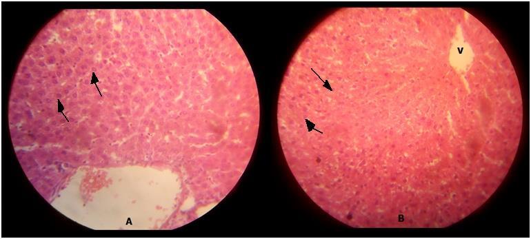
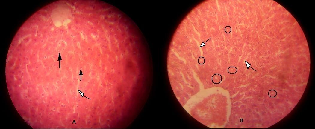
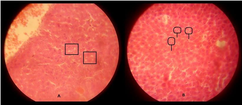
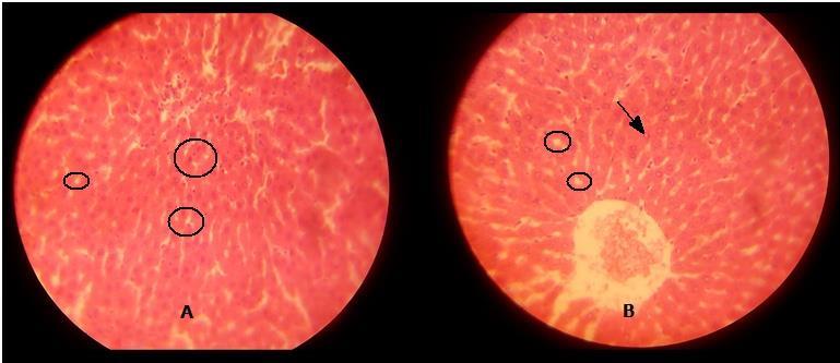
TABLE 2: TABLE SHOWING THE HISTOLOGICAL EFFECTS OF IBUPROFEN ON THE LIVER OF ADULT WISTAR RATS
GROUP IBUPROFEN (ml/kg) MICROSCOPIC EXAMINATION
CONTRO A 0 showing distinct hepatocytes and central vein.
GROUP B1 0.18 Showing reduced hepatocyte population and parenchymal population.
GROUP B2 0.18 showing nuclear vacuolation
GROUP C1 0.32 showing parenchymal vacuolation.
GROUP C2 0.32 showing pyknotic nuclei.
GROUP D1 0.40 showing eosinophillic nuclei and tissue crevices
GROUP D2 0.40 showing tissue vacuolation and tissue crevices
The above table explains the photomicrograph (below) of the control and test animals administered Ibuprofen at various doses and duration.
ISSN 2348-313X (Print)
International Journal of Life Sciences Research ISSN 2348-3148 (online)
Vol. 10, Issue 4, pp: (15-21), Month: October - December 2022, Available at: www.researchpublish.com
ISSN 2348-313X (Print)
International Journal of Life Sciences Research ISSN 2348-3148 (online) Vol. 10, Issue 4, pp: (15-21), Month: October - December 2022, Available at: www.researchpublish.com
The present results show that administration of Ibuprofen decreases the weight gain of wistar rats when compared with the control group. However, there were increases in weight of both control and test group before after acclimatization.
The mechanism of this decrease in weight gain by the administration of Ibuprofen is yet to be determined. The inhibition of lipoprotein lipase activity, increased energy expenditure, inhibition of nutrient absorption from the gastrointestinal tract, and suppression of the appetite (Dyer, 1994) are some likely reasons for the reductions in body weight and growth retardation upon administration of Ibuprofen.
Following the histological observation of this study, liver tissue damages expressed in several forms were presented with administration of graded doses of Ibuprofen for three weeks (acute) as well as for six weeks (chronic). Specifically, mild to severe glomerular degeneration, tubular wall disruptions, vacuolation, glomerular shrinkage, tubular congestion, exudation, parenchymal erosion, pykrosis, inflammatory cell infilteration, infarction with palour, and edema were presented. In addition, these renal damages were observed to be dependent on dose and duration of Ibuprofen administration.
Irrespective of the variations, it is clear that continous and prolong Ibuprofen administration have the potential to induce damagestotheliver.InaveryrecentexperimentonIbuprofentreatmentcarriedoutby Aprioku et al., 2014 ontheevaluation of toxicological profile of Ibuprofen in wistar rats, there was a detected cellular injury to the liver and observed renal toxicity.
Also, in the same experimental investigation, the heart was reported to be free of any form of injury or toxicity following administration of clinical doses of ibuprofen.
Although the control section presented some vacuolations, the comparative examination of the sections indicated that there were obvious features of alteration in the cytoarchitecture of the test group sections compared to control. The changes observed in the test group indicated that they existed irrespective of the dosage or duration, but there severity was dosage and duration dependent.
Considering the observed effects of Ibuprofen on the histology of the liver of test animal, it is recommended that its administration should be strongly regulated as it may cause cellular injuries to the liver. Furthermore, there is need for further research in this domain.
[1] Aprioku, E.J., and Uche, N.W. (2013): The influence of hemodialysis on the pharmacokinetics of ibuprofen and its major metabolites. Journal of Clinical Pharmacology 26(3):184-190.
[2] Aprioku, S.S., McCullough, K.F. and Nicholson, J.S. (2014): The pharmacological properties of ibuprofen, an antiinflammatory, analgesic and antipyretic agent. Archive of International Pharmacodynamic Therapy. 178(1):115-129.
[3] Bennett, S.K., Sen, P. and Ray, A. (1996): Central nervous system. In: Das PK editor. Pharmacology, 2nd ed., Elsevier, New Delhi, p. 268.
[4] Bradbury, F. (2004): How important is the role of the physician in the correct use of a drug? An observational cohort study in general practice. Internation Journal of Clinical Pratice. 144:27-32.
[5] Burke, A., Smyth, E. and FitzGerald, G.A. (2006): Analgesic-anti pyretic and anti inflammatory Agents; Pharmacotherapy of gout. In: Bruntom LL, Lazo JS and Parker KL (editors). Goodmans and Gilman’s the pharmacological basis of therapeutics. 11th ed., McGraw Hill, New York. p. 676-700.
[6] Capone, W.G., Brater, D.C. and Jhonson, A.R. (2007): Non steroidal anti inflammatory, anti pyretic analgesics. In: Goth’s medical pharmacology. 13th ed., Mosby year book. St: Louis Baltimore Booston. P. 567-570.
ISSN 2348-313X (Print)
International Journal of Life Sciences Research ISSN 2348-3148 (online) Vol. 10, Issue 4, pp: (15-21), Month: October - December 2022, Available at: www.researchpublish.com
[7] Dyer, P.J. (1994): Apoptosis induction in gastric mucous cells in vitro: lesser potency of Helicobacter pylori than Escherichia coli lipopolysaccharide, but positive interaction with ibuprofen. Journal of Endotoxin Research. 12(1):4756.
[8] Green K.F. (2001): Non steroidal anti inflammatory drugs for heavy bleeding or apin associated with intra uterinedevice use. Cochrane Database System Review. 18(4).
[9] Higuchi, V.V., Olkkola, K.T., Leino, K., Lundgren, S., Neuvonen, P.J. and Rane, A. (2009): Effects of the antifungals voriconazole and fluconazole on the pharmacokinetics of s-(+)- and R-(-)-Ibuprofen. Antimicrobial Agents Chemotherapy. 50(6):1967-1972.
[10] Kupper, S., Pukrittayakamee, S., Supanaranond, W., Kuile, F., Ruprah M and Sura T, (2010). Fever in uncomplicated Plasmodium falciparum malaria: randomized double-‘blind’ comparison of ibuprofen and paracetamol treatment. Tropical and Medical Hygiene. 89(5):507-509.
[11] Muntener, I., Minic, M., Dawood, M.Y., Akin, M.D., Spann, J. and Niland, N.F. (2010): Comparison of the efficacy and safety of nonprescription doses of naproxen and naproxen sodium with ibuprofen, acetaminophen, and placebo in the treatment of primary dysmenorrhea: a pooled analysis of five studies. Clinical Therapy. 24(9):1384-1400.
[12] Reynolds, H.P., Dale, M.M. and Ritter, J.M. (1999): Anti-inflammatory and immunosuppressant drugs. In: Pharmacology. 5th ed., Churchil Livingstone Edinburgh London. p. 248.
[13] Traversa, K.P. and Praticò, D. (1995): Novel therapeutic opportunities for Alzheimer’s disease: focus on nonsteroidal anti-inflammatory drugs. FASEB Journal. 19(12):1592-1601.
[14] Xavier, M.M., Lichenstein, D.R. and Signh, G. (2002): Gastrointestinal toxicity of non steroidal anti inflammatory drugs. England Journal of Medicine. 340(24):1888-1899.
[15] Yokoyama, D.M., Monaghal. J., Streete, P., Jones, A.L. and Dargan, P.I. (2013): Fourty five years of ibuprofen use. Critical care, 10:44.