


16/03/24
Dear readers,
Welcome to the second edition of the STAHS MedVetDent magazine! These articles have been written by aspiring medics, dentists and vets from the STAHS community, and we hope the huge variety of articles reflects the vast range of individual passions and interests within each field, and that there is something to interest every prospective reader.
Additionally, we would like to thank all the talented writers that dedicated their time and effort this half term to contribute to this edition; all your hard work is evident and greatly appreciated.
All that’s left to say is we hope that you enjoy reading this magazine as much as we have whilst editing, and we would love to hear feedback for our next edition in the summer term!
Heidi and Sabrin
By: Eleena Hearne
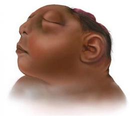
Anencephaly is a rare brain defect where a child is born with parts of their brain and skull missing. It is estimated that around 1 in 4,600 babies are born with this condition in the US annually and is unfortunately considered one-hundred percent fatal for the child, with most affected infants only living for up to a few hours or days if they are lucky Anencephaly is known as a neural tube defect (NTD) which occurs when the neural tube does not close properly during embryogenesis The neural tube is responsible for helping the formation of a baby’s brain, skull, spinal cord and backbones as it forms and closes. When this tube fails to close properly, an NTD arises. Anencephaly, specifically, is a result of the upper part of the neural tube not closing all the way, which has repercussions of the front part of the brain and the cerebrum (the part responsible for thinking and coordinating) failing to form with the remaining parts of the brain often being left bare, not covered by bone or skin (Figure 1). Active causes are unknown, but it is thought that a mother’s diet, medication or random mutations in the baby’s chromosomes could give rise to such a disease
Regarding prevention, folic acid is a highly renowned substance believed to help prevent NTDs, and taking 400mg a day is recommended to greatly reduce the risk of developing any sort of complications in the womb Ever since the US has started encouraging the use of folic acid, the number of pregnancies resulting in neural tube defects has decreased by 28%
This condition is usually quite easy to spot prior to birth as it happens so early on in embryonic development. Ultrasounds can usually detect it by looking at the cranial vault which provides a reliable diagnosis. Despite there being little investigation into families who chose to terminate the pregnancy, there have been reports of termination rates at around 43% or greater. As this is one of the most lethal congenital defects regarding such a sensitive part of the body, there are no known treatments and almost all infants with anencephaly die shortly after birth However, there was a case where an anencephalic infant survived for 28 months completely medically unassisted As shown in the image, she had a visible skull defect from which exposed tissue protruded, not covered by scalp She never required any surgical interventions or any other form of life sustaining treatment but did suffer from seizures which became more frequent towards the end of her life. As expected, she was slow to develop cognitively and physically, not reaching milestones in either sector expected of her age and only weighed around 9kg by the end of her 28 months.
Despite her sad ending, she is greatly respected and admired by many medical professionals Her contribution to the understanding of this illness has been crucial and has brought researchers another step closer to finding treatments to help future infants suffering with the same lethal disease
Facts about Anencephaly | CDC
· Facts about Neural Tube Defects | CDC
· Neural Tube Defects, Folic Acid and Methylation - PMC (nih.gov)
· Case Report: Prolonged unassisted survival in an infant with anencephaly - PMC (nih.gov)
By: Sabrin Osman
Emergency Medicine is centred around the treatment, evaluation and management of individuals from all walks of life with acute illnesses or injuries that require immediate medical attention ER Doctors are trained to handle a broad range of medical conditions, because of the wide range of situations they are presented with each day. ER doctors specialise in providing rapid initial assessments, diagnoses and initiating the first steps of care before diverting the patients to the next steps. Because of the vigorous 24/7-time schedule and fast-paced, frantic environment of the Emergency Department, ER physicians are required to stay on top of new treatments and are expected to continuously learn and stay current with new advancements in medical knowledge, technology and treatment modalities Whilst this is typical to some other specialities within medicine, what differentiates emergency medicine from other specialities and what distinguishes it as a speciality in its own right is because of the patients The ER sees a diverse myriad of patients suffering from severe injuries, mental health problems, and complicated diseases, unlike countless other specialities An array of cases mirrors the varied experiences individuals face in their lifetime. From chronic conditions to trauma, the ER encompasses a limitless range of health-related problems.
Ultimately Emergency Medicine serves as a microcosm of life. The emergency room reflects on the challenges and complexities common to the human experience, a theatre where the curtains never close, and the stage is almost set for life trials and uncertainties. Within the dynamic environment pulses the urgency of life-threatening medical crises Time is
both a precious commodity and a relentless adversary Emergency physicians juggle multiple cases simultaneously, with both the skill and power to alter the impending, immediate course of health trajectories. The aspect which stood out particularly to me is the beautiful showcase of the power of immediate action reflecting on the urgency of some medical cases. The profession is a continuous progression of learning, adaptation and improvisation and one that I too hopefully will have the opportunity to do.
The emergency room is populated not only by healthcare professionals but also by a diverse population of patients - from all walks of life The interactions between patients and physicians are what excite me the most – the interactions are not scripted, and the dialogues are unfiltered and genuine People from different walks of life converge, emphasising the connections of humanity Every case has its storyline, each one adding to the depths of each physician’s career. Each story leaves a life-lasting effect and impact, shaping their medical career for the better. Patients are the centre focus of physician’s choices, and medical decisions- with their best interest in mind.
Whilst emergency medicine is a critical and rewarding field, it’s important to acknowledge that it’s not without its challenges Especially when it comes to its losses and tragedies The nature of their work stems from the unique difficulties they face, and unfortunately, the urgency of the situations is sometimes so critical that they can lead to heart-breaking outcomes Emergency physicians often find themselves making high-stakes decisions in critical situations.
The weight of these decisions can sometimes be overwhelming. especially when the outcome is unfavourable The emotional challenge that comes with the responsibility of making life-anddeath choices is an aspect of the field many doctors struggle with This paired with the grief that may potentially follow as a result of the quick, intense connections with the patients and families can be extremely overwhelming. This is why it’s incredibly important to find the right balance between the empathy needed to truly connect with your patients, as well as the need for professional detachment for their own mental well-being.
Despite physicians’ best efforts and incredible dexterity and skills, emergency physicians may face situations where they have limited control over outcomes This lack of certainty can be particularly difficult to navigate, partly because of the high standards physicians hold themselves to, but also because they strive to provide the best possible care to their patients at all times However, it’s important to acknowledge that there is sometimes only so much that they can do, especially as a consequence of the unpredictable nature of emergencies.
Another challenge akin to emergency medicine is the difficulty of communicating bad news. Whilst this is an inherent part of emergency medicine, the difficulty lies with delivering information with a mixture of clarity and sensitivity, all while managing their own emotional
response. The difficulty also lies with the rapid decision making which adds an extra layer of complexity, which leaves physicians to deliver lifechanging news in situations where there is great uncertainty with diagnosis and prognosis The significance of choosing the right words is a skill so paramount to the role of emergency medicine Navigating the complex dynamics of family reactions, grief, anger and denial can be extremely difficult, especially in the high-pressure environment of the emergency room. Over time the cumulative effect of repeatedly delivering bad news may contribute an overwhelming toll on their career- a truly sad aspect because over time it may overshadow and distort what an incredibly rewarding profession it is because of the immediate connection to the patients.
The microcosm of emergency medicine, where life’s most crucial and dramatic moments unfold, highlights the significance of emergency medicine and exposes it as a speciality in its own right As we reflect on the experiences, it's truly eye-opening and incredible to witness the skill emergency physicians possess- a truly remarkable and inspiring feat We owe so much to them, for their attention, care, empathy and compassion, and I hope that I can become a doctor who resembles their skill but most importantly dedication to their patients.
Citations
-EMRA and CORD Student Advising Guide- Ch. 1 - Choosing Emergency Medicine (no date) Ch. 1 - Choosing Emergency Medicine. Available at: https://www.emra.org/books/msadvisingguide/choosing-emergencymedicine
By: Anya Ahuja
Neurofibromatosis can be broken down to understand what it means as follows: neuro- means relating to the nervous system, fibroma- means a non-cancerous growth consisting of fibrous tissue and -osis means disease Therefore, neurofibromatosis is a group of genetic disorders that result in the formation of tumours anywhere in the nervous system – brain, spinal cord and nerves – and skin Neurofibromatoses are often benign (non-cancerous) with generally mild symptoms but 10% of all neurofibromatosis cases are cancerous, meaning they have the ability to spread throughout the body. There are three major types of neurofibromatoses: type 1, type 2 and schwannomatosis, of which types 1 and 2 are most common.
Neurofibromatosis type 1, also known as von Recklinghausen disease, makes up around 96% of all neurofibromatosis cases and its incidence is 1 in 3000 The clinical presentation for neurofibromatosis 1 is often neurofibromas (small tumours under the skin), café-au-lait (CAL) spots, bond deformities and optic gliomas (brain tumours pressing on the optic nerve which is responsible for vision) CAL spots are pigmented patches of skin with a brown colour different from the tone of the rest of your skin. Neurofibromatosis type 1 is caused by a mutation in the tumour suppressor gene neurofibromin 1 (NF1) which codes for the production of neurofibromin. This mutation can be a deletion or point mutation, meaning a section of the gene has been deleted and is no longer translated, causing a loss of function of the protein which the gene is responsible for. In this case, neurofibromin is a protein that regulates many cellular processes and dopamine levels. Neurofibromin is a highly conserved protein, and we know this because human neurofibromin is 100% identical to its counterpart in chimpanzees, and the fact that this protein has remained unchanged through evolution and natural selection tells us that it is a vital protein with key functions
Showing just how detrimental the complete lack or malfunction of this protein can be. Neurofibromatosis type 2 is much less common than type 1 and common symptoms include gradual hearing loss, ringing tinnitus, balance issues and headaches It is characterised by schwannomas (rare nervous system cancer that grows on Schwann cells), specifically bilateral vestibular schwannomas which are associated with both hearing nerves and present in 90-95% of patients with type 2 neurofibromatosis, and meningiomas (tumours on the brain and spinal cord’s protective membranes called meninges) which are seen in around 50% of patients. Signs of these tumours range from numbness or weakness of limbs to seizures and facial drops. The NF2 gene is usually inherited as a dominant allele meaning family history plays a key role in diagnosing this disease However, there are occurrences where the patient has no family history of neurofibromatosis meaning that a mutation of the NF2 gene has occurred and the protein that the neurofibromin 2 gene codes for – Merlin – which is a tumour suppressor, is no longer being produced or is malfunctioning due to alterations in structure. The incidence of neurofibromatosis 2 is around 1 in 40,000 making it an extremely rare disorder. Schwannomatosis is a type of neurofibromatosis most prevalent among people around the age of 25-30 and results in multiple cranial, spinal and peripheral tumours or Schwannomas leading to intense pain and even neurological dysfunction. Key symptoms of schwannomatosis include chronic pain, numbness of limbs, tingling sensations and loss of muscle Research shows that 15% of people with the condition have inherited it and these cases are referred to as familial cases
In cases where there is no family history of schwannomatosis, it can be referred to as a sporadic case, which just means that there doesn’t appear to be a pattern of incidence. In sporadic cases, a random mutation in either the SMARCB1 or LZTR1 genes can cause cells to divide and grow uncontrollably and form schwannomas The epidemiology reports of schwannomatosis show that its occurrence is around 1 in 40,000 people
Cancer is a disease more prevalent than most of us realise yet there is still no definite cure It affects 1 in 3 people at least once in their lifetimes and caused over 25% of all deaths in the UK in 2020. Cancer cells are formed when healthy cells lose the normal regulatory mechanisms that control cell growth and experience loss of differentiation, meaning that they lose the unique characteristics that allow cells to specialise or differentiate into a different type of cell. This then results in a cancerous growth, otherwise known as a tumour. A tumour can either be benign (non-cancerous) or malignant, which means it has the potential to metastasise, which is the process by which a cancer can migrate and spread to other body tissues Malignant cells are rapid and uncontrollable and do not die as normal cells do due to a mutation in their genetic makeup
As all three of these forms of neurofibromatosis are primarily genetic disorders, genetic testing is available for people interested in having children who may be affected
However, genome profiling is an extremely costly process ranging from hundreds to sometimes thousands of pounds, which can make it inaccessible to people in developing countries. Genetic testing is covered by the NHS in the UK if referred for it by a hospital specialist due to a suspected genetic health condition.
Around 10% of all neurofibromatosis cases, primarily type 1, progress to stage IV which is malignant cancer meaning the cancer cells can spread to other regions of the patient’s body through metastasis and these cases would receive treatments such as chemotherapy, radiation and possibly surgery but face a poor prognosis of around 5 years. However, there is ongoing research into new treatment styles and a variety of modern and innovative treatments including personalised precision medicine and nuclear medicine.
https://www.mayoclinic.org/diseases-conditions/neurofibromatosis/symptoms-causes/syc-20350490
https://www.ncbi.nlm.nih.gov/books/NBK459329/
· https://www.mayoclinic.org/diseases-conditions/meningioma/symptoms-causes/syc-20355643
· https://www.nidcd.nih.gov/health/vestibular-schwannoma-acoustic-neuroma-and neurofibromatosis#:~:text=Bilateral%20vestibular%20schwannomas%20affect%20both,first%20time%20in%20their%20family
· https://www.ncbi.nlm.nih.gov/pmc/articles/PMC7692384/ https://my.clevelandclinic.org/health/diseases/22627-cafe-au-lait-spots https://www.ncbi.nlm.nih.gov/books/NBK470350/#_article-25787_s4_ https://www.hopkinsmedicine.org/health/conditions-and-diseases/neurofibromatosis/schwannomatosis# https://www.ncbi.nlm.nih.gov/pmc/articles/PMC2666272/# https://www.ncbi.nlm.nih.gov/pmc/articles/PMC2708144/
By: Alannah Jayamanna
The FDA (Food and Drug Administration), a federal agency of the Department of Health and Human Services in the US, have confirmed that injecting a substance called poly-L-lactic acid could reduce the effects of aging on the cheeks and face and is used for volume restoration in these areas. The substance is called poly-L-lactic acid and is a biodegradable polymer as well as an ‘absorbable, semi-permanent injectable implant that can gradually restore the volume and stimulate collagen formation’ as said by the National Library of Medicine The FDA has approved Sculptra (the company that first produced this substance) for the aesthetic use of PLLA since 2009 to use this substance to correct the appearance of fine lines, nasolabial folds (smile lines) and wrinkles in the cheeks
As we age, both the collagen and fat in the face decrease and thin, causing the formation of wrinkles and a loss of elasticity in the skin, which can lead to a rough appearance. Collagen is a fibrous protein that is in the dermis of the skin and gives it structure while elastin (also found in the dermis) ensures the skin maintains its shape. Because of this, you can imagine the effect this has on the skin if either the collagen, elastin or hyaluronic acid underneath the skin's epidermis decrease. If the collagen matrix becomes fragile and starts to wear down, as a result of ageing, it also causes the other features to deplete PLLA is known as a bio-stimulatory dermal filler as it stimulates the skin to make collagen which fills the spaces where the fat has thinned over time This process is called neocollagensis Sculptra has claimed that the use of poly-L-lactic acid improves the quality of the skin by ‘helping to firm sagging skin for a more radiant and glowing appearance’ and according to a study, 96% of patients showed improvements within as early as three months of receiving the treatment.
PLLA stimulates collagen by stimulating the fibroblasts (a type of tissue that contributes to the formation of connective tissue) and thickening the dermis as well as building the skin structure.
According to Sculptra, however, there can be some side effects such as swelling, pain and itching in the injection site and that it cannot be administered to everyone, and medical history must be considered before proceeding with the treatment. The effect also only lasts for two years and over time converts to carbon dioxide and water and is therefore relatively harmless to the individual. The buildup of the collagen takes time over more than one injection and a few sessions are needed in order to achieve the best possible results
HA fillers are another type of temporary filllers that has increased in popularity over the past few years. They are made of mainly hyaluronic acid and take the form of a gel-like substance which has effects for 6 months to a year before the skin on the face starts to naturally absorb it as it is already a naturally occurring feature of the skin’s dermis.
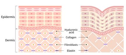
Here are some main differences between PLLA and HA fillers that are relevant to this article:
HA fillers stimulate a small amount of collagen which are most effective in filling soft tissue rather than stimulating collagen like PLLA does
PLLA fillers take a series of sessions whereas HA fillers take a larger dosage of the product in one session only · HA fillers are made from hyaluronic acid which is a naturally occurring substance in our bodies. Whereas in PLLA fillers, the main ingredient is polyL-lactic acid.
Overall, the discovery and use of Poly-L-lactic acid in cosmetic dermatology has proved to be both popular and effective with clinics all over the world supplying this type of treatment As it is a temporary and safer option in comparison to some other treatments, many people who are finding that they are having a decrease in volume in the face, may want to opt for this filler instead
Citations
-https://www dermatologytimes com/view/fda-gives-the-green-light-for-cheek-wrinkle-solution -https://www.asds.net/skin-experts/skin-treatments/injectables/injectable-poly-l-lactic-acid
https://www.ncbi.nlm.nih.gov/pmc/articles/PMC7564527/#:~:text=It%20increases%20the%20collagen%20by,o f%20applications%20in%20the%20future.
https://www.fda.gov/ --> FDA website
By: Sophia Adams
Every year, approximately 200,000 people die from glioblastomas
The average length of survival after diagnosis is a mere 8 months.
Only 6 9% of those diagnosed survive past 5 years.
Glioblastomas (GBMs) are the most common type of cancerous brain tumour in adults, making up 32% of all primary tumour diagnoses in England between 1995 and 2017. They are grade 4 tumours (according to WHO classifications), the highest grade, meaning they are the fastest growing tumours, often return after treatments, and can spread to other parts of the brain and even the spinal cord There are two types of glioblastomas; primary, which are developed de novo, and secondary, which are progressions of low-grade or anaplastic astrocytomas - primary glioblastomas are more common and aggressive, typically developing in older patients, with secondary glioblastomas appearing more in younger patients
Currently, the main treatment methods include a combination of surgery, radiotherapy and chemotherapy. Surgery is the main treatment method, as even if your surgeon is unable to fully remove the tumour, removing even small parts of it can help slow down the progression of the tumour, and also help relieve symptoms, but only results in stable or improved outcomes (compared to the preoperative assessment) 77 7% of the time Furthermore, despite positive surgical outcomes, patients often also observe a neurocognitive decline and therefore lowered quality of life post-surgery Radiotherapy involves aiming X-rays, gamma rays or photons at the tumour to try and kill the cancerous cells, therefore shrinking the tumour and alleviating pressure from the brain
Finally, chemotherapy involves the usage of cytotoxic (anti-cancer) chemicals, usually after surgery has been tried first, and can be given intravenously, as a tablet or directly into your brain or spine The most common types of chemotherapy drugs for glioblastoma treatment are temozolomide and carmustine, with the former being taken in pill form and the latter intravenously. However, it can be difficult for chemotherapy drugs to even reach glioblastomas, all due to our blood-brain barrier
The blood-brain barrier (BBB) is a physiological barrier between the blood and the CNS, serving to limit the accumulation of harmful substances in the brain by preventing their passage from blood to the brain, but also being key in maintaining brain homeostasis. This means that chemotherapy drugs are often blocked by the blood-brain barrier and cannot treat tumours, even if the drugs are effective against glioblastoma cells in Petri dishes (without the BBB) In the past, researchers have explored several strategies to overcome the BBB, but each has had significant risks, including heat damage and excessive toxicity - fortunately, new nanotechnology can help address this problem.
In a recent study, Dr Bachoo and his colleagues at UT Southwestern Medical Centre and the University of Texas at Dallas, have found a new way to overcome the BBB without generating excessive heat. This new approach uses gold nanoparticles covered in antibodies against a main protein in the BBB complex, which when injected intravenously into animal models, attach to junction proteins in the BBB This means that once targeted by a precise wavelength of laser light, these nanoparticles will vibrate and open up the BBB without generating heat, and this strategy has been named optoBBTB
Researchers used two types of genetically engineered mice which had mutations apparent in humans with glioblastomas, firstly carrying out optoBBTB and then applying dye to see if it spread to the tumour, overcoming the BBB, which it did Subsequently, the researchers then used the chemotherapy drug paclitaxel instead of dye, and after three cycles of paclitaxel (after optoBBTB), three days apart each, they found that these mice lived up to 50% longer in comparison to other mice who received either a placebo or paclitaxel intravenously without optoBBTB first
Although much more vigorous testing and drug trials are needed before optoBBTB can be applied in human patients, this is extremely promising, as emerging methods to open up the blood-brain barrier will enable far more chemotherapy drugs to reach and treat glioblastomas, hopefully improving the survival rate of this aggressive cancer

Figure 1:image from https://www.utsouthwestern.edu/newsroom/articles/year2023/oct-nanotechnology-helps-chemo.html
https://braintumor.org/events/glioblastoma-awareness-day/aboutglioblastoma/#:~:text=Glioblastoma%20Facts%20%26%20Figures&text=It%20is%20estimated%20that%20more,to%20be%20only%208%20months https://academic.oup.com/neuro-oncology/article/1/1/44/1113761
https://www.utsouthwestern.edu/newsroom/articles/year-2023/oct-nanotechnology-helps-chemo.html
https://www.ncbi.nlm.nih.gov/pmc/articles/PMC9415007/ https://www.gliocure.com/en/patients/glioblastoma/#:~:text=About%20200%2C000%20people%20die%20each,9%2C000%20in%20the%20United%20States https://www.moffitt.org/cancers/glioblastoma/treatment/radiation/#:~:text=During%20radiation%20therapy%20for%20glioblastoma,alleviate%20pressure%20on %20the%20brain
https://www.ncbi.nlm.nih.gov/pmc/articles/PMC9954589/#:~:text=In%2077.7%25%20of%20the%20cases,surgery%20than%20a%20single%20technique https://braintumourresearch.org/blogs/types-of-brain-tumour/glioblastoma-multiforme-gbm https://www.ncbi.nlm.nih.gov/pmc/articles/PMC6981899/#:~:text=The%20BBB%20is%20a%20physiological,the%20brain%20to%20the%20blood https://www.cancerresearchuk.org/about-cancer/brain-tumours/brain-and-spinal-cord https://www.cancerresearchuk.org/about-cancer/brain-tumours/treatment/chemotherapy-treatment
By: Aneesa Patel
Cancer is a wider term for diseases which are characterised by random cell division with many risk factors being environmental, physical and genetics. It is globally known as a significant health problem and one of the leading causes of death.
In particular, lung cancer is a prevalent cause of cancer mortality worldwide, affecting around 2 1 million individuals and causes 1 8 million deaths per year The most common cases of lung cancer are NSCLC (non-small cell lung cancer) and SCLC (small cell lung cancer), with NSCLC making up 85% of patients and SCLC making up 15% of patients NSCLC has 4 stages: stage 1, which is the most common in the lung, stage 2 which extends to lymph nodes close to the lung, stage 3 which spreads further into the lymph nodes and the middle of the chest and stage 4 where cancer has spread broadly throughout the body. The survival rates for a patient at stage 1 is around 90% but drops to 10% at stage 4. Patients with SCLC have as 30% chance of developing a modest illness and a 10% chance of developing a severe disease.
Common cancer treatments include surgery, chemotherapy, targeted therapy, radiation therapy, hormone therapy and immunotherapy Whilst treatments such as chemotherapy and radiation therapy can inhibit cell growth and multiplication (cytostasis) and cause damage to a cell (cytotoxicity), these treatments are also known to have severe effects and have high risks of recurrences. Common side effects include neuropathies (where nerves can become damaged), fatigue, hair loss and damage to the functioning of the lungs and heart.
More recent cures for cancers such as lung cancer has stemmed from nanotechnology.
Nano particles are particles (generally 1-100nm in size) with unique properties which
are usually not found in bulk samples of the same material and are classified (depending on their size) into the four groups: 0D, 1D, 2D or 3D. There is a range of properties of nano materials, such as, a high surface area to volume ratio, unique fluorescence properties, enhanced electrical conductivity, spectral shirt or optical absorption and super paramagnetic behaviour. As a result, nano materials, within medicines, can be used in drug transportation, increased permeability, controlled release and improved biocompatibility Nanotechnology can directly and selectively target cancerous cells and neoplasms, heighten the therapeutic efficacy of radiation-based treatments and guide surgical resection of tumour
Moreover, nano materials can be classified into several categories: nano particles, liposomes, solid lipid nano particles, nano structured lipid carriers, nano emulsions, dendrimers, graphene and metallic nano particles.
Liposomes are made up of lipids and have a closed spherical lipid bilayer which forms and inner cavity. In turn, this can hold aqueous solutions. The two lipid layers are made up of two phospholipid sheets with a hydrophobic tail and a hydrophilic head and the hydrophobic ends are attracted to one another whilst the other end is directed to the surrounding water This forms a double layer of the phospholipid molecules and in turn prevents the inner solution (Figure 1) This inner solution can then be transported with the liposome to where it is needed Currently, around 12 liposome-based medicines have been approved of for medical use in various phases of clinical studies.
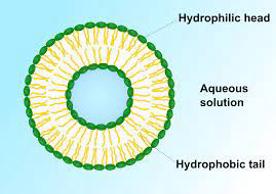
1: Liposome formed by phospholipids in aqueous solution (Wikipedia)
One form of treatment for lung cancer involving liposomes is Liposomal dry powder. It is a promising possible treatment for lung cancer and is an anti cancer medication delivered through the lungs, as a dry nano particulate powder. A trial of docetaxel (DTX) which was encapsulated in folic acid-conjugated liposomes was effective at targeting the tumour. This was seen in the fact that the re-dispersed liposomes obtained after the re dispersion of inhaled dry powders differed from the initial liposomes which concluded that Liposomal Dry powder formulations can be deposited all over the lung and so can be used as a treatment for lung cancer.
One of the main advantages of nanotechnology (liposomes in particular) over other cancer treatments is targeted delivery which revolves around the precise targeting of specific cancer cells which in turn reduces the toxicity in normal cells. This is achieved in two ways: passive targeting or active targeting. Passive targeting is the use of enhanced permeability and retention effect (EPR) whereas active targeting is the binding of certain ligands which recognise cell surface markers. Many ligand structures have been formed depending on the target site such as antibodies, peptides, small molecules and aptamers. Ultimately, this advantage increases the probability of effective treatment as well as decreasing the risk to the patient.
Additionally, lipid based nano carriers are similar to the membrane component of cells and therefore have the advantage of having fewer side effects when being absorbed into cells Lipid based nano carriers can also be administered by a variety of routes for drug delivery such as oral, parenteral, ophthalmic, intranasal and dermal routes. Whilst there is a multitude of different ways the treatment can be administered, the oral route Is the most popular due to its limited invasiveness, low cost and lowest risk of adverse effects making liposomal dry powder more suitable.
Despite the advantages to the use of this technology, there are many challenges that are arguably faced The main issues in relation to cancer nano medicine is the development of immunological and haematological drawbacksfor example, autoimmunity, inflammation, suppression of bone marrow etc This is because nano particles can have a lethal effect on normal cells within the body depending on the therapy being provided for the tumorous cells.
Whilst these challenges exist, the use of nanotechnology and the trials into its use are still progressing, making it a promising form of treatment for the future.
Citations:
https://www.ncbi.nlm.nih.gov/pmc/articles/PMC544578/ https:/www.ncbi.nlm.nih.gov/pmc/articles/PMC8645667/ https:/jhoonline.biomedcentral.com/articles/10.1186/s13045-021-01096-0 https:/www cancer gov/nano/cancernanotechnology/treatmenthttps:/www.ncbi.nlm.nih.gov/pmc/articles/PMC8805951/https:/www .tandfonline.com/doi/full/10.2147/IJN.S406415
By: Nyah Mistry
A new age of innovation and development in dental treatment has been brought about by the intersection of 3D printing technology and dentistry 3D printing has completely changed the way that dental prosthetics, implants, and surgical guides are made Its capacity to create complex structures layer by layer has allowed for unmatched precision and customisation. 3D printing provides a smooth process that combines with digital dental practices, simplifying treatment planning and execution, from digital design to clinical implementation. This article explores the evolution, uses, and revolutionary possibilities of 3D printing in dentistry, a field that is always changing.
The field's unwavering pursuit of innovation is demonstrated by the historical growth of 3D printing in dentistry 3D printing technology first gained traction in sectors including automotive and aerospace engineering in the late 1980s But in the early 2000s, its prospective uses in dentistry grew more and more apparent The creation of specific 3D printing materials for the dentistry field marked a significant turning point in the field by making it possible to create precise dental prototypes and models. The creation of dental prosthetics, such as crowns, bridges, and dentures, with unmatched precision and customisation was made possible by later developments in printing technology. As the technology advanced, it revolutionised a number of dental specialties, including orthodontics, implantology, endodontics, and periodontics
Current Applications:
Dental Prosthetics:
The creation of dental prosthetics has been transformed by 3D printing, which has many advantages over conventional production techniques. These days, dental labs use 3D printing
technology to create prosthetic items like crowns, bridges, and dentures with remarkable precision and personalisation. Clinicians may ensure the best possible fit and function for each patient and produce superior cosmetic results by digitally designing prosthetic restorations and printing them directly from digital models. The capacity to tailor prosthetic devices to the unique anatomy and preferences of each patient improves treatment outcomes and increases patient satisfaction. Moreover, 3D printing streamlines the prosthetic construction process by allowing quicker turnaround times and minimising the need for several appointments
Orthodontics:
3D printing is essential for improving patient comfort and treatment planning in orthodontics. Using 3D printing technology, orthodontists may create customised devices that are suited to each patient's individual dental architecture, such as clear aligners, retainers, and orthodontic appliances. Because digital scans of the patient's teeth can be instantly turned into printable models, 3D printing in orthodontics eliminates the need for messy and painful traditional impressions.
The design and production of surgical guides and dental implants have also been completely revolutionised by the use of 3D printing in implantology Using 3D printing technology, dental implants which support dental prosthetics as artificial tooth roots can be precisely tailored to match the patient's natural tooth. Furthermore, during implant insertion procedures, 3D-printed surgical guides offer clinicians vital support, guaranteeing the best possible implant positioning and angulation for long-term success. This decreases surgery time, increases implant placement accuracy, and lowers the risk of complications.
Endodontics and Periodontics:
Additionally, 3D printing provides creative answers for a range of treatment modalities in endodontics and periodontics, such as periodontal therapy and root canal therapy. The efficiency and results of treatment can be enhanced by 3D printing customised endodontic equipment, like as files and reamers, to perfectly match the anatomy of the root canal system Furthermore, the creation of tissue scaffolds and periodontal surgical guides has been made possible, which promotes accurate and consistent results in periodontal procedures In these specialised areas of dentistry, endodontists and periodontists can improve treatment precision, reduce invasiveness, and maximise patient results.
Advantages and Disadvantages:
Benefits:
Customisation: 3D printing in dentistry offers the key benefit of delivering customised treatment solutions for patients 3D printing allows physicians to produce personalised dental appliances that accurately correspond to the individual anatomical characteristics and treatment requirements of each patient, in contrast to conventional manufacturing techniques that typically use standardised prosthetic devices. This degree of customisation enhances the fit, comfort, and functionality of dental prosthesis, leading to improved treatment results and patient contentment.
Conciseness: 3D printing technology enables the fabrication of intricate dental structures with outstanding precision and accuracy. Clinicians can accomplish precise details and fine surface finishes that are hard to produce with traditional means by employing digital design tools and high-resolution printing procedures This high level of accuracy is especially important in fields like implantology and orthodontics, where flawless fit and alignment are crucial for optimal treatment results
orthodontic appliances, and surgical guides, resulting in faster turnaround times and fewer chair-side appointments compared to traditional procedures. Moreover, optimising material consumption and reducing waste leads to cost savings for both dentists and patients.
Material Selection: Choosing the right materials with the necessary qualities for dental uses is still a major obstacle in 3D printing Various materials including as resins, ceramics, and metals are used for dental 3D printing, each with distinct properties and constraints. Clinicians need to consider biocompatibility, mechanical strength, and aesthetics when selecting materials for dental procedures to provide the best treatment results and maintain patient safety.
Regulatory Compliance: Compliance with regulations and standards for utilising 3D printing in dentistry is an additional obstacle for dental professionals As 3D printing technology advances in dentistry, regulatory agencies like the GDC and FDA are responsible for creating guidelines and regulations to guarantee the safety and effectiveness of 3D-printed dental products Dentists must stay updated on regulatory regulations and adhere to current standards to reduce potential risks and guarantee patient safety.
Expertise and Instruction: Proficient use of 3D printing technology in dentistry necessitates specialised training and competence. Dental professionals need to master digital design tools, printer operation, and post-processing processes to properly utilise 3D printing technology in their work. Continuing education and training are crucial to be informed about improvements in 3D printing technology and emerging best practices Dental professionals can enhance patient care by investing in training and professional development to fully utilise the advantages of 3D printing technology
Efficiency in Time and Cost: 3D printing simplifies production processes and lowers treatment costs by removing the necessity for physical labour and numerous manufacturing procedures. 3D printing enables the quick production of dental prosthesis,
Advancements in 3D printing technology and specialised dental materials are anticipated to transform dental practice in the future. This involves creating biocompatible materials and
enhancing printing methods for prostheses, implants, and tissue engineering. We expect that 3D printing will be more smoothly incorporated into regular dental processes, from scanning at the chair-side to creating unique equipment in-house There are abundant research opportunities in fields like regenerative dentistry and personalised medicine, leading to innovative discoveries and improved patient care
The key findings emphasise the significant influence of 3D printing technology on different areas of dental practice, such as prosthetics, orthodontics, implantology, endodontics, and periodontics.
Customising dental devices, achieving precise results, and improving production processes has greatly improved patient care and treatment results. The future impact of 3D printing in dentistry is evident With rapid improvements in technology and materials, 3D printing is poised to become a crucial tool in enhancing dental care and enhancing patient results Dental professionals are likely to fully utilise 3D printing by embracing improvements and investigating new research opportunities to satisfy their patients' changing requirements and bring about a new era of innovation in dentistry.
Citations:
Schweiger, J., Edelhoff, D. and Güth, J.-F. (2021). 3D Printing in Digital Prosthetic Dentistry: An Overview of Recent Developments in Additive Manufacturing. Journal of Clinical Medicine, 10(9), p.2010. doi:https://doi.org/10.3390/jcm10092010.
Paras, A. (2023). The Future of Dentistry: How 3D Printing is Changing the Industry. [online] Institute of Digital Dentistry. Available at: https://instituteofdigitaldentistry.com/3d-printing/the-future-of-dentistry-how-3d-printing-is-changing-the-industry/. Eplus3D. (n.d.). 5 Advantages and 2 Challenges of 3D Printing in Dentistry. [online] Available at: https://www.eplus3d.com/5-advantagesand-2-challenges-of-3d-printing-in-de.html [Accessed 7 Mar. 2024].
Célio Figueiredo-Pina and Ana Paula Serro (2023). 3D Printing for Dental Applications. Materials, [online] 16(14), pp.4972–4972. doi:https://doi.org/10.3390/ma16144972.
Lebi, R. (2023) Good news for dental labs and 3D printing - Dentistry. https://dentistry.co.uk/2023/09/08/the-developments-in-3dprinting-that-spell-good-news-for-dental-labs/.
Ebner, F. (2022) 3D printing – the pinnacle of productivity? - Dentistry. https://dentistry.co.uk/2022/10/02/3d-printing-the-pinnacle-ofproductivity/.
By: Zara Bhatti
Oral health is a term referring to the overall health of teeth, gums and the whole facial system that allows us to speak, smile and chew Good oral hygiene is an important practice as it helps to prevent gum disease, tooth decay and bad breath However, your oral health has a much wider impact than just on the mouth, as oral health has been proven to be linked to both your physical and mental wellbeing It is said that your dentist can detect underlying conditions such as heart disease, cancer and diabetes from just a check-up. This article will discuss the wider, more inconspicuous implications of poor oral hygiene, revealing the crucial importance of "good" teeth on your overall health and wellbeing. A well-known, and extremely common, consequence of poor oral health is gum disease. Failure to maintain good oral practices, such as daily brushing and flossing, means bacteria that cause gingivitis, the earliest stage of disease, and periodontitis, a serious gum infection that damages the soft tissue around teeth, reach high and concerning levels Not only does this have serious implications in terms of oral health, including tooth decay, bad breath and bleeding gums, multiple studies have recorded that people with an unhygienic oral cavity tend to have higher rates of heart disease and stroke. The mouth is a key access point to internal body organs; bacteria building in the mouth can travel through the bloodstream, causing inflammation and so an increased risk of hardened (atherosclerosis) arteries and increased blood pressure (hypertension).
Alarmingly, if fatty deposits block a blood vessel to the heart, a heart attack can occur; if the brain’s blood supply is cut off, a stroke can occur. Moreover, poor dental health not only affects the brain in terms of likelihood of strokes – substances released from infected gums can damage brain cells and may even lead to memory loss It seems that dementia, or even Alzheimer’s, can be tracked back to gingivitis in the mouth These conditions are known to have a degenerative, detrimental and distressing
effect on a person, without a known cure. Sadly, periodontitis has also been linked to pregnancies – according to the Centers for Disease Control and Prevention (CDC), children of mothers who have high levels of untreated cavities are more than three times likely to develop cavities themselves CDC also suggests that it has been associated with poor pregnancy outcomes in general, including premature births and low birth weight – this is not completely understood yet. It’s also possible for bacteria in the oral cavity to be breathed into the lungs leading to respiratory infections, such as pneumonia and acute bronchitis.
On the other hand, the impact of poor oral health is not limited to just physical health complications. In our society, there is an existence of dental phobia and anxiety – many patients have some sort of fear and, in turn, stop visiting their dentist regularly It’s evident that frequent check-ups are essential and irregular visits can cause conditions in the mouth to worsen It seems that the pressure that exists over oral health only deters patients’ further from attending to problems due to a growing anxiety around dentistry. Undoubtedly, the stigma around “ugly” teeth exist and has only been heightened by social media – there is this extreme stress to have the “perfect” smile which includes straight, evenly-shaped and pearlywhite teeth. Not fitting into this very specific, and often unachievable, beauty standard incites a feeling of self-embarrassment, shame and low self-esteem. If these emotions are ongoing, it can sometimes even lead to depression – a serious mental health illness Another example of a consequence of the pressure of a “good” oral cavity is the growth in popularity of “Turkey Teeth” Many feel compelled into changing their teeth, however, they may lack the funding or would rather not pay expensive prices to undergo
drastic changes to their teeth. This is leading to several resorting to undergoing a procedure of gaining very white and square crowns in Turkey. Despite being done for aesthetic reasons, patients often face gum infections, everlasting tooth pain, rotting teeth and so much more. The influence and exposure to unrealistic cosmetic expectations for teeth can make patients feel inclined into causing more serious damage to not only their teeth but their overall health
Despite the serious potential consequences of poor oral hygiene, including implications on physical wellbeing, preventative steps can be taken to protect against such diseases: brushing teeth twice daily for at least two minutes each time can help remove remaining food and plaque, flossing daily maintains healthy gums, a healthy diet and a limit on sugary foods and drinks to fight tooth decay, avoiding alcohol consumption and smoking is key, and finally schedule frequent dental check-ups.
Nevertheless, combatting the mental health consequences of poor oral health has a much more complicated solution which involves a wider acceptance in society of difference rather than constantly conforming to a beauty standard. Overall, it’s evident that oral health affects much more than just your teeth as it impacts how well your body and internal organs function, and how you perceive yourself in the world
Citations:
https://www perfectsmile-dental com/news/how-ignoring-dental-health-can-cause-bighealth-
problems/#:~:text=Generally%2C%20people%20with%20gum%20disease,cardiovascular%20 disease%20or%20kidney%20failure.
https://www.mayoclinic.org/healthy-lifestyle/adult-health/in-depth/dental/art-20047475
https://newsroom.cigna.com/medical-conditions-with-surprising-connection-oral-health https://www.dentalhealth.org/mental-illness-and-oral-health
https://www.cdc.gov/oralhealth/publications/features/pregnancy-and-oralhealth.html#:~:text=Pregnancy%20and%20Periodontal%20Disease&text=Periodontitis%20ha s%20also%20been%20associated,birth%20and%20low%20birth%20weight.&text=However% 2C%20how%20periodontitis%20may%20lead,safe%20and%20important%20during%20preg nancy.
By: Komachi Paine
Skin allergies are one of the most prevalent and challenging canine health concerns According to statistics, approximately 10-15% of dogs suffer from skin allergies, making it one of the most common reasons for vet visits [1]. Of these skin allergies, Canine Atopic Dermatitis (cAD) affects 3-15% of the canine population, with some breeds even being genetically predisposed [1]. Symptoms of cAD include pruritus (severe itching), erythema (redness of the skin caused by inflammation), alopecia (hair loss), and recurrent skin infections, all of which significantly damage the quality of life for affected dogs
In this article, I will cover current and possible future treatments for cAD, as well as preventative measures
What are the causes of cAD?
According to research, around half of dogs born to atopic parents will develop cAD [2]. Dogs who are affected by cAD without atopic parents are likely to have allergic reactions due to dust mites, pollen, mould spores, and food allergies [3].
Traditional Treatments:
Traditional treatments such as steroids are commonly used in managing cAD, as they often rapidly alleviate itching, inflammation, and skin lesions Steroids are also relatively affordable compared to some newer therapies, making them accessible to a wider range of pet owners However, steroids do have notable disadvantages: prolonged or high-dose steroid use can lead to adverse effects such as a damaged immune system, weight gain, polyuria (excessive urination), and polydipsia (excessive thirst), as well as possible future drug resistance and steroid withdrawal when treatment is terminated. Thus, whilst steroids offer rapid relief, their potential adverse effects and limited long-term benefits warrant caution from both vet and owner.
Monoclonal Antibodies and Janus Kinase Inhibitors:
According to research, treatments such as Monoclonal antibodies and JAK inhibitors offer fewer systemic side effects compared to traditional immunosuppressive drugs [4].
How do monoclonal antibodies work?
Monoclonal antibodies are produced in labs and are designed to target specific components of the immune system involved in the allergy response. They bind and neutralise key inflammatory mediators, like interleukins or cytokines, that contribute to the progression of the allergic reaction How do Janus Kinase (JAK) Inhibitors work?
JAK Inhibitors block the family of Janus Kinase enzymes which are involved in the production of inflammatory cytokines Inhibiting these enzymes slows down the inflammatory process
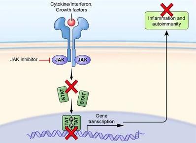
Diagram showing the mechanism of action of a JAK inhibitor in blocking Janus Kinase enzymes to prevent an inflammatory response. [8]
Both of these treatments work to reduce inflammation, which in turn provides rapid relief and long-term management of symptoms. However, further research is necessary to evaluate their independent long-term efficacy. For now, monoclonal antibodies and JAK inhibitors are normally used in conjunction with other treatments, as they work mainly to calm nflammation and itching, which will improve the response from treatments like ASIT and diet regulation
Allergen-specific immunotherapy (ASIT) has been seen to benefit 60-70% of dogs with cAD by offering long-term control of allergic symptoms [5] Recent advancements in ASIT formulations, including sublingual (under the tongue) and oral immunotherapy options, have expanded treatment options for dogs with cAD. Canine patients are often more complacent with these alternative routes compared to traditional routes like injections, making it a viable option, especially when ASIT treatment requires frequent dosage. Additionally, to the long treatment period, ASIT can be expensive due to the extent of testing and consultations, and is for the majority only applicable to environmental allergens such as pollen, dust mites, and mould spores So, although the scope of ASIT does not cover all cAD cases, it has shown long-term efficacy in its constituent cases
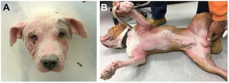
Image of a dog presenting with erythema, alopecia, and excoriations on the (A) face, pinnae, ear canals and eyelids, as well as (B) paws, axillae, ventrum, and inguinum. (Courtesy of the Veterinary Service of the William R. Pritchard Veterinary Medical Teaching Hospital at the University of California, Davis.) [5]
Personalised medicine and advanced allergen testing methods allow vets to identify specific triggers responsible for a dog's allergic reactions. Intradermal skin testing (injections within the top layer of the skin) and IgE antibody testing enables vets to identify exactly which allergens are to blame, meaning customised immunotherapy treatments or allergen avoidance strategies can be put in place This way, life-style and diet changes can go hand in hand with managing allergies without the need for long-term medication
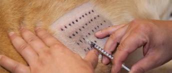
showing an Intradermal skin test. (Courtesy of Steve Design/shutterstock.com)
Currently, PRP is most commonly used for healing musculoskeletal injuries, soft tissue injuries, and Osteoarthritis management, however, it has emerged as a promising therapy for cAD [6], as it contains a high concentration of growth factors and cytokines that promote tissue repair and regulate inflammatory response. Whilst research on PRP therapy for cAD is ongoing, preliminary studies suggest that PRP may offer as an effective treatment for dogs, especially with refractory atopic dermatitis (a type of cAD that does not respond to conventional treatment).
Diet plays a crucial role in managing and preventing cAD, as the avoidance of identified or potential allergens can help to reduce the severity or even prevalence of allergic reactions Officials recommend diets incorporating protein sources that the dog has not been exposed to before, hydrolysed protein diets (protein has been processed down into smaller fragments), and elimination diets. [7] Additionally, nutritional supplements such as omega-3 fatty acids, and
antioxidants can support skin health and reduce inflammation, which can help soothe symptoms naturally in dogs (although not targeting the root cause itself)

“Hydrolysis of proteins is the process which cleaves the peptide bonds of the different amino acids that make up a protein, thereby disrupting the protein structure to remove as many allergenic epitopes as possible. Photo credit: Royal Canin SAS” [9]
Selective breeding is a proactive approach to tackling the root cause of cAD In fact, studies have identified genetic predispositions to cAD in certain breeds, including Labrador Retrievers, Boxers, and Bulldogs [4]. This increased susceptibility in some breeds means that there could be genetic markers associated with cAD, which once identified can be selectively bred out of gene pools, to reduce the prevalence of cAD.
To conclude, it is evident that there are a variety of different treatments for cAD, as well as promising future treatments on the horizon However, I think that it is vital to weigh-up the accessibility of these treatments, considering the fact that skin allergies affect at least 10% of dogs [1]. The financial burden that can come with diagnosing and managing cAD, through consultations, diagnostic tests, medications, and specialised diets, can amount to hundreds or even thousands of pounds annually for pet owners. Especially during the UK’s cost of living crisis, these treatments are becoming unaffordable for some, leaving many dogs untreated and suffering needlessly This highlights the urgency of exploring alternative and preventive measures, as well as perhaps further advertising information about genetically predisposed breeds, improved diet, and improved life-style practices to mitigate the consequences of severe skin allergies in our beloved pets.
[1] CE;, H.A. (2001) The ACVD Task Force on canine atopic dermatitis (i): Incidence and prevalence, Veterinary immunology and immunopathology. Available at: https://pubmed.ncbi.nlm.nih.gov/11553375/ (Accessed: 23 February 2024).
[2] Shaw SC;Wood JL;Freeman J;Littlewood JD;Hannant D;, S. (2004) Estimation of heritability of atopic dermatitis in Labrador and Golden Retrievers, American journal of veterinary research. Available at: https://pubmed.ncbi.nlm.nih.gov/15281664/ (Accessed: 23 February 2024).
[3] Picco F;Zini E;Nett C;Naegeli C;Bigler B;Rüfenacht S;Roosje P;Gutzwiller ME;Wilhelm S;Pfister J;Meng E;Favrot C;, F. (2008) A prospective study on canine atopic dermatitis and food-induced allergic dermatitis in Switzerland, Veterinary dermatology. Available at: https://pubmed.ncbi.nlm.nih.gov/18477331/ (Accessed: 23 February 2024).
[4] Dreschler, Y. (2024) Canine Atopic Dermatitis: Prevalence, Impact, and Management Strategies, Veterinary medicine (Auckland, N.Z.). Available at: https://www.ncbi.nlm.nih.gov/pmc/articles/PMC10874193/ (Accessed: 23 February 2024).
[5] Outerbridge, C.A. and Jordan, T.J.M. (2021) Current knowledge on canine atopic dermatitis: Pathogenesis and treatment, Advances in small animal care. Available at: https://www.ncbi.nlm.nih.gov/pmc/articles/PMC9204668/ (Accessed: 23 February 2024).
[6] K, S. (2021) Therapeutic Potential of Platelet-Rich Plasma in Canine Medicine, Archives of Razi Institute. Available at: https://www.ncbi.nlm.nih.gov/pmc/articles/PMC8791002/ (Accessed: 23 February 2024).
[7] Olivry, T. (2015) Treatment of canine atopic dermatitis: 2015 updated guidelines from the International Committee on Allergic Diseases of Animals (ICADA), BMC veterinary research. Available at: https://www.ncbi.nlm.nih.gov/pmc/articles/PMC4537558/ (Accessed: 23 February 2024).
[8] Outerbridge, C.A. and Jordan, T.J.M. (2021a) Current knowledge on canine atopic dermatitis: Pathogenesis and treatment, Advances in small animal care. Available at: https://www.ncbi.nlm.nih.gov/pmc/articles/PMC9204668/#:~:text=Canine%20atopic%20dermatitis%20(CAD)%20is,against%20environmen tal%20allergens%20%5B1%5D. (Accessed: 24 February 2024).
[9] Lima, L. (2022) Food hypersensitivity and hydrolysed protein diets, Veterinary Practice. Available at: https://www.veterinarypractice.com/article/food-hypersensitivity-and-hydrolysed-protein-diets (Accessed: 24 February 2024).
