Use of artificial intelligence to improve batteries • Locomotor advances in early dinosaurs • Earthquakes in the American Northeast • Innovative new treatments for cancer
ALSO FEATURING Staples students’ projects from the 2023-2024 section of the Scientific Research class.
Editor-in-Chief
Samuel Zwick-Lavinsky, ’25
Editorial Board
William Boberski, ’25
Abraham Lobsenz, ’25
Zachary Gottlieb, ’25
Gray McGuinness, ’26
Reia Bhardwaj, ’26
Nolan Francis, ‘26
Layout Editor
Gray McGuinness, ’26
Advertising Coordinator
Nolan Francis, ’27
Writers
Nicholas Penna, ’25
Luke Cooper, ’27
Gray McGuinness, ’26 Nolan Francis, ’26
Also featuring posters created by students in the 2023-2024 Scientific Research class at Staples High School.
The Staples STEM Journal is made possible by the generous support of Staples High School, as well as contributions from advertisers. Learn more in the final few pages of this issue.
Interested in writing for us? Keep an eye out at the Club Fair in September, or email sz1010382@students.wesportps.org for more information.
Instagram: @staples_stem_journal
Issuu: @staplesstemjournal
We thank our excellent staff advisor, Mrs. Amy Parent, for her support and guidance.
Cover image from Unsplash, a subsidiary of Getty Images.
The Staples STEM Journal provides an outlet for individuals to share their STEM interests with the Staples High School community, and aims to broaden public interest and knowledge in these fields, both at Staples and elsewhere.
Table of Contents
How AI is helping scientists make discoveries in battery science......................5 Luke Cooper, ‘27
The past, present, and future of earthquakes in the northeastern United States.............8 Gray McGuinness, ‘26
Locomotor advances in early dinosaurs helped them outcompete other species...........13 Nicholas Penna, ‘25
A new dawn in cancer treatment: Targeted therapies offer promise and precision......15 Nolan Francis, ‘26 Scientific Research class projects.....................................................................................18
Letter from the Editor
Dear Reader,
Thank you for reading the Staples STEM Journal’s spring 2024 issue! I’m very excited to begin next year with our new leadership core, as well as my first issue as Editor-in-Chief. As we welcome the younger generation of Stem writers, researchers, and future scientists to lead the Staples STEM Journal, which has long served as an outlet for students to broaden their knowledge of STEM interests and publish their research, we must also thank the older mentors that have led us to this point in the development of the STEM journal as well as our fantastic faculty advisor Ms. Amy Parent. Without them, we wouldn’t be able to publish this issue detailing everything from efficiency of batteries to advances in research on early dinosaurs.
Furthermore, this is a very special publication of Staples STEM Journal, as we have included for the first time the work of students taking the Scientific Research class, who worked all year on their own research and experimentation, which is now being publicized in the Staples STEM Journal. We are grateful for this opportunity to showcase the hard work of these students and their extended commitment to this project.
The club would also not be possible without the commitment of all our writers, editors, and advertising coordinators, who raise funds to print publications like these. Our presentation series, which has been going strong into the spring, has created the opportunity for those in the club and not in the club to learn from a professional scientist about how they conduct and write about research in the real world. We hope to continue these presentations, with a special focus on creating connections in the Westport community and beyond! If you work in a STEM field and would be interested in presenting to the club, please reach out to me (sz1010382@students.westportps.org) or Ms. Parent (aparent@westportps.org) for further details.
Finally, I’d like to highlight the skill and development of our writers’ scientific literacy. In a world in which science is coming to the forefront of all fields, the skills of writing in fact become all the more important.
We hope you enjoy the issue!
- Samuel Zwick-Lavinsky, ‘25 Editor-in-Chief, Staples STEM Journal
How AI is helping scientists make discoveries in battery science
Luke Cooper, ‘27
The demand for safer and more efficient battery technologies has never been more urgent, given the rapid expansion of renewable energy sources and the proliferation of electric vehicles. However, the traditional approach to material discovery for batteries involves extensive laboratory experimentation with trial and error, and this can take years. When scientists from the Pacific Northwest National Laboratory (PNNL) and Microsoft were faced with this problem while trying to create a new battery material, they turned to artificial intelligence (AI). With the help of artificial intelligence, researchers were able to screen 32 million materials and single out 18 candidates, in only 80 hours. A task of this magnitude would have taken a team of humans decades to complete.
How batteries work
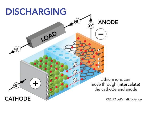
Before we dive into how the experiment worked, we need to learn more about how batteries work. On the left is a diagram showing the inner workings of a cell phone battery . As seen in the diagram, electrons move from one end of the wire to the other. Once they cross a load such as a light, the negatively charged electrons power the load, but at the same time turn into a positively charged electron. The positively charged electron then moves through the positive electrode (cathode) and travels through the electrolyte where it turns negative. After that the electron moves through the negative electrode (anode) and goes back through the wire to power the load. The problem with most batteries today is that the electrolyte is liquid which is unsafe because the liquid often leaks from the battery, which is toxic to touch. This is why scientists are working towards a solid electrolyte battery. Solid electrolytes, however, are hard to make because they require a balance of conductivity between the electrolytes and the positive and negative electrodes. This is what the scientists from PNNL and Microsoft were trying to solve in creating a solid electrolyte battery.
Why do we need new batteries?
As discussed above, Lithium-ion batteries are used in most electronics today. These batteries are good because they have a long lifespan and are compact. However, they have many problems such as overheating, leakage, and limited lithium supply. Lithium-ion batteries also contain cobalt, which is mostly mined through child labor. These children are forced to go into dangerous mines without proper safety equipment and work grueling hours. Also according to Aman Purewal, “The children in the mines are paid roughly six cents (USD) per day.”
There are solid state alternatives to Lithium-ion batteries that do not use cobalt. However, these batteries have their own problems. For example, Nickel-cadmium is a solid electrolyte battery that does not use cobalt. This type of battery is hard to damage, has a high energy density, so it can hold a lot of energy and can release a lot of energy in a short amount of time. However, nickel-cadmium batteries contain toxic materials, which pollute the environment when disposed of, and can not be used often because of their long charge time. From this list of commonly used batteries, it is obvious that there is no perfect battery type and all batteries have their pros or cons. This is why researchers from Microsoft and the Pacific Northwest National Laboratory wanted to find a new material to be used in solid state batteries.
The experiment
The researchers utilized Microsoft’s Azure Quantum Elements platform to conduct a screening of potential battery materials. Microsoft’s Azure Quantum Elements platform is a very high-tech platform with many AI algorithms and high-performance computing resources. The diagram on the right shows the steps researchers took to get down to their final electrolyte. The researchers started with entering a dataset with over 32 million materials into the platform. The materials were then filtered through many different AI algorithms which filtered through the materials based on predefined criteria, such as stability, conductivity, and cost-effectiveness. This screening process ended with only 18 promising materials. These 18 promising materials were then put through more screening processes directed by the scientists until they found the best material. But what was this material?
The results
With help from AI, researchers were able to identify a new solid-state electrolyte material that could
be used in batteries. This material is a combination of sodium, lithium, yttrium, and chloride ions. With these combinations of elements you create a solid state electrolyte which uses 70% less lithium, no cobalt and still has all the same benefits from a normal Lithium-Ion battery. Also according to Victoria Atkinson, this new battery material could “drastically reduce the price and environmental impact of these batteries in the future.” While further testing is required, the successful application of this AI in battery research gives the field hope on how AI can help humans in different parts of science.
References
“Scientists used AI to build a low-lithium battery from a new material that took just hours to discover” from Live Science: Conover, J. (2022, January 11).
“Artificial intelligence helped scientists create a new type of battery” from Science News: https://www.sciencenews.org/article/artificial-intelligence-new-battery Calma, J. (2024, January 16). Artificial intelligence helped scientists create a new type of battery. Science News. Retrieved from https://www.sciencenews.org/article/artificial-intelligence-new-battery Calma, J. (2024, January 9). AI helps scientists create new battery type in under 80 hours. CoinGeek. Retrieved from https://coingeek.com/ai-helps-scientists-create-new-battery-type-in-under-80-hours/ Gizbot. (n.d.). AI might soon be able to predict your EV battery degradation. Gizbot. Retrieved from https://www.gizbot.com/electric-vehicles/ai-might-soon-be-able-to-predict-your-ev-batterydegradation-082829.html
Let’s Talk Science. (n.d.). How Does a Lithium-Ion Battery Work? Let’s Talk Science. Retrieved from https://letstalkscience.ca/educational-resources/stem-explained/how-does-a-lithium-ion-bat tery-work
Thermo Fisher Scientific. (n.d.). Electrolyte Materials in Lithium-Ion Batteries. Thermo Fisher Scientific. Retrieved from https://www.thermofisher.com/blog/materials/electrolyte-materials-in-lithium-ion-batteries/ Lardinois, F. (2024, January 9). Microsoft puts Azure Quantum Elements to work. Tech Crunch. https://techcrunch.com/2024/01/09/microsoft-puts-azure-quantum-elements-to-work/? guccounter=1&guce_referrer=aHR0cHM6Ly93d3cuZ29vZ2xlLmNvbS8&guce_refer rer_sig=AQAAAIJamTdwefugcDlaYGqjKXXcGS4Jon7zwYPm4M9NNFHQ50K_J03quOC QDzsZxuZ2mhpeDhL6IKhLghnH1_IwM1ObGwUtFFMkLLj22LzQ-VRkGwt3kZYKVai4w pAzDTZZhSBIZx_cnZtjwcGXhC730QPV3hPRUj-sLgsvLp7A8vLr
The past, present, and future of earthquakes in the northeastern United States
Gray McGuinness, ‘26
At about 10:23 am on April 5th, 2024, an earthquake with a magnitude of 4.8 on the Richter scale rattled the Northeast and mid-Atlantic United States. It lasted only about thirty seconds, was relatively benign, and its only real consequences were reports of some people’s fragile glassware shattering; many people didn’t even notice it. It has long been a common misconception that the Northeast is seismically inactive. However, this isn’t true; no region can entirely be free of seismic activity. While it is true that earthquakes rarely happen in this part of the country, was this really geologically unusual?
Earthquakes in the Northeast are few and far between, and one with a magnitude of 4.8 is even less common on human timescales. A 4.8 magnitude earthquake is quite common, for example, in California. However, in the Northeast, this caliber of earthquake is really only a once-or-twice-in-a-lifetime event. This was many people in our area’s first experience with an earthquake. But, geologically speaking, this is certainly not unprecedented. In fact, about 1100 million years ago (Mya) the northeastern United States was one of the most seismically active places on the planet.
1100 Mya, the Laurentian and Amazonian plates converged, creating the supercontinent Rodinia.
The result of this was the uplifting of what we now call the Appalachian Mountains. The collision induced significant geological instability, produced new faultlines, and was the beginning of significant seismic activity in the area that was, for the first time, identifiably the geologic forerunner of the Northeast. Extreme activity continued for another 600 Mya or so, until Rodinia began to separate back into the continents that exist in the modern era, though the new North America stayed together. At this point, a large deposit of bedrock was formed a few miles beneath the surface of the area, provoked by the collision and breakup of Rodinia. About 200300 Mya, significant tectonic movements in the Northeast ground to a halt. Today, many of the earthquakes that we experience here are almost entirely due to the periodic redistribution and shifting of that layer of bedrock, not due to subduction, or any other more dramatic seismic processes, with the exception of certain fault lines in the New York metropolitan area. This has the effect of producing small magnitude events every so often.
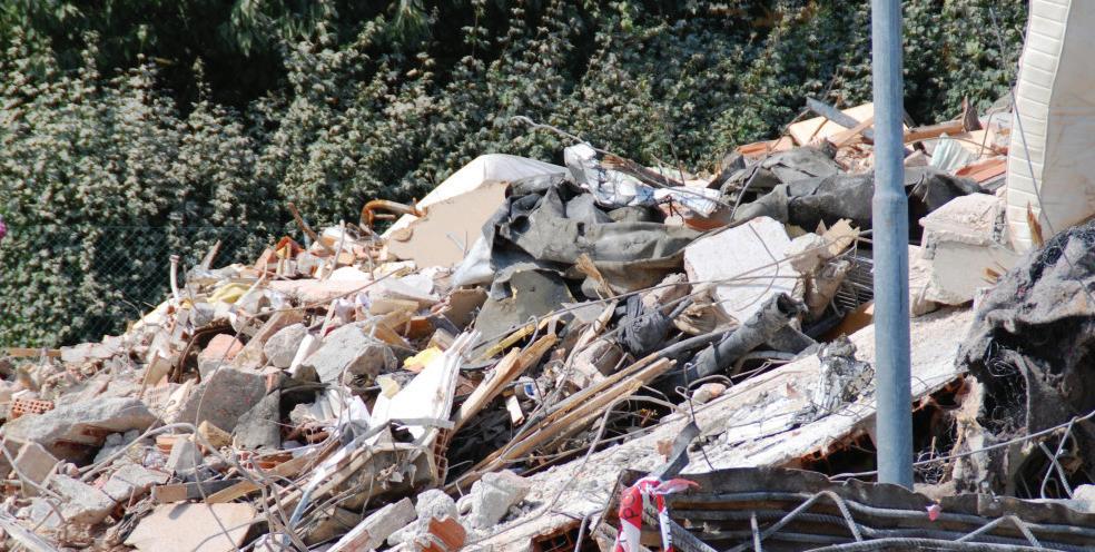
Earthquakes in the recent history of our area seem to have been relatively moderate in magnitude. Earthquakes with an extremely high magnitude leave significant amounts of physical evidence behind, including the raising or recession of land, shoreline alterations, sediment deposits, and others. None of that evidence has ever been found in the Northeast. Since European contact, there have been several notable instances of medium-high magnitude earthquakes in the Northeast. In 1755, for example, a damaging earthquake struck near Cape Ann, north of Boston, with an estimated magnitude of about 6.2 on the Richter scale. This earthquake was particularly damaging, collapsing houses and chimneys, causing springs to dry up and rivers to change course, and even producing a small tsunami. It was felt as far away as the Carolinas to the south and in Nova Scotia to the north. In 1783, a magnitude 5.3 earthquake struck in New Jersey. It was felt in a wide area, by people from New Hampshire to western Pennsylvania, including, notably, George Washington and his army in New York City, at the height of the American Revolution. These events in particular demonstrate a notable geologic quirk of the Northeast; the crust of our region is unusually dispersive. Very simply, when there is an earthquake in the Northeast, the vibrations can be felt much further from the epicenter than they can in most other areas. As the most densely populated part of the United States, if there is a high-
magnitude earthquake here, its most damaging effects will impact people over a far greater area than they would if the earthquake were to happen in San Francisco, for example.
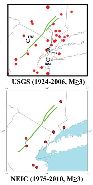
The New York metropolitan area has, notably, been subject to more earthquakes than most other parts of the Northeast. This is due to its proximity to the Ramapo fault line (Figure 2), which is a medium-small sized fault located in western New Jersey and eastern Pennsylvania. It is theoretically capable of producing an earthquake with a magnitude of up to 6.0 or 7.0, which would be a significant earthquake in this part of the country. Though, as previously stated, this doesn’t seem to have taken place with any kind of regularity in the recent geologic past, as there is essentially no physical evidence of such a traumatic event occurring here. An earthquake of that magnitude would have the capacity to significantly reshape the land, and that has clearly not happened recently. The Ramapo fault line has been the source of a number of relatively significant earthquakes in recent times, including April 5th’s earthquake.
Tectonic activity in the Northeast is prevalent, though typically mild. Accordingly, April 5th’s magnitude 4.8 was well within the realm of normal for our region. Actually, this particular earthquake was much less severe than previous ones. With the potential output of the Ramapo fault in mind, it is entirely possible that another earthquake of 6.0 or more could take place, though this is impossible to predict with any accuracy. In some cases, it is possible to guess, based on what we know about previous earthquakes, roughly how often one might take place in a certain area. However, there seems to be no recognizable pattern to when or where earthquakes take place along the Ramapo fault, or any other nearby faultline.
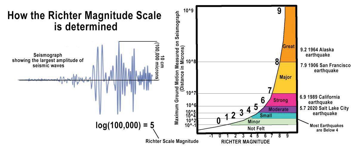
It is important to note that the Richter scale is logarithmic. Therefore, each level of intensity on the Richter scale is a magnitude of 10 times more extreme than the previous one. So, if an earthquake of, for instance, magnitude 6.0, were to take place, we would experience the effects of April 5th’s earthquake, but tenfold (see Figure 4). A magnitude 6 or 7 earthquake, which is, as previously stated, an entirely possible event in the tri-state area, would be an extremely damaging event. The 1989 Loma Prieta earthquake, which devastated California’s Bay Area, was measured as being 6.9 on the Richter scale. That earthquake collapsed numerous bridges, liquified soil, and caused at least 60 deaths and $6 billion worth of damage. But, earthquakes are quite common in California; they were relatively prepared. That sort of event, extrapolated onto New York City, would be much worse. New York’s population density is far higher, its buildings taller, and its population and infrastructure totally unprepared for the possibility of a damaging earthquake. While researchers have determined that it is theoretically possible for the Ramapo fault, or other nearby faults, to produce an earthquake of a magnitude similar to that of Loma Prieta, there is little indication that that sort of an event could take place here in the near future. Given that there seems to have been few earthquakes of that caliber in the recent past in the New York area, it would seem unlikely that one will happen anytime soon. However, it remains impossible to predict earthquakes with any certainty.
In summary, earthquakes are not an unusual occurrence in the Northeast. While they are typically not severe, that does not mean that they are uncommon. April 5th, 2024’s earthquake was not a deviation from what we know about the geology of the Northeast. Rather, it was completely within the precedent of previous, similar earthquakes that have happened in the Northeast over the past few decades. While it was of a slightly higher magnitude than is typical, this is simply a matter of probability; sometimes the slips are large, sometimes they are small. There is no reason to believe that this particular earthquake is an indication of any new trend or any increase in tectonic activity in our area. April 5th’s earthquake was, almost certainly, business as usual; unremarkable for geologists who study the Northeast.
References
American Museum of Natural History. (n.d.). New York City & Regional Geology. AMNH. https://www.amnh.org/research/physical-sciences/earth-and-planetary-sciences/public-out reach/new-york-city-geology
Appalachian Basin Geology | EARTH 109 Fundamentals of Shale Energy Development. (n.d.). College of Earth and Mineral Sciences at Pennsylvania State University. https://www.e-education.psu.edu/earth109/node/973
Clark, S. H. B., U.S. Geological Survey, & U.S. Department of the Interior. (n.d.). Birth of the Mountains: The geologic story of the Southern Appalachian Mountains. https://pubs.usgs.gov/gip/birth/birth.pdf
Federal Emergency Management Agency, Pakiser, P. A., United States Geological Society, National Earthquake Hazard Reduction Program, New Jersey Geological and Water Survey, New Jersey Department of Environmental Protection, New Jersey Department of Transportation, & American Association of State Highway and Transportation Officials. (2019). State of New Jersey 2019 All-Hazard Mitigation Plan. State of New Jersey. https://www.nj.gov/njoem/mitigation/pdf/2019/mit2019_section5-5_Earthquake.pdf
Kafka, A. [Alan kafka]. (2011, January 8). File:NYC Seis.png. Wikipedia Commons. https://commons.wikimedia.org/wiki/File:NYC_Seis.png Lithium6ion. (2010, November 26). File:RamapoFaultSystem.png. Wikipedia Commons. https://commons.wikimedia.org/wiki/File:RamapoFaultSystem.png Nietzke Adamo, L., Wright, J., Bourke, J., Miller, K., Kinney, S., & Schlische, R. (2024, April 19). What Was it Like Being a Rutgers Geologist During a Magnitude 4.8 Earthquake? Rutgers Climate and Energy Institute (RCEI).
https://rcei.rutgers.edu/what-was-it-like-being-a-rutgers-geologist-during-a-magnitude-4-8earthquake/ Northeast States Emergency Consortium. (n.d.). Earthquakes Hazards. https://nesec.org/earthquakes-hazards/ Solar, G., & Us, C. (2024, April 5). What Causes Earthquakes in the Northeast like the Magnitude 4.8 One in New Jersey? Scientific American. https://www.scientificamerican.com/article/what-causes-earthquakes-in-the-northeast/ Stokem, S. (n.d.). April 5 earthquake: the strongest in NJ in over 240 years. The Lancer Ledger. https://lancerledger.com/10705/news/april-5-earthquake-the-strongest-in-nj-in-over-240years/
United States Geological Survey. (n.d.-a). Geology of the New York Region.
Locomotor advances in early dinosaurs helped them outcompete other species
Nicholas Penna, ‘25
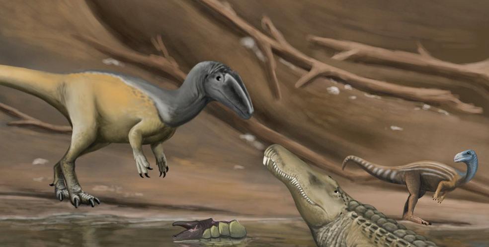
The Triassic period was a unique time in the history of our planet where life took up some of the strangest most bizarre alien forms. the competition was fierce and a catastrophic Permian extinction, killed 70 percent of life on land, paleontologists are puzzled as to why out of all of these remarkable animals, dinosaurs were the ones who rose to conquer the earth. A new study led by University of Bristol researcher Amy Shiply suggests that the dinosaur’s secret weapon was dynamic locomotion.
The dinosaur’s success can partially be attributed to their erect posture. According to the paper, ”In a sprawling posture, movement of the limbs causes lateral bending of the vertebral column which compresses the lungs and limits ventilation. An erect posture positions the limbs underneath the body, so this bending does not occur, thus removing any respiratory constraint and allowing increased efficiency during locomotion” (Shipley, 02, 07, 2024).
This new type of locomotion is similar to humans; without the compression of the lungs, the organism can be far more active than its competitors, which is part of the reason that dinosaurs rose to dominance. Researchers believe that these adaptations are tied to endothermy, as both dinosaurs and mammals that have these adaptations are endotherms.
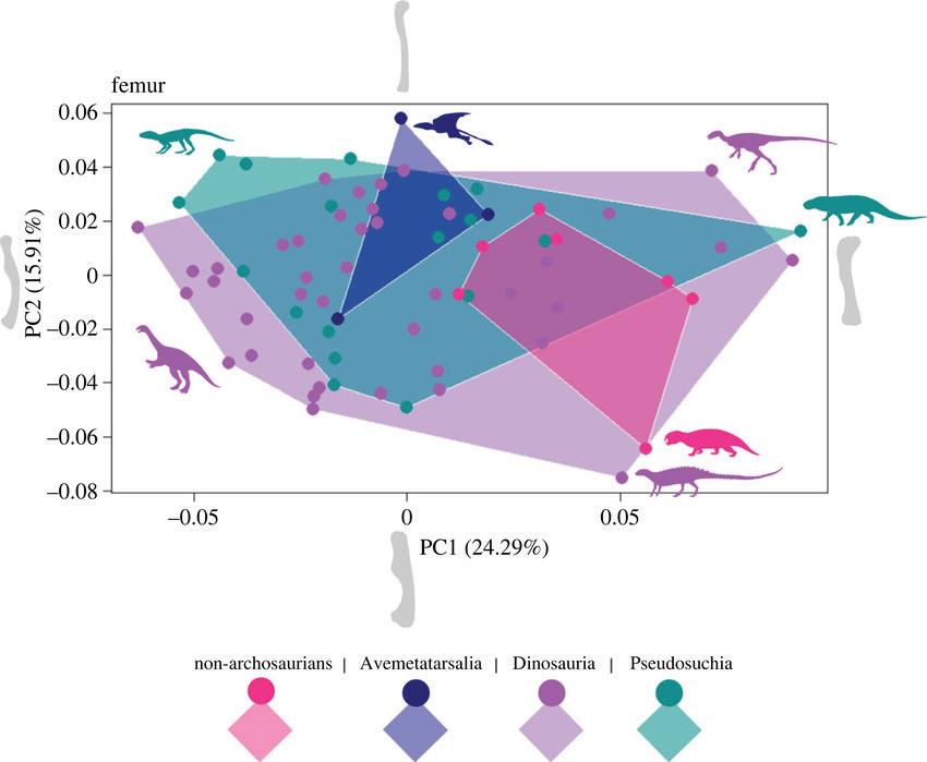
This study looked at 208 different species from the group Archosauromorpha from the late Permian to the early Triassic (259.1-174.1 Mya). For each of the 208 selected animals they examined their hindlimbs and forelimbs. They found that the dinosaurs show much more variation in limb bone shape and show a general trend of disparity as time goes on.
Next, the researchers looked at an index of quadrupedality (walking on all fours) and cursoriality.
(running ability). To find out the quadripedality and cursoriality of the animals they used the quadripedality index which assesses the comparative length of the fore limb to the hind limb. If the animal has a shorter forelimb then it is bipedal. They also used the cursoriality index which measures the length of metatarsal III to the femur. A longer metatarsal is better for running making it more cursorial. They found that most dinosaurs were not quadrupedal; rather they were bipedal. They also found that dinosaurs and their close relatives the avemetatarsalians had the highest cursoriality. This shows that the dinosaurs were more bipedal and cursorial, whereas psedosuchains were less cursorial and quadripedal. Interestingly, this adaptation for bipedality and corporality was not shared by all dinosaurs, especially towards the end Triassic and Jurassic when large quadripedal forms such as sauropods and sauropodomorphs began to emerge. However, many dinosaurs were both bipedal and cursorial. Additionally, dinosaurs had a much more erect posture, which is more beneficial and allowed them to reach such gargantuan sizes.
Overall this study shows how the right adaptations mixed with the right conditions are the perfect recipe for a clade to take over the world.
References
Shipley, Amy E., et al. “Locomotion and the early mesozoic success of archosauromorpha.” Royal Society Open Science, vol. 11, no. 2, Feb. 2024 https://doi.org/10.1098/rsos.231495.
@Paleotaku. #Sciart #Paleoart #Dinosaur Gojirasaurus and Machaeroprosopus. X (formerly Twitter), 12, Aug. 2021. 03:20
A new dawn in cancer treatment: Targeted therapies offer precision and promise
Nolan Francis, ‘26
Introduction
For decades, the fight against cancer has relied heavily on chemotherapy, a broad-spectrum approach that often leaves a trail of devastation in its wake. Healthy cells, alongside cancerous ones, bear the brunt of chemotherapy’s attack, leading to a multitude of debilitating side effects like fatigue, nausea, and hair loss. However, a revolutionary shift is underway with the development of targeted anti-cancer agents. These drugs hold immense promise for a more precise and effective approach to cancer treatment, offering the potential for significantly fewer side effects and a brighter outlook for patients.
Monoclonal antibodies
At the heart of cancer lie mutations in genes that control cell growth and division. These mutations unleash uncontrolled cellular proliferation, leading to the formation of tumors. Targeted anti-cancer agents, as the name suggests, are like microscopic assassins wielding the knowledge of these specific abnormalities. They exploit these very mutations, interfering with the molecular pathways that fuel cancer’s growth and survival.
This targeted approach takes on several forms. Monoclonal antibodies, for example, act like highly trained immune system mimics. Engineered to biologically recognize and bind to specific proteins adorning the surface of cancer cells, these antibodies trigger a multi-pronged attack. They can flag the cancer cells for destruction by the body’s immune system or directly inhibit their ability to grow and divide.
Small molecule drugs
Another weapon in the targeted therapy arsenal is small-molecule drugs such as Elevidys. Such drugs were developed by injecting genes with a micropipette directly into a living mammalian cell or other exposed cells to a precipitate of DNA containing the desired genes.These drugs, meticulously designed to fit into specific pockets on proteins within cancer cells, disrupt essential cellular processes. By occupying these critical sites, they can dismantle signaling pathways or hinder DNA replication, ultimately leading to the demise of the cancer cell.
The significance of targeted anti-cancer agents lies not just in minimizing side effects. Compared to traditional chemotherapy’s indiscriminate assault, targeted therapy offers a laser-like focus. This precise approach has the potential to achieve superior tumor shrinkage and significantly improve patient outcomes.
Conclusion
The field of targeted cancer therapy is on a fast track, constantly evolving as researchers tirelessly identify new mutations and molecular pathways unique to different cancers. This ongoing quest fuels the development of even more precise and potent targeted agents, offering a continually refined arsenal in the fight against this complex disease.
However, it’s crucial to acknowledge that targeted therapy is not a magic bullet. Cancer, with its multitude of underlying causes, presents a multifaceted challenge. Not all cancers have readily identifiable targets, and even for those that do, resistance can develop over time.
Despite these limitations, the future of cancer treatment gleams brighter with the continued development of targeted anti-cancer agents. As researchers delve deeper into the molecular underpinnings of cancer, they will continue to unlock new targets and refine existing therapies. This paves the way for a more personalized approach to cancer treatment, offering patients a greater chance of successful outcomes and a significantly improved quality of life.
References
American Cancer Society (2023, January 11). Targeted drug therapy. Retrieved from https://www.cancer.org/cancer/managing-cancer/treatment-types/targeted-therapy.html
National Cancer Institute (2022, October 26). Targeted cancer therapies. Retrieved from https://www.cancer.gov/about-cancer/treatment/types/targeted-therapies
Beckers, T (2021, September 29). Small molecules in targeted cancer therapy: advances, challenges, and future perspectives. Retrieved from https://www.nature.com/articles/s43018-023-00664-2
Frost, H (2022, December). Patient attrition in Molecular Tumor Boards: A Review. Retrieved from www.medrxiv.org/content/10.1101/2021.10.07.21264241v2.full.pdf
Message from outgoing editor William Boberski
Dear Reader,
Thank you for joining us for the STEM Journal’s spring 2024 issue! While I am no longer this issue’s Editor-in-Chief, I am still excited to share the research that our club’s writers, and the larger Staples science community, have to offer. In the following section, the STEM Journal is publishing posters and abstracts from the student-scientists of this year’s inaugural Scientific Research Honors class.
Scientific Research Honors provides the opportunity for students to independently research, design, carry out, and present an original scientific experiment. Each project poster represents a genuine contribution to scientific knowledge in a diverse array of fields, and is the result of countless hours of reading through scientific literature, practicing laboratory techniques, collaborating with peers and teachers, and presenting at science fairs.
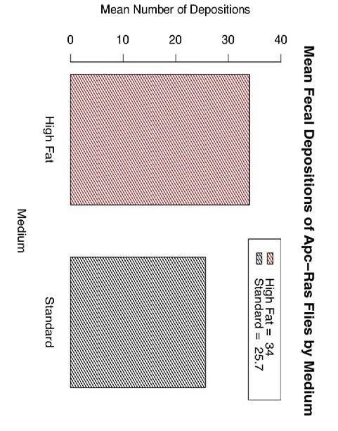
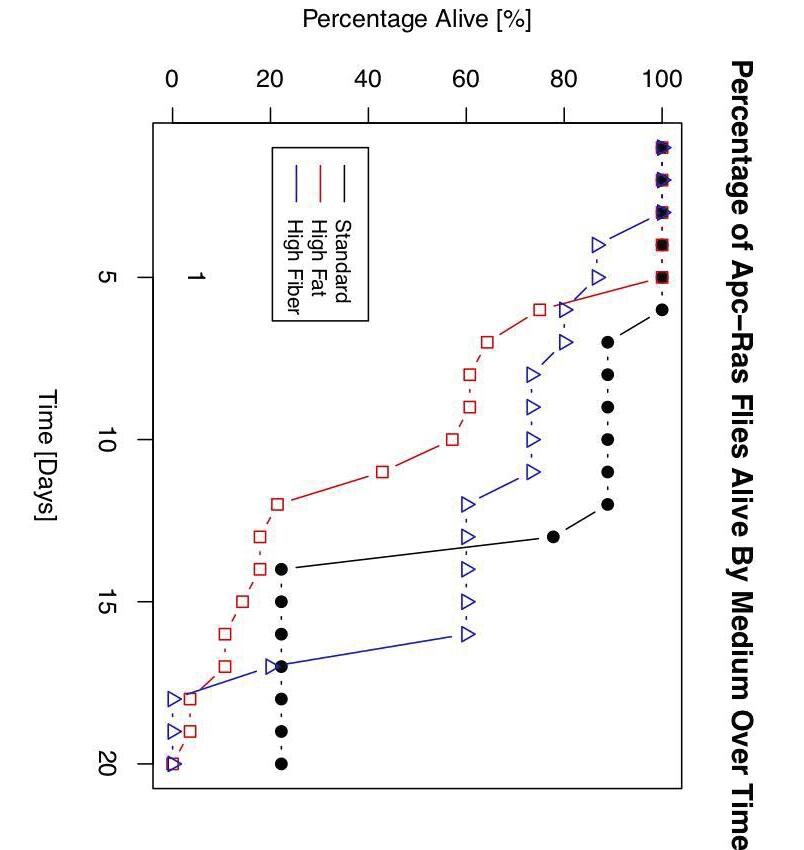
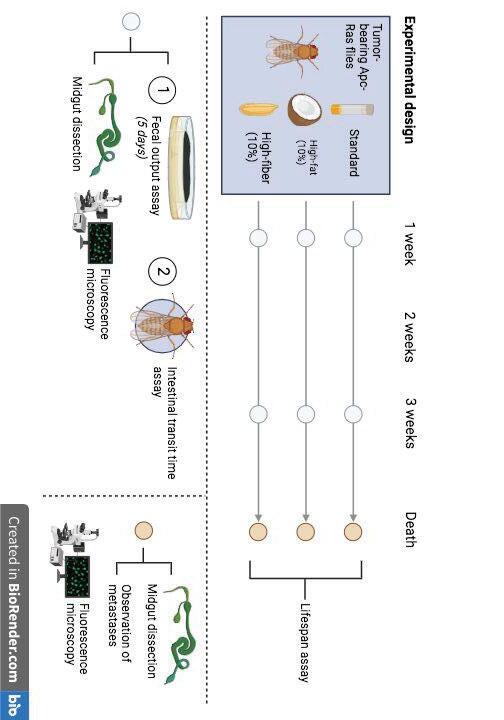
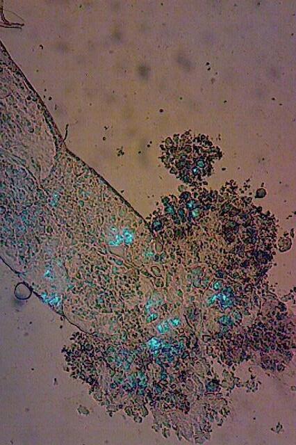
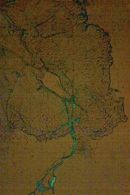
I took Scientific Research Honors this year, where I studied the effects of diet on a fruit fly model of colorectal cancer, and I plan to continue my project in the class next year. Ever since my first review of scientific literature last summer, the research, critical thinking and writing skills I developed as a writer and editor for the STEM Journal have been relevant to my project. While designing an original experiment seemed daunting at first, it rests on the same principle as the fascinating articles you just read: let your curiosity guide you to a paper or field of study, and keep asking questions! As budding scientists, we aspire to lead with curiosity, and I hope that this STEM Journal issue inspires you to do the same.
- William Boberski, ‘25 Outgoing Editor
The next few pages consist of projects produced by students in the 2023-2024 Scientific Research class at Staples. The work of the following students has been featured:
• Ellen Ou ‘24
• Lucas Nilsson ‘27
• Annam Olasewere ‘25
• Tessa Cassell ‘24
• Alex Esser ‘25
• Sebastian Rodriguez ‘25
• Julianna Gallo ‘25
• Kendra Chang ‘25
• Sierra Denkin ‘26
• William Boberski ‘25
• Nolan Francis ‘26
• Sara De Pinho ‘25
● Process for algae will be similar, although no nitric acid will be used; algae will be centrifuged and filtered out and solution will be analyzed as is.
● When soil or media is extracted and analyzed, it will be done through flame atomic emission spectrometry
● Solution will be filtered, and then sent to WCSU for testing
● Soil will be obtained, then digested in 3M nitric acid (Fig 4)
● Algae will also be stained with Trypan blue at the end of treatment to analyze cell death levels.
● Earthworm health and death rate (if applicable) will be monitored
● Algae will be analyzed every week by OD680 in order to determine density changes in response to the heavy metal. ( Fernandez )
(Fig 2) Analysis
● Algae will be introduced to copper concentrations of 1.15 mg/L (Braglia, 2021)
● Earthworms and mushrooms will be introduced to copper concentrations of 40 mg/kg (Xiao, 2022) (Fig 2)
● Copper (a representation of heavy metal ions) will be dissolved into distilled water in the form of CuCl 2 and added to soil or media where various bioremediators are growing (Fig 2) ( Guo )
Copper Concentrations
○ Earthworms will be kept in soil, which will then be contaminated (Fig 3)
○ Mushrooms will be grown in Blooming Acres All in One Grain Growth Bags, then transferred to soil for remediation
will be transferred into contaminated soil when mature
○ Algae will be grown in the Caroline Labs AlgaGro Freshwater Medium
● Green algae (Chlorella vulgaris) will be analyzed at stagnant and maximum growth phases, which was determined by their optical density. Previous studies showed maximum growth phases at OD680 of approximately 0.4 and a stagnant growth phase at 1.2 (Fernandez).
Bioremediator growth
METHODS
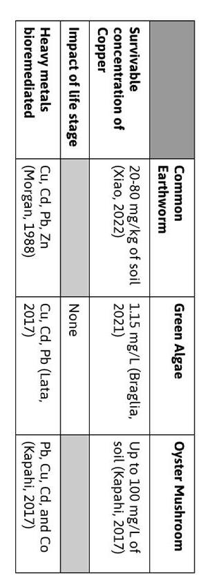
Table 1: Review of previous studies referenced by methodology and experiment development
● Additionally, there are no previous studies comparing the effectiveness of them directly in the same study, or analyzing the impact of life stage on removal effectiveness (Table 1)
● Bioremediators chosen because of prevalence, hardiness, and lack of toxicity
● Bioremediators have been proven to work in the past on cadmium levels (Lata, 2017), and similar species have been observed to work with copper as well (Morgan, 1988) (Table 1)
● Three bioremediators will be compared: Common earthworms (Morgan, 1988), Oyster mushrooms (Lata, 2017), and Green algae (Lata, 2017) (Table 1)
● Copper is considered less dangerous than cadmium and lead, therefore this laboratory analysis will use copper ions (Amaroso, 2020)
● Many heavy metals, such as cadmium (Cd), copper (Cu), zinc (Zn ) and lead (Pb), are similar because they have a 2+ charge and are part of the same family of element. (Amaroso, 2020)
● Bioremediation is the process of using plants, animals, or other biotic factors to help remove pollutants from the environment.
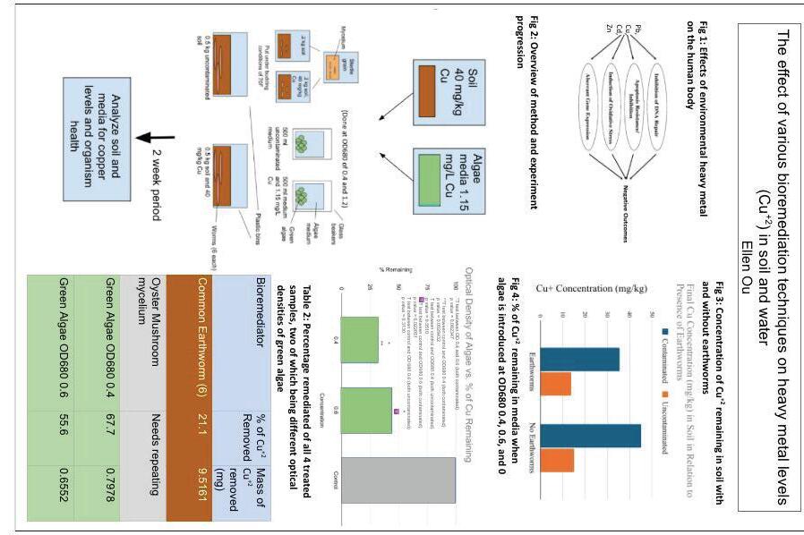
● Heavy metals are sometimes used in paints and batteries (Fig 1), and prolonged exposure or digestion can lead to serious health complications (Fig 1). Removing heavy metals from soil with machinery is a slow, expensive process (Xu, 2020).
INTRODUCTION AND BACKGROUND
https://www.ncbi.nlm.nih.gov/pmc/articles/PMC9055442/ [2] Andjelkovic, M., Buha Djordjevic, A., Antonijevic, E., Antonijevic, B., Stanic, M., Kotur-Stevuljevic, J., Spasojevic-Kalimanovska, V., Jovanovic, M., Boricic, N., Wallace, D., & Bulat, Z. (2019). Toxic Effect of Acute Cadmium and Lead Exposure in Rat Blood, Liver, and Kidney. International Journal of Environmental Research and Public Health 16 (2), 274. https://doi.org/10.3390/ijerph16020274 [3] Lata, S., Kaur, H., & Mishra, T. (2019). Cadmium bioremediation: A review. International Journal of Pharmaceutical Sciences and Research https://www.researchgate.net/publication/339712646_CADMIUM_BIOREMEDIATION_A_R EVIEW [4] Pitt, M. J. In Bretherick’s Handbook of Reactive Chemical Hazards 6th ed.; Urben, P. G. Ed.; Butterworth-Heinemann Ltd: Oxford, 1999; Vol. 2, pp 307-312. [5] Large scale gold refining by the aqua regia acid method (n.d.). Shor International. Retrieved August 28, 2023, from sdfsdfs https://www.ishor.com/large-scale-gold-refining-using-aqua-regia#:~:text=The%20ni tric%20acid%20from%20the,chlorides%20unchanged%20and%20in%20solution. [6] Santoro, A., Held, A., Linsinger, T., Perez, A., & Ricci, M. (2017, April). Comparison of total and aqua regia extractability of heavy metals in sewage sludge: The case study of a certified reference material. ScienceDirect https://www.sciencedirect.com/science/article/pii/S0165993616303922?ref=cra_js_challe nge&fr=RR-1 [7] Reusch, W. (2013, May 5). Mass spectrometry . Michigan State University Chemistry. Retrieved August 28, 2023, from https://www2.chemistry.msu.edu/faculty/reusch/virttxtjml/spectrpy/massspec/masspec1.h tm
[1] Xu, M., Liu, Y., Deng, Y., Zhang, S., & Hao, X. (2020). Bioremediation of cadmium-contaminated paddy soil using an autotrophic and heterotrophic mixture. PubMed
CITED
To Dr. Baluha, for generously agreed to help analyze soil and media samples using the Flame Atomic Emission Spectrometer at Western Connecticut State University. WORK
To Mrs. Parent, for tons of lab and technical support and help editing my abstract and poster, as well as suggesting methodologies and giving invaluable feedback at every stage of this project
ACKNOWLEDGEMENTS
○ Through studying species we already know as widespread, it can broaden understanding about the potential of common organisms in removing heavy metals from the environment
● Many bioremediation techniques have difficulty being implemented due to concerns regarding biodiversity and invasive species
● It is anticipated that the bioremediation effectiveness in copper uptake will reflect upon their proficiency at remediating other heavy metals as well
● Though data was not statistically significant, it might imply a correlation that can be further explored between the life stage of algae and its bioremediation rate.
● Bioremediation is a promising solution to environmental heavy metal
IMPLICATIONS and FUTURE
RESEARCH
● However, in terms of total mass removed overall, evidence showed that earthworms might be the most efficient. (Table 2)
● Overall, green algae was the bioremediator that exhibited the most copper removal by percentage (Table 2)
○ Uncontaminated soil showed a 9.3% decrease in Cu +2 likely due to there being small levels of Cu +2 in local soils used. (Figure 3)
○ Contaminated soil treated with earthworms displayed a 21.1% change in Cu +2 (Figure 3). This implies higher efficiency (mg/day)
○ Some actually seemed more active, although this may have been due to erroneous factors
● Earthworms exhibited little change in physiology or survival after being introduced to copper contaminated environments
○ Over a 2 week period, samples were analyzed but data was incongruous, leading to an inability to process it.
● Oyster mushrooms were observed to be able to grow in Cu +2
○ Therefore, there is not enough evidence to suggest that life stage has a positive effect on the rate of bioremediation of green algae.
■ However, the difference in remediation rates should be noted for further study
○ After performing a single tailed T Test, it can be concluded that his is not statistically significant data
○ Green algae treatment at OD680 0.4 led to a 67.7% change in Cu +2 compared to a control, while green algae treatment at .6 led to a 55.6% change (Figure 4)
● Green algae proved to be effective in remediating
Cu +2 contamination
RESULTS
● Precision, recall and mean average precision (mAP) were measured on both pre-processed and raw models. (B Juba, 2019)
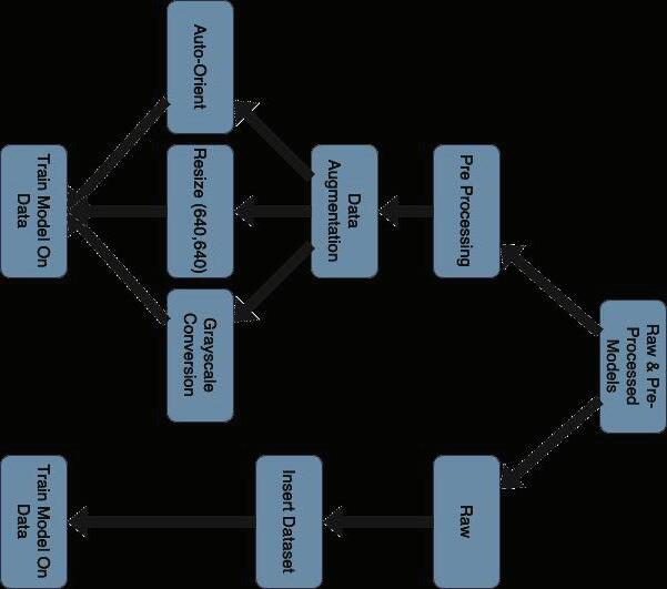
● Pre-processing techniques for improving image clarity and consistency include grayscale conversion, auto-orientation and resizing. (Figure 2) These are vital for enabling accurate detection and classification of traffic signs by AI models. (E Karantoumanis, 2022)
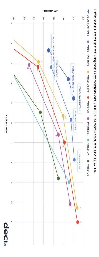
● Deep learning models for object detection such as You Only Look Once (YOLO) are known for speed and accuracy. YOLO NAS is their newest and most advanced open source object detection model.(J Terven, 2023) (Figure 1)
● The rising importance of adhering to wrong-way traffic signs, calls to question the reliability of noticing wrong-way traffic. (RV Ponnaluri, 2016)
● In 2022, Connecticut experienced an increase in wrong-way driving incidents, leading to 23 fatal collisions among various vehicle types(News8, 2024).
In 2022, Connecticut witnessed a surge in wrong-way collisions among all vehicles, totaling 23 fatal accidents ( News8, 2024 ). The gravity of this statistic calls into question the reliability of noticing wrong way traffic signs on our roads. (RV Ponnaluri, 2016) This project investigates the accuracy of different approaches of training modern image interpretation models to detect traffic sings, including “wrong way” signs. Pre-processing methods like grayscale conversion, auto-orientation and resizing to (640, 640) are applied to YOLO (You Only Look Once), utilizing the COCO dataset (J Terven, 2023; TY Lin, 2014) Raw, and pre-processing techniques were studied measuring their performance through mean average precision (mAP), precision and recall. This approach aims to evaluate the efficacy and efficiency of pre-processing recognizing traffic signs under varied conditions. (B Juba, 2019) It is hypothesized that while pre-processing will benefit the YOLO model, due to the advancements in AI, it will be minimal. In this study, it was observed that the model trained with pre-processed data exhibited a 1.4% higher mean Average Precision (mAP) and a 2.4% increase in recall compared to the model trained on raw data. This slight improvement demonstrates that although pre-processing can enhance the model's ability to correctly identify and locate objects, the advancements in artificial intelligence, specifically in YOLO models, are significant enough that such enhancements may contribute minimally. This suggests an evolving capability in AI models that functions more akin to human visual processing, which might eventually reduce the need for intensive pre-processing steps.
● E. Karantoumanis, V. Balafas, M. Louta and N. Ploskas, "Computational comparison of image preprocessing techniques for plant diseases detection," 2022 7th South-East Europe Design Automation, Computer Engineering, Computer Networks and Social Media Conference (SEEDA-CECNSM), Ioannina, Greece, 2022, pp. 1-5
● Juba, B., & Le, H. S. (2019). Precision-Recall versus Accuracy and the Role of Large Data Sets. Proceedings of the AAAI Conference on Artificial Intelligence 33 (01), 4039-4048. https://doi.org/10.1609/aaai.v33i01.33014039
● RV Ponnaluri, (2016), The odds of wrong-way crashes and resulting fatalities: A comprehensive analysis, Accident Analysis & Prevention Volume 88, March 2016, Pages 105-116
● Lin, TY. (2014). Microsoft COCO: Common Objects in Context, https://link.springer.com/chapter/10.1007/978-3-319-10602-1_48
● J Terven, (2023), A Comprehensive Review of YOLO Architectures in Computer Vision: From YOLOv1 to YOLOv8 and YOLO-NAS, https://www.mdpi.com/2504-4990/5/4/83
References / Works Cited
● Thanks also to the various AI experts and AI forums that have helped with the coding and research for this project
● Special thanks to Amy Parent for her support in creating throughout the research project.
Methodology Background
● The findings of this project highlight the potential for modern machine learning models, like YOLO, to be integrated into low-cost, in-vehicle systems for real-time “wrong way” traffic sign detection, such as those shown in Figure 6, thereby potentially reducing the incidence of wrong-way driving accidents.
Implications
Figure: 2 Data Processing Steps
Nilsson
● The slight improvement indicates that the models are so advanced and therefore do not require pre-processing.
● The model trained with pre-processed data displayed a 1.4% better mean Average Precision (mAP) and a 2.4% increase in recall. ● This indicates a slight enhancement in its ability to correctly identify and locate objects.
Results
● The each model with mAP, precision and recall in order to compare the models' efficiency and effectiveness in recognizing traffic signs under varied conditions.
● Train both You Only Look Once (YOLO) models on each dataset. (Figure 4)
● Apply pre-processing techniques (grayscale conversion, auto-orientation, and resizing to 640, 640). (Figure 3)
● Load Images from COCO dataset loaded onto model.
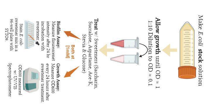
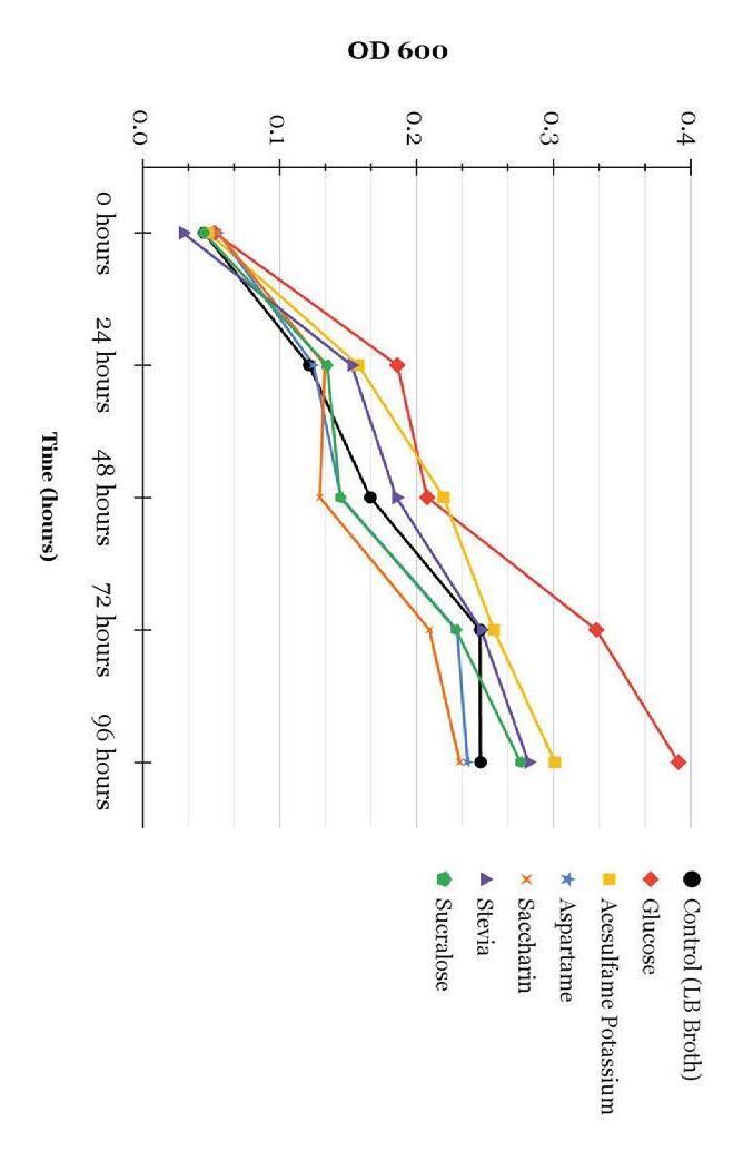
1. Growth Assessment : E. coli will be streaked on agar plates, colonies transferred to liquid media, than treated with the corresponding sweeteners, and growth measured via OD600 analysis every 24 hours for 3 days (Shil & Chichger, 2021)
A stock solution of E. coli strain ATCC 10536 will be grown until reaching an optical density (OD) of approx 0.1, then diluted to OD = 0.01 in 20% LB Broth at a concentration of 8x10 8 cells/mL and treated with 1mM concentrations of Ace-K, Aspartame, Sucralose, Saccharin, Stevia, and Glucose for 24 hours (Fig. 2)
Aspartame Decreases ( Shahriar et al. 2020) [0.00022 mM ]. Increase ( Shil & Chichger 2021) [0.1mM].
Acesulfame Potassium Shown to decrease ( Wang 2018) & increase ( Shahriar et al. 2020) [12.43 mM, 0.00022 mM].
Glucose Increases (Bren et al., 2016) [11.1 mM]. Decreases (Jackson et al., 2002) [11.1 mM].
2. Biofilm Formation Analysis : SweetenerE. coli mixtures will be plated in a 96 well plate with 1.2 x 10 6 cells per well, planktonic cells with then be removed from biofilms via PBS washing 24 hours post treatment, and absorbance will be quantified using SYTO 9 Stain at 600 nm (Li et al., 2022). Sweetener
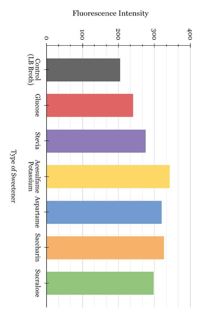
† Values were statistically significant, determined by a one-tailed t-test compared to stevia, wi/ p-values < 0.001.
**Values were statistically significant, determined by a one-tailed t-test compared to glucose, w/ p-values < 0.0005.
*Values were statistically significant, determined by a one-tailed t-test compared to LB Broth, w/ p-values < 0.005.
Sweetener Type
● This will be the first study to compare the same concentration of all AS to glucose in a singular study.
● Ace-K shown to both decrease and increase E. coli growth (Wang et al., 2018; Shahriar et al., 2020; Table 1)
● The effects of AS on E. coli has been examined by several researchers, although not collectively in one study, and at different concentrations, yielding conflicting results (see Table 1).
● The impact of natural and artificial sweeteners on E. coli is summarized in Table 1. Most AS decrease E. coli growth and increase biofilm formation
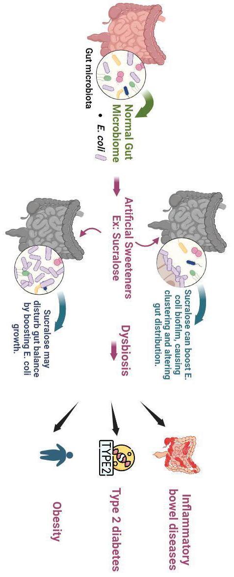
● Studying biofilm is crucial; AS-induced changes in E. coli may promote pathogenicity in the gut microbiome (Shil & Chichger, 2021)..
● The study aims to investigate the impact of saccharin, sucralose, aspartame, Ace-K, and stevia (common AS used in foods and beverages) on E. coli growth and biofilm formation compared to glucose or no sugar.
● AS impacts E. coli in the gut microbiome, where E. coli can comprise up to 5% of the microbial population. This is significant as changes in the gut microbiome, known as dysbiosis (via growth and biofilm formation), have been associated with conditions such as obesity, type 2 diabetes, and inflammatory diseases (Foster-Nyarko, 2022; see Fig. 1).
● Despite health risks, FDA approves these sweeteners (Shum & Georgia, 2021).
● AS, 13,000 times sweeter than sugar, can increase cravings and raise risks of various health issues (Bartolotto, 2015).
● 2 in 5 US adults, and 1 in 4 US children use artificial sweeteners (AS) daily (increasing annually) (Sylvetsky et al., 2017).
*The value observed after 24 hours for Ace-K was statistically significant, as determined by a one-tailed t-test compared to LB Broth (control), with a p-value of 0.00575.
I am grateful to Mrs. Amy Parent for her constructive feedback and support during the research process, as well as her invaluable assistance with the statistical analysis of the data.
Guide sweetener choices to minimize harm on blood sugar, gut, and health as depicted in Figure 1, while favoring natural sugars if necessary. W o r k c i t e d Li, Z., Gao, J., Guo, Y., Cui, Y., Wang, Y., Duan, W., & Wu, Z. (2022). Enhancement of antibiotic resistance dissemination by artificial sweetener acesulfame potassium: Insights from cell membrane, enzyme, energy supply and transcriptomics. Journal of Hazardous Materials 422 126942. https://doi.org/10.1016/j.jhazmat.2021.126942 Pang, M. D., Goossens, G. H., & Blaak, E. E. (2021). The impact of artificial sweeteners on body weight control and glucose homeostasis. Frontiers in Nutrition 7 https://doi.org/10.3389/fnut.2020.598340 Sharma, A., Amarnath, S., Thulasimani, M., & Ramaswamy, S. (2016). Artificial sweeteners as a sugar substitute: Are they really safe? Indian Journal of Pharmacology 48 (3), 237. https://doi.org/10.4103/0253-7613.182888 Shil, A., & Chichger, H. (2021). Artificial sweeteners negatively regulate pathogenic characteristics of two model gut bacteria, E. coli and E. faecalis. International Journal of Molecular Sciences 22 (10), 5228. https://doi.org/10.3390/ijms22105228 I m p l i c a t
● Despite also increasing biofilm formation in comparison to both the negative and positive controls, stevia, notably, induced lower biofilm formation than other artificial sweeteners, suggesting a potentially better sugar alternative (Fig. 4).
● All artificial sweeteners significantly increased biofilm formation compared to LB broth and glucose-treated E. coli ( Fig. 4).
● Growth assay trends generally aligned with literature, except for saccharin, for which there is a trend in inhibiting growth but it did not achieve statistical significance (n=2 due to resource constraints) (Fig. 3).
● Sweeteners had no significant impact on bacterial growth compared to LB broth control, except for Acesulfame Potassium (Ace-K) at 24 hours (p = 0.00575) (Fig. 3).
Figure 5: Study
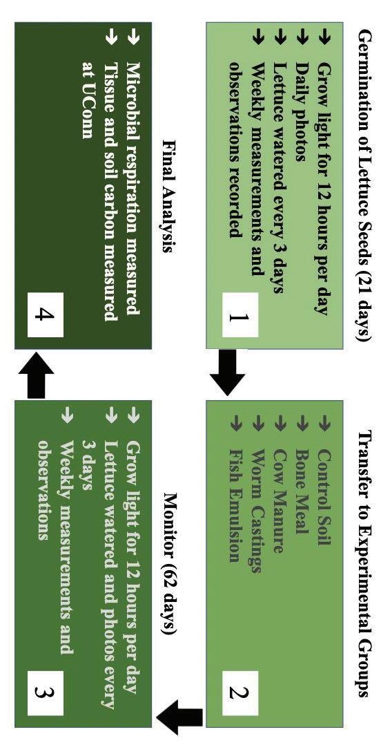
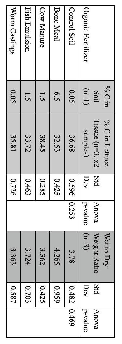
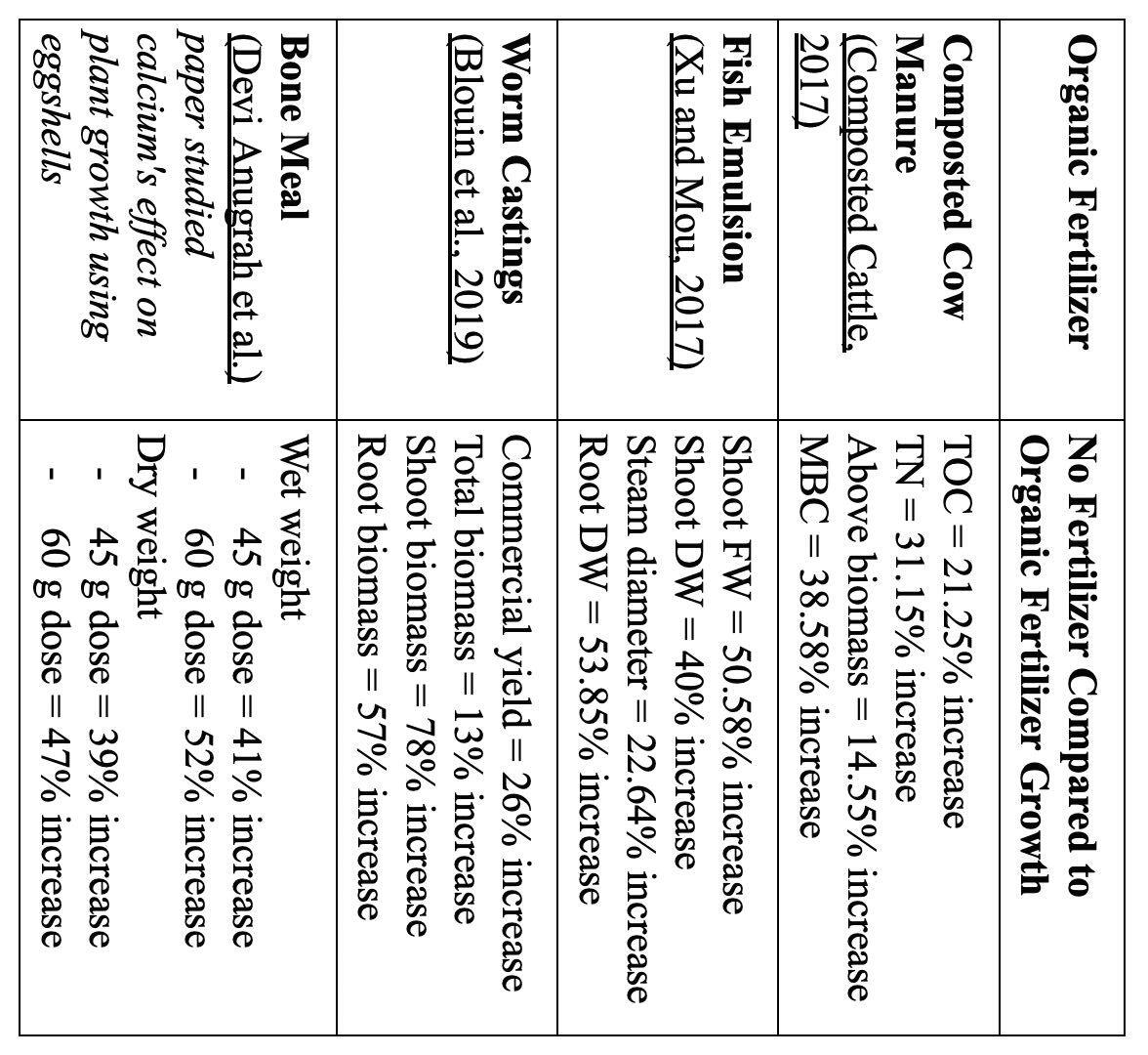
➔ The lettuce and soil samples from each fertilizer were measured by vario macro analyzer at UCONN
➔ Despite the differences in C levels in the soil, these variations did not translate into notable differences in lettuce growth, carbon sequestration, or soil microbial activity.
➔ Day 64, lettuce was cut at the stem and weighed. The soil was evaluated for CO2 levels with a Solvita test kit for 24 hours.
➔ No differences in wet to dry weight issues were noted between groups (ANOVA p-value 0.469)
➔ The lack of correlation between the C in soil, tissue, microbial respiration and plant growth suggest that the impact of these fertilizers most likely need to be studied on a longer timeline or used on a different type of crop.
➔ Germinated seedlings were transferred to larger size pots, each fertilizer pot had 3 germinated groups. Lettuce grew over 64 days (grow lights were on for 12 hours per day, progress photos and watering were every 3 days, and plant size and observations were recorded once per week.)
➔ Seedlings grew in germination pots for 21 days (grow lights were on for 12 hours, daily progress photos taken, seedlings were watered every 3 days and plant size and observations were recorded once per week.
➔ Organic fertilizers were compared to control in a multi-step process (figure 5).
Methodology: Lettuce Growth Measured in Organic Fertilizers
➔ Prior research demonstrated that organic fertilizers enhance C in soil more than synthetic (Nakhro,2010), but no studies have directly compared the impact on soil microbial activity and C sequestration in lettuce s (table 1)
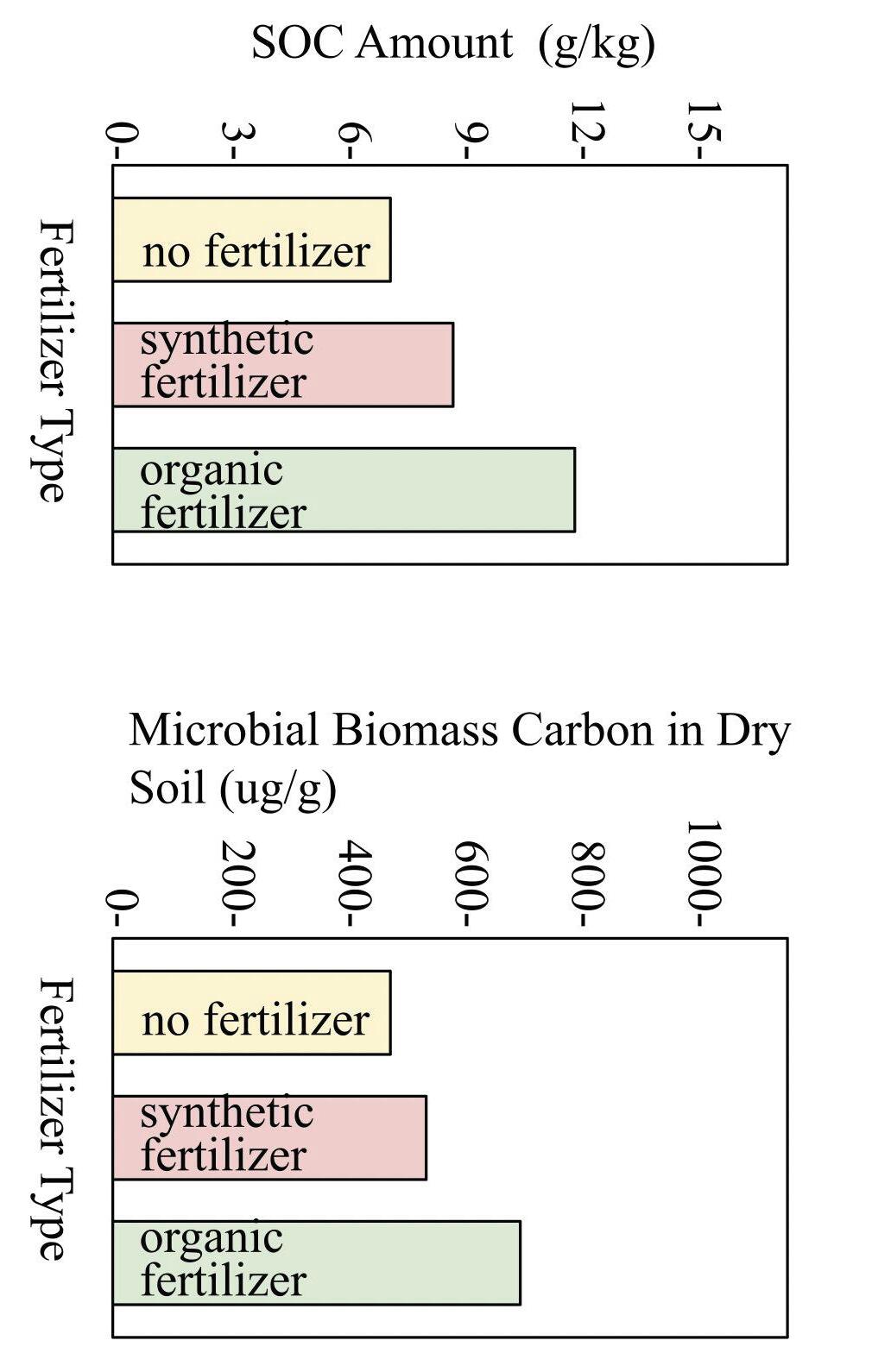
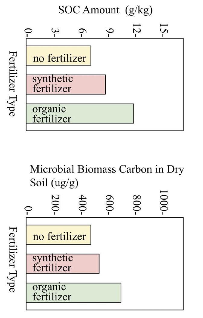
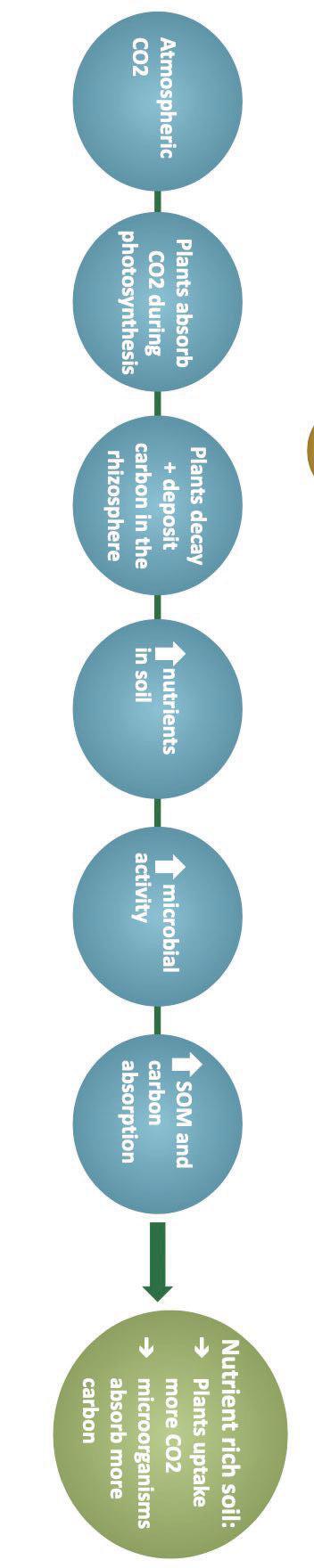
➔ The carbon present in living cells of microorganisms (microbial biomass carbon) is highest with an organic fertilizer amendment; therefore, microorganisms will absorb the most carbon with organic fertilizers (He et al., 2023) (figure 4)
➔ In one year, soil with an organic fertilizer sequestered more soil organic carbon (SOC) when compared to synthetic fertilizer and no fertilizer; therefore, organic fertilizers increase carbon sequestration (Nakhro,2010) (figure 3)
➔ The more nutrient rich the soil, the more microbial activity and carbon absorption; therefore nutrient rich soils increases carbon uptake in soil (Adingo et al., 2021) (figure 2)
➔ Due to microbes in the soil, soil is one of the largest carbon holders, and it has the potential to reduce carbon in the atmosphere (Mason et al., 2023)
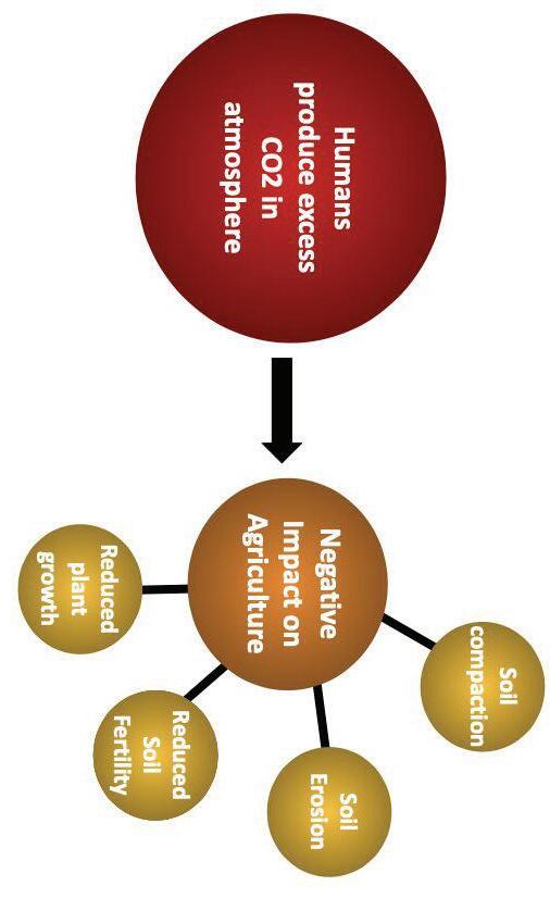
➔ Humans produce >40 billion metric tons of carbon dioxide per year (Lindsey & Dahlman, 2023), resulting in climate change which impacts agriculture (figure 1)
Background
➔ % C in lettuce were all similar (ANOVA p-value 0.253)
➔ Across all four fertilizers and the control soil, microbial respiration reached the maximum limit depicted on the CO 2 chart with no difference (data not shown)
➔ Although other studies revealed organic fertilizers increase C uptake and plant biomass when compared to control soil, this study did not have similar findings.
➔ The highest percent C in soil was bone meal soil at 6.5% C; both the fish emulsion and cow manure soil C levels were 1.5%, and worm castings matched control soil at 0.05% C
Implications
support, and collaboration. Thank you Uconn for analyzing the C in lettuce tissue and soil. Adingo, Samuel, et al. Environmental Science 15 Oct. 2021, https://doi.org/10.7717/peerj.12131. Accessed 22 Aug. 2023. Blouin, Manuel, et al. Springer Link link.springer.com/article/10.1007/s13593-019-0579-x#citeas. Accessed 20 Sept. 2023. 5 Sept. 2017. PubMed Central https://doi.org/10.3389/fmicb.2017.01702. Accessed 20 Sept. 2023. Devi Anugrah, Robert, et al. IOP Science iopscience.iop.org/article/10.1088/1755-1315/755/1/012001/pdf. Accessed 20 Sept. 2023. Earth Science www.earthsciencegrowing.com/products/natural-bone-meal/. Accessed 23 Sept. 2023. He, Hao, et al. Catena vol. 221, no. A, Feb. 2023, https://doi.org/10.1016/j.catena.2022.106784. Accessed 13 Sept. 2023. Lindsey, Rebecca, and Luann Dahlman. Edited by Jessica Blunden. Climate.gov 18 Jan. 2023, www.climate.gov/news-features/understanding-climate/climate-change-global-temperature#: ~:text=Highlights,2°%20F%20in%20total. Accessed 21 Aug. 2023. Nakhro, N., and M.S Dkhar. Journal of Agronomy vol. 9, no. 3, 15 June 2010, pp. 102-10, https://doi.org/10.3923/ja.2010.102.110. Accessed 20 Dec. 2023. Panettieri, Marco, et al. CATENA vol. 214, no. July, July 2022. ScienceDirect www.sciencedirect.com/science/article/abs/pii/S0341816222002442. Accessed 22 Aug. 2023. Smith, K.A. Soil Use and Management vol. 15, no. 2, June 1999. British Society of Soil Science bsssjournals.onlinelibrary.wiley.com/doi/epdf/10.1111/j.1475-2743.1999.tb00068.x. Accessed 21 Aug. 2023. Xu, Chenping, and Beiquan Mou. HortTechnology vol. 27, no. 4, Aug. 2017, https://doi.org/10.21273/HORTTECH03723-17. Accessed 20 Sept. 2023. TOC = total organic carbon | TN = total nitrogen | MBC = microbial content | FW = fresh weight | DW = dry weight
➔ Weekly measurements and observations were not different between groups (data not shown)
➔ The study results are summarized in Table 2.
Results
et al.,
Tessa Cassell
Figure 1: Dangers of Excess CO2 (Hamidov et al.)
Figure 2: Importance of increasing Nutrients and Microorganisms in Soil (Adingo
2021)
Figure 3: Carbon Uptake in Meadow Soil (He et al., 2023)
Figure 4: Microbial Biomass Carbon Measured in Paddy
Design
Table 2:
https://doi.org/10.1016/j.maturitas.2011.12.009
Jungbauer, A., & Medjakovic, S. (2012). Anti-inflammatory properties of culinary herbs and spices that ameliorate the effects of metabolic syndrome. Maturitas, 71(3), 227–239.
Aditi, K., Singh, A., Shakarad, M. N., & Agrawal, N. (2022). Management of altered metabolic activity in Drosophila model of Huntington's disease by curcumin. Experimental biology and medicine (Maywood, N.J.), 247(2), 152–164. https://doi.org/10.1177/15353702211046927
Ehrhardt, B., El-Merhie, N., Kovacevic, D., Schramm, J., Bossen, J., Roeder, T., & Krauss-Etschmann, S. (2022). Airway remodeling: The Drosophila model permits a purely epithelial perspective. Frontiers in allergy, 3, 876673. https://doi.org/10.3389/falgy.2022.876673
Works cited
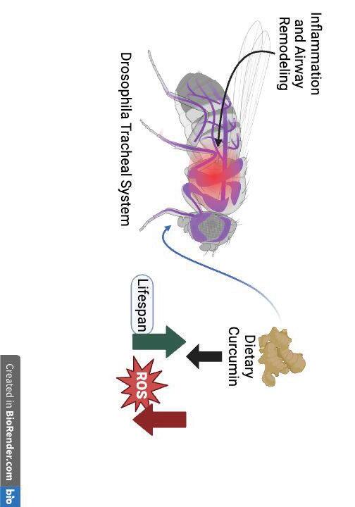
● As curcumin, is anti inflammatory and has shown positive results in both drosophila models of other inflammatory diseases and murine models of asthma, it is anticipated to increase lifespan, and decrease ROS levels in an anoxia asthma model in drosophila (figure 2).
Hypothesis
● When compared to Canton-S and Oregon-R strains, Berlin-K Drosophila showed the most significant difference in mortality and ROS levels between normoxic and hypoxic (2% oxygen for 24 hrs) conditions (Malacrida et al., 2022)
● Drosophila has homologs with around 75% of human disease genes (Roeder et al., 2009); has orthologs with the entire canonical set of asthma susceptibility genes excluding those associated with adaptive immunity
● Simpler airway structure and lack of adaptive immunity allow greater focus on innate immune (inflammatory) processes (Ehrhardt et al., 2022)
● Easier obtainability and shorter life cycles make testing cost and time efficient
Drosophila
Melanogaster :
● In Drosophila with Huntington's disease, 10µM dietary Curcumin improved median lifespan and reduced ROS levels in adipose tissue (Aditi et al., 2022)
● Has been shown to mediate symptoms in various diseases such as asthma, arthritis, and hypertension in rats (Chawla et al., 2022)
● Found in turmeric
Curcumin:
● Reactive oxygen species (ROS) are known inflammatory biomarkers (Stone et al., 2022)
● In a chronic asthma mice model, activation of the hypoxia response exacerbated asthma phenotype, airway remodeling, and airway inflammation (Ahmad et al., 2012)
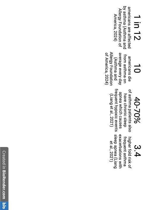
● Asthma is common pulmonary disease, affecting 1 in 12 people in the U.S. (Asthma and Allergy Foundation of America, 2024) involving symptoms such as wheezing, coughing, breathlessness causing chronic inflammation and remodeling of the airway (Ehrhardt et al., 2022) and severe cases can lead to hospitalization or death (figure 1).
Asthma:
Background
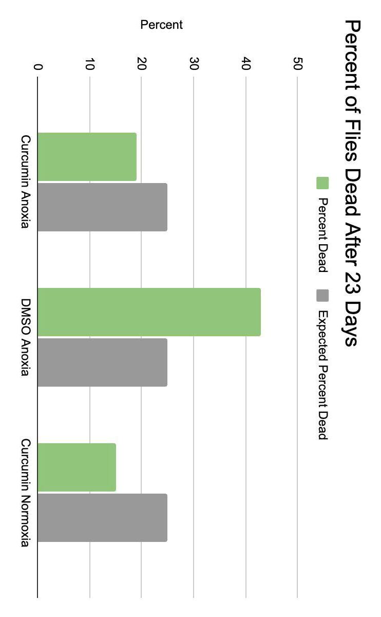
Chi square, p=0.04
● Thank you to Mrs. Parent, who supported me through every step of the process, giving me valuable advice on my work, teaching me how to work with drosophila and in a lab, and devoting her time to help with my experiment
Acknowledgements
● Will be performed using 2',7'-dichlorodihydrofluorescein diacetate (DCFDA) as a ROS dependant, fluorescence emitting stainer in a 96 well plate
● The supernatant will then be extracted and analysed in a fluorescence spectrometer
● The homogenate will be centrifuged at 1600 xg for 10 min
● Flies will be homogenized on ice in 20 mM Tris buffer using manual homogenizers
ROS Fluorescence Spectroscopy to determine the amount of reactive oxygen
species:
Future Work
● May eventually show pharmaceutical potential
● If curcumin is proven to be a reliable agent in mediating asthma and airway inflammation in drosophila, it might be a possible new treatment for asthma or other pulmonary diseases
● May show potential to reduce airway inflammation in Drosophila
● Curcumin may have a positive impact on drosophila survival in anoxia models
Implications
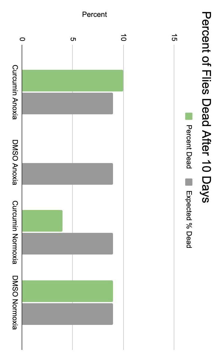
● After 23 days, the DMSO anoxia group showed a death percentage above the expected amount, whereas the curcumin anoxia and curcumin normoxia groups were both consistent in being less than the expected amount (p=0.04)
● After 10 days, the DMSO normoxia group showed a death percentage inline with the expected amount (p=0.2)
○ After 23 days, the p-value was 0.04 (significant difference between groups
● x ○ After 10 days, the p-value was 0.2 (no difference between groups)
○ At 14 days, the DMSO control group became moldy
● Calculated % dead after 10 and 23 days (figures 5 and 6)
Results
● Flies that escape or die due to human interference were removed from the study
● Number of deaths with be tracked daily
● Flies were transferred once every 4-7 days or when deemed necessary
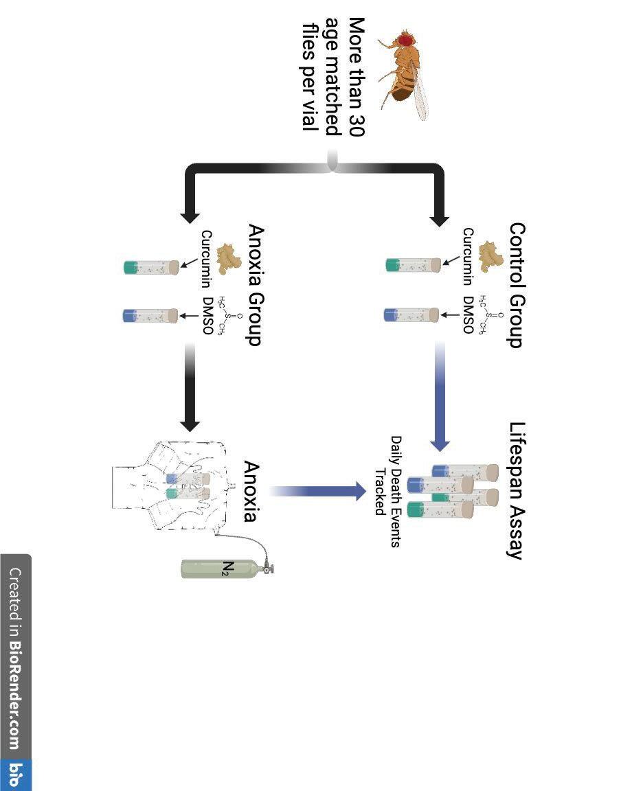
● Aged matched flies of corresponding experimental and control groups were split into 4 vials of at least 30 flies each
Lifespan
Assay:
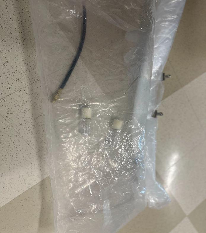
● Exposure lasted for 18 minutes or until flies were motionless at bottom of vial
(Krishnan et al., 1996)
● Fly vials were placed inside a glove bag connected to N 2 tank to evacuate all oxygen (figure 4)
Anoxia:
● Flies were split based on diet and anoxia exposure (See figure 3)
● 1 curcumin and 1 DMSO vial were exposed to anoxic conditions
● Age matched flies raised on corresponding diets were transferred into 4 vials, 2 with curcumin diet and 2 with DMSO diet.
Study
Design:
● Flies kept at 23C ±3C (room temperature)
● Transferring was done either live or on ice every seven days and as needed when vials became moldy or to avoid new flies hatching with old flies
● Berlin-K drosophila were raised on standard fly medium with either 10 uL of DMSO or 10 uL of 10 uM Curcumin in DMSO, both in 20 mL of DI water and 0.5 oz of standard drosophila media (Carolina Biologicals)
Fly
Cultures:
By: Alex Esser
effect of dietary curcumin on the lifespan of Drosophila
Methods
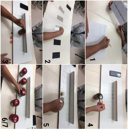
● Test 2 is Jebsen Taylor hand functionality where volunteers perform ADL tasks such as writing and moving objects) (fig 4b)
● Test 1 is block and box where volunteers move boxes from one box to another (fig 4a)
● Volunteers will perform each hand functionality test during a 90 minute session with each of the 3 models over separate days and different model orders
● Print failures and time will be evaluated (Malformations, stringing, holes, etc)
● Study will follow a comparative study format where 3 3D models will be sized to volunteers, printed, and subjected to functionality and strength tests
4a:Box and Block test
● 3 models will be scaled to fit 3 volunteers adequately; institutional review board has reviewed, consents will be obtained (fig 3)
● Volunteers will be measured using a low technology, basic sizing method (fig 2)
Methods
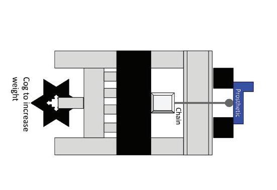
● Vernier Materials tester has the properties to test material strength (Vernier Structures & Materials Tester 2023)
● Functionality tests (Box and Block test and Jebsen Taylor) have been used to test prosthetics and serve as inexpensive and reliable tests for functionality (Hermansson 2023)
4b: Jebsen Taylor test
Figure 5: Strength test using vernier materials tester
Work cited Bijadi, S., de Bruijn, E., Tempelman, E. Y., & Oberdorf, J. (n.d.). Application of multi-material 3D printing for improved functionality and modularity of open source low-cost prosthetics: A case study. Application of multi-material 3D printing for improved functionality and modularity of open source low-cost prosthetics: A case study https://doi.org/10.1115/dmd2017-3540 Cabibihan, J.-J., Alkhatib, F., Mudassir, M., Lambert, L. A., Al-Kwifi, O. S., Diab, K., & Mahdi, E. (2021). Suitability of the openly accessible 3D printed prosthetic hands for war-wounded children. Frontiers in Robotics and AI 7 https://doi.org/10.3389/frobt.2020.594196 Parry-Hill, J. L., & Ashbrook, D. L. (2016, April 20). Challenges and Opportunities in DFO-AT: A Study of e-NABLE Retrieved November 20, 2023, from http://scholarworks.rit.edu/article/1808 Thomas, A., & Muñecas, T. (2022). A rehabilitation protocol for the use of a 3D-printed prosthetic hand in pediatrics: A case report. Journal of Hand Therapy https://doi.org/10.1016/j.jht.2022.10.010 Tian, L., Zheng, J., Cai, Y., Bin Abdul Halil, M. F. K., Thalmann, N. M., Thalmann, D., & Li, H. (2021). Fast 3D modeling of prosthetic robotic hands based on a multi-layer deformable design. International Journal of Bioprinting , 8 (1), 406. https://doi.org/10.18063/ijb.v8i1.406
● The low technology, basic sizing method is more accessible so it was used in this studying to model those conditions. (Ashbrook 2016)
Assess functionality with volunteers through functionality tests
Assess strength through vernier materials tester
Special thanks to Amy Parent for constant aid and support through the research process, and Humphrey Wong and Leonel Rodriguez for the donation of materials as well as peer editing and assistance making the poster.
Acknowledgements
Gaining objective data on how sizing impacts different models using an easy to follow sizing method, will provide valuable insights to printers in third world countries or war zones where cheap prosthetics are required ( Mahdi 2021 ).
Implications
● While Fusion 360 and the blender method are more comprehensive compared to the basic sizing method, both are unrealistic in war zones and third world countries. ( Simon J, 2022 )
● There is 3 different sizing methods, Fusion 360, blender, and the basic sizing method (Fig 2).
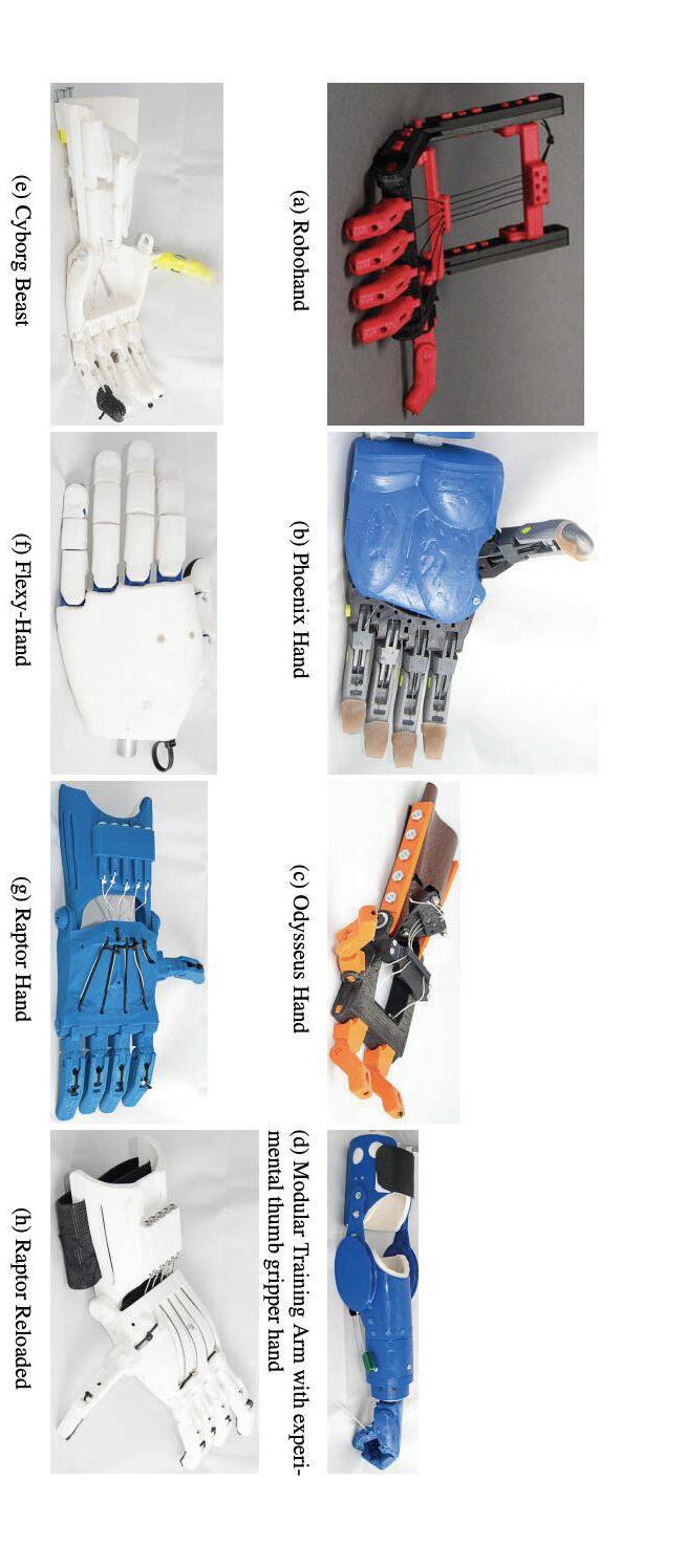
○ There are over 30 models with little objective guidance on sizing or preferred model for specific patients.(Oberdorf 2020)
● Functionality and strength data are limited for E-Nable 3D printed prosthetics with most studies suggesting lack of confidence in sizing and making the prosthetics (Oberdorf 2020)
● E-Nable has been the most successful at the making of 3D prosthetics
Figure 6: Comparison of different sized models in the space between the palm and the fingers
● 3D printing offer low cost and time requirements (Fig 1)(Mahdi 2021)
○ Government produced prosthetics can cost upwards of 50,000
1: Comparison of cost and time for different prosthetics.
● Prosthetics take time to produce and are costly (Fig 1) (Mahdi 2021)
● War zones increase the need for prosthetics (Mahdi 2021)
Background
Figure 2: Low technology, basic sizing method
● Data collection is ongoing; results are anticipated prior to presentation.
● Bigger Models will be weaker because less dense, but more functional
● Smaller models will be stronger because more dense, but less functional
● Geometry of prosthetic changes, therefore overall functionality changes
● The change in size affects functionality because the grasp changes with size (fig 6)
Hypothesis
Sebastian Rodriguez
The effect of changes in model and model sizes on the functionality in terms of grasping positions, material strength, and grip strength of 3D printed hand actuated prosthetic limbs.
● Strength will be evaluated by Vernier material tester. The cog turns to increase the weight and the more weight the more force is imposed on the object in the bridge. A computer program measures the amount of newtons before the object bends or breaks (fig 5) (Vernier Structures and materials tester 2023)
Methods continued
Figure
Figure 4: Functionality Tests Kinetic hand
Figure 3: Study overview
● Cost-effective and natural, these supplements offer alternatives to pharmaceutical interventions.
● Probiotics present in supplements and foods like yogurt, along with curcumin found in common supplements, provide readily available options.
● Additive or synergistic effects from combined supplements could offer a holistic approach to mitigate adverse effects of obesity.
● Xu, J., Du, P., Liu, X., Xu, X., Ge, Y., & Zhang, C. (2023). Curcumin supplementation increases longevity and antioxidant capacity in Caenorhabditis elegans. Frontiers in pharmacology, 14, 1195490. https://doi.org/10.3389/fphar.2023.1195490 Implications
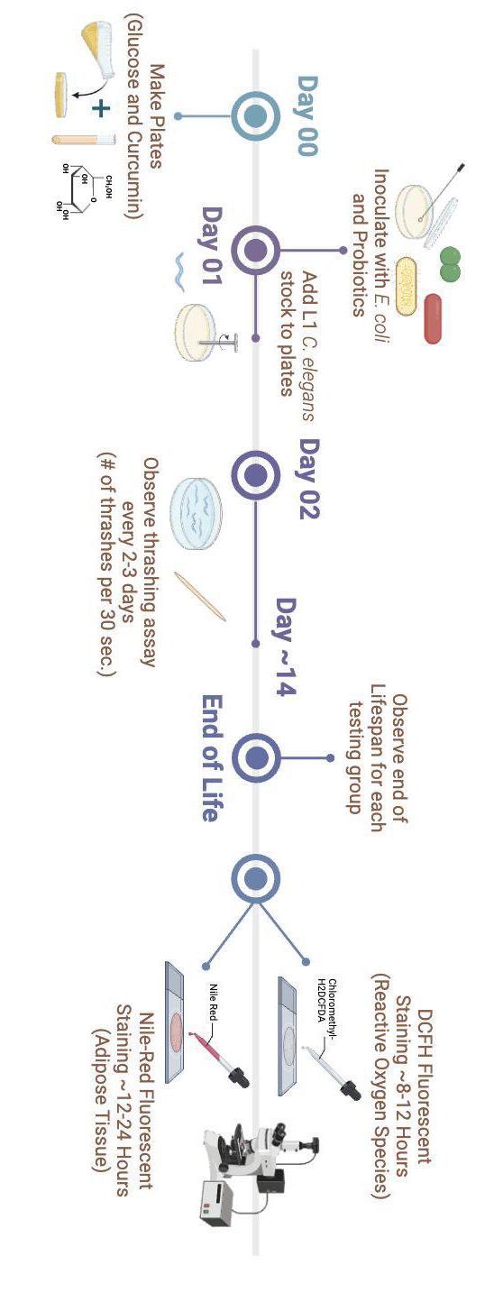
○ Nile Red staining: dye powder added to DMSO mixture, diluted to 600µM applied to worms to measurement adipose tissue.
● Núñez, S., Moliner, C., Valero, M. S., Mustafa, A. M., Maggi, F., Gómez-Rincón, C., & López, V. (2023). Antidiabetic and anti-obesity properties of a polyphenol-rich flower extract from Tagetes erecta L. and its effects on Caenorhabditis elegans fat storages. Journal of physiology and biochemistry, 79(2), 427–440. https://doi.org/10.1007/s13105-023-00953-5
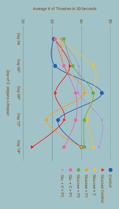
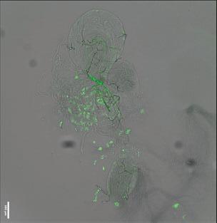
● NIle red assay measured for each group at the end of the C. elegans lifespan (Figure 3)
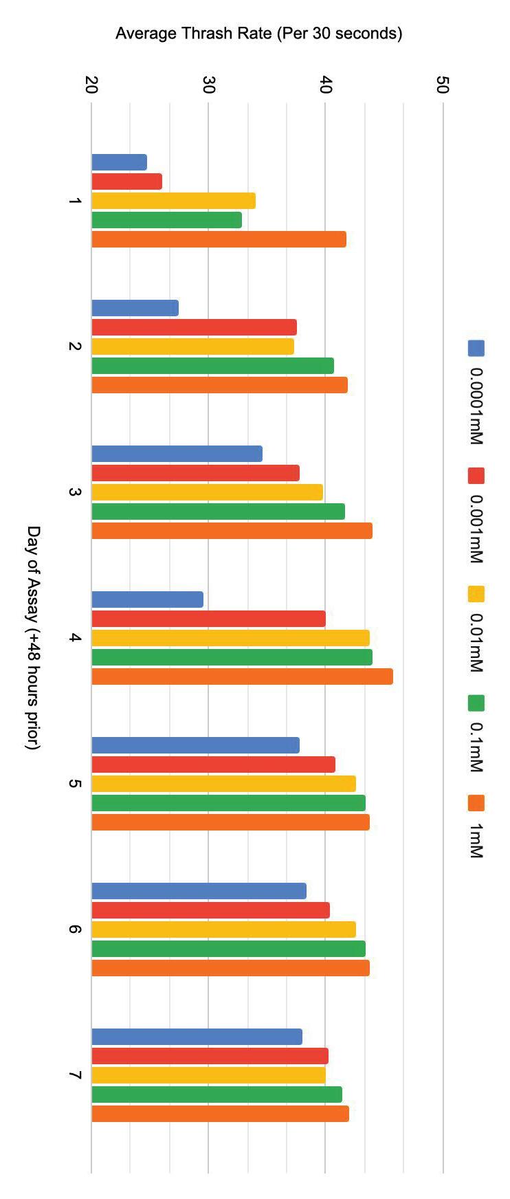
○ Worms placed in M9 buffer, 1 thrash is a full back and forth motion, counted (in 30 sec. under dissecting microscope)
● Thrashing assay was monitored in 5 worms from each group every 2-3 days as a marker of motility and quality of life
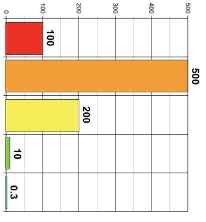
● Put on NGM with supplements added (Figure 3)
● 6 experimental groups of glucose induce L1 C. elegans established, one control group un-induced (Figure 3).
Started Experimentation:
● C. elegans were prepared from a stock plate, larvae were given glucose diet to induce obesity until the L1 stage and then were transferred to supplemented platesmaintained glucose diet.
● LGG was recorded as P1, LLC was recorded as P2
● Plates were inoculated with OP50, if added probiotics, streaked the plate with 2.5mL of each.
● 25ml poured into each 9cm plate allowed NGM to set.
● Ellulu, M. S., Patimah, I., Khaza'ai, H., Rahmat, A., & Abed, Y. (2017). Obesity and inflammation: the linking mechanism and the complications. Archives of medical science : AMS, 13(4), 851–863. https://doi.org/10.5114/aoms.2016.5892 ● Hasan, N., & Yang, H. (2019). Factors affecting the composition of the gut microbiota, and its modulation. PeerJ, 7, e7502. https://doi.org/10.7717/peerj.7502 ● Khanna, D., Khanna, S., Khanna, P., Kahar, P., & Patel, B. M. (2022). Obesity: A Chronic Low-Grade Inflammation and Its Markers. Cureus, 14(2), e22711. https://doi.org/10.7759/cureus.22711 ● Liu, B. N., Liu, X. T., Liang, Z. H., & Wang, J. H. (2021). Gut microbiota in obesity. World journal of gastroenterology, 27(25), 3837–3850. https://doi.org/10.3748/wjg.v27.i25.3837 ● Lobstein, T., Jackson-Leach, R., Powis, J., Brinsden, H, Gray, M. (2023) World Obesity Atlas. World Obesity Federation, 2023. https://data.worldobesity.org/publications/?cat=19
● Nematode Growth Medium Agar (NGM) melted, no treatment (control), 80µM glucose (G) (obese worms) and/or 25µM curcumin (C) added as indicated.
Plate preparation (Figure 3):
(Figure 2) Methodology
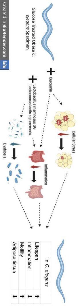
● It is anticipated that curcumin will lower the stress of inflammation on obese C. elegans and the probiotics will regulate the gut microbiome stopping the negative reactive cycle of obesity.
● The individual positive impacts of curcumin and probiotics in N2 C. elegans are clear, effects of combination treatment will be at least additive but hopefully synergistic (Figure 2).
● Using curcumin and probiotics to treat glucose obesity has never been done, combined treatment has never been studied (Table 1)
● C. elegans is good model for testing. Their digestive tract, which hosts a gut microbiome genetically comparable to a human and adipose cells which function in the same way as a humans.
● Curcumin in C. elegans has been found to extend their lifespan, increase cell healing, mitochondrial replication and has the ability to reduce oxidative stress on the cells of the C. elegans (Table 1) (Hernández-Cruz, 2023, Xu, 2023)
Table 1: Impact of Supplements
● Curcumin aids in fortifying the cellular walls and stopping the peroxidation of lipids and proteins increasing cellular life and longevity in C. elegans (Table 1) (Xu, 2023)
● Lactobacillus rhamnosus GG (LGG) has been studied in C. elegans and has proven to boost the immune system from infections (Table 1) (Yun, 2022)
● The probiotic Lactococcus lactis ssp cremoris (LLC) acts as a probiotic when in the human gut ( Jones, 2019).
● Probiotic supplementation in both C. elegans and humans, has been found to have positive results on decreasing dysbiosis (Table 1) (Yun., 2022, Barathikannan, 2022)
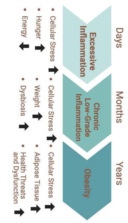
● The gut microbiome is closely linked to inflammation and obesity (Yun, 2022)
● The chronic inflammation that an unhealthy lifestyle or the initial stages of obesity creates can perpetuate obesity and lead to other health complications (Figure 1) (Ellulu, 2017)
1)
● Inflammation is one of the causes and symptoms of obesity (Figure
● Obesity os a medical condition characterized by the accumulation of adipose tissue in the body, it has become so apparent that it is now considered a global crisis.
Figure 1: Negative Cycle of Inflammation
Works Cited
Acknowledgements
● Worms will be treated with 250 μM of DMSO dissolved Chloromethyl-H2DCFDA dye and 100 μl of M9 buffer. Will then be incubated for 4 hours, then centrifuged to remove supernatant. To then measure fluorescence intensity and collect data a Flex Station 3 Reader will be used.
ROS assay, per group at end of lifespan
(Figure 3)
Future Work
● Fluorescence was visible in the glucose control group, and some was visible in the obese worms that were treated with P1 and P1+C (data not shown) Background
Nile Red Adipose Staining Results (Figure 5):
I would like to thank my teacher and guide, Mrs. Amy Parent for being such helpful and invaluable asset throughout the entire research process. I would also like to thank Dr. Lisa Merrill for her valuable opinions and contribution to my project. Investigating
● The N2 un-induced control group of worms showed no fluorescence, as well as the obese G+C treated worms, G+P2+C and G+P2 obese C. elegans groups
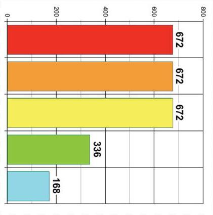
● Daily averages and standard deviations found through ANOVA. The ANOVA showed that data collected between all treatment groups on days 06, 08, 12, and 14 present statistically significant difference in avg. thrashing among groups. P-Value < ⍺ -level 0.01
● Daily averages and standard deviations for each treatment group were then combined to create an overall average threshing rate for each treatment group.
○ Obese worms without treatment (G Ctrl): 32 ± 4.2 mean thrashes
○ Obese worms treated with probiotic 1(G+P1): 35 ± 2.4 mean thrashes
○ Obese worms treated with curcumin and probiotic 1 (G+C+P1): 36 ± 2.6 mean thrashes
○ N2 un-induced worms (Ctrl): 36 ± 2.7 mean thrashes
○ Obese worms treated with probiotic 2 (G+P2): 40 ± 3.1 mean thrashes
(G+C+P2): 41 ± 3.2 mean thrashes
○ Obese worms treated with curcumin and probiotic 2
● The data for the other treatment groups were as follows
of Curcumin and Probiotics and their Effects on the Longevity, Motility, Inflammation, and Fat Deposits of with Obesity.
● The greatest mean thrashing rate was
42 ± 2.6 thrashes per 30 seconds, observed in the obese worms treated with curcumin (G+C supplementation)
● Therefore, diacetyl’s ability to slow B. minutum’s growth must be experimentally determined using dosing curve (Table 1) (Figure 3)
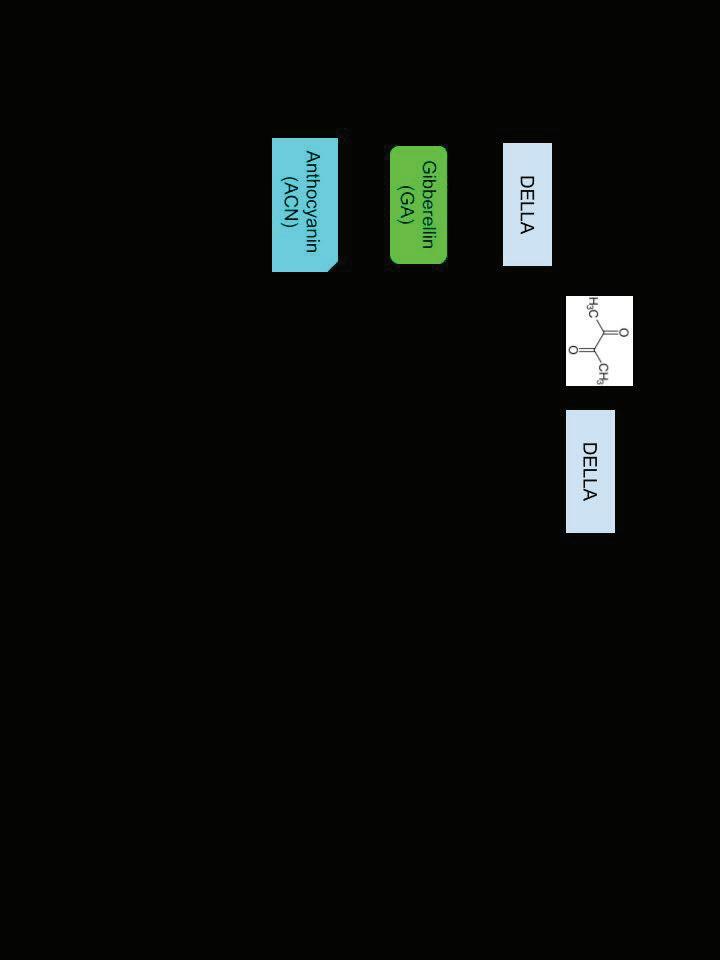
● In the Arabidopsis thaliana plant, DELLA proteins inhibit GA proteins. Exogenous diacetyl was added to the plant, causing DELLA promotion and subsequent slowed growth through GA inhibition. The addition of exogenous diacetyl on B. minutum has not been tested before (Achard et al., 2009, Morcillo et al., 2020)
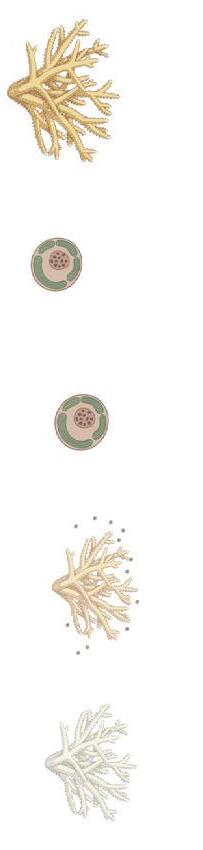
● Breviolum minutum, an algae species associating to corals like Acropora tenuis has been shown to contain a gibberellin (GA) protein pathway, which regulates stress and growth levels (Wu et al., 2022)
Background
● This study aims to determine if long-term thermotolerance happen through heat acclimation treatments during inhibited algal growth (Figure 3)
2018)
● Algal thermotolerance has been increased short-term through heat acclimation treatments exposing coral-symbiont systems to sublethal temperatures → long-term thermotolerance not achieved (Gibbin et al.,
Background
Habitat
Table 1:
● Coral bleaching is becoming more frequent because of global warming, leading to damaging effects (Figure 2) (McGowan and Theobald, 2023)
● If phenotypic plasticity is proven to increase under influence heat treatments and slowed growth from diacetyl, epigenetically modified symbionts can be reintroduced to coral since, if compatible, coral associate with the most thermotolerant available symbionts. This will form a more thermotolerant coral-symbiont system without immune response barriers (Bowling, 2022, Matthews et al., 2017)
● Future research will quantify phenotypic plasticity through experiments such as reactive oxygen species tests
● From preliminary research results, at lethal temperatures, higher replication rates from 10μM diacetyl groups indicate greater survivability, possibly due to phenotypic plasticity (Table 3, Figure 3)
● The diacetyl dosing curve confirmed B. minutum’s response to diacetyl → a new discovery (Table 1, Figure 3)
Conclusions
Applications
● Four experimental groups with 10mL media and initial 2x10^5 cells/mL density experienced various types of heat treatments (Table 2) ● 10μM diacetyl proliferated the most over the lethal treatment time period (Table 3)
● 3μM and 10μM were chosen for the heat acclimation experiment (Table 2)
● From dosing curve results, 3μM and 10μM diacetyl slowed growth the most (proof of concept achieved) (Table 1)
● Hemocytometer density counts under the microscope at 100X objective were used to evaluate cell growth each day (Table 1, 3)
● Coral bleach when their algal symbionts release tissue-damaging reactive oxygen species when stressed by environmental conditions such as increased temperatures (Figure 1, 2) (Douglas, 2003)
The Problem: Coral Bleaching
Figure 2:
● To confirm B. minutum ’s response to diacetyl, a dosing curve was performed using hemocytometers with 10mL media and 2x10^5 cells/mL (0.3μM to 30 μM) (Table 1)
● B. minutum cells were cultured and maintained in 250mL L1 media, supplemented with 50mg/L kanamycin and streptomycin at 24°C before transferring to 10mL samples with 2x10^5 cells/mL (Wu et al., 2022) (Table 1, 2)
Experimental Plan and Results
Kendra Cheng
● Thank you to Professor Huazhong Shi for providing wild type A. Thaliana seeds
● Thank you to Ms. Amy Parent for research guidance
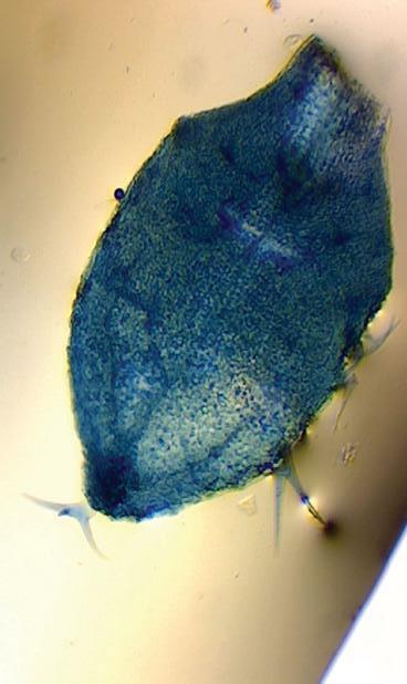
● Thank you to Xueyi Chen for further information on Anthocyanin extraction and Trypan Blue staining
● Thank you to Professor Huiming Zhang for guidance about Diacetyl and A. Thaliana
Acknowledgements
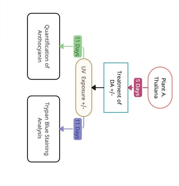
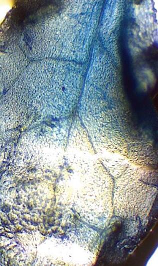
● It was hypothesized that when measuring trypan blue staining, those treated with DA under UV stress would show less staining
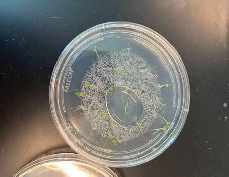
● Arapdiposis thaliana is a plant that has been used in many studies due to their short growth time and knowledge about them available
● With increased UV light resistance, this treatment may be applied to further studies in plants and vegetation
● DA has not been studied on UV light resistance, however with mechanism it is predicted to help support UV light resistance
● ACN is a plant pigment that provides UV light resistance (Zhao et al, 2022)
● Diacetyl (DA) increases DELLA levels, causing further ACN accumulation due to fewer GA molecules being present in order to inhibit ACN accumulation (Marsillo et al, 2020) (Figure 2)
● Decreased GA leads to increase in anthocyanin (ACN) levels (Morcillo, 2020)
● DELLA proteins downregulate the Gibberellin (GA) signalling pathway (Binenbaum et al, 2018)
● With UV light increases, there is a rising concern about crop success (Bernard et al, 2020)
Images of plants in petri dish 11 DAT. Plants grown under UV have higher stems and smaller leaves as compared to those grown under growth light. There also seems to be less leaves grown when under UV. There is a slight difference in leaf size and color when plants under UV are treated with DA. As expected, plants treated with DA with the growth light (GL DA) show less plant growth likely due to gibberellin downregulation.
● With climate change, UV light is increasing due to ozone shrinking (Bernard et al, 2020) (Figure 1)
Background
With increasing climate change, UV Light is increasing generating an abiotic stress for plants. A plants ability to resist abiotic stressors is vital in their survival. The chemical compound diacetyl(DA) may protect from abiotic stress, as DA treatment has been associated with increasing levels of Anthocyanin(ACN) (Marsillo et al,2020). ACN is increased through the gibberellin(GA) signaling pathway. DELLA proteins, which negatively regulate GA, increases with treatment of diacetyl. GA negatively regulates ACN accumulation, with less GA more ACN is present. ACN provides resistance to UV light. Arabidopsis thaliana was grown in petri dishes containing a 0.5 strength MS Medium containing 1.5% sucrose/0.7% agar for a 5 day germination period, then transferred to petri dishes with fresh medium treated with 9.7ug/ml free space of DA. Seedlings were either grown under a traditional grow light for control or were introduced to UV light stress exposed for 12 hours every 24 hour cycle. 11 days after treatment (DAT), UV light and control plants had ACN levels quantified by putting grounded leaves into an extraction buffer and measuring optical density(OD) at 657nm and 530nm using a UV/VIS spectrometer. Dead cells were stained with Trypan Blue staining and observed under light through a microscope, in order to assess cell death. While certain functions of DA within the abiotic stress system is unknown, it is anticipated that ACN will increase with the treatment of DA and cell death will decrease, therefore increasing stress resistance. When trypan blue was analyzed, there may be a higher death rate in cells of plants not treated with DA when exposed to UV light. In contrast, plants grown in control light showed a higher death rate when treated with DA suggesting that DA is only protective during times of stress. Based on this data, diacetyl treatment might be a valuable additive to treat agricultural crops, which could possibly assist them with different stressors. Abstract
(Shi et al, 2021)
The ozone layer absorbs UV light waves emitted by the sun, decreasing the frequency of the rays entering the earth's atmosphere. When the ozone shrinks, this absorption decreases allowing higher frequency rays reach the surface. When there is high absorbance, the UV light rays are a lower frequency and plants do not get damaged. When the frequency is high, plants are put under stress and damaged. Specifically, UV-C light (Wavelength of 200-280 nm) is seen damaging DNA when the high energy photons come into contact with DNA inside plants.
An image of Trypan Blue results when studying plants exposed to UV. On the top is a image taken under a microscope of a plant treated with UV stress, and below is one treated with UV along with DA. Trypan Blue stains dead cells in leaves, causing the dead cells to appear blue when observed
DA increases DELLA, which then plays multiple roles in abiotic stress responses. DELLA negatively regulates GA, which negatively regulates ACN. ACN provides stress resistance to UV.
Barnes, P.W. (2022, February 21). Environmental effects of stratospheric ozone depletion, UV radiation, and interactions with climate change: UNEP Environmental Effects Assessment Panel, Update 2021. Retrieved October 2, 2023, from https://pubmed.ncbi.nlm.nih.gov/32364555/ Bernard, G.H. (2023, April 21). Stratospheric ozone, UV radiation, and climate interactions. Retrieved October 3, 2023, from https://pubmed.ncbi.nlm.nih.gov/37083996/ Binenbaum, J. (2018, March 9). Gibberellin Localization and Transport in Plants. ScienceDirect. Retrieved October 2, 2023, from https://pubmed.ncbi.nlm.nih.gov/29530380/ Marsillo, R. J. (2020). Rhizobacterium-derived diacetyl modulates plant immunity in a phosphate-dependent manner. Retrieved October 2, 2023, from https://pubmed.ncbi.nlm.nih.gov/31802519/ Morcillo, R. (2020, April 2). Bacteria-derived diacetyl enhances Arabidopsis phosphate starvation responses partially through the DELLA-dependent gibberellin signaling pathway. Retrieved October 4, 2023, from https://pubmed.ncbi.nlm.nih.gov/32183580/ Shi, C. (2021, November 3). How plants protect themselves from ultraviolet-B radiation stress. Retrieved February 29, 2024, from https://pubmed.ncbi.nlm.nih.gov/34734275/ Zhao, S. (2022, October 23). Anthocyanin Accumulation Provides Protection against High Light Stress While Reducing Photosynthesis in Apple Leaves. Retrieved February 28, 2024, from https://pubmed.ncbi.nlm.nih.gov/36293472/
Works Cited
● Preliminary results suggest down-regulating of gibberellin using diacetyl may be protective against UV light.
Implications
● Anthocyanin levels will also be quantified.
● In the future, an assay which provides a numerical quantification of cell death will be used.
Future work
● This data is preliminary and the study must be repeated.
Plants grown for 11 days +/DA and +/UV light are shown prior to trypan blue staining (Figure 4) Trypan Blue staining showed that with treatment of DA, plants under UV stress had less cell death. It was observed that the top image of a leaf exposed to UV stress with no treatment has a far more significant death count than one treated with DA. (Figure 5) This shows that with DA treatment the leaves shown show less death when exposed to UV than those who were not treated with DA.
Results
Leaves were transferred to ethanol overnight then once extracted covered in 60% glycerol Relative cell death was qualitatively assessed through amount of blue staining on leaves (Figure 4)
Leaves harvested were soaked in Trypan blue (0.4 g trypan blue dissolved in 80 ml PBS) solution for 1h
Trypan blue solution was used to stain dead cells
Trypan Blue Staining
Diacetyl Treatment Plants were planted, then germinated for 5 days, then treated after the germination period. In order to treat DA, 1 ml of agar was placed on the top of the lid of the petri dish at the start of treatment Diacetyl was mixed with the agar with a concentration of Dish were sealed with parafilm for the treatment period Assays were done 11 days after treatment (DAT) (Fig 3)
To prepare medium, MS powder was mixed in distilled water along with 1.5% sucrose and 0.7% agar at a ½ strength concentration and autoclaved Plants were exposed to UV light will be grown in a separate box containing a UV-B lamp(Figure 3)
● A.Thaliana seeds sterilized using a 45% bleach and 5% Tween20 solution Seeds transported to petri dishes containing 0.5 MS medium prepared with 16ml of media, or ⅓ of the petri
Plant Growth and Abiotic Stress
Methods
Figure 3: Study Design
Figure
Denkin
Figure 5: Trypan Blue UV Results
● Imaged using Leitz Dialux 22 fluorescence microscope; two samples imaged using Leica THUNDER Imager fluorescence microscope at UConn's Advanced Light Microscopy Facility (AMLF), and processed in Fiji/ImageJ Fecal Deposition Assayonce a week, 3 random flies from each genotype from lifespan assay transferred to small Petri dishes with the corresponding medium with 0.5% bromophenol blue, for 72 hours; Petri dishes scanned ( Martorell et al., 2014 ); number of depositions analyzed using T.U.R.D. ( Wayland et al., 2014 )
Midgut Dissection and Imaging1 fly from each genotype dissected for midgut once a week in 1 X PBS buffer ( Bangi et al., 2016 ), and fixed in 1 X PBS buffer, 4% paraformaldehyde, 4% BSA, and 0.1% Triton X-100 ( Micchelli et al., 2014 ); dissections will be controlled for age and the number of days on diet as more data is collected
● Lifespan data is reproducibly normalized and graphed using the "R" programming language in RStudio
● Flies transferred into vials with plastic scaffolding to minimize the flies sticking in the medium ( Liao et al., 2021 ), and maintained in an incubator at approximately 25 o C ( Martorell et al., 2014 )
( Lambeau et al., 2017 )
● Media prepared by adding 10% coconut oil (high-fat) or 10% psyllium husk (high-fiber) to Bloomington standard cornmeal medium ( Liao et al., 2020 )
● Straight-winged offspring (both sexes) were selected for mosaic analysis with a repressible cell marker (MARCM) and heat-shocked in 37 bath for 1 hour
25 o C
Lifespan Assay25 freshly occluded Ras females and 5 Apc males crossed, transferred every 3 days, and maintained in an incubator at approximately
● Transgenic flies gifted from Dr. Andreu Casali in Spain were propagated Formula 4-24 Instant Medium from Carolina Biological, and live-transferred every 7-10 days (Figure 1)
Methodology
Time did not permit high fiber yet
Figure created in RStudio n = 3 (for both groups)
Deposition Assay
Figure 4: Fecal
● The study aimed to validate an Apc-Ras Drosophila system to model colorectal cancer, and to provide preliminary evidence about the impact of diet on cancer progression n = 9 n = 28 n = 15
● It was anticipated that high-fat diets would be correlated with a shorter lifespan, more tumors, and fewer fecal depositions compared to high-fiber diets ( Cheng et al., 2020 )
Hypotheses
Ağagündüz, Duygu, et al. "Understanding the Role of the Gut Microbiome in Gastrointestinal Cancer: A Review." Frontiers in Pharmacology , vol. 14, 24 Jan. 2023, https://doi.org/10.3389/fphar.2023.1130562. Anand, Preetha, et al. "Cancer Is a Preventable Disease That Requires Major Lifestyle Changes." Pharmaceutical Research , vol. 25, no. 9, 15 July 2008, pp. 2097-116, https://doi.org/10.1007/s11095-008-9661-9. Bangi, Erdem, et al. "Functional Exploration of Colorectal Cancer Genomes Using Drosophila." Nature Communications , vol. 7, no. 1, 29 Nov. 2016, https://doi.org/10.1038/ncomms13615. Cheng, Yiwen, et al. "The Intestinal Microbiota and Colorectal Cancer." Frontiers in Immunology vol. 11, 30 Nov. 2020, https://doi.org/10.3389/fimmu.2020.615056. Janssen, Klaus–Peter, et al. "APC and Oncogenic KRAS Are Synergistic in Enhancing Wnt Signaling in Intestinal Tumor Formation and Progression." Gastroenterology , vol. 131, no. 4, Oct. 2006, pp. 1096-109, https://doi.org/10.1053/j.gastro.2006.08.011. Lambeau, Kellen V., and Johnson W. McRorie. "Fiber Supplements and Clinically Proven Health Benefits." Journal of the American Association of Nurse Practitioners , vol. 29, no. 4, Apr. 2017, pp. 216-23, https://doi.org/10.1002/2327-6924.12447. Liao, Sifang, et al. "Impact of High-fat Diet on Lifespan, Metabolism, Fecundity and Behavioral Senescence in Drosophila." Insect Biochemistry and Molecular Biology , vol. 133, June 2021, p. 103495, https://doi.org/10.1016/j.ibmb.2020.103495. Martorell, Òscar, et al. "Conserved Mechanisms of Tumorigenesis in the Drosophila Adult Midgut." PLoS ONE , vol. 9, no. 2, 6 Feb. 2014, p. e88413, https://doi.org/10.1371/journal.pone.0088413. Micchelli, Craig A. "Whole-mount Immunostaining of the Adult Drosophila Gastrointestinal Tract." Methods vol. 68, no. 1, June 2014, pp. 273-79, https://doi.org/10.1016/j.ymeth.2014.03.022. Mirzoyan, Zhasmine, et al. "Drosophila Melanogaster: A Model Organism to Study Cancer." Frontiers in Genetics vol. 10, 1 Mar. 2019, https://doi.org/10.3389/fgene.2019.00051. Ren, Lingli, et al. "The Role of Intestinal Microbiota in Colorectal Cancer." Frontiers in Pharmacology vol. 12, 19 Apr. 2021, https://doi.org/10.3389/fphar.2021.674807. Sung, Hyuna, et al. "Global Cancer Statistics 2020: GLOBOCAN Estimates of Incidence and Mortality Worldwide for 36 Cancers in 185 Countries." CA: A Cancer Journal for Clinicians , vol. 71, no. 3, 4 Feb. 2021, pp. 209-49, https://doi.org/10.3322/caac.21660. Wayland, Matthew T., et al. "Spotting the Differences: Probing Host/microbiota Interactions with a Dedicated Software Tool for the Analysis of Faecal Outputs in Drosophila." Journal of Insect Physiology , vol. 69, Oct. 2014, pp. 126-35, https://doi.org/10.1016/j.jinsphys.2014.05.023.
Works Cited
I'm grateful to Dr. Andreu Casali, of the Institut de Recerca Biomedica in Lleida, Spain, for his mentorship and generous gift of the transgenic Drosophila model. I'm also grateful to Dr. Christopher O'Connell and Dr. Emery Ng at the UConn Advanced Light Microscopy Facility for the gift of their time and expertise, and to Ms. Amy Parent, my Scientific Research Honors teacher, for critical review and support throughout the research process.
● Evidence for the beneficial impact of a high-fiber diet , or the deleterious impact of a high-fat diet , would support dietary interventions to ameliorate CRC progression
Figure created in BioRender
Midgut Dissection and Imagingsuggests that flies on high-fat diets may have more tumors than flies on high-fiber diets , and that flies on high-fiber diets may have the fewest tumors out of the other diets (Figure 5)
● This preliminary data is promising, appears to support the hypothesis, and merits additional study, but confident conclusions can't be drawn because analysis is still ongoing
○ Ras oncoprotein → tumorigenesis through MAPK cascade; increases intracellular β-catenin, which leads to Wnt signaling in Apc-mutant cells ( Janssen et al., 2006 )
○ Apc genemore than 60% of human colonic adenomas and carcinomas were due to APC loss of function mutations ( Janssen et al., 2006 )
○ Ras gene64% of CRC patients experience a misregulation of RTK/RAS ( Bangi et al., 2016 ), and 50% of colorectal adenomas/carcinomas carry activating mutation in RAS protooncogene ( Janssen et al., 2006 )
Lifespan Assaytrend suggests that tumor-bearing flies on high-fat diets may die sooner than flies on both other diets, and that flies on high-fiber diets may die sooner than flies on standard diets (Figure 3) Fecal Output Assaysuggests that flies on high-fat diets may have more fecal depositions than flies on standard medium (Figure 4). Research is ongoing for larger populations and the high-fiber diet
Conclusion and Acknowledgements
○ Transgenic Apc-Ras model allows comparison of high-fat/high-fiber on cancer progression ( Martorell et al., 2014 ) ( Bangi et al., 2016 )
● Midgut is made up of a single layer of epithelial cells, replenished by intestinal stem cells, which is analogous to human colon ( Mirzoyan et al., 2018 )
● Drosophila offer a model to evaluate the impact of specific nutrients on CRC/gut health
● Fat causes higher colonic crypt cell proliferation, carcinogenic DNA alkylation/damage, and gut barrier breakdown, while soluble fiber lowers fecal pH, inhibits bacterial proliferation, enhances apoptosis, and reduces inflammation ( Cheng et al., 2020 )
● There is a lack of quantitative evidence on the impact of specific nutrients
● Multiple studies have shown Western diets (low fiber, high fat, high protein) lead to dysbiosis in comparison with MedDiet (high fiber, high unsaturated fatty acids, high plant protein); dysbiosis associated with inflammation, CRC ( Agagunduz et al., 2023 )
● Estimated that 70% of CRC deaths are linked to diet ( Anand et al., 2008 )
● Environmental risk factors (diet, obesity, substance use) modulate the intestinal microbiome, which can change the risk of CRC development ( Ren et al., 2021 )
● Colorectal cancer (CRC) is the third most frequently diagnosed cancer worldwide, but the second highest cause of death ( Sung et al., 2021 )
Background
Results
The Effect of Diet Composition on Lifespan and Colorectal Cancer
Figure 3: Lifespan Assay
Figure 1 : An example vial of Ras flies
Figure created in RStudio
P= 0.40059
References
in muscle and decreased ROS levels.
It is expected that C. elegans treated with vitamin D will be stronger and show more mobility. Future studies will likely show increased Ca
Hypothesis:
Given positive data for vitamin D, it is anticipated that C. elegans treated with vitamin D will be stronger, live longer, show more mobility, show increased Ca2+ and have decreased ROS (Ellwood 2018).
Acknowledgements: Special thanks to Dr. Nathaniel Szewczyk and Alexa Tasoula for the donation of materials and methods, as well as continued support. And Amy Parent for her statistical and ongoing aid with the research process.
elegans provides supportive evidence for supplementation in DMD patients.
Given data in children supplemented with vitamin D (figure 2), it is expected that DMD C. elegans will show increases in muscle strength (Morcillo 2020)
Current studies looked at other vitamins/amino acids showing some increased longevity, while others show no impact. No experiments have evaluated vitamin D in this model system. (Ellwood 2018)
Dystrophin Gene modification decreases longevity in C. elegans.
C. elegans with mutations in the dystrophin gene display differences in movement compared to wild type (Fig. 3) (Achrad 2009).
C. Elegans
Vitamin D offers promising benefits for muscle function in DMD, being safe and inexpensive (NHS 2022).
Positive correlation between vitamin D and muscle strength shown both children and DMD mice (Figure 2) (Bruce, 2021; Morcillo 2020).
Studies demonstrate beneficial effects of vitamin D in improving muscle function in elderly individuals and other muscle atrophy conditions (Zhu, 2020 ).
Importance of Studying Vitamin D
Caused by a nonsense frameshift mutation in the DMD gene, producing the defective dystrophin protein (NHS, 2022) leading to physical limitations, reduced life expectancy, higher Ca 2+ (NHS 2022) and increased inflammation (Fig 1) (Gibbin 2018).
Affects nearly 1 in every 3,500 male births worldwide (Clinic, 2022)
Duchenne Muscular Dystrophy (DMD) is an x-linked genetic disorder that results in progressive muscle wasting and weakness.
P= 0.00731
P= 0.01323
P= 0.00003
P= 0.00110
P= 0.00113 P= 0.00001 P= 0.00004
P= 0.07977
P= 0.00043 P= 0.00495 P= 0.00256 Figure 6: Thrash Rate vs. Time
Figure 1: Effects of DMD over Time - Figure 3: differences between DMD vs Wild type C.
Figure 4: Study Design Timeline
Figure 5: Image of Thrash Test
Bacterial Transformation into 5 Alpha E coli : 20 μL of the 5-alpha nonpathogenic E coli was mixed with 2 μL of the generated plasmid. Mixture was incubated on ice for 30 min with a heat shock for 30 sec at 42 o C. Cells were resuspended in 950 μL of Super Optimal broth with Catabolite repression (SOC) media and placed to shake for 1hr. Then the colonies were allowed to grow overnight on warmed Ampicillin (AMP) bacteria plates at 37 o C.Colonies were harvested.Sequencing using Genewhiz and analysis using Benchling: To get confirm identity of plasmids, mRNA, and assess the outcomes of the electroporation.mRNA synthesis: The kit “HiScribe® T7 mRNA Kit with CleanCap® Reagent AG” (NE BioLabs) was used. Nucleosides (ATP, GTP, UTP, and CTP) (all 5 μL) were catalyzed by the enzyme T7 polymerase.
Gibson Assembly: Using the “Gibson Assembly® Cloning Kit” (NE BioLabs), took the PCR ETAA1-AAD with gibson assembly vector (1.5 μL), Master mix (5μL), and deionized water. Mixture went into a thermocycler at 50 o C for 1 hour, stored at -20 o C for nonpathogenic 5-alpha nonpathogenic E coli transformation (Gibson, 2009, 343-345).
Concentration Determination: Using The Nanodrop Spectrophotometer (Egli Lab), a droplet (1.5μL) of the sample was used to determine yield, giving the concentration of the DNA (Desjardins, 2010)
Gel Electrophoresis: 2% agarose mixed with Tris base, acetic acid, and EDTA (TAE), heated in a standard microwave for 3 minutes. Then, 10 μL of “GelRed®” added to strain the DNA. After 30 minutes, gel was loaded with PCR samples of ETAA1-AAD and 6x loading dye. Samples run for 30 at 100 volts, and analyzed in UV projection. (Isbir, 2013)
PCR Purification: Using the kit “QIAquick PCR Purification Kit” (Qiagen) 30 μL purified sample was produced (Zhou, 2018)
DNA template (1 μL). (Smith, 2022)
Polymerase Chain Reaction (PCR): Protocol from New England Biolabs (NEB) was followed. PCR amplification was done in an Eppendorf Thermal Cycler using dNTPs (1 μL per reaction), the Q5 polymerase enzyme (0.5 μL), Q5 Polymerase buffer (10 μL), and the plasmid
PCR to Mouse Oocyte Electroporation (Fig.4)
Study was designed through a multi-step process from
Methods
Focusing on its ATR activating domain makes it even more efficient as well, only using one of many specific functions of the protein. (Fig. 3)
● ETAA1, a nuclear regulator, was chosen from a wide range of ATR activating proteins because of its high efficiency and easy accessibility to gibson cloning.
● Ataxia telangiectasia and Rad3-related protein (ATR) is activated using proteins like ETAA1 to latch onto an ATR Interacting Protein (ATRIP) that lays on the Replication Protein A’s (RPAs) of single stranded DNA (Fig. 3)
● ATR, when activated, slows down replication and the cell cycle (Fig. 2)
● Abnormal DNA replication is called “Replication Stress”, slowing down the replication fork (Palmerola, 2022; Zeman, 2014) (Fig. 2)
Replication stress is proposed to lead to DNA replication errors and aneuploidy. (Palmerola, 2022) (Fig. 1)
Fig. 5a: eGFP in Mouse Zygotes 18hr Post RNA Delivery Fig. 6a: Development Up To The Blastocyst Stage Assessed ~96 hrs after Electroporation
● Early stage zygotes replicate a mass amount of DNA during the 1, 2, and 4 cell stages (Zeman, 2014).
Acknowledgements : Thank you to my collaborators!!! Fig. 2: Use of ATR During Replication Stress
Supplemental bibliography provided for all methodological citations due to space limitations. McSwine, D. (2023, November 7).Retrieved January 2, 2024, from https://www.sciencedirect.com/science/article/pii/S2589004222001742 Palmerola, K. L. (2022). Replication stress impairs chromosome segregation and preimplantation development in human embryos. Cell, 185, 2988-3007. Rainey, M. D. (2020, September 1). ATR Restrains DNA Synthesis and Mitotic Catastrophe in Response to CDC7 Inhibition. PubMed. Retrieved August 22, 2023, from https://pubmed.ncbi.nlm.nih.gov/32877678/ Yang, M. (2021, September 21). Depletion of aneuploid cells in human embryos and gastruloids. Nature. https://www.nature.com/articles/s41556-021-00660-7 Zeman, M. K., & Cimprich, K. A. (2014). Causes and Consequences of Replication StressPMC. NCBI. Retrieved January 4, 2024, from https://www.ncbi.nlm.nih.gov/pmc/articles/PMC4354890/ Work cited
Fig.
Fig. 5b: How mRNA Delivery of ETAA1-AAD Affects the Timing of Mitotic Entry ns ns
Aneuploidy Kills 40-60% of embryos and IVF fails 50% of the time, primarily caused by aneuploidy (Brosens, 2022; Yang, 2021) This study has proven non-toxicity in ETAA1-AAD expression and therefore ATR Activation in zygotes, with no harm done to mitosis or blastocyst development. In continued future experiments, mouse cells’ DNA replication can be artificially stressed using Aphidicolin, an enzyme inhibitor, to mimic stressors in humans, eventually leading to test of concept in human eggs This study provides a strong basis for further investigation into what might help to save zygotes that otherwise would’ve died in IVF
4: Study
Design
Implications
● ETAA1’s ATR Activating Domain (0.349 KB) will be studied to evaluate its impact on ATR activation, affecting mitotic entry and blastocyst outcomes (Fig.4)Green Fluorescent Protein (eGFP) mRNA was successfully delivered into the mouse oocytes as shown by the visible fluorescence 18 hrs after delivery, past mitosis. (Fig.5a)Average mitotic entry for ETAA1-AAD treated-oocytes (n=25) was 16.28hrs, compared to 15.93, 16.27, and 16.08hrs for factor groups with no electroporation (control 1)(n=15)(p=0.331), electroporation no mRNA(control 2)(n=11)(p=>0.999) eGFP mRNA (n=18)(p=0.789). There is no significance in their differences (Fig.5b).Blastocysts were able to develop ~96 hrs after electroporation observed through microscopic and photographic evidence (Fig.6a)Oocytes electroporated with ETAA1-AAD mRNA had 96% blastocyst survival rate compared to control 1 (86%, p=0.501), control 2 (100%, p=0.278), and eGFP (94.4%, p = 0.811). There is no significance in their survival between groups. (Fig.6b)
Results
Mice
Oocyte
Electroporation: using their “Electroporation Needle Machine”. ~5μL of Gene Pulser® electroporation buffer used in combination with the mouse oocyte along with 400μL of each factor (ETAA1-AAD, Control 1, Control 2, and eGFP mRNA) (Aschheim, 2017; Gu, 2018) Methods (Cont.)
The reaction contained the DNA template (10 μL), RNA cap (4 μL), T7 polymerase (10 μL), 10x buffer (5 μL), and nucleus-free H2O (51 μL), amounting to 100 μL. (Berdanier, 2005)
Fig. 1: Nondisjunction During Mitosis in Zygote Cell Division