
22 minute read
Further Personalize CAR T Cell Treatment


Tanvi Gorre1*, Pabir Patra2,3 , Bhushan Dharmadhikari4,c
Introduction
Advertisement
CAR T cell therapies are the newest innovation in leukemia cancer therapies in the last few decades. Follicular cancer, a type of cancer that is treated through CAR T cell therapy, is a form of cancer that presents near blood vessels and, when it transforms, can be aggressive. Although this therapy has led to progress against this condition, it does not always lead to full remission. Furthermore, many experience cytokine storms that cause not only serious discomfort to the patient, but also death and inflammatory conditions. Follicular lymphoma patients can also have different cell ratios in their cancer microenvironment, which can also affect the effectiveness of the treatment. In this study, we created a comprehensive model of the follicular lymphoma microenvironment that can be modified to a patient’s unique cell ratio. This model then allows scientists to simulate how CAR T cells will interact with the patient’s cancer microenvironment and, as a result, change the secretion rates of varying factors in CAR T cells to increase remission rates in follicular lymphoma patients. In this particular study, we changed the secretion rate of Granzyme A in CAR T cells. Throughout the experiment, we measured the efficiency of the CAR T cell therapy using the cell ratio in the microenvironment, the follicular lymphoma motility, and dendritic cell interactions. Our findings can have applications not only to make adjustments to current treatments of follicular lymphoma, but also to introduce the idea of modifying CAR T cells on a molecular scale to improve CAR T cell treatment efficiency
1 Staples High School, Westport, CT 06880, United States; tanvigorre@gmail.com
* Presenting author
2 Department of Biomedical Engineering, University of Bridgeport, CT 06604, United States; ppatra@bridgeport.edu
3 Department of Mechanical Engineering, University of Bridgeport, CT 06604, United States; ppatra@bridgeport.edu
4 Department of Electrical & Computer Engineering & Technology, Minnesota State University Mankato, MN 56001 , bhushan.dharmadhikari@mnsu.edu c Corresponding author
Methods
Our model was formulated using the cell ratio in Burkitt's lymphoma, a lymphoma with a similar cell ratio to follicular lymphoma. The ratio of cells in the simulation were 50 follicular lymphoma cells: 2 dendritic cells: 5 M2 cells: 1 M1 cell: 1 T regulatory cell, as shown in Figure 1 and in Figures 2 and 3 with the latter showing the microenvironment as demonstrated in CompuCell3D [1]. The Table 1 and the Figure 1 also shows the cell ratio used in the CompuCell3D simulation. The cell ratio, however, varies from patient to patient and was obtained from an average of 22 samples [2]. The secretion rate of the varying factors for each cell type was estimated using the concentration of different factors in the anaplastic lymphoma, which is similar to the follicular lymphoma microenvironment without any CAR T cell involvement [3]. Then, the concentration of these factors was divided by the number of cells that secrete that particular factor in order to get the secretion rate of each factor for each cell type. It should be noted that there were other factors and cell types present in the study that were referenced; however, due to their minimal impact or small concentration in the microenvironment, they were excluded from the simulation. These values are listed in Table 1 along with information regarding how the different factors affect the lymphoma microenvironment.
Table 1 tabulates the various factors involved in this computation as well as the secretion rates of these factors by the cells in the simulation. These values were calculated by dividing the environmental concentration of these factors by the number of cells in the computation that secrete said factor. It was assumed that each cell type that secretes the factor has the same secretion rate, as there was no information to be found from the scientific community on the specific secretion rates of factors by the cells in the follicular lymphoma microenvironment [13]
Factor Table
Factor Field Name Properties
Interleukin 6 IL 6 type 1
Reduces number of migratory immune cells to lymphoma site [4], [5]
Hedgehog ligand Hh
Prevents lymphoma apoptosis [6], [7]
Interleukin 6 IL 6 type 2
Increases number of migratory immune cells to lymphoma site [8], [9]
0.0385 per M2, Treg, and Intratumoral dendritic cell [3]
0.05 per MSC, Intratumoral dendritic, and reticular cell [3]
0.27 per M1 cell [3]
Interleukin 2 IL2 Promotes NK and T cells activation and cell growth [10], [11]
Interleukin 4 IL4 Decreases CD8+ cell growth; increases follicular lymphoma cell growth; increased stromal cell proliferation; regulated lineage of DCs in microenvironment [12]
Interleukin 10 IL10
Inhibits IL2 and IFN gamma production; Downregulates M1 activity and inhibits expression of CD80 and CD86 which are present on M1 cells which interact with CD28 on CAR T cells and increase CAR T cell activity [13]–[17]
1.89 per CAR T cell [3]
0.3695 per M2 and Treg cell [3]
0.097 per M2 and Treg cell [3]
Interleukin 12 IL12
Upregulates IFN gamma production in T and NK cells and increases T cell exhaustion; increases treg activity [18]
IFN gamma gamma Decreases volume and spontaneous creation of lymphomas [16], [19]
TGF beta beta Increased stromal and endothelial cell Infiltration [20]–[23]
CCL19, CCL21, CXCL12
FRsecretion Increase interactions with T zone which allows for more naïve T and B cells to become assimilated into environment as immunosuppressants and increases B cell migration to area [24], [25]
Interleukin 15 IL15 Increase STAT-5 FL activation [26], [27]
TNF alpha alpha Increases FRC differentiation and increases M2 polarization [29]
0.0765 per Intratumoral dendritic, M2, and Follicular cell [3]
0.3045 per CAR T cell [3]
0.1165 per M2 cell [3]
0.05 per Reticular cell [3]
CXCL1, CXCL2, and CXCL12
MSCsecretion Increase cancer proliferation as well as possibility of metastasis [17]
0.0555 per MR and Intratumoral dendritic cell [3]
0.0585 per Intratumoral dendritic, follicular, and MSC cell [3]
0.05 per MSC cell [3]
There were several aspects of follicular lymphoma that were excluded from the model because it would require an environment larger than the scope of the model, or it was not possible because there is not enough information available. The T-zone within lymph nodes is one aspect of the microenvironment that was excluded. Reticular cells produce factors CCL19, CCL21, and CXCL12, which cause increased interactions between the T-zone B cells and therefore the follicular lymphoma cells [24]. In lymphomas that are resistant to CAR T cell therapy, genes FADD, BID, CASP8, and TNFRSF10B were deplete and gained genes that resist cell death such as CFLAR, BIRC2, and TRAF2. Lymphomas typically gain these genes from severe inflammation or stress in the microenvironment caused by CAR T cell therapy [30]. CAR T cell resistant cancers can also take the form of cancer stem cells that can be undifferentiated, allowing them to change the antigens they present on their cell membrane [31]. This phenomenon is called lineage switchingmediated target antigen loss and can render CAR T cell therapy useless [31]. This environment impacts the CAR T cell as it results in diminished cytotoxicity, minimal T cell differentiation, and aerobic glycolysis, Thus, when this occurred, it decreased the cell growth of CAR T cells. This phenomenon was taken into account in the model by setting a threshold of interleukin 6 from M1 cells that can cause follicular lymphoma CAR T cell resistance.
Results
Cell ratios can be used to monitor the status of the microenvironment and the success of the CAR T cell therapy. In Figure 2a, the cell population of a certain cell type are monitored over the duration of the experiment, which is 1000 Monte Carlo steps with each Monte Carlo step equal to 30 minutes in real time. In figure 2b we monitored cell motility of lymphoma cells are directly correlated with metastasis and we found that at a monte carlo step of 238 all cell motility in lymphoma cells went back to their baseline motility. In Figure 2c, we modeled dendritic cell interactions which are vital for the status of the microenvironment. Dendritic cells vary in their activity based on their interactions with other cells. For instance, when dendritic cells interact with pro tumor cells, it causes the dendritic cell to secrete pro-tumor factors such as IL12 and TNF alpha [32]. When dendritic cells interact with anti-tumor cells, the dendritic cells secrete anti- tumor factors such as IL2.
Discussion
In this study, we explored how modifying the Granzyme A secretion rate in CAR T cells can improve patient conditions through CompuCell modeling; however, this approach can be used for the patient’s unique cell ratio and can vary different secretion rates in the CAR T cell to improve remission. We could not demonstrate the full complexity of this microenvironment, but we included the parts of the microenvironment most impactful to follicular lymphoma growth. The importance of a cell or factor was determined by its concentration and the significance of its impact on the microenvironment.
From the results, we concluded that 0.2 pg per monte carlo step relative to the other factors is the Granzyme A secretion rate of CAR T cells that was the most beneficial, as it decreased the follicular lymphoma population significantly without creating a highly pro-inflammatory microenvironment. It also caused decreased cell motility in follicular lymphoma cells, which decreased the likelihood of metastasis, or the state in which cancer travels to other parts of the body.
However, this Granzyme A secretion rate is specific to the cell ratio used in the computational representation of the follicular lymphoma microenvironment. This model that we have created of this microenvironment can be altered based on the cell ratios of individual patients, allowing for a further specialized and more effective treatment as shown in other studies. Others have done work predicting how cancer microenvironments will react to treatments, and our study provides an in-depth demonstration of how follicular lymphoma will respond based on the cell ratio. Changes to the secretion rates of these factors can be achieved by changing the polyadenylation to the mRNA sequences that code for granzyme and granzyme A in the CAR T cell. Our findings demonstrate how CAR T cell therapy can be further specialized for treatment success.

References
Header Image:
Krebs: Chronik des fortschritts. (2019, June 23). Pharma Fakten. https://pharma-fakten.de/news/787-krebs-chronik-des-fortschritts/
[1] M. H. Swat, G. L. Thomas, J. M. Belmonte, A. Shirinifard, D. Hmeljak, and J. A. Glazier, “Multi-Scale Modeling of Tissues Using CompuCell3D,” Methods Cell Biol, vol. 110, pp. 325–366, Jan. 2012, doi: 10.1016/B978-0-12-388403-9.00013-8.
[2] M. Granai et al., “Immune landscape in Burkitt lymphoma reveals M2-macrophage polarization and correlation between PD-L1 expression and non-canonical EBV latency program,” Infect Agent Cancer, vol. 15, no. 1, May 2020, doi: 10.1186/s13027-020-00292w.
[3] F. Knörr et al., “Blood cytokine concentrations in pediatric patients with anaplastic lymphoma kinase-positive anaplastic large cell lymphoma,” Haematologica, vol. 103, no. 3, pp. 477–485, Feb. 2018, doi: 10.3324/haematol.2017.177972.
[4] D. de Jong and T. Fest, “The microenvironment in follicular lymphoma,” Best Pract Res Clin Haematol, vol. 24, no. 2, pp. 135–146, Jun. 2011, doi: 10.1016/J.BEHA.2011.02.007.
[5] E. Bettelli et al., “Reciprocal developmental pathways for the generation of pathogenic effector TH17 and regulatory T cells,” Nature, vol. 441, no. 7090, pp. 235–238, May 2006, doi: 10.1038/nature04753.
[6] G. Casella et al., “IL4 induces IL6-producing M2 macrophages associated to inhibition of neuroinflammation in vitro and in vivo,” Journal of Neuroinflammation, vol. 13, no. 1, Jun. 2016, doi: 10.1186/s12974-016-0596-5.
[7] G. Basile Carballo, J. Ribeiro Honorato, G. Pinto Farias de Lopes, and T. Cristina Leite de Sampaio Spohr, “A highlight on Sonic hedgehog pathway”, doi: 10.1186/s12964-018-0220-7.
[8] C. Dierks et al., “Essential role of stromally induced hedgehog signaling in B-cell malignancies,” Nature Medicine, vol. 13, no. 8, pp. 944–951, 2007, doi: 10.1038/nm1614.
[9] K. Nakajima et al., “A central role for Stat3 in IL-6-induced regulation of growth and differentiation in Ml leukemia cells,” The EMBO Journal, vol. 15, no. 14, pp. 3651–3658, 1996.
[10] Y. Yamanaka, K. Nakajima, T. Fukada, M. Hibi, and T. Hirano, “Differentiation and growth arrest signals are generated through the cytoplasmic region of gp130 that is essential for Stat3 activation,” EMBO Journal, vol. 15, no. 7, pp. 1557–1565, Apr. 1996, doi: 10.1002/j.1460-2075.1996.tb00500.x.
[11] T. Miyazaki et al., “Three Distinct IL-2 Signaling Pathways Mediated by bcl-2, c-myc, and Ick Cooperate in Hematopoietic Cell Proliferation,” 1995.
[12] S. H. Ross and D. A. Cantrell, “Signaling and Function of Interleukin-2 in T Lymphocytes,” Annu Rev Immunol, vol. 36, pp. 411–433, Apr. 2018, doi: 10.1146/annurev-immunol-042617-053352.
[13] V. Spieler et al., “Targeting interleukin-4 to the arthritic joint,” Journal of Controlled Release, vol. 326, pp. 172–180, Oct. 2020, doi: 10.1016/J.JCONREL.2020.07.005.
[14] J. Cortes and R. Kurzrock, “Interleukin-10 in non-Hodgkin’s lymphoma,” Leukemia and Lymphoma, vol. 26, no. 3–4. Informa Healthcare, pp. 251–259, 1997. doi: 10.3109/10428199709051774.
[15] J. K. Riley, K. Takeda, S. Akira, and R. D. Schreiber, “Interleukin-10 receptor signaling through the JAK-STAT pathway. Requirement for two distinct receptor-derived signals for anti-inflammatory action,” Journal of Biological Chemistry, vol. 274, no. 23, pp. 16513–16521, Jun. 1999, doi: 10.1074/jbc.274.23.16513.
[16] A. H. S-y, S. H-y Wei, A. L-f Mui, A. Miyajima, and K. W. Moore, “Functional Regions of the Mouse Interleukin-10 Receptor Cytoplasmic Domain,” 1995. [Online]. Available: https://journals.asm.org/journal/mcb
[17] M. A. Meraz and † J Michael White, “Targeted Disruption of the Stat1 Gene in Mice Reveals Unexpected Physiologic Specificity in the JAK-STAT Signaling Pathway,” 1996.
[18] S. J. Rodig et al., “Disruption of the Jak1 Gene Demonstrates Obligatory and Nonredundant Roles of the Jaks in Cytokine-Induced Biologic Responses,” 1998.
[19] Z.-Z. Yang et al., “IL-12 upregulates TIM-3 expression and induces T cell exhaustion in patients with follicular B cell non-Hodgkin lymphoma,” The Journal of Clinical Investigation, vol. 122, 2012, doi: 10.1172/JCI59806.
[20] F. Castro, A. P. Cardoso, R. M. Gonçalves, K. Serre, and M. J. Oliveira, “Interferon-gamma at the crossroads of tumor immune surveillance or evasion,” Frontiers in Immunology, vol. 9, no. MAY. Frontiers Media S.A., May 04, 2018. doi: 10.3389/fimmu.2018.00847.
[21] P. A. Guerrero and J. H. McCarty, “TGF-β Activation and Signaling in Angiogenesis,” in Physiologic and Pathologic AngiogenesisSignaling Mechanisms and Targeted Therapy, InTech, 2017. doi: 10.5772/66405.
[22] B. Tirado-Rodriguez, E. Ortega, P. Segura-Medina, and S. Huerta-Yepez, “TGF-β: An important mediator of allergic disease and a molecule with dual activity in cancer development,” Journal of Immunology Research, vol. 2014, 2014, doi: 10.1155/2014/318481.
[23] M. Bakkebø, K. Huse, V. I. Hilden, E. B. Smeland, and M. P. Oksvold, “TGF-β-induced growth inhibition in B-cell lymphoma correlates with Smad1/5 signalling and constitutively active p38 MAPK,” BMC Immunology, vol. 11, Dec. 2010, doi: 10.1186/1471-2172-11-57.
[24] Z. Z. Yang et al., “TGF-β upregulates CD70 expression and induces exhaustion of effector memory T cells in B-cell non-Hodgkin’s lymphoma,” Leukemia, vol. 28, no. 9, pp. 1872–1884, 2014, doi: 10.1038/leu.2014.84.
[25] F. Mourcin et al., “Stromal cell contribution to human follicular lymphoma pathogenesis,” 2012, doi: 10.3389/fimmu.2012.00280.
[26] P. Amé -Thomas et al., “Human mesenchymal stem cells isolated from bone marrow and lymphoid organs support tumor B-cell growth: role of stromal cells in follicular lymphoma pathogenesis,” 2007, doi: 10.1182/blood-2006-05.
[27] A. Mishra, L. Sullivan, and M. A. Caligiuri, “Molecular pathways: Interleukin-15 signaling in health and in cancer,” Clinical Cancer Research, vol. 20, no. 8, pp. 2044–2050, Apr. 2014, doi: 10.1158/1078-0432.CCR-12-3603.
[29] J. A. Johnston et al., “Tyrosine phosphorylation and activation of STAT5, STAT3, and Janus kinases by interleukins 2 and 15,” 1995.
[30] J. Cheng et al., “Understanding the Mechanisms of Resistance to CAR T-Cell Therapy in Malignancies,” Frontiers in Oncology, vol. 9. Frontiers Media S.A., Nov. 21, 2019. doi: 10.3389/fonc.2019.01237.
[31] Z. Yu, T. G. Pestell, M. P. Lisanti, and R. G. Pestell, “Cancer stem cells,” International Journal of Biochemistry and Cell Biology, vol. 44, no. 12. Elsevier Ltd, pp. 2144–2151, 2012. doi: 10.1016/j.biocel.2012.08.022.
[32] F. Veglia and D. I. Gabrilovich, “Dendritic cells in cancer: the role revisited,” Current Opinion in Immunology, vol. 45. Elsevier Ltd, pp. 43–51, Apr. 01, 2017. doi: 10.1016/j.coi.2017.01.002.
4. The Ever-Changing Brain: Daily Use of Digital Technology and Its Effects on Neuroplasticity Wan ‘Aliyah, Global Jaya School
Fictional stories as we know them are fabricated by novelists who have the power to create and control the plot. Almost like fiction, the human brain possesses an incredible ability to rewire itself: it is the author that writes and the pencil that draws its version of literature.
This phenomenon is referred to as neuroplasticity, which is the capability of neural networks inside the brain to adapt, reorganise and change their behaviour in response to new experiences, information, stimuli or damage [1]. It is the reason bad habits remain ingrained in the brain and that even valuable skills can be lost over time indeed, a “use it or lose it” system.
Structural neuroplasticity involves reshaping individual neurons and changing their physical structure due to learning and experience. The neurons in the brain send and receive electrical impulses connected by a synapse acting as a bridge between one neuron and another. While repetition and practice strengthen these existing synapses connections within the brain, new experiences and skills can form new ones this is known as neurogenesis [2]. In contrast, weakened synaptic connections that remain unused in the brain are eliminated during synaptic pruning to keep its processes more efficient [2]. Due to its malleability, the brain can easily be influenced, trained and controlled by external factors, including an individual’s lifestyle and habits [3].
Researchers examine neuroplasticity through neuronal changes in the brain using different types of magnetic resonance imaging (MRI). Neuronal changes can be categorised by grey and white matter in the brain. They are responsible for the conduction, processing, and transmission of information as well as the speed of the impulse. They may influence MRI signals, making it the perfect modality to measure neuroplasticity [4].
In a world where technology is a significant aspect of personal lives, it has become a habit and a lifestyle that many cannot recall a life without instant connectivity to the internet. Modern cognition such as navigation, calculation, communication and memorisation rely on technology. From accessibility to productivity, it provides all the facets that facilitate daily activities by either enhancing or extending mental capabilities. Everyday exposure to technological devices, media and the internet influences the way individuals access and engage with information. It stimulates alteration in their brain activity to a profound degree. The biological changes induced by exposure to technology have led researchers to examine the response and transformation that occur in human brains and their cognitive ability.
Technology and Language in Developing Brains
An area of neuroplastic interest is whether intensive digital media usage affects the development of processes linked with language. This study estimates white-matter integrity in the brain of preschool children using a standardised 15-item screening tool known as ScreenQ. The ScreenQ scores were then statistically correlated with the children’s diffusion tensor MRI measurement and cognitive test scores.


The overall white matter volume results showed a significant correlation between ScreenQ scores, lower fractional anisotropy (FA) and higher radial diffusivity (RD). The FA and RD indicate the fibre tract in the whole-brain images, suggesting a link between intensive early childhood digital media use and poorer microstructural integrity of white matter tracts. It is most prominent in areas involved in language and the comprehension of speech: the Broca and Wernicke areas in the brain. The researchers observed lower executive functions and literacy abilities in the study despite matching age and average household income. As a result of digital media usage, the fibre tracts in language areas of the children’s brain were not developed to their full extent [6].
Media Multi-Tasking: Attention and Memory
Technological accessibility has blurred the lines between online and offline activities and allows us to perform multiple concurrent tasks. Skimming past information with superficial interest and shifting from one activity to another could result in short-term memories. This habit trains the brain to diminish the internalisation and retention of information. That is because the brain does not actively engage in the processes necessary for forming long-term memories [7]. At the same time, this also affects an individual’s attentional functioning.
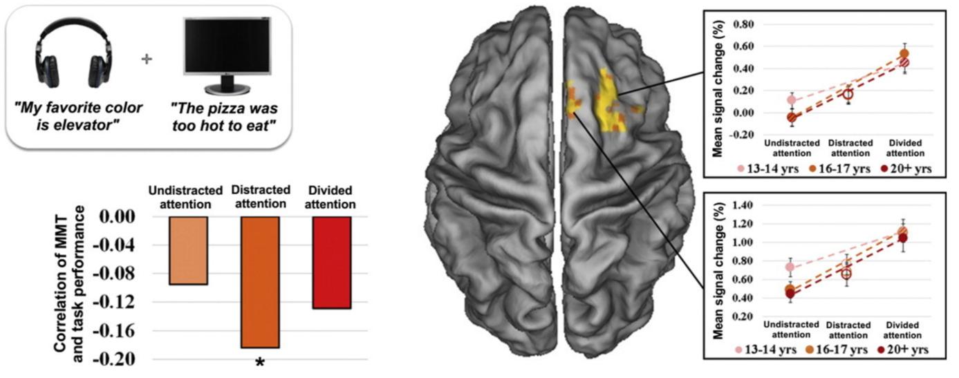
A study found that those who engage in frequent media multi-tasking (MMT) in their daily lives experience tend to perform poorer in several cognitive tasks, especially for sustained attention [7]. In the study, 149 adolescents and young adults were assigned speech-listening and reading assignments that required them to maintain attention in the presence of distractor stimuli. The participants’ performance was measured using functional magnetic resonance imaging (fMRI).
Based on the study, a higher media multi-tasking score was associated with poor performance and increased brain activity in the right prefrontal regions. Despite the increased activity, the performance was lower than light media multi-taskers. The findings suggest that heavy media multi-taskers require a greater cognitive effort to maintain concentration when faced with distractor stimuli. In terms of structure, higher internet usage and heavy media multi-tasking levels are related to decreased grey matter in prefrontal regions. The reduction in grey matter possibly indicates the unpracticed skill in maintaining goals and focusing in the face of a distraction [9].
Internet Usage: Changes in Reward Pathways
On top of it all, ever wondered why switching off social media may be challenging? According to research, these social media habits might be due to neuroplasticity [10]. When an urge is satisfied, dopamine, a neurotransmitter that gives pleasure, is released. This chemical is essential to neuroplastic change as it assists in building neural connections that reinforce that particular habit. This reward pathway motivates people to seek and repeat the activity. In this case, the reward pathway shift could lead to addiction to the internet and social media [11].
Positive Neuroplasticity
Despite these potentially harmful brain-health effects, studies also show that video games and digital technological experiences provide opportunities for cognitive sharpening through brain-strengthening neural exercises [12].
The Benefits of Video Games
Video games can be both detrimental and beneficial depending on the time spent playing and the training of skills involved. A study in 2013 conducted by Germany’s Max Planck Institute for Human Development and St. HedwigKrankenhaus investigated the effects of video games on the brain. Results that were measured using MRI revealed that playing Super Mario 64 increases grey matter volume in the right hippocampus (HC), right dorsolateral prefrontal cortex (DLPFC) and cerebellum. These brain regions are responsible for memory formation, strategic planning, spatial navigation, and fine motor skills. The changes in grey matter volume were more pronounced in participants who were eager to play, reflecting the role of motivation in the learning process as it builds and strengthens synapses connections [14]. Thus, the cognitive demands in mastering video games have led to the formation of new synaptic connections in brain sites responsible for spatial navigation, planning and decision making [2]. It is advantageous to encourage positive changes in neural networks such as this.

Improving Neuroplasticity
The neuroplastic change consists of five components: challenge and novelty, intention, specific attention, repetition and intensity, and time of the practice [15]. Begin by selecting an activity that is new, challenging and relevant. Committing to the engagement of new healthy exercises and habits will lead to favourable long-lasting neuroplasticity changes. The list of activities to choose from is massive! It may include learning something new such as an instrument or a new language, personal expression in different forms of creativity, travelling and exploring new places, meeting new people and much more. Even planning a healthy diet or workout routine or fixing an unhealthy sleeping schedule is one step closer to a positive change. Don’t forget to get plenty of sleep as well, as it facilitates brain plasticity growth and development.
As neuroplasticity continues to promote both positive and negative changes, it emphasises the importance of personal balance and repetition to maintain essential and practical skills in the future. Exposure to digital media and technology is inevitable. Although the focus of neuroplasticity was centred on the negative aspects of technology, numerous positive elements have yet to be discussed. Taking advantage of the efficiencies of new technologies could prove to be a future asset. Rewiring the human brain and its plasticity depends on an individual and the lifestyle they choose to follow.
References
Header Image: Alimran. (2020, June 08). Neuroplasticity and CNS reorganization: Alimran Medical Center. Retrieved June 14, 2022, from https://alimranmed.com/2020/06/08/neuroplasticity-and-cns-reorganization/
[1] Rugnetta, M. (n.d.). Neuroplasticity. Retrieved June 13, 2022, from https://www.britannica.com/science/neuroplasticity
[2] Gamma, E. (2021, March 24). What is brain plasticity? Retrieved June 13, 2022, from https://www.simplypsychology.org/brainplasticity.html
[3] Cherry, K. (2022, February 18). How brain neurons change over time from life experience. Retrieved June 13, 2022, from https://www.verywellmind.com/what-is-brain-plasticity-2794886
[4] Reid, L., Boyd, R., Cunnington, R., & Rose, S. (2015, December 29). Interpreting intervention induced neuroplasticity with fmri: The case for Multimodal Imaging Strategies. Retrieved June 13, 2022, from https://www.ncbi.nlm.nih.gov/pmc/articles/PMC4709757/#:~:text=One%20very%20popular%20modality%20used,a%20compari son%20or%20resting%20state.
<5> Hutton, J et al., (2020, February 12). A novel, composite measure of screen-based media use in young children (ScreenQ) and associations with parenting practices and Cognitive Abilities. Retrieved June 13, 2022, from https://www.nature.com/articles/s41390-020-07651#:~:text=ScreenQ%20is%20a%2015%2Ditem,scores%20reflecting%20greater%20non%2Dadherence.
[6] The impact of the Digital Revolution on human brain and behavior: Where do we stand? (2022, April 01). Retrieved June 13, 2022, from https://www.tandfonline.com/doi/full/10.31887/DCNS.2020.22.2/mkorte
[7] Anderson, I. (2013). Thinking in 140 Characters: The Internet, Neuroplasticity, and Intelligence Analysis. Retrieved from https://digitalcommons.usf.edu/cgi/viewcontent.cgi?article=1268&context=jss
<8> Salmela, V., Hietajärvi et al., (2016, July 01). Media multitasking is associated with distractibility and increased prefrontal activity in adolescents and young adults. Retrieved June 14, 2022, from https://www.sciencedirect.com/science/article/abs/pii/S1053811916300441
[9] Firth, J et al., (2019, May 06). The "Online Brain": How the Internet may be changing our cognition. Retrieved June 13, 2022, from https://www.ncbi.nlm.nih.gov/pmc/articles/PMC6502424/
[10] What social media is really doing to your brain. (2017, October 10). Retrieved June 13, 2022, from https://www.nib.com.au/thecheckup/what-social-media-is-really-doing-to-your-brain
[11] 3 ways negative neuroplasticity hurts you. (2017, November 12). Retrieved June 13, 2022, From https://thebestbrainpossible.com/mental-health-neuroplasticity-brain-habits-negative/
[12] Small, G et al., (2020, June). Brain health consequences of digital technology use. Retrieved June 13, 2022, from https://www.ncbi.nlm.nih.gov/pmc/articles/PMC7366948/?report=classic
<13> Watson, A. (n.d.). Super mario changes your brain. Retrieved June 13, 2022, from https://www.nationalelfservice.net/diagnosis/brain-imaging/super-mario-changes-your-brain/
[14] Video game playing found beneficial for the brain. (2013, November 01). Retrieved June 13, 2022, from https://www.kurzweilai.net/video-game-playing-found-beneficial-for-the-brain
[15] Call, M. (2019, August 08). Neuroplasticity: How to use your brain's malleability to improve your well-being. Retrieved June 14, 2022, from https://accelerate.uofuhealth.utah.edu/resilience/neuroplasticity-how-to-use-your-brain-s-malleability-to-improve-your
5. Magnesium Fact Check: How is Magnesium Helpful for the Bone and the Muscle?
Jihyeon Choi & Eun Gyul Hwang, Global Jaya School
Have you ever experienced shaking in the muscles below your eye? It is called eye twitching and can be caused by magnesium deficiency and persistent stress, which leads to an imbalance in magnesium levels. This stops your eyelid muscles from relaxing properly, so your eyelid muscle twitches. There are many other similar medical phenomena associated with magnesium. In fact, magnesium plays a critical role in our body [3].
Magnesium is a chemical element with the symbol Mg and atomic number 12. It is an alkaline-earth metal of group 2 on the periodic table and the lightest structural metal. In addition, it is the eighth-most abundant element in Earth’s crust, making up approximately 2.5 percent [7].
This element is an essential mineral for the body. Its compounds are widely used in construction and medicine. Furthermore, magnesium is one of the elements essential to all cellular life and processes [7].
A healthy body needs proper nutrition; the required magnesium consumption depends on age and sex. For example, 14 to 18 year-old teen girls are required to consume 360 mg of magnesium per day while boys require 410 mg per day. Consuming the recommended amount is important because magnesium supports many bodily functions including nerve signals, muscle contraction, energy production, blood pressure, and more [10].
Bone consists of 60% magnesium. The mineral also contributes to bone-building cells and the parathyroid hormone, which balances calcium levels. In fact, it has been found that a higher intake of magnesium resulted in greater bone mineral density. A Women’s Health Initiative study of 73,684 postmenopausal women found that a lower magnesium intake was associated with lower bone mineral density throughout the entire body [8].
Magnesium is also involved in bone growth and repair, directly modifying the structure and the size of the bone crystal. Furthermore, magnesium reduces and controls the concentrations of parathyroid hormone (PTH) and vitamin D, which are highly relevant to bone disorders [13]. In summary, magnesium is a mineral that plays an important role in maintaining healthy bones by contributing to increased bone density and preventing the onset of osteoporosis

Since magnesium works closely with calcium, it is important to have an appropriate ratio of both in order for them to be effective. A good rule of thumb is a 1:2 magnesium and calcium ratio. For example, if you take 400 mg of magnesium, you should also take 800 mg of calcium [11].
In addition, magnesium is helpful for the muscular system. It helps regulate muscle contraction and works as a natural calcium blocker to help muscles relax. Inside the muscle, calcium binds proteins, such as troponin C and myosin, to alter the shape of the two proteins, therefore causing contraction. In contrast, magnesium targets the same binding sites as calcium in order to relax the muscle. Therefore, if the body does not have the proper amount of magnesium to compete with calcium, the muscles contract too much and start to cramp or spasm [12]
Magnesium helps the muscle by lessening the build-up of lactic acid, the chemical that causes muscular tension. Magnesium does this by allowing the muscle to get the oxygen that it needs [2]. Twitches, tremors, and muscle cramps are signs of magnesium deficiency. While occasional twitches are common, you should see your doctor if your symptoms persist. Muscle cramps are sudden, involuntary contractions that occur in various muscles. These contractions are often painful and can affect different muscle groups. Commonly affected muscles include those in the back of the leg and thigh [4].
Fatigue is a condition characterized by physical or mental exhaustion and is another symptom of magnesium deficiency. Typically, rest will resolve the symptoms; however, severe or persistent fatigue may be a sign of a health problem. Myasthenia gravis is another sign of magnesium deficiency. Myasthenia gravis most commonly affects the muscles that control most ports of the body including the eyes and eyelids, facial expressions, chewing, swallowing, and speaking [1].
There are different types of magnesium. Magnesium chloride is a magnesium salt that includes chlorine. This compound is easily absorbed in the digestive tract. It is usually taken in the form of a capsule or tablet but it is also contained in products like lotions and ointments. This type of magnesium is generally used for relaxing muscles. Another type that is useful to muscular health is magnesium sulfate. It is a form of magnesium that is combined with sulfur and oxygen. Magnesium sulfate can be consumed through the digestive system or it can be dissolved in bath water and absorbed by the skin. It is sometimes contained in skin care lotions. This magnesium is generally used for achy muscles and relieves stress. There are also magnesium citrate, magnesium oxide, magnesium lactate, magnesium malate, magnesium taurate, magnesium L-threonate, magnesium glycinate, and magnesium orotate, which are helpful for digestive disorders, mental diseases, and cardiovascular diseases [4].
The recommended daily amount of magnesium is 300 mg to 500 mg for adults [11]. Sometimes people can take in too much magnesium in antacids or laxatives. Taking more than the recommended dose can cause vomiting, diarrhea, abdominal cramps, and irregular heartbeat in extreme cases [9].
References
Header Image:
Fungsi Magnesium, Mengatur Fungsi Organ Hingga Cegah Depresi. (2019, November 19). [Photograph]. SehatQ. https://www.sehatq.com/artikel/pentingnya-manfaat-magnesium-bahkan-bisa-cegah-depr esi
[1] BSc, A. A., PhD. (2022, April 12). 7 Signs and Symptoms of Magnesium Deficiency. Healthline. https://www.healthline.com/nutrition/magnesium-deficiency-symptoms#TOC_TITLE_H DR_5
[2] Escaping Gravity Ltd. Collaborator. (2022, May 9). Magnesium for muscle tension how it works and how to choose the right type of supplement. BetterYou. https://betteryou.com/blogs/health-hub/how-magnesium-supplements-can-relieve-muscle -tension
[3] Eye twitching due to magnesium deficiency | Biolectra® Magnesium. (2022, March 14). Biolectra Magnesium. https://www.biolectra.com/magnesium-deficiency/consequences/eye-twitching/#:%7E:te xt=Magnesium%20relaxes%20muscles.,in%20other%20words%2C%20eye%20twitchin g.0
[4] Higuera, V. (2019, August 27). What Causes Muscle Cramps? Healthline. https://www.healthline.com/health/musclecramps#diagnosis
[5] Hill, R. A. D. (2019, November 21). 10 Interesting Types of Magnesium (and What to Use Each For) Healthline. https://www.healthline.com/nutrition/magnesium-types#10.-Magnesium-orotate
<6> Huebsch, T. (2020, June 26). VIDEO: Frightening calf cramp appears to have a life of its own. Canadian Running Magazine. https://runningmagazine.ca/sections/training/angel-bermudez-calf-cramp/0/ https://chem.libretexts.org/Bookshelves/Inorganic_Chemistry/Supplemental_Modules_an d_Websites_(Inorganic_Chemistry)/Descriptive_Chemistry/Elements_Organized_by_Blo ck/1_sBlock_Elements/Group__2_Elements%3A_The_Alkaline_Earth_Metals/Z012_C hemistry_of_Magnesium_(Z12)
[7] Libretexts. (2020, August 22). Chemistry of Magnesium (Z=12). Chemistry LibreTexts.
[8] Magnesium. (2021, October 14). The Nutrition Source. https://www.hsph.harvard.edu/nutritionsource/magnesium/
[9] Magnesium deficiency. (2021, May). Symptoms, Causes, Treatment & Prevention | Healthdirect. https://www.healthdirect.gov.au/magnesium-deficiency
[10] Office of Dietary Supplements - Magnesium. (2021, March 22). National Institutes of Health. https://ods.od.nih.gov/factsheets/Magnesium-Consumer/#:%7E:text=Magnesium%20is% 20a%20nutrient%20that,protein%2C%20bone%2C%20and%20DNA
[11] Osteoporosis: Calcium and Magnesium. (2019, May 11). Spineuniverse. https://www.spineuniverse.com/conditions/osteoporosis/osteoporosis-calcium-magnesiu m
[12] Raman, M. R. S. (2018, June 9). What Does Magnesium Do for Your Body? Healthline. https://www.healthline.com/nutrition/whatdoes-magnesium-do#muscle-function
[13] S. (2019, December 30). Magnesium and Bone Health. Ask The Scientists. https://askthescientists.com/magnesium-bone-health/ https://www.suidkaapforum.com/News/Article/LifeStyle/love-your-bones-20170711
<14> Suid-Kaap Forum News. (2016, October 19). Suid-Kaap Forum News. Suid-Kaap Forum.


