
3 minute read
Ultrasound had high accuracy in measuring hip joint capsule thickness


Ultrasound had high accuracy in measuring hip joint capsule thickness
Reviewer: Dr Michelle Fenech, FASA | ASA SIG: Musculoskeletal
Authors: Gao G, Fang H, Zhou K, Mo Z, Liu J, Meng L, Wang J and Xu Y
Why the study was performed
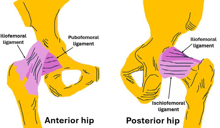
The hip joint capsule plays a crucial role in hip joint stability. The iliofemoral, pubofemoral and ischiofemoral ligaments contribute to the hip joint capsule and can become thickened with various hip conditions. Ultrasound can demonstrate the anterior hip joint capsular morphology and measure the capsule thickness which can be used to diagnose hip joint pathology, guide patient management and evaluate treatment outcomes, and guide hip joint injections and aspirations. This study compared the thickness of the hip joint capsule on the same patient as measured by magnetic resonance imaging (MRI) and ultrasound to determine their correlation (Figure 1).
How the study was performed
Magnetic resonance and ultrasound imaging of the hips of 307 patients (mean age 38.6 years) was retrospectively reviewed, and the anterior hip joint capsular thickness was measured and compared using Bland-Altman analysis and two-tailed paired t-tests. The capsule thickness (depth) was measured where the anterior iliofemoral ligament folded back on itself at the junction of the femoral head and neck (Figure 2).

For MR imaging, the patient was supine with legs extended; for ultrasound imaging, the patients were positioned supine, with their leg of interest in the ‘frog leg’ position (knee flexed and externally rotated). The transducer was positioned along the long axis of the femoral neck.
What the study found
Significant correlation between MRI and ultrasound capsular thickness measurements was demonstrated (p < 0.001). The mean capsular thickness on MRI was 5.0 mm ± 1.2 mm, and on ultrasound was 5.0 ± 1.5 mm. Ultrasound is consistent with MRI for investigating the thickness of the anterior hip joint capsule.
Relevance to clinical practice
Ultrasound of the anterior hip joint can not only be used to identify fluid and pathological material within the hip joint recess but can demonstrate the capsule itself which is comprised of ligaments. It is important for sonographers to appreciate the anatomy of the iliofemoral, pubofemoral (both anterior hip) and ischiofemoral (posterior hip) ligaments, and identify their presence on imaging.
Ultrasound can be used clinically as a substitute for MRI in assessing hip joint capsule thickness


