
9 minute read
MEDBULLETINS

from Issue 39

MEDBULLETIN
Advertisement
ALZHEIMER’S MicroRNAs IMPROVING THE ARDS PATIENT A THERAPEUTIC DISCOVERY EXPERIENCE
ADRIAN WONG
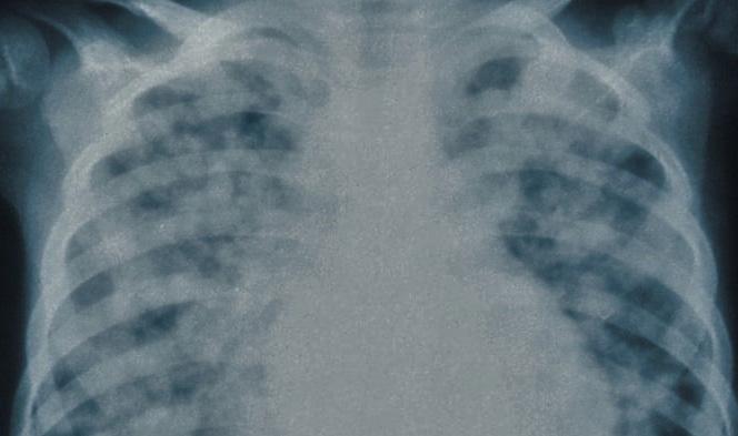
Acute respiratory distress syndrome (ARDS) is a condition caused by tissue insults that can severely damage the respiratory system.1 Approximately 10% of intensive care patients, totaling 3 million people worldwide, suffer from ARDS,2 largely affecting patients with pneumonia, sepsis, and trauma.3 Patients with ARDS usually undergo supportive treatment, which often involves mechanical ventilation.4 However, mechanical ventilation can result in structural damage to the lung, termed ventilator-induced lung injury (VILI).5 A recent study suggests that microRNAs, which are small, non-coding RNA molecules involved in post-transcriptional regulation as well as the formation of many pulmonary diseases, may provide a new strategy for preventing VILI.3
Bobba et al. hypothesized that microRNA-146a (miR-146a) may control the mechanisms in alveolar macrophages that lead to VILI, and tested that hypothesis using in vitro and in vivo models. In mouse models, they discovered that injurious mechanical ventilation caused a tenfold increase in miR-146a levels in alveolar macrophages, while miR-146a knockout mice lacking the microRNA subtype suffered greater lung injury. Nevertheless, the endogenous increase in miR-146a expression was insufficient to completely alleviate lung damage. Alveolar macrophages were then treated with lipid nanoparticles containing pre-miR-146a in both in vitro and in vivo conditions. miR-146a levels increased 100-fold in vitro and 10,000-fold in vivo, enough to prevent significant inflammation caused by pressure changes. In animal models, elevated miR-146a levels led to significant improvements in lung function.3
This study builds on existing research indicating the alteration of microRNA expression by mechanical ventilation, suggesting that microRNAs may be involved in VILI pathogenesis.6 Moreover, miR-146a has been shown to regulate inflammation caused by mechanical ventilation in lung epithelial cells, indicating its potential as a treatment target.7 Overall, the findings of this study suggest that delivering miR-146a through lipid nanoparticles to supplement physiological levels may prove effective in ameliorating VILI and improving the ventilation treatment experience for ARDS patients.3
1. Bos LDJ, Artigas A, Constantin J-M, Hagens LA, Heijnen N, Laffey JG, et al. Precision medicine in acute respiratory distress syndrome: workshop report and recommendations for future research. Eur Respir Rev. 2021;30(159):200317. Available from: doi:10.1183/16000617.0317-2020. 2. Fan E, Brodie D, Slutsky AS. Acute respiratory distress syndrome: advances in diagnosis and treatment. JAMA. 2018;319(7):698. Available from: doi:10.1001/jama.2017.21907. 3. Bobba CM, Fei Q, Shukla V, Lee H, Patel P, Putman RK, et al. Nanoparticle delivery of microRNA-146a regulates mechanotransduction in lung macrophages and mitigates injury during mechanical ventilation. Nat Commun. 2021;12(1):289. Available from: doi:10.1101/796557. 4. Fan E, Needham DM, Stewart TE. Ventilatory management of acute lung injury and acute respiratory distress syndrome. JAMA. 2005;294(22):2889. Available from: doi:10.1001/jama.294.22.2889. 5. Slutsky AS, Ranieri VM. Ventilator-induced lung injury. N Engl J Med. 2013; 369(22):2126–36. Available from: doi:10.1056/ NEJMra1208707. 6. Vaporidi K, Vergadi E, Kaniaris E, Hatziapostolou M, Lagoudaki E, Georgopoulos D, et al. Pulmonary microRNA profiling in a mouse model of ventilator-induced lung injury. Am J Physiol Lung Cell Mol Physiol. 2012;303(3):L199–207. Available from: doi:10.1152/ajplung.00370.2011. 7. Huang Y, Crawford M, Higuita-Castro N, Nana-Sinkam P, Ghadiali SN. miR-146a regulates mechanotransduction and pressureinduced inflammation in small airway epithelium. FASEB J. 2012;26(8):3351–64. Available from: doi:10.1096/fj.11-199240.
CANNABINOIDS
ELUCIDATING THE EFFECTS OF CBD AND THC ON DRIVING
ADRIAN WONG
With the increasing legalization of cannabis and use of medicinal cannabis worldwide,1,2 concerns regarding driving under the influence of cannabis (DUIC) have grown.1 Cannabis ingestion has been linked to poor performance on divided-attention tasks, decision-making, and sudden reactions.3 As such, DUIC has been associated with an increased risk of collision.3 However, most relevant studies have focused on cannabis containing tetrahydrocannabinol (THC), while the effects of cannabidiol (CBD), another prominent substance in cannabis products, have been given less attention.4
A recent study, conducted by the University of Sydney and Maastricht University, sought to delineate the effects of both THC and CBD on driving performance and cognition.5 Arkell et al. tested whether inhalation of cannabis containing 13.75 mg of only THC, only CBD, or a mix of THC and CBD (THC/CBD) affected driving performance in healthy adults who occasionally used cannabis. The primary outcome was standard deviation of lateral position (SDLP), which assesses control of a vehicle; a higher value indicates less control, with more weaving and swerving. From 40 to 100 minutes after consumption, the THC and THC/CBD groups saw an increase in SDLP of more than 2 cm (86.94 cm and 85.51 cm respectively) compared with placebo conditions (84.41 cm), while the CBD group (84.07 cm) did not see any significant changes. From 240 to 300 minutes after consumption, the difference in SDLP between all groups was negligible, indicating that THC had no effect during this time.5
These findings indicate that while THC diminished short-term driving performance, CBD itself had no negative effect.5 Contrary to popular belief, CBD did not negate the effects of THC on driving performance.4,5 The findings of this study invite further research into the effects of THC and CBD on driving performance in regular cannabis users, including medicinal cannabis users, as well as research involving a greater range of cannabinoid doses.4,5
1. Watson TM, Mann RE. International approaches to driving under the influence of cannabis: a review of evidence on impact. Drug Alcohol Depend. 2016;169:148–55. Available from: j.drugalcdep.2016.10.023. 2. WHO Expert Committee on Drug Dependence. Cannabis plant and cannabis resin. Section 5: epidemiology. 2018. Available from: https://www.who.int/medicines/access/controlled-substances/Section5.CannabisPlant.Epidemiology.pdf?ua=1. [cited 2021 Feb 28] 3. Chow RM, Marascalchi B, Abrams WB, Peiris NA, Odonkor CA, Cohen SP. Driving under the influence of cannabis: a framework for future policy. Anesth Analg. 2019;128(6):1300–8. Available from: doi:10.1213/ANE.0000000000003575. 4. Cole TB, Saitz R. Cannabis and impaired driving. JAMA. 2020; 324(21):2163. Available from: doi:10.1001/jama.2020.18544. 5. Arkell TR, Vinckenbosch F, Kevin RC, Theunissen EL, McGregor IS, Ramaekers JG. Effect of cannabidiol and Δ9tetrahydrocannabinol on driving performance: a randomized clinical trial. JAMA. 2020;324(21):2177. Available from: doi:10.1001/jama.2020.21218.

HUNTINGTON’S
HEAT SHOCK PROTEINS FOR TREATING HUNTINGTON’S
NURI SONG
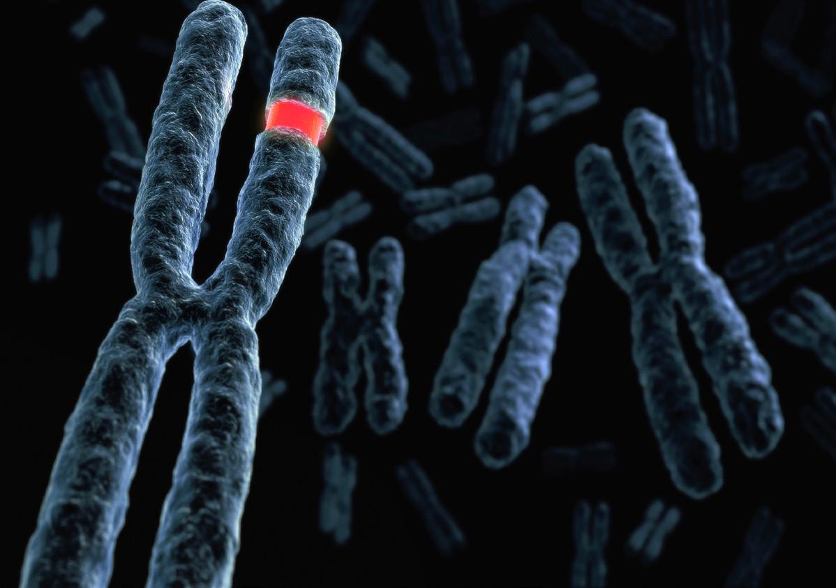
Huntington’s disease is an autosomal dominant genetic disorder caused by the abnormal repetition of the DNA sequence CAG on the Huntingtin (HTT) gene.1 This trinucleotide mutation (with a minimum of 36 repeats) results in the aggregation of a faulty glutamine-based protein in the brain, causing a range of debilitating symptoms and, ultimately, death.2 The onset of Huntington’s disease ranges from 30 to 50 years old and patients suffer from dementia, behavioural and psychiatric disturbances, and involuntary motor movements known as chorea.2 Following pneumonia, the most common cause of death is suicide.2 Although a range of therapeutic interventions are available for Huntington’s patients, the pathophysiology of the condition remains unclear, and, as a result, there is no current cure.
Researchers led by Patrick van der Wel et al., from the University of Groningen in the Netherlands, have successfully revealed the structure of a heat shock protein (HSP), yielding insight into the treatment of Huntington’s.3 HSPs are highly conserved proteins with regulatory and protective roles in the cell.4 A notable class of HSPs is HSP40, which plays a critical role in regulating protein aggregation. The focus of van der Wel’s team was to visualize DnaJB8, a subtype of HSP40 known to bind to other proteins with elongated chains of glutamine amino acids, similar to the HTT protein in Huntington’s. Until recently, its structure has been unclear, preventing researchers from understanding the mechanism of HSPmediated protein deaggregation. Using solid-state NMR spectroscopy, van der Wel et al. successfully visualized HSP40. Based on its structure, researchers predict that DnaJB8 binds to protein aggregates, and activates another class of HSPs, HSP70, which inhibits further aggregation.
Researchers hypothesize that enhancing the activity of DnaJB8 or its analogues will reduce protein aggregation in Huntington’s patients, and alleviate symptoms. Although more research is required, insight into the mechanism has paved the way for future studies in the quest to cure Huntington’s.
DEPRESSION
INSIGHT INTO THE NEURONAL CIRCUITRY
NURI SONG
Major depression is one of the most common mental health conditions in Canada, with an estimated 1.7 million adults experiencing at least one episode.1 Prevalence rates are highest among 18–25 year olds, with increased risk in females (twice the risk of males) and individuals with a family history of depression.2 With existing treatments, only about onethird of patients achieve full remission.3 Despite its prevalence and growing attention, the underlying neuronal mechanisms of depression are poorly understood.
Recent research by Fang et al. has found that agouti-related peptide (AgRP) neurons, a small group of neurons nestled in the arcuate nucleus of the hypothalamus, likely contribute to depression.4 When subjecting mice to chronic, unpredictable stress (resulting in an animal model of depression), AgRP neurons decreased in frequency of firing, and increased in firing irregularities. Moreover, when using a synthetic small molecule agonist to stimulate AgRP neurons, Fang et al. noted the reversal of standard depressive symptoms, such as inability to experience pleasure. As a result, Fang et al. have demonstrated both that chronic stress causes AgRP dysfunction, and that AgRP neurons are key to the neural circuitry underlying depression.
This discovery has important implications for future medications used to treat depression. A common current medication is Prozac, which regulates mood by inhibiting serotonin uptake, but results in sleep disturbance due to low serotonin levels. Shifting treatment focus to AgRP may yield more targeted treatments to mitigate such side effects. It is important to note that AgRP neurons are implicated in other physiological processes including stimulating metabolism and increasing food-seeking behaviour.5 Treatments targeting AgRP may therefore lead to side effects such as weight gain.5 With that said, further research is needed to elucidate to what extent AgRP neurons are implicated in depression as a result of chronic stress.
1. 1. HTT - Huntingtin - Homo sapiens (Human) - HTT gene & protein [Internet]. Available from: https://www.uniprot.org/uniprot/ P42858. [cited 2021 Feb 28]. 2. Roos RA. Huntington’s disease: A clinical review. Orphanet J Rare Dis. 2010; 5:40. Available from: doi:10.1186/1750-11725-40. 3. 3. New insight into protein structures that could treat Huntington’s disease [Internet]. ScienceDaily. Available from: https:// www.sciencedaily.com/releases/2021/02/210212113926.htm. [cited 2021 Feb 28]. 4. Li Z & Srivastava P. Heat-shock proteins. Curr Protoc Immunol. 2004; 58;1:A.1T.1-6. Available from: doi:10.1002/0471142735.ima01ts58.
1. 1. Fast Facts about Mental Illness [Internet]. CMHA National. Available from: https://cmha.ca/fast-facts-about-mental-illness. [cited 2021 Feb 28]. 2. Albert PR. Why is depression more prevalent in women? J Psychiatry Neurosci. 2015; 40(4):219–21. Available from: doi:10.1503/jpn.150205. 3. Ionescu DF, Rosenbaum JF, Alpert JE. Pharmacological approaches to the challenge of treatment-resistant depression. Dialogues Clin Neurosci. 2015; 17(2):111–26. Available from: doi:10.31887/DCNS.2015.17.2/dionescu. 4. Fang, X., Jiang, S., Wang, J. et al. Chronic unpredictable stress induces depression-related behaviors by suppressing AgRP neuron activity. Mol Psychiatry. 2021. Available from: doi:10.1038/s41380-020-01004-x. 5. Essner RA, Smith AG, Jamnik AA, Ryba AR, Trutner ZD, Carter ME. AgRP neurons can increase food intake during conditions of appetite suppression and inhibit anorexigenic parabrachial neurons. J Neurosci. 201; 37(36):8678–87. Available from: doi:10.1523/JNEUROSCI.0798-17.2017.


